Lake EMS Basic EKG Review: Dreaded Heart Blocks EMS Basic EKG Review: Dreaded Heart Blocks The Lake...
239
Lake EMS Basic EKG Review: Dreaded Heart Blocks The Lake EMS Quality Development Team
Transcript of Lake EMS Basic EKG Review: Dreaded Heart Blocks EMS Basic EKG Review: Dreaded Heart Blocks The Lake...

Lake EMS Basic EKG Review:Dreaded Heart Blocks
The Lake EMSQuality Development Team

This program is the Intellectual Property ofLake Emergency Medical ServicesUse of this program is limited to training and Quality Education only
Captain Mike Hilliard, Lake EMS Training Officer2761 West Old Highway 441, Mount Dora, FL 32757-3500
352/383-4554 (w); 352/735-4475 (f); [email protected]

This program In this program we will review the basic
components of heart block rhythms Then we’ll demonstrate a way to assess a heart block
and to accurately identify it, easily!

Our disclaimer opinion With respect to the many revered instructors and
authors who teach electrocardiology rhythm assessment, there are many differences in opinion regarding things such as heart rates for rhythms So we defined our own parameters with the blessings of the
Lake County Medical Director, Pushpal R. Banerjee, D.O.

Our solution Consequently, our Basic EKG Online review meets the
criteria as set forth by our Quality Development Department: John Simpson, Chief Operations Officer Michael R. (Mike) Hilliard, Non-Clinical/Non-Quality Training
Officer Jamie A. Lowery, District Chief, Field Training Coordinator Scott Temple, Clinical Training Officer Julie Treadwell, Clinical Quality Officer
And our Medical Director: Pushpal R. (Paul) Banerjee, D.O.

Basic wave breakdown Please understand this is an interpretation
review, not a diagnostic patient assessment Always treat the patient and not the monitor
P-wave: Atrial depolarization QRS-complex: Ventricular depolarization T-wave: Ventricular repolarization

1st Axiom of EMS And if you forget to treat the patient and are
considering treating the monitor, remember the first axiom of EMS:

1st Axiom of EMS And if you forget to treat the patient and are
considering treating the monitor, remember the first axiom of EMS: If you’re not sure what to do,

1st Axiom of EMS And if you forget to treat the patient and are
considering treating the monitor, remember the first axiom of EMS: If you’re not sure what to do, ask your EMT what the
other paramedics would do in a similar situation.

“Hey, that looks like…” Many of us were taught
how to visually recognize EKGs
We were taught a simple process of 5-steps that help define the rhythm characteristics; however, over time we returned to the visual recognition

For rhythm assessment review Please complete the Lake EMS Online Review:
Basic EKG Review, Atrial Rhythms Before we begin, we’ll perform a short review of
the Atrioventricular (AV) Node and the PR Interval

Normal Impulse ConductionAV Node
Sinus Node
AV Node
BundleOf His
Right BundleBranch
Left BundleBranch
Purkinje fibers

Normal Impulse Conduction
AV NodeAtrioventricular (AV) Node delays (holds) electrical impulse
Sinus Node
AV Node
BundleOf His
Right BundleBranch
Left BundleBranch
Purkinje fibers

Normal Impulse Conduction
AV NodeAllows ventricles to fill with blood from atria (two bottom chambers of the heart)
Sinus Node
AV Node
BundleOf His
Right BundleBranch
Left BundleBranch
Purkinje fibers

Impulse Conduction & the EKGSinoatrial node
AV nodeP
PR

The PR Interval
Atrial depolarization +
delay in AV node
Delay allows time for atria to empty completely before the ventricles contract
P
Q
R
S
T
PR

Mini-review Let us take a moment to review the normal
assessment criteria of Normal Sinus Rhythm

Normal Sinus Rhythm NSR is the normal rhythm produced when the
SA node initiates the cardiac electrical impulse It is what we compare most rhythms against

Normal Sinus Rhythm1. Rate:2. Rhythm:3. P-waves:4. PRI:5. QRS:
60 – 99, on average Regular Normal Normal Narrow

Normal Sinus Rhythm
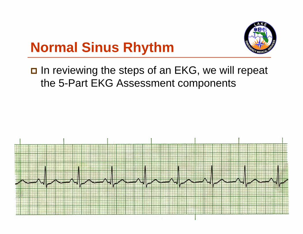
Normal Sinus Rhythm In reviewing the steps of an EKG, we will repeat
the 5-Part EKG Assessment components

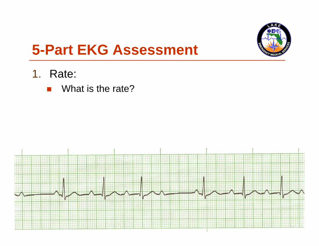
5-Part EKG Assessment1. Rate:
QRS in 6-second strip, multiply x 102. Rhythm:
QRS distances consistent throughout strip3. P-waves: (in the entire strip being assessed):
Are P-waves present? Do they look like a small rounded hill? Is there a P for every QRS? Is there a QRS for every P? Does each P looks like all the others? Is each P the same distance from the QRS?
4. P to R Interval (PRI): 0.12 to 0.20 seconds
5. QRS-Complexes: Narrow: <0.12-seconds (3 small boxes) Wide: >0.12-seconds

5-Part EKG Assessment1. Rate:
What is the rate?

5-Part EKG Assessment1. Rate:
80-bpm

5-Part EKG Assessment2. Rhythm:
Is the rhythm regular or irregular?

5-Part EKG Assessment2. Rhythm:
Regular

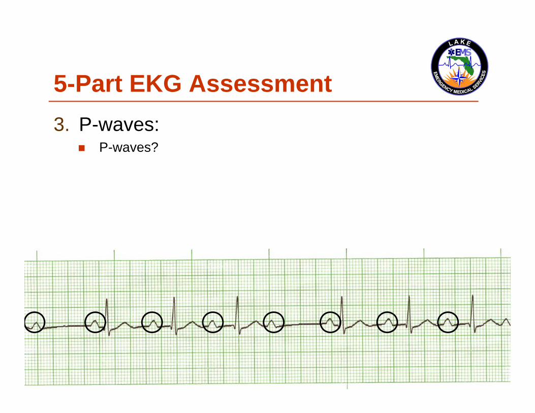
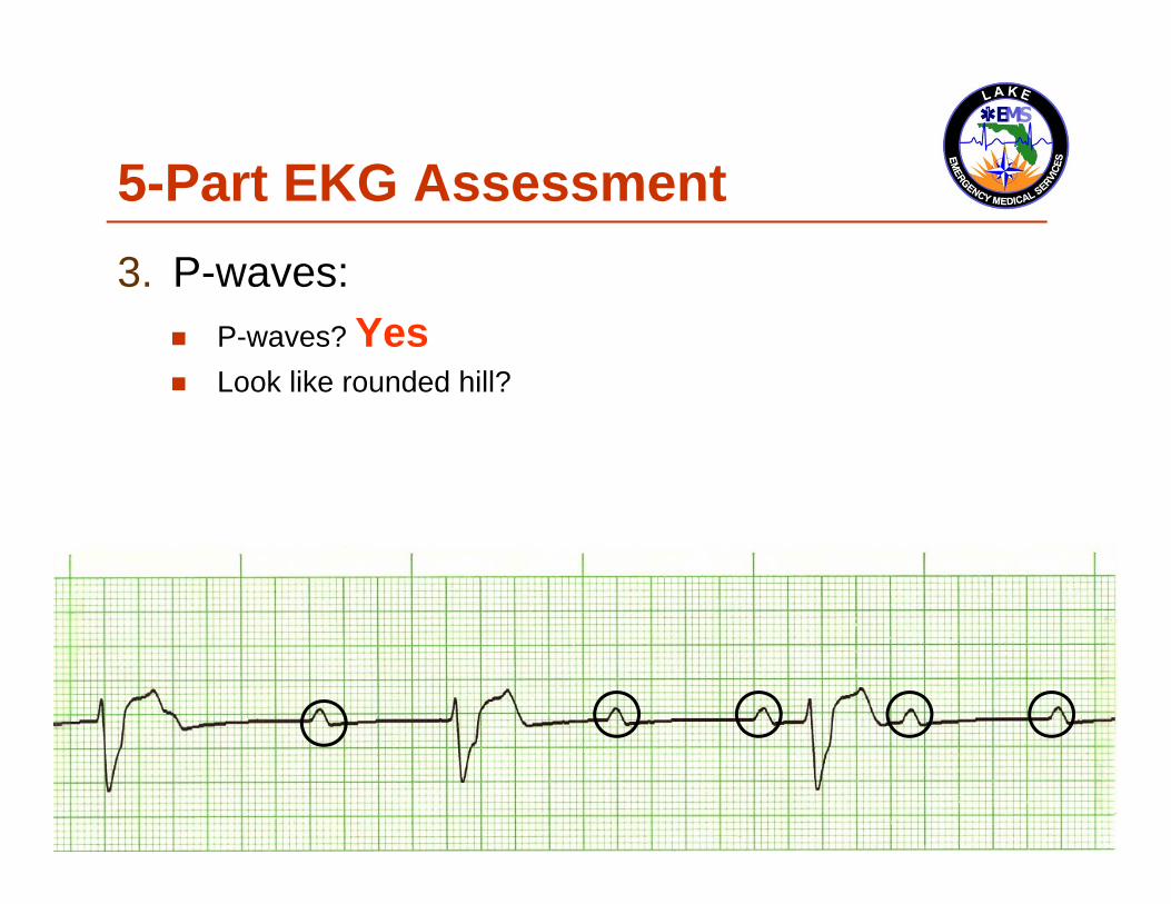
5-Part EKG Assessment3. P-waves:
Are P-waves present? Do they look like a small
rounded hill? Is there a P for every
QRS?
Is there a QRS for every P? Does each P looks like all the
others? Is each P the same distance
from the QRS?

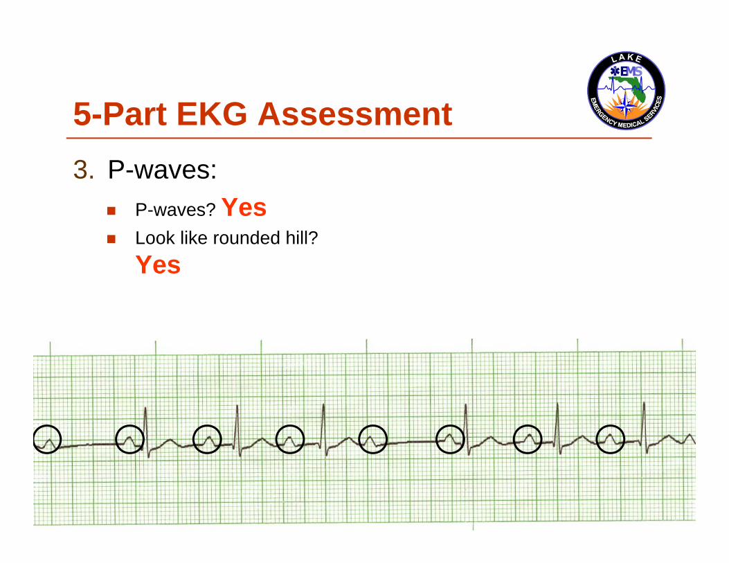
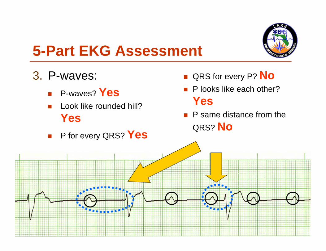
5-Part EKG Assessment3. P-waves:
P-waves? Yes Look like rounded hill?
Yes P for every QRS? Yes
QRS for every P? Yes P looks like each other? Yes P same distance from the
QRS? Yes

5-Part EKG Assessment4. P to R Interval (PRI):
Is the PRI between 3-5 small boxes?

5-Part EKG Assessment4. P to R Interval (PRI):
Yes, 0.20-seconds

Part 4: PRI(not Public Radio International)
P to R Interval (PRI): 0.12 to 0.20 seconds (3-5 small boxes)

Part 4: PRI(not Public Radio International)
P to R Interval (PRI): 0.12 to 0.20 seconds (3-5 small boxes)
The PRI is a window into the effectiveness of the AV Node

Part 4: PRI(not Public Radio International)
P to R Interval (PRI): 0.12 to 0.20 seconds (3-5 small boxes)
The PRI is a window into the effectiveness of the AV Node AV Node has the duty to delay the atrial impulse to
allow for better ventricular filling

5-Part EKG Assessment5. QRS-Complexes:
Is QRS narrow or wide?

5-Part EKG Assessment5. QRS-Complexes:
Narrow, 0.08-seconds

This is Normal Sinus Rhythm1. Rate:2. Rhythm:3. P-waves:4. PRI:5. QRS:
80 Regular Normal Normal Narrow

And now… For something completely different

Ladies and Gentlemen…

Ladies and Gentlemen…Heart Blocks

Ladies and Gentlemen…Heart Blocks (yuck)

The heart block challenge For decades I have taught Advanced Cardiac
Life Support (ACLS) Provider, Refresher, and Instructor Candidate courses to pre-hospital and in-hospital medical personnel

Training Centers/Training Sites AHA Training Centers/Training Sites often have
participants take a pre-requisite EKG rhythm recognition exam to demonstrate their ability to accurately recognize and identify these core rhythms before entering an ACLS class

Start the dreading So the two (2) dreaded questions that we hear
from students before they take an EKG test are…

Dreaded Heart Blocks1. How many questions can I get wrong and still
pass? And,

Dreaded Heart Blocks1. How many questions can I get wrong and still
pass? And,2. How many Heart Blocks are there on the test?

Dreaded Heart Blocks1. How many questions can I get wrong and still
pass? And,2. How many Heart Blocks are there on the test?
They are hoping both answers are the same

This program In this program we will review the basic
components of heart block rhythms Then we’ll demonstrate a way to assess a heart block
and to accurately identify it, easily!

This program In this program we will review the basic
components of heart block rhythms Then we’ll demonstrate a way to assess a heart block
and to accurately identify it, easily! At times you will hear the term AV
(Atrioventricular) and heart blocks

This program In this program we will review the basic
components of heart block rhythms Then we’ll demonstrate a way to assess a heart block
and to accurately identify it, easily! At times you will hear the term AV
(Atrioventricular) and heart blocks For the purposes of this program they are
interchangeable

First Degree Heart Block 1st degree heart block is simply a delay in
passage of the impulse This delay usually occurs at the level of the AV node
1st degree heart blocks are characterized by PR intervals longer than 0.20 second

1st degree heart block 1. Rate:2. Rhythm:3. P-waves:4. PRI:5. QRS:
40-100 Regular Normal This interval is prolonged Narrow

5-Part EKG Assessment1. Rate:
What is the rate?

5-Part EKG Assessment1. Rate:
60-bpm

5-Part EKG Assessment2. Rhythm:
Is the rhythm regular or irregular?

5-Part EKG Assessment2. Rhythm:
Regular

5-Part EKG Assessment3. P-waves:
Are P-waves present? Do they look like a small
rounded hill? Is there a P for every
QRS?
Is there a QRS for every P?
Does each P looks like all the others?
Is each P the same distance from the QRS?

5-Part EKG Assessment3. P-waves:
P-waves? Yes Look like rounded hill?
Yes P for every QRS? Yes
QRS for every P? Yes P looks like each other?
Yes P same distance from the
QRS? Yes

5-Part EKG Assessment4. P to R Interval (PRI):
Is the PRI between 3-5 small boxes?

5-Part EKG Assessment4. P to R Interval (PRI):
No, 7.5 small boxes; 0.30-seconds

5-Part EKG Assessment5. QRS-Complexes:
Is QRS narrow or wide?

5-Part EKG Assessment5. QRS-Complexes:
Narrow, 0.08-seconds

This is 1st degree heart block1. Rate:2. Rhythm:3. P-waves:4. PRI:5. QRS:
60 Regular Normal Prolonged Narrow

Occasionally… We hear of paramedics saying that they have a
borderline 1st degree heart block

Occasionally,… We hear of paramedics saying that they have a
borderline 1st degree heart block 1st degree heart block is or it isn’t; there is no
borderline

Also,… Please remember that 1st degree heart block is a
rhythm category in and of itself

Also,… Please remember that 1st degree heart block is a
rhythm category in and of itself It is not a normal sinus rhythm with a 1st degree
heart block

Also,… Please remember that 1st degree heart block is a
rhythm category in and of itself It is not a normal sinus rhythm with a 1st degree
heart block (You don’t call 3rd degree heart block a idioventricular
rhythm with disassociated P-waves, do you?)

Documentation If you want to describe the severity of the rhythm
remember it is not the rhythm that presents itself, it is the patient

Documentation If you want to describe the severity of the rhythm
remember it is not the rhythm that presents itself, it is the patient
Quantifiers that can be used are: Patient has chest pain Heart rate of 30-bpm BP is 62/P

Second degree heart block,Mobitz type I (Wenckebach) 2nd degree heart block, Mobitz type I, is
characterized by a progressive prolongation of the PR interval This means the PRI becomes longer complex to
complex Then a single impulse is blocked, and the
pattern is repeated

Second degree heart block,Mobitz type I (Wenckebach) 2nd degree heart block, Mobitz type I, is
characterized by a progressive prolongation of the PR interval This means the PRI becomes longer complex to
complex Then a single impulse is blocked, and the
pattern is repeated Key word: pattern

Blocking By the complex being blocked we mean that an
impulse is held by the AV Node until it is unable to continue to carry the electricity into the ventricles What we see is a P-wave with no corresponding QRS

Karel Frederick Wenckebach, MD
First identified the rhythm and it was subsequently named for him
It is also called 2nd degree heart block, Mobitz type I For a synopsis on Dr. Wenckebach, please read:
http://circ.ahajournals.org/cgi/reprint/113/25/f97.pdf

2nd degree heart block, Mobitz type I1. Rate:2. Rhythm:3. P-waves:
4. PRI:
5. QRS:
Ventricular < atrial rate Irregular Has blocked P-wave
> 1 P to QRS
Progressive increase in PR interval until P-wave is blocked
Narrow
5-Part EKG Assessment1. Rate:
What is the rate?

5-Part EKG Assessment1. Rate:
60-bpm

5-Part EKG Assessment2. Rhythm:
Is the rhythm regular or irregular?

5-Part EKG Assessment2. Rhythm:
Irregular ≠

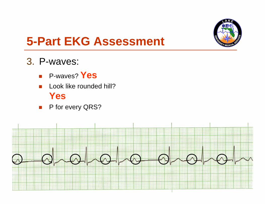
5-Part EKG Assessment3. P-waves:
Are P-waves present? Do they look like a small
rounded hill? Is there a P for every
QRS?
Is there a QRS for every P?
Does each P looks like all the others?
Is each P the same distance from the QRS?
5-Part EKG Assessment3. P-waves:
P-waves?

5-Part EKG Assessment3. P-waves:
P-waves? Yes

5-Part EKG Assessment3. P-waves:
P-waves? Yes Look like rounded hill?
5-Part EKG Assessment3. P-waves:
P-waves? Yes Look like rounded hill?
Yes
5-Part EKG Assessment3. P-waves:
P-waves? Yes Look like rounded hill?
Yes P for every QRS?

5-Part EKG Assessment3. P-waves:
P-waves? Yes Look like rounded hill?
Yes P for every QRS? Yes

5-Part EKG Assessment3. P-waves:
P-waves? Yes Look like rounded hill?
Yes P for every QRS? Yes
Yes?

5-Part EKG Assessment3. P-waves:
P-waves? Yes Look like rounded hill?
Yes P for every QRS? Yes
Yes? You betcha!

5-Part EKG Assessment3. P-waves:
P-waves? Yes Look like rounded hill?
Yes P for every QRS? Yes
Yes? You betcha! Every QRS does
have a P-wave; however,…

5-Part EKG Assessment3. P-waves:
P-waves? Yes Look like rounded hill?
Yes P for every QRS? Yes
QRS for every P?

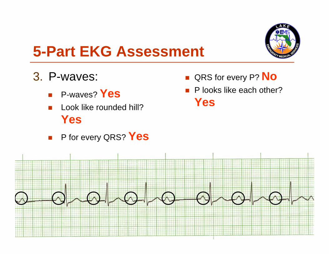
5-Part EKG Assessment3. P-waves:
P-waves? Yes Look like rounded hill?
Yes P for every QRS? Yes
QRS for every P? No

5-Part EKG Assessment3. P-waves:
P-waves? Yes Look like rounded hill?
Yes P for every QRS? Yes
QRS for every P? No P looks like each other?
5-Part EKG Assessment3. P-waves:
P-waves? Yes Look like rounded hill?
Yes P for every QRS? Yes
QRS for every P? No P looks like each other?
Yes

5-Part EKG Assessment3. P-waves:
P-waves? Yes Look like rounded hill?
Yes P for every QRS? Yes
QRS for every P? No P looks like each other?
Yes P same distance from the
QRS?

5-Part EKG Assessment3. P-waves:
P-waves? Yes Look like rounded hill?
Yes P for every QRS? Yes
QRS for every P? No P looks like each other?
Yes P same distance from the
QRS? No

5-Part EKG Assessment4. P to R Interval (PRI):
Is the PRI between 3-5 small boxes?

5-Part EKG Assessment4. P to R Interval (PRI):
No, varies in length

5-Part EKG Assessment5. QRS-Complexes:
Is QRS narrow or wide?

5-Part EKG Assessment5. QRS-Complexes:
Narrow, 0.08-seconds

This is 2nd degree heart block,Mobitz type I1. Rate:2. Rhythm:3. P-waves:4. PRI:5. QRS:
60 Irregular Too many P-waves to QRSs Varies Narrow

Second degree heart block,Mobitz type II A hallmark of this type of 2nd degree heart block,
Mobitz type II, is that the PR interval does not lengthen before a dropped beat

Second degree heart block,Mobitz type II 1. Rate:2. Rhythm:3. P-waves:
4. PRI:5. QRS:
Ventricular is < atrial rate Regular or irregular Has blocked P-wave
> 1 P to QRS
Constantly normal or prolonged Narrow or wide

5-Part EKG Assessment1. Rate:
What is the rate?

5-Part EKG Assessment1. Rate:
50-bpm

5-Part EKG Assessment2. Rhythm:
Is the rhythm regular or irregular?

5-Part EKG Assessment2. Rhythm:
Irregular ≠

5-Part EKG Assessment3. P-waves:
Are P-waves present?

5-Part EKG Assessment3. P-waves:
P-waves? Yes

5-Part EKG Assessment3. P-waves:
P-waves? Yes Look like rounded hill?

5-Part EKG Assessment3. P-waves:
P-waves? Yes Look like rounded hill?
Yes

5-Part EKG Assessment3. P-waves:
P-waves? Yes Look like rounded hill?
Yes P for every QRS?

5-Part EKG Assessment3. P-waves:
P-waves? Yes Look like rounded hill?
Yes P for every QRS? Yes

5-Part EKG Assessment3. P-waves:
P-waves? Yes Look like rounded hill?
Yes P for every QRS? Yes
QRS for every P?

5-Part EKG Assessment3. P-waves:
P-waves? Yes Look like rounded hill?
Yes P for every QRS? Yes
QRS for every P? No

5-Part EKG Assessment3. P-waves:
P-waves? Yes Look like rounded hill?
Yes P for every QRS? Yes
QRS for every P? No P looks like each other?

5-Part EKG Assessment3. P-waves:
P-waves? Yes Look like rounded hill?
Yes P for every QRS? Yes
QRS for every P? No P looks like each other?
Yes

5-Part EKG Assessment3. P-waves:
P-waves? Yes Look like rounded hill?
Yes P for every QRS? Yes
QRS for every P? No P looks like each other?
Yes P same distance from the
QRS?

5-Part EKG Assessment3. P-waves:
P-waves? Yes Look like rounded hill?
Yes P for every QRS? Yes
QRS for every P? No P looks like each other?
Yes P same distance from the
QRS? Yes
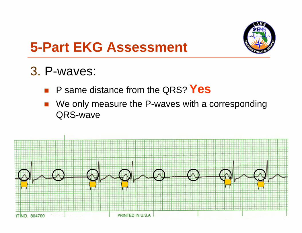
5-Part EKG Assessment3. P-waves:
P same distance from the QRS? Yes We only measure the P-waves with a corresponding
QRS-wave

5-Part EKG Assessment3. P-waves:
Sometimes you can actually see them wave

5-Part EKG Assessment4. P to R Interval (PRI):
Is the PRI between 3-5 small boxes?

5-Part EKG Assessment4. P to R Interval (PRI):
Yes, 0.20-seconds

Yes? Is that correct? Is the PRI normal?

Yes? Is that correct? Is the PRI normal?
Indeed it is

5-Part EKG Assessment4. P to R Interval (PRI):
Yes, every PRI is the same

5-Part EKG Assessment4. P to R Interval (PRI): These P-waves have no QRS and
thereby no PRI Must have P and QRS waves

5-Part EKG Assessment5. QRS-Complexes:
Is QRS narrow or wide?

5-Part EKG Assessment5. QRS-Complexes:
Narrow, 0.08-seconds

This is 2nd degree heart block,Mobitz type II1. Rate:2. Rhythm:3. P-waves:4. PRI:5. QRS:
50 Irregular Too many P-waves to QRSs Normal Narrow

Third degree heart block/complete heart block 3rd degree heart block indicates complete
absence of conduction between atria and ventricles
3rd degree heart block is characterized by a complete dissociation between P waves and QRS complexes

Third degree heart block/complete heart block Both terms are acceptable:
3rd degree heart block Complete Heart Block

The heart The ventricles do not receive any innervations
from the atrium Subsequently, the ventricles must begin to fire
as they have the ability to create their own impulse AKA automaticity

Automaticity and Inherent Myocardial Cell Firings Remember the initiated heart rate from the
ventricles is a back-up system; it is here to keep us alive: SA Node: 60-150 bpm (Not working) AV Junction: 40-60 bpm (Not choosing to work) Ventricles: 30-40 bpm
These are normal values, other rates can and do occur at times
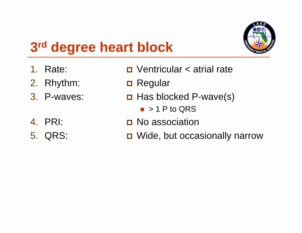
3rd degree heart block1. Rate:2. Rhythm:3. P-waves:
4. PRI:5. QRS:
Ventricular < atrial rate Regular Has blocked P-wave(s)
> 1 P to QRS
No association Wide, but occasionally narrow

5-Part EKG Assessment1. Rate:
What is the rate?

5-Part EKG Assessment1. Rate:
30-bpm

5-Part EKG Assessment2. Rhythm:
Is the rhythm regular or irregular?

5-Part EKG Assessment2. Rhythm:
Regular

5-Part EKG Assessment3. P-waves:
Are P-waves present? Do they look like a small
rounded hill? Is there a P for every
QRS?
Is there a QRS for every P?
Does each P look like all the others?
Is each P the same distance from the QRS?

5-Part EKG Assessment3. P-waves:
P-waves?

5-Part EKG Assessment3. P-waves:
P-waves? Yes
5-Part EKG Assessment3. P-waves:
P-waves? Yes Look like rounded hill?

5-Part EKG Assessment3. P-waves:
P-waves? Yes Look like rounded hill?
Yes

5-Part EKG Assessment3. P-waves:
P-waves? Yes Look like rounded hill?
Yes P for every QRS?

5-Part EKG Assessment3. P-waves:
P-waves? Yes Look like rounded hill?
Yes P for every QRS? Yes

5-Part EKG Assessment3. P-waves:
P-waves? Yes Look like rounded hill?
Yes P for every QRS? Yes
QRS for every P?

5-Part EKG Assessment3. P-waves:
P-waves? Yes Look like rounded hill?
Yes P for every QRS? Yes
QRS for every P? No

5-Part EKG Assessment3. P-waves:
P-waves? Yes Look like rounded hill?
Yes P for every QRS? Yes
QRS for every P? No P looks like each other?

5-Part EKG Assessment3. P-waves:
P-waves? Yes Look like rounded hill?
Yes P for every QRS? Yes
QRS for every P? No P looks like each other?
Yes

5-Part EKG Assessment3. P-waves:
P-waves? Yes Look like rounded hill?
Yes P for every QRS? Yes
QRS for every P? No P looks like each other?
Yes P same distance from the
QRS?
5-Part EKG Assessment3. P-waves:
P-waves? Yes Look like rounded hill?
Yes P for every QRS? Yes
QRS for every P? No P looks like each other?
Yes P same distance from the
QRS? No

5-Part EKG Assessment4. P to R Interval (PRI):
Is the PRI between 3-5 small boxes?

5-Part EKG Assessment4. P to R Interval (PRI):
No, varies
PRI The PRI is a window into the effectiveness of the
AV Node AV Node has the duty to delay the atrial impulse to
allow for better ventricular filling

5-Part EKG Assessment5. QRS-Complexes:
Is QRS narrow or wide?

5-Part EKG Assessment5. QRS-Complexes:
Wide, nearly 0.20-seconds

5-Part EKG Assessment5. QRS-Complexes:
Wide, nearly 0.20-seconds What a normal PRI should look like

This is 3rd degree heart block 1. Rate:2. Rhythm:3. P-waves:4. PRI:5. QRS:
30 Regular No QRS for each P-wave Varies Wide
Dreaded Heart Blocks From my testing experiences, most people can
identify that the rhythm is a heart block What they cannot accurately and consistently do is to
correctly identify which of the 2nd or the 3rd degree heart block it is
Now we’ll demonstrate a way to assess a heart block and to accurately identify it, easily!

American Heart Association
On page 65 of the 2nd Edition, Textbook of Advanced Cardiac Life Support, it offers the greatest algorithm for differentiating 2nd and 3rd
degree heart block Now some of you may not have a readily available
copy of your 1987 textbook so feel free to make an appointment with me and you can see my copy

2 Step Heart Block Analysis
More P’s than QRSs
PRI same in all complexes?
no
QRSs regular?
no
yes
yes
yes
2nd degree Heart Block, Mobitz II
3rd degree Heart Block
2nd degree Heart Block, Mobitz I (Wenckebach)

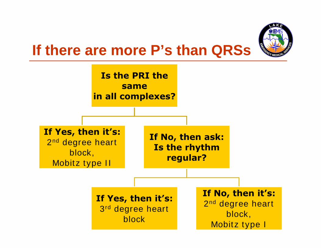
Another way to view (If there are more P’s than QRSs)
Is the PRI the same
in all complexes?
If Yes, then it’s:2nd degree heart
block,Mobitz type II
If No, then ask:Is the rhythm
regular?
If Yes, then it’s:3rd degree heart
block
If No, then it’s:2nd degree heart
block,Mobitz type I

This is an example of what peoplelook like after learning this process

Practice time
Practice timeAnd the crowd goes crazy

Practice timeAnd the crowd goes crazy, -ish

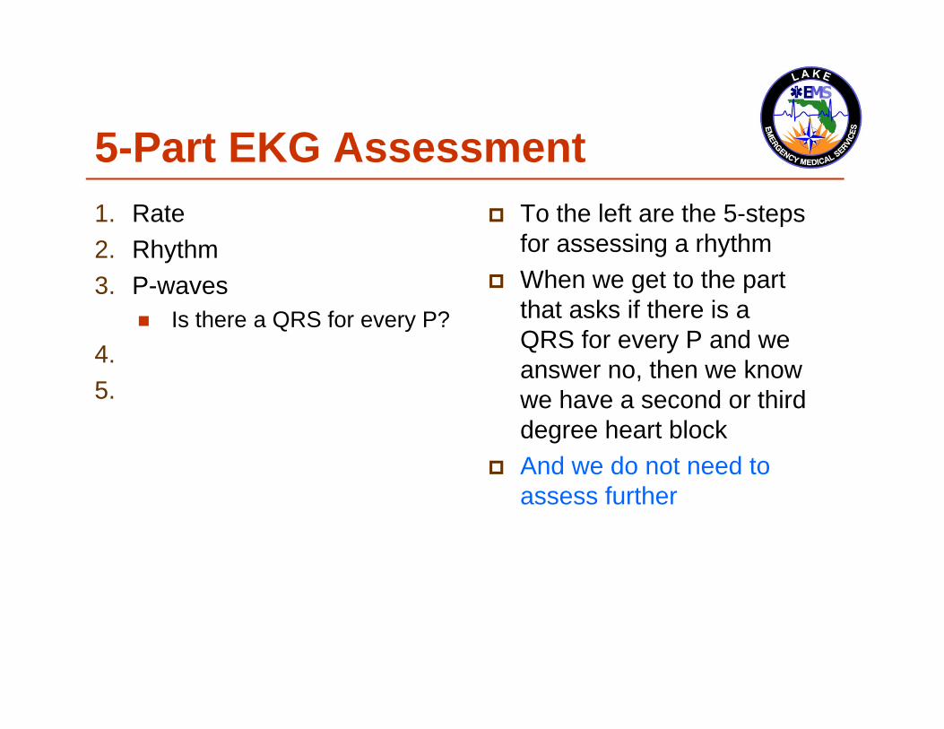
5-Part EKG Assessment1. Rate2. Rhythm3. P-waves4. P to R Interval (PRI)5. QRS-Complexes
To the left are the 5-steps for assessing a rhythm

5-Part EKG Assessment1. Rate2. Rhythm3. P-waves
Is there a QRS for every P?4. P to R Interval (PRI)5. QRS-Complexes
To the left are the 5-steps for assessing a rhythm
When we get to the part that asks if there is a QRS for every P and we answer no,

5-Part EKG Assessment1. Rate2. Rhythm3. P-waves
Is there a QRS for every P?4. P to R Interval (PRI)5. QRS-Complexes
To the left are the 5-steps for assessing a rhythm
When we get to the part that asks if there is a QRS for every P and we answer no,then we know we have a second or third degree heart block
5-Part EKG Assessment1. Rate2. Rhythm3. P-waves
Is there a QRS for every P?4. P to R Interval (PRI)5. QRS-Complexes
To the left are the 5-steps for assessing a rhythm
When we get to the part that asks if there is a QRS for every P and we answer no, then we know we have a second or third degree heart block
And we do not need to assess further

5-Part EKG Assessment1. Rate2. Rhythm3. P-waves
Is there a QRS for every P?4. P to R Interval (PRI)5. QRS-Complexes
Is the PRI the samein all complexes? If Yes, then it’s: 2nd degree
heart block,Mobitz type II
If No, then ask: Is the rhythm regular? If Yes, then it’s: 3rd degree
heart block If No, then it’s: 2nd degree
heart block,Mobitz type I

Sample rhythms Lets give it a try

Sample rhythms Lets give it a try Remember, we are looking for the sign of more
than one P-wave to a QRS-wave

Whatissit? Number 1
If there are more P’s than QRSsIs the PRI the
samein all complexes?
If Yes, then it’s:2nd degree heart
block,Mobitz type II
If No, then ask:Is the rhythm
regular?
If Yes, then it’s:3rd degree heart
block
If No, then it’s:2nd degree heart
block,Mobitz type I

Whatissit? Number 1 Is the PRI the same in all complexes?

Whatissit? Number 1 Is the PRI the same in all complexes?
No

Whatissit? Number 1 Is the PRI the same in all complexes?
No Is the rhythm regular?

Whatissit? Number 1 Is the PRI the same in all complexes?
No Is the rhythm regular?
No

Whatissit? Number 1 Is the PRI the same in all complexes?
No Is the rhythm regular?
No: 2nd degree heart block, Mobitz type I

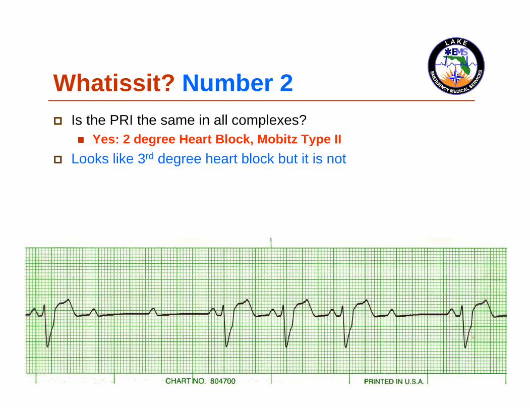
Whatissit? Number 2

Whatissit? Number 2 Is the PRI the same in all complexes?

Whatissit? Number 2 Is the PRI the same in all complexes?
Yes: 2 degree Heart Block, Mobitz Type II
Whatissit? Number 2 Is the PRI the same in all complexes?
Yes: 2 degree Heart Block, Mobitz Type II Looks like 3rd degree heart block but it is not

Whatissit? Number 2 Is the PRI the same in all complexes?
Yes: 2 degree Heart Block, Mobitz Type II Looks like 3rd degree heart block but it is not This is why we have such great success using the algorithm

Whatissit? Number 3

Whatissit? Number 3 Is the PRI the same in all complexes?

Whatissit? Number 3 Is the PRI the same in all complexes? Does it matter?

Whatissit? Number 3 Is the PRI the same in all complexes? Does it matter? Nope

Whatissit? Number 3 Is the PRI the same in all complexes? Does it matter? Nope, because those are not P-waves

Whatissit? Number 3 Is the PRI the same in all complexes? Does it matter? Nope, because those are not P-waves They are F-waves also known as Flutter waves

Whatissit? Number 3 Is the PRI the same in all complexes? Does it matter? Nope, because those are not P-waves They are F-waves also known as Flutter waves This is Atrial Flutter

Whatissit? Number 4

Whatissit? Number 4 Is the PRI the same in all complexes?

Whatissit? Number 4 Is the PRI the same in all complexes?
No

Whatissit? Number 4 Is the PRI the same in all complexes? No Is the rhythm regular?

Whatissit? Number 4 Is the PRI the same in all complexes? No Is the rhythm regular?
Yes: 3rd degree heart block

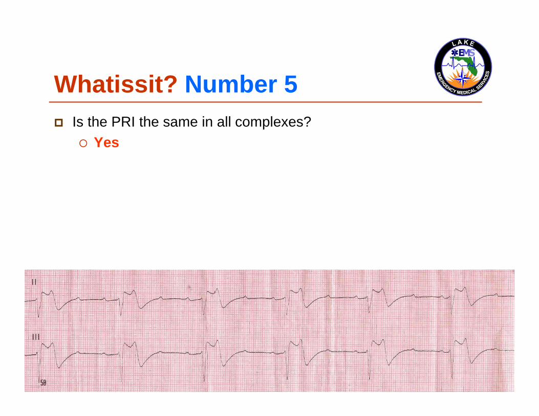
Whatissit? Number 5

Whatissit? Number 5 Is the PRI the same in all complexes?
Whatissit? Number 5 Is the PRI the same in all complexes?
Yes

Whatissit? Number 5 Is the PRI the same in all complexes?
Yes: 2 degree Heart Block, Mobitz Type II

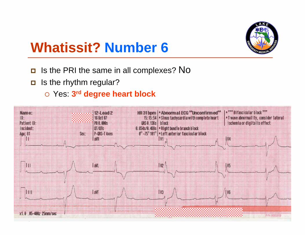
Whatissit? Number 6

Whatissit? Number 6 Is the PRI the same in all complexes?

Whatissit? Number 6 Is the PRI the same in all complexes?
No

Whatissit? Number 6 Is the PRI the same in all complexes? No Is the rhythm regular?

Whatissit? Number 6 Is the PRI the same in all complexes? No Is the rhythm regular?
Yes
Whatissit? Number 6 Is the PRI the same in all complexes? No Is the rhythm regular?
Yes: 3rd degree heart block

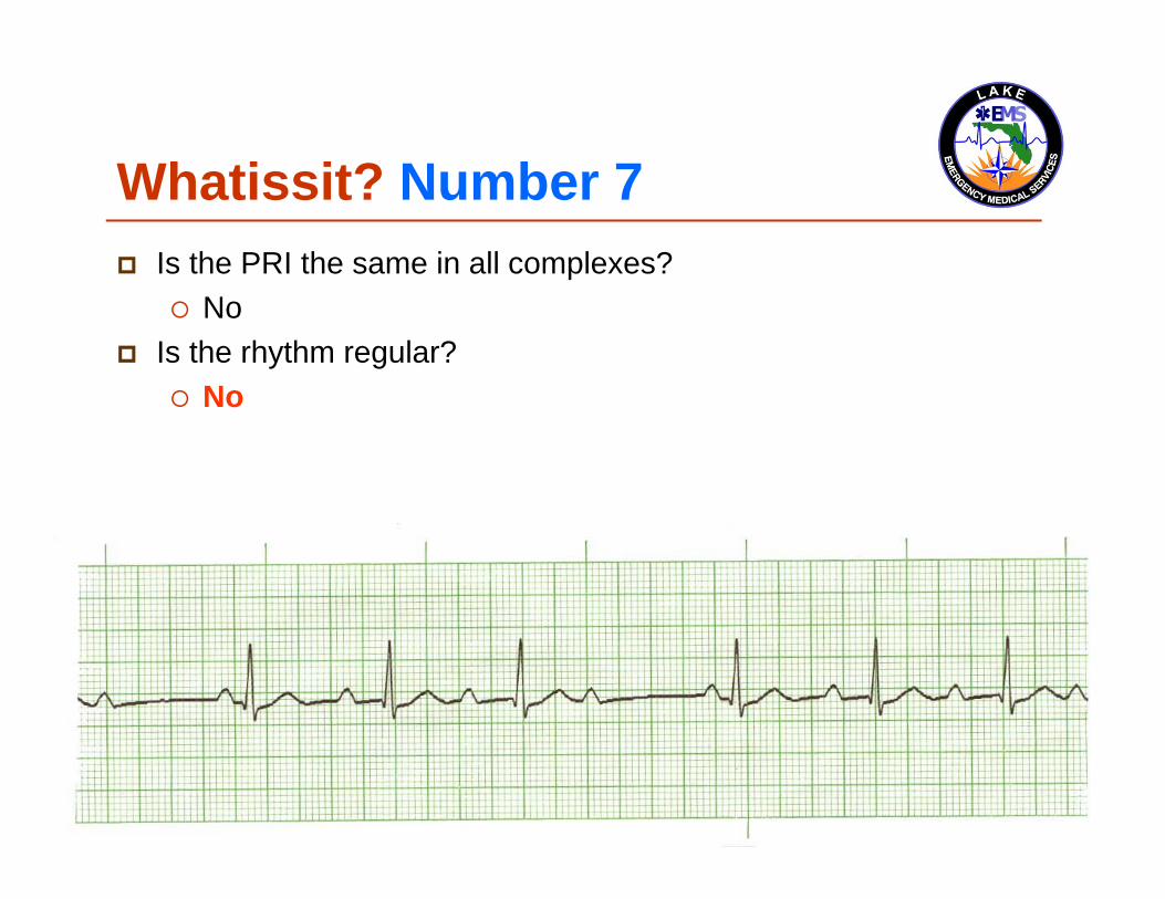
Whatissit? Number 7

Whatissit? Number 7 Is the PRI the same in all complexes?

Whatissit? Number 7 Is the PRI the same in all complexes?
No

Whatissit? Number 7 Is the PRI the same in all complexes?
No Is the rhythm regular?
Whatissit? Number 7 Is the PRI the same in all complexes?
No Is the rhythm regular?
No

Whatissit? Number 7 Is the PRI the same in all complexes?
No Is the rhythm regular?
No: 2nd degree Heart Block, Mobitz Type I

Whatissit? Number 8
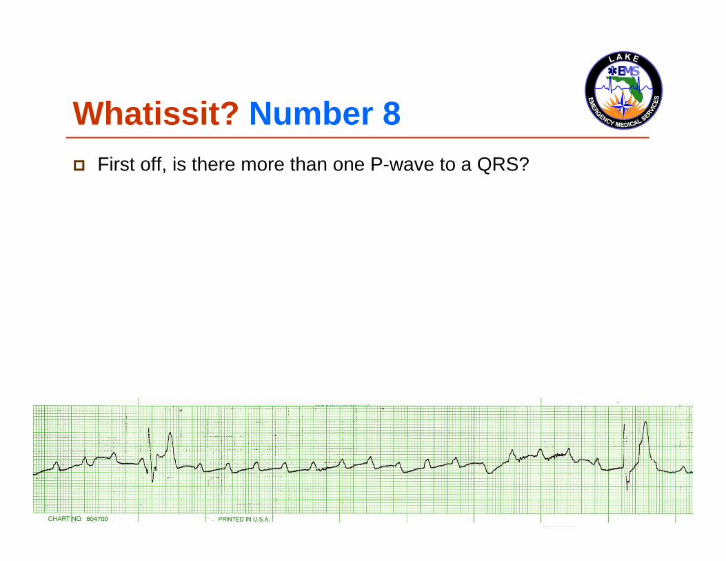
Whatissit? Number 8 First off, is there more than one P-wave to a QRS?

Whatissit? Number 8 First off, is there more than one P-wave to a QRS? Take your time

Whatissit? Number 8 First off, is there more than one P-wave to a QRS? Take your time
You can do it

Whatissit? Number 8 So yes, this is a heart block

Whatissit? Number 8 Is the PRI the same in all complexes?

Whatissit? Number 8 Is the PRI the same in all complexes?
No

Whatissit? Number 8 Is the PRI the same in all complexes?
No Is the rhythm regular?

Whatissit? Number 8 Is the PRI the same in all complexes?
No Is the rhythm regular?
Hard to tell but it was, which leads us to identify the rhythm as…

Whatissit? Number 8 Is the PRI the same in all complexes?
No Is the rhythm regular?
Yes: 3rd degree heart block

Background story 1985

Background story 1985
Feel free not to shout out, “Hey, that was before I was born!”

Background story 1985
Feel free not to shout out, “Hey, that was before I was born!”
Elderly patient with 2nd degree heart block, Mobitz type II, slow heart rate and very symptomatic

Background story My great partner, Shawn Metayer, and I
administered 0.5-mg Atropine

Background story My great partner, Shawn Metayer, and I
administered 0.5-mg Atropine TCP did not exist in EMS at that time

Background story My great partner, Shawn Metayer, and I
administered 0.5-mg Atropine TCP did not exist in EMS at that time
Atropine at that time was not contraindicated in late stage Heart Blocks by the American Heart Association

Background story Did the Atropine work?
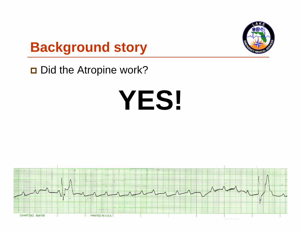
Background story Did the Atropine work?
YES!

Background story Did the Atropine work?
YES! Atropine is an vasolytic and allows the SA Node
to fire more

Background story Did the Atropine work? 140 Atrial rate
A six (6) second strip

Background story Did the Atropine work? 140 Atrial rate, albeit a 0-ventricular rate
A six (6) second strip

Background story Did the Atropine work? 140 Atrial rate, albeit a 0-ventricular rate The patient remained conscious the entire time
A six (6) second strip

Background story Despite our best efforts, the patient survived

Background story Despite our best efforts, the patient survived In all reality, we were providing the best care we
could at that time

This is the end Stay tuned for the last installment of Basic EKG
Review: Ventricular Rhythms

This program is the Intellectual Property ofLake Emergency Medical ServicesUse of this program is limited to training and Quality Education only
Captain Mike Hilliard, Lake EMS Training Officer2761 West Old Highway 441, Mount Dora, FL 32757-3500
352/383-4554 (w); 352/735-4475 (f); [email protected]

Lake EMS Basic EKG Review:Dreaded Heart Blocks
The Lake EMSQuality Development Team