Lactate dehydrogenase + 38 ATP + 2 ATP. How does lactate dehydrogenase perform its catalytic...
23
Lactate dehydrogenase + 38 ATP + 2 ATP
-
Upload
juliana-nelson -
Category
Documents
-
view
237 -
download
2
Transcript of Lactate dehydrogenase + 38 ATP + 2 ATP. How does lactate dehydrogenase perform its catalytic...
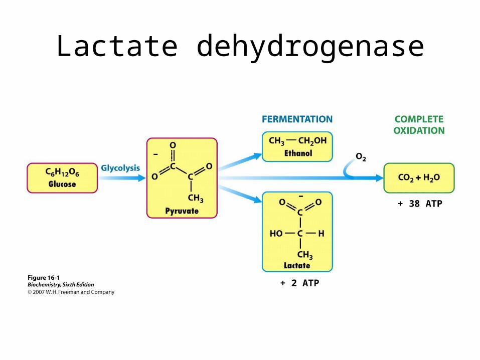
Lactate dehydrogenase
+ 38 ATP
+ 2 ATP
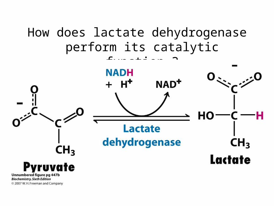
How does lactate dehydrogenase perform its catalytic function ?
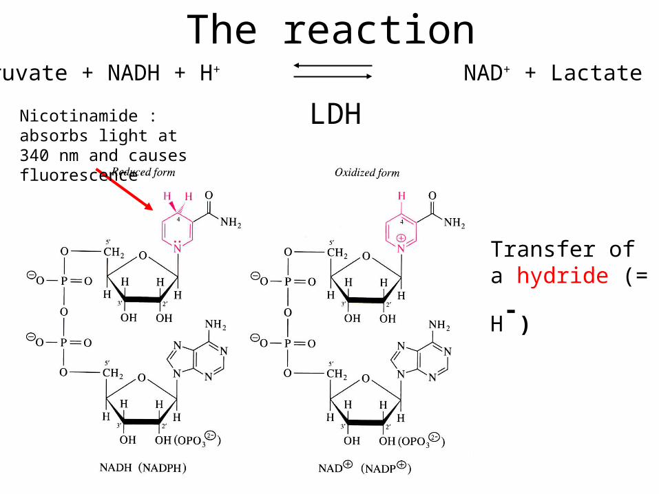
The reaction
LDH
Pyruvate + NADH + H+ NAD+ + Lactate
Transfer of a
hydride (= H-)
Nicotinamide : absorbs light at 340 nm and causes fluorescence
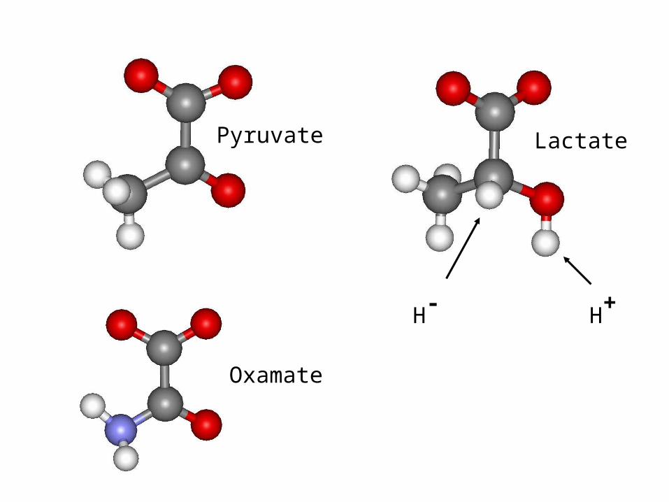
Pyruvate
Oxamate
Lactate
H- H+
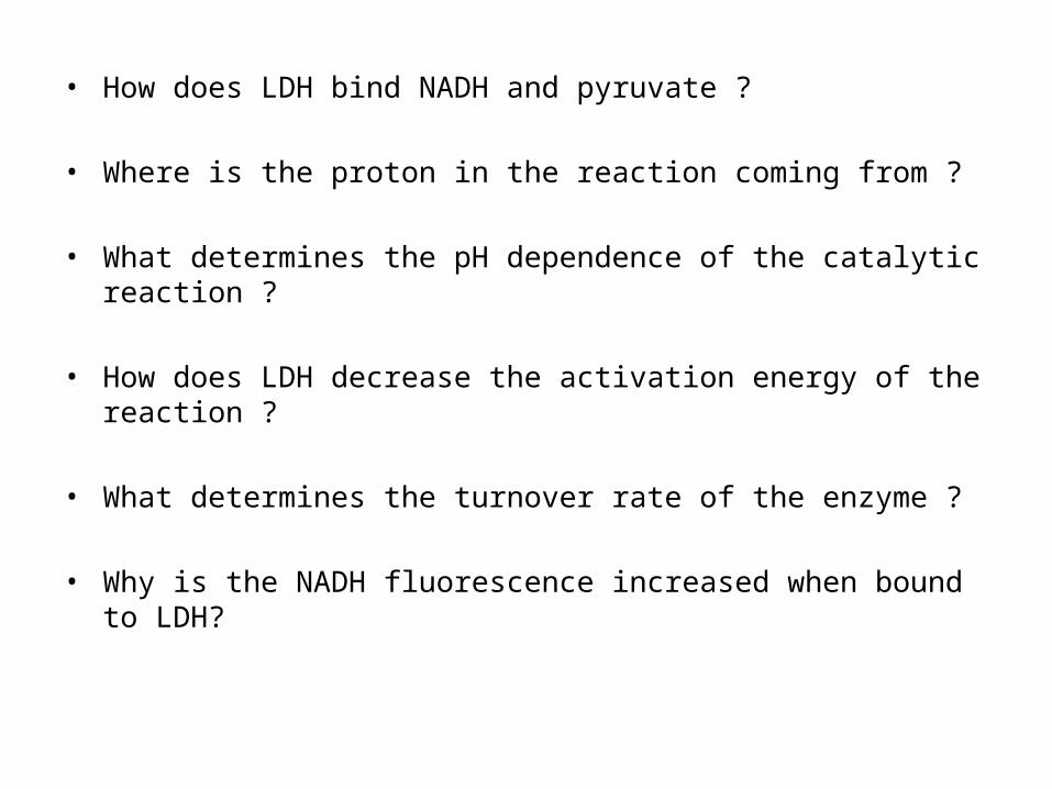
• How does LDH bind NADH and pyruvate ?
• Where is the proton in the reaction coming from ?
• What determines the pH dependence of the catalytic reaction ?
• How does LDH decrease the activation energy of the reaction ?
• What determines the turnover rate of the enzyme ?
• Why is the NADH fluorescence increased when bound to LDH?
Properties of the enzyme are determined by the properties and spatial arrangement of amino acids in the 3D-structure of the protein:
• Position• Size• Polarity• Interactions• Chemical properties
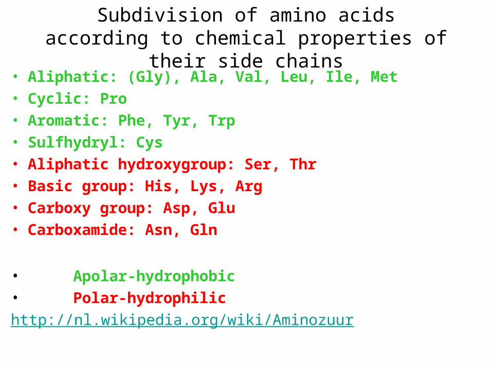
Subdivision of amino acids according to chemical properties of their side chains
• Aliphatic: (Gly), Ala, Val, Leu, Ile, Met• Cyclic: Pro • Aromatic: Phe, Tyr, Trp• Sulfhydryl: Cys• Aliphatic hydroxygroup: Ser, Thr• Basic group: His, Lys, Arg• Carboxy group: Asp, Glu• Carboxamide: Asn, Gln
• Apolar-hydrophobic • Polar-hydrophilic
http://nl.wikipedia.org/wiki/Aminozuur
The 3D-structure of a protein can be determined by X-ray diffraction
• The protein is crystallyzed• The protein crystals are irratiated with X-rays• X-rays are diffracted by the atoms in the protein• From the diffraction pattern, the position of each atom in
the protein (except for H-atoms) is determined (electron density map).
• The structure of the protein is reconstructed from the electron density map, up to 0.2 Å accuracy. This gives the exact position of each amino acid , substrates, etc. in the 3D-structure
The 3D-structure of a protein can be determined by X-ray diffraction
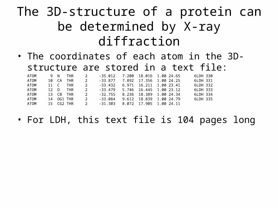
• The coordinates of each atom in the 3D-structure are stored in a text file:ATOM 9 N THR 2 -35.012 7.200 18.016 1.00 24.65 6LDH 330ATOM 10 CA THR 2 -33.877 7.892 17.356 1.00 24.25 6LDH 331ATOM 11 C THR 2 -33.432 6.971 16.211 1.00 23.41 6LDH 332ATOM 12 O THR 2 -33.479 5.746 16.445 1.00 23.12 6LDH 333ATOM 13 CB THR 2 -32.755 8.236 18.389 1.00 24.34 6LDH 334ATOM 14 OG1 THR 2 -33.084 9.612 18.839 1.00 24.79 6LDH 335ATOM 15 CG2 THR 2 -31.303 8.072 17.905 1.00 24.11
• For LDH, this text file is 104 pages long
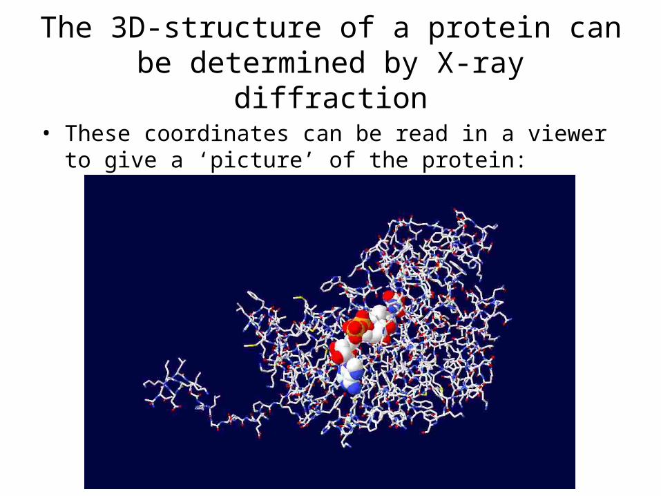
The 3D-structure of a protein can be determined by X-ray diffraction
• These coordinates can be read in a viewer to give a ‘picture’ of the protein:
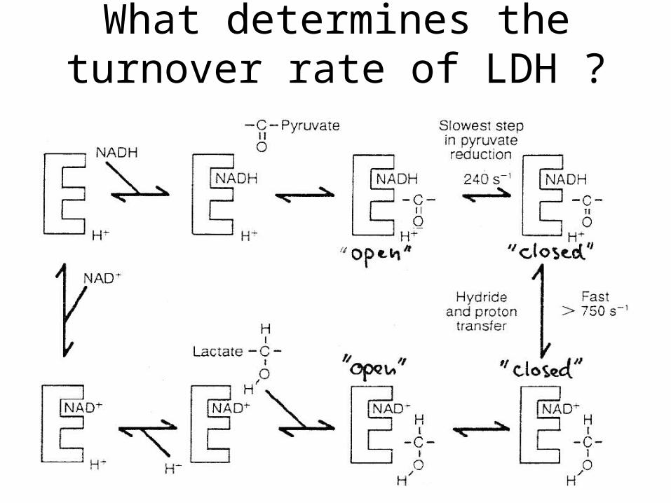
What determines the turnover rate of LDH ?
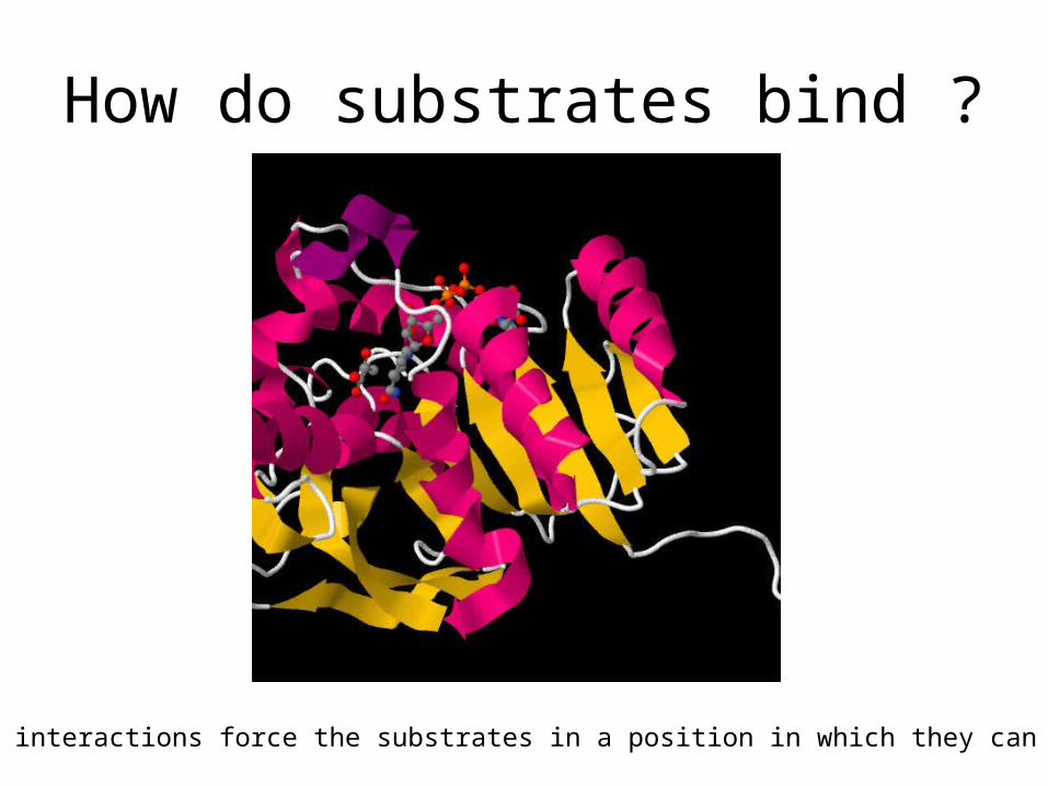
How do substrates bind ?
What interactions force the substrates in a position in which they can react ?
Non-covalent interactions in proteins
• Electrostatic (or ionogenic) interactions
• Hydrogen bridges
• Vanderwaals interactions
• Hydrophobic interactions
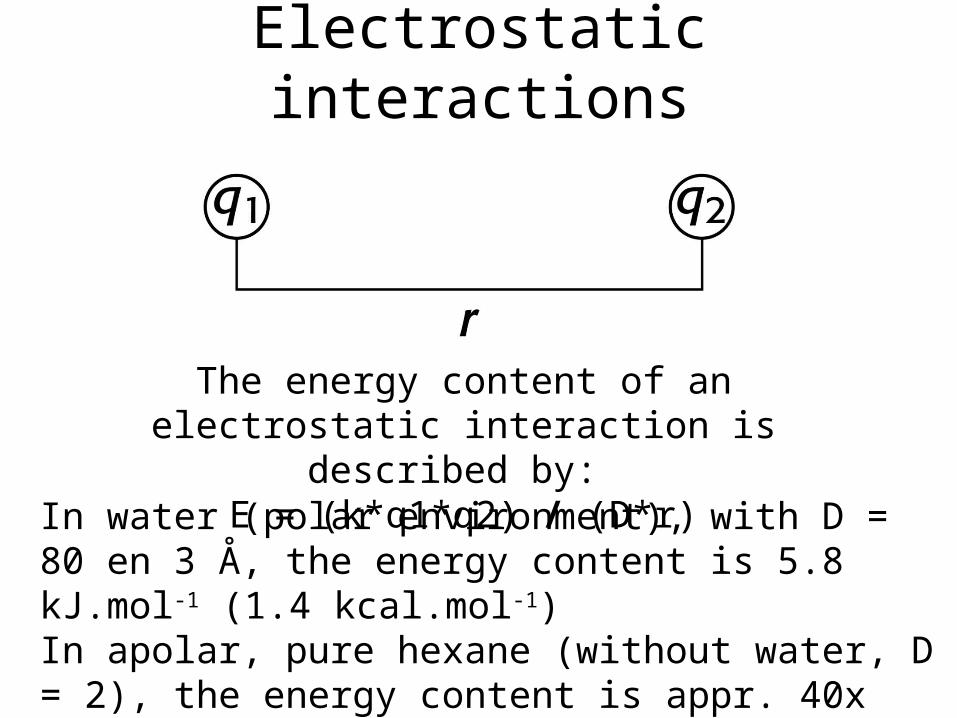
Electrostatic interactions
The energy content of an electrostatic interaction is described by:
E = (k*q1*q2) / (D*r)In water (polar environment), with D = 80 en 3 Å, the energy content is 5.8 kJ.mol-1 (1.4 kcal.mol-1)In apolar, pure hexane (without water, D = 2), the energy content is appr. 40x higher.
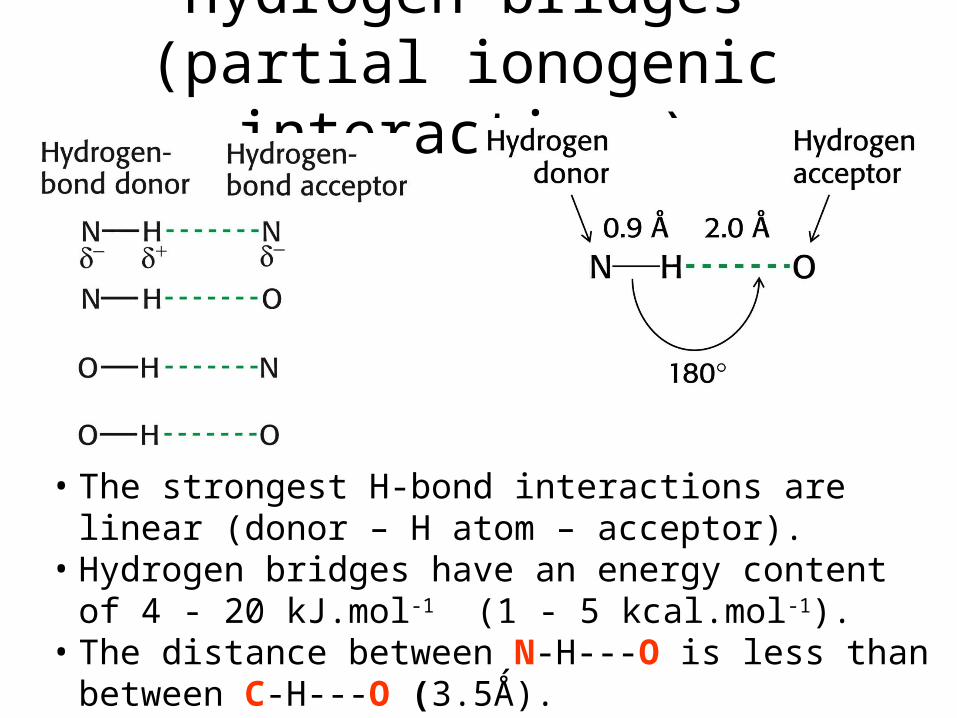
Hydrogen bridges (partial ionogenic interactions)
• The strongest H-bond interactions are linear (donor – H atom – acceptor).
• Hydrogen bridges have an energy content of 4 - 20 kJ.mol-1 (1 - 5 kcal.mol-1).
• The distance between N-H---O is less than between C-H---O (3.5Ǻ).
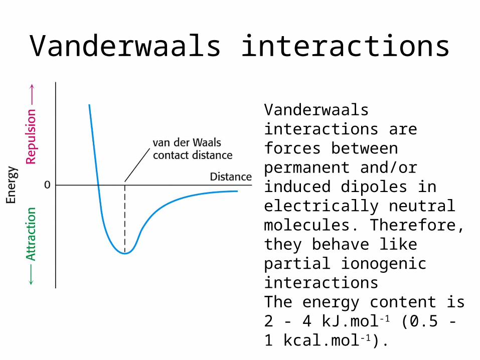
Vanderwaals interactions
Vanderwaals interactions are forces between permanent and/or induced dipoles in electrically neutral molecules. Therefore, they behave like partial ionogenic interactionsThe energy content is 2 - 4 kJ.mol-1 (0.5 - 1 kcal.mol-1).

Hydrophobic interactions
• Association of apolar groups/molecules in water results in the release of water molecules that surround the apolar surface in a stiff, ice-like structure.
• The released water molecules have more possibilities to interact with other water molecules in solution.
• This results in an increase of the entropy (S) of the water in: G = H - TS. This results in a decrease in the free energy. Hydrophobic interactions are responsible for clustering of amino acids with hydrophobic residues in the center of a protein.
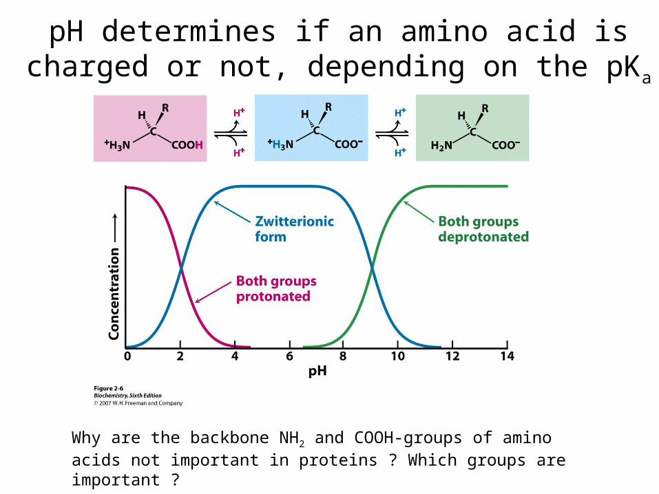
pH determines if an amino acid is charged or not, depending on the pKa
Why are the backbone NH2 and COOH-groups of amino acids not important in proteins ? Which groups are important ?
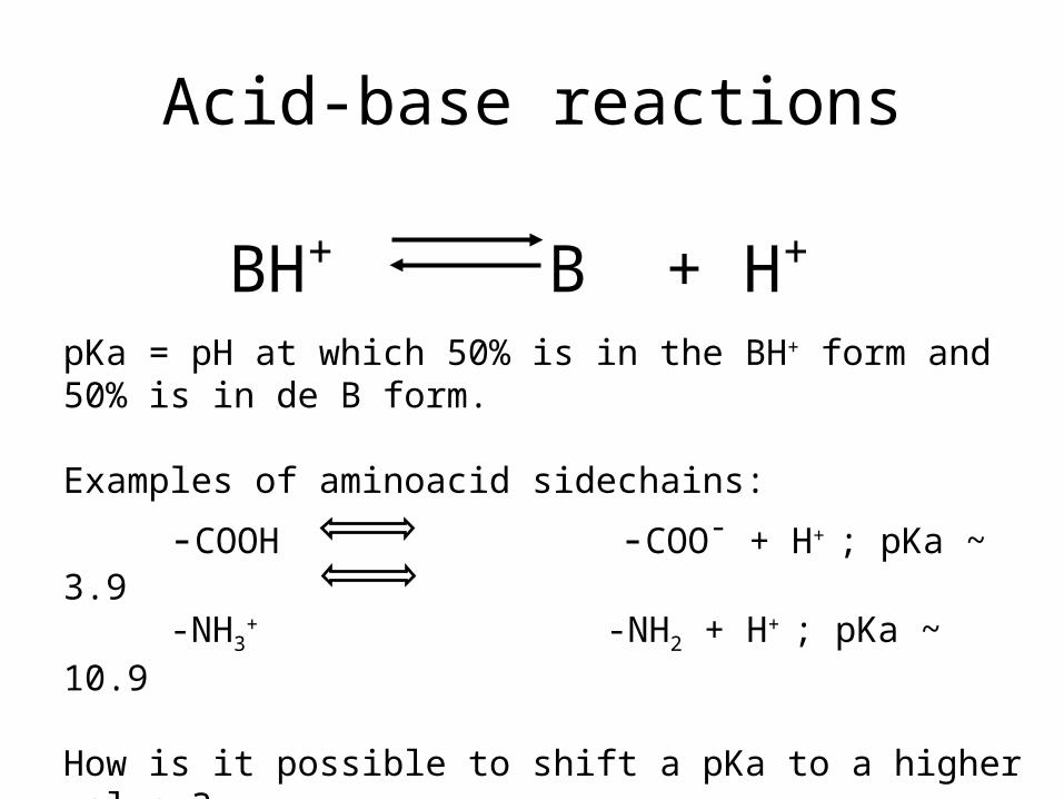
Acid-base reactions
BH+ B + H+
pKa = pH at which 50% is in the BH+ form and 50% is in de B form.
Examples of aminoacid sidechains:
-COOH -COO- + H+ ; pKa ~ 3.9-NH3
+ -NH2 + H+ ; pKa ~ 10.9
How is it possible to shift a pKa to a higher value ?
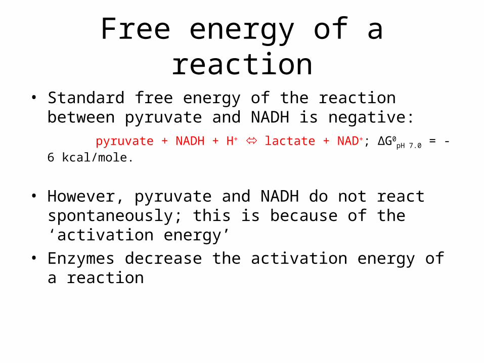
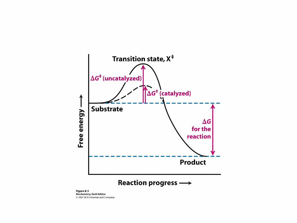
Free energy of a reaction
• Standard free energy of the reaction between pyruvate and NADH is negative:
pyruvate + NADH + H+ lactate + NAD+; ΔG0pH 7.0 = - 6 kcal/mole.
• However, pyruvate and NADH do not react spontaneously; this is because of the ‘activation energy’
• Enzymes decrease the activation energy of a reaction
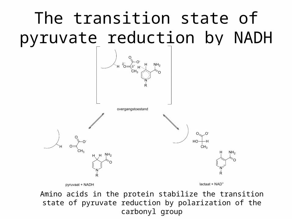
The transition state of pyruvate reduction by NADH
Amino acids in the protein stabilize the transition state of pyruvate reduction by polarization of the carbonyl group