laboratoryyg methods in diagnosis of -...
63
Transcript of laboratoryyg methods in diagnosis of -...

laboratory methods in diagnosis of y gviral infections in
immunocompromised patientsimmunocompromised patientsMasoud Parsania, Ph.D.Masoud Parsania, Ph.D.
Department of Microbiology, Tehran Medical Sciences Branch, Islamic Azad University, Tehran, Iran.
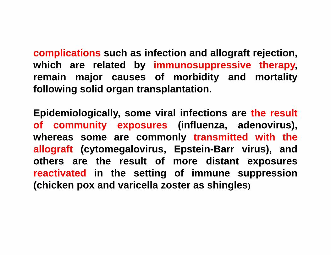
complications such as infection and allograft rejection,which are related by immunosuppressive therapy,remain major causes of morbidity and mortalityremain major causes of morbidity and mortalityfollowing solid organ transplantation.
Epidemiologically some viral infections are the resultEpidemiologically, some viral infections are the resultof community exposures (influenza, adenovirus),whereas some are commonly transmitted with theallograft (cytomegalovirus, Epstein-Barr virus), andothers are the result of more distant exposuresreactivated in the setting of immune suppressionreactivated in the setting of immune suppression(chicken pox and varicella zoster as shingles)
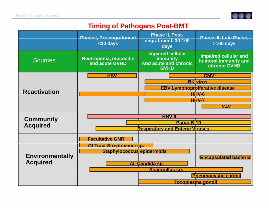
HHV-6 in Transplant Patients
Phase I Pre-engraftment Phase II, Post- Phase III Late Phase
Timing of Pathogens Post-BMTPhase I, Pre engraftment
<30 days engraftment, 30-100 days
Phase III, Late Phase, >100 days
Neutropenia, mucositis and acute GVHD
Impaired cellular immunity
And acute and chronicImpaired cellular and
humoral immunity and h i GVHD
Sources and acute GVHD And acute and chronic GVHD chronic GVHD
•Reactivation
HSVBK virus
CMV+
EBV Lymphoprolferative disease•Reactivation
C it
VZV
HHV-6
HHV-6HHV-7
Community Acquired Respiratory and Enteric Viruses
Facultative GNR
HHV-6Parvo B-19
EnvironmentallyAcquired
Aspergillus sp
Staphylococcus epidermidisEncapsulated bacteria
All Candida sp.
GI Tract Streptococci sp.
Aspergillus sp.Pneumocystic carinii
Toxoplasma gondiiUMICH BMT Program, 2005
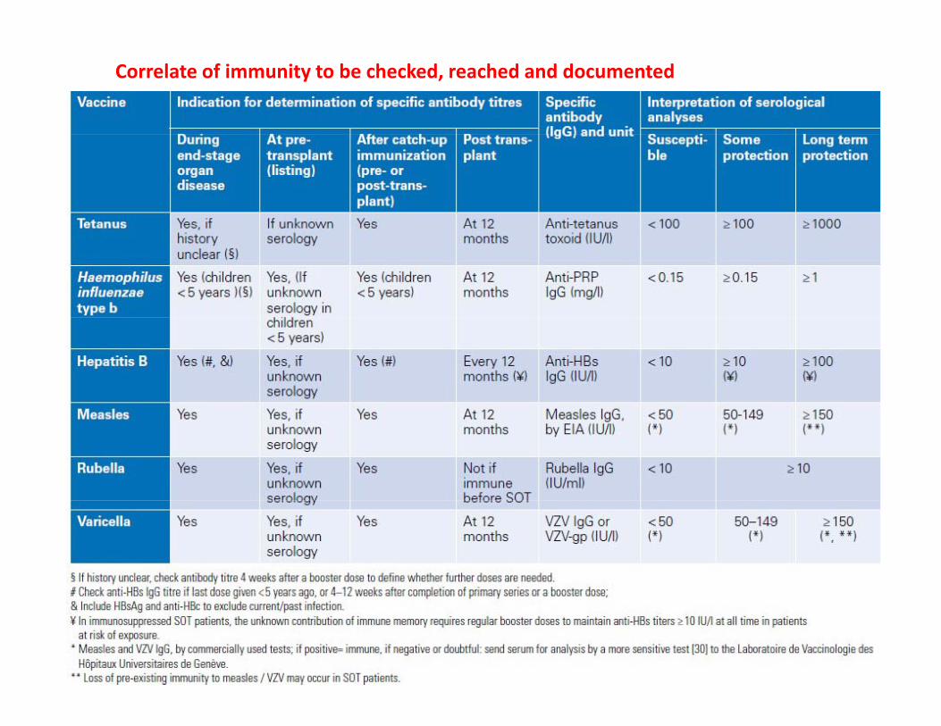
Correlate of immunity to be checked, reached and documented
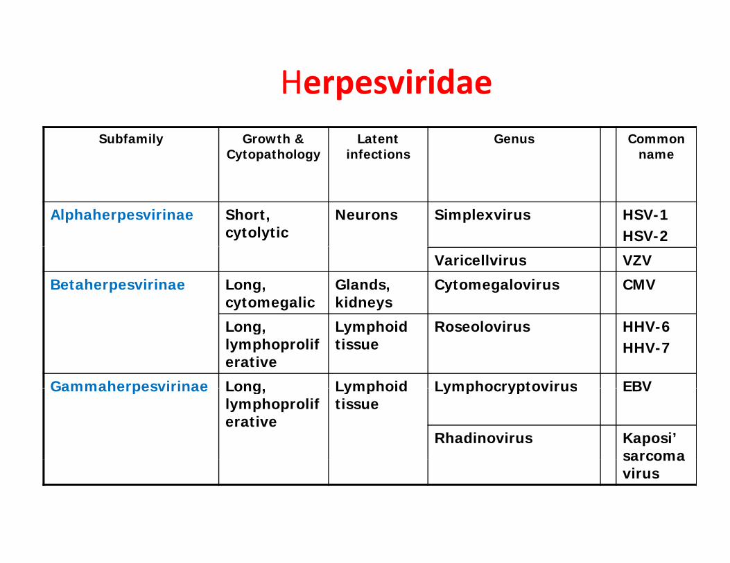
HerpesviridaeSubfamily Growth &
CytopathologyLatent
infectionsGenus Common
name
Alphaherpesvirinae Short, cytolytic
Neurons Simplexvirus HSV-1HSV-2
Varicellvirus VZV
Betaherpesvirinae Long, cytomegalic
Glands, kidneys
Cytomegalovirus CMV
Long, lymphoproliferative
Lymphoid tissue
Roseolovirus HHV-6HHV-7
Gammaherpesvirinae Long Lymphoid Lymphocryptovirus EBVGammaherpesvirinae Long, lymphoproliferative
Lymphoid tissue
Lymphocryptovirus EBV
Rhadinovirus Kaposi’ sarcomasarcoma virus
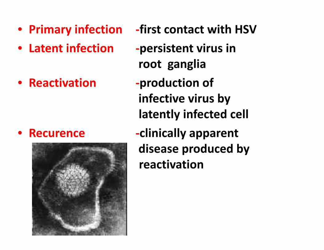
• Primary infection ‐first contact with HSV• Latent infection persistent virus in• Latent infection ‐persistent virus in
root ganglia• Reactivation ‐production of
infective virus by l l f d lllatently infected cell
• Recurence ‐clinically apparent disease produced by reactivation
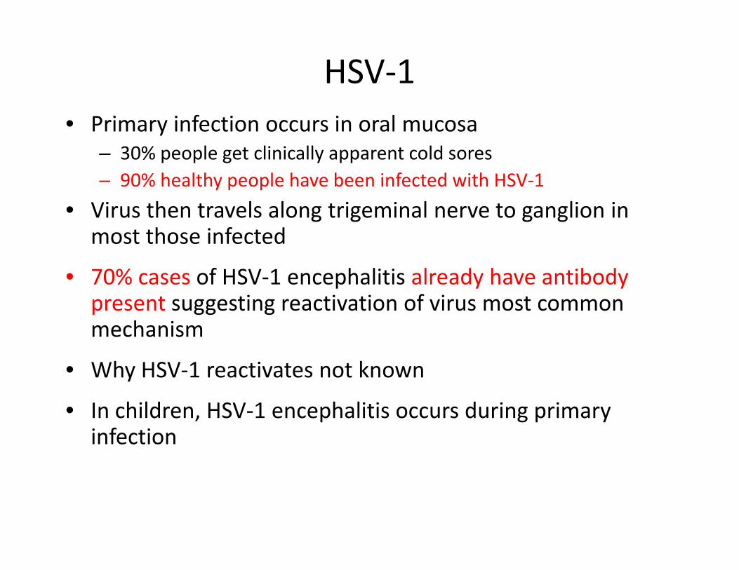
HSV‐1• Primary infection occurs in oral mucosa
– 30% people get clinically apparent cold sores90% healthy people have been infected with HSV 1– 90% healthy people have been infected with HSV‐1
• Virus then travels along trigeminal nerve to ganglion in most those infected
• 70% cases of HSV‐1 encephalitis already have antibody present suggesting reactivation of virus most common
h imechanism
• Why HSV‐1 reactivates not known
• In children, HSV‐1 encephalitis occurs during primary infection
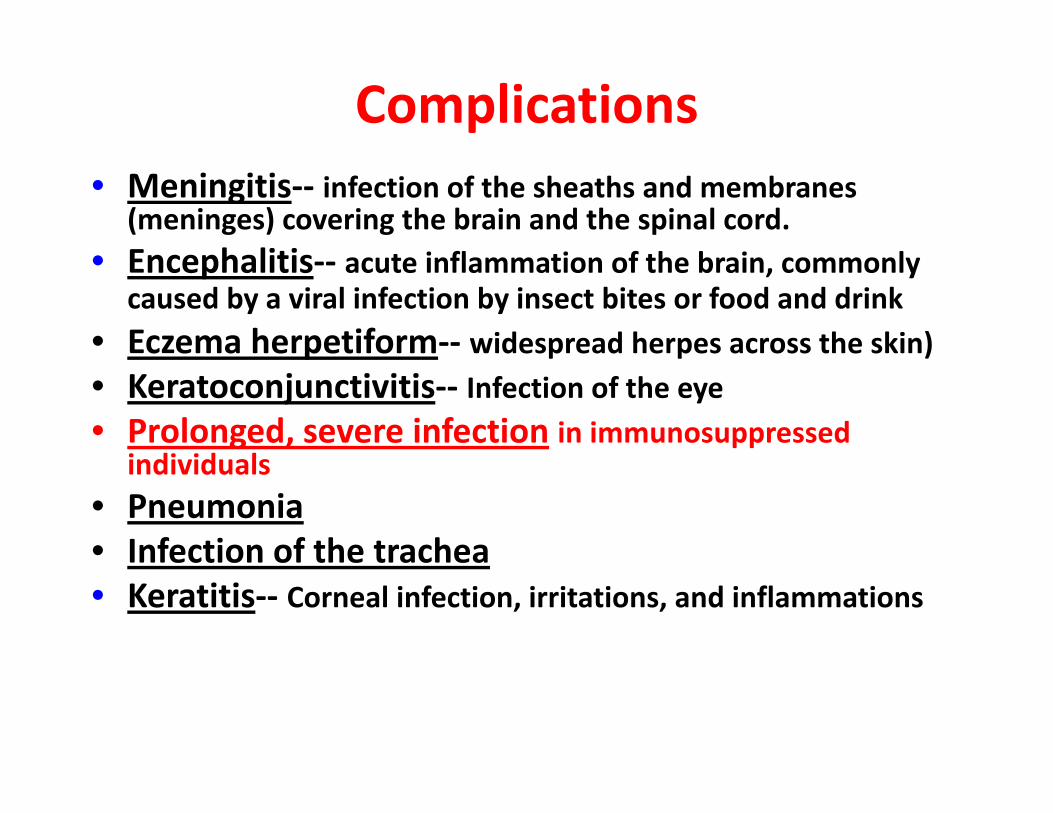
Complications• Meningitis‐‐ infection of the sheaths and membranes
(meninges) covering the brain and the spinal cord. E h liti i fl i f h b i l• Encephalitis‐‐ acute inflammation of the brain, commonly caused by a viral infection by insect bites or food and drink
• Eczema herpetiform‐‐ widespread herpes across the skin) p p p )• Keratoconjunctivitis‐‐ Infection of the eye• Prolonged, severe infection in immunosuppressed
i di id lindividuals • Pneumonia • Infection of the tracheaInfection of the trachea• Keratitis‐‐ Corneal infection, irritations, and inflammations
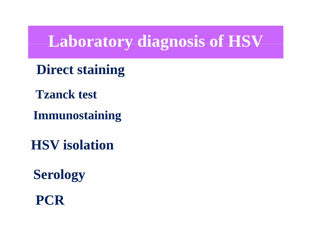
Laboratory diagnosis of HSVLaboratory diagnosis of HSV Direct staining
Tzanck test
ect sta g
Immunostaining
HSV isolation
Serology
PCR
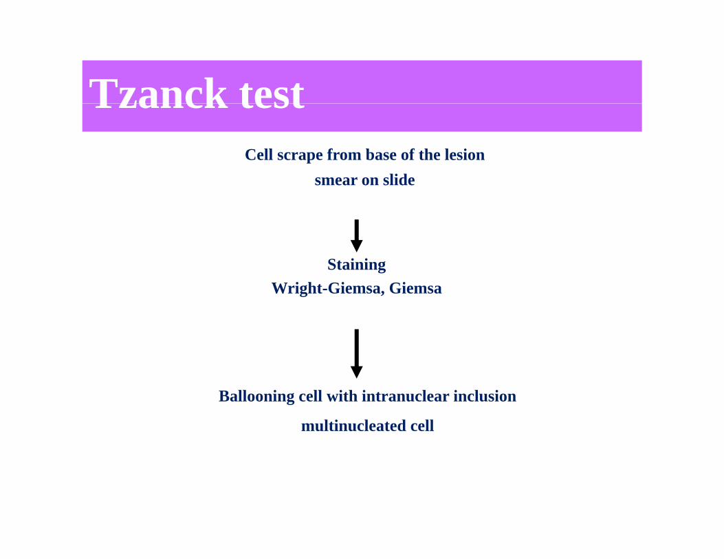
Tzanck testTzanck testCell scrape from base of the lesion
smear on slide
StainingWright-Giemsa, Giemsa
Ballooning cell with intranuclear inclusion
multinucleated cell
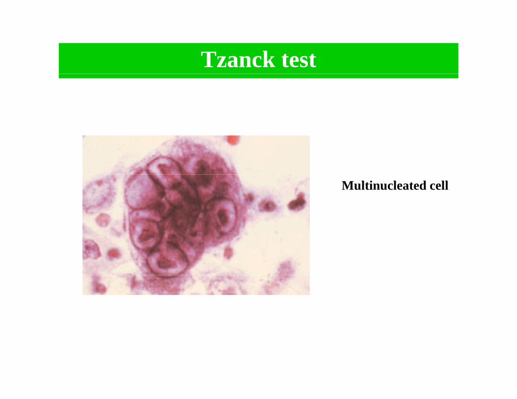
Tzanck test
Multinucleated cell
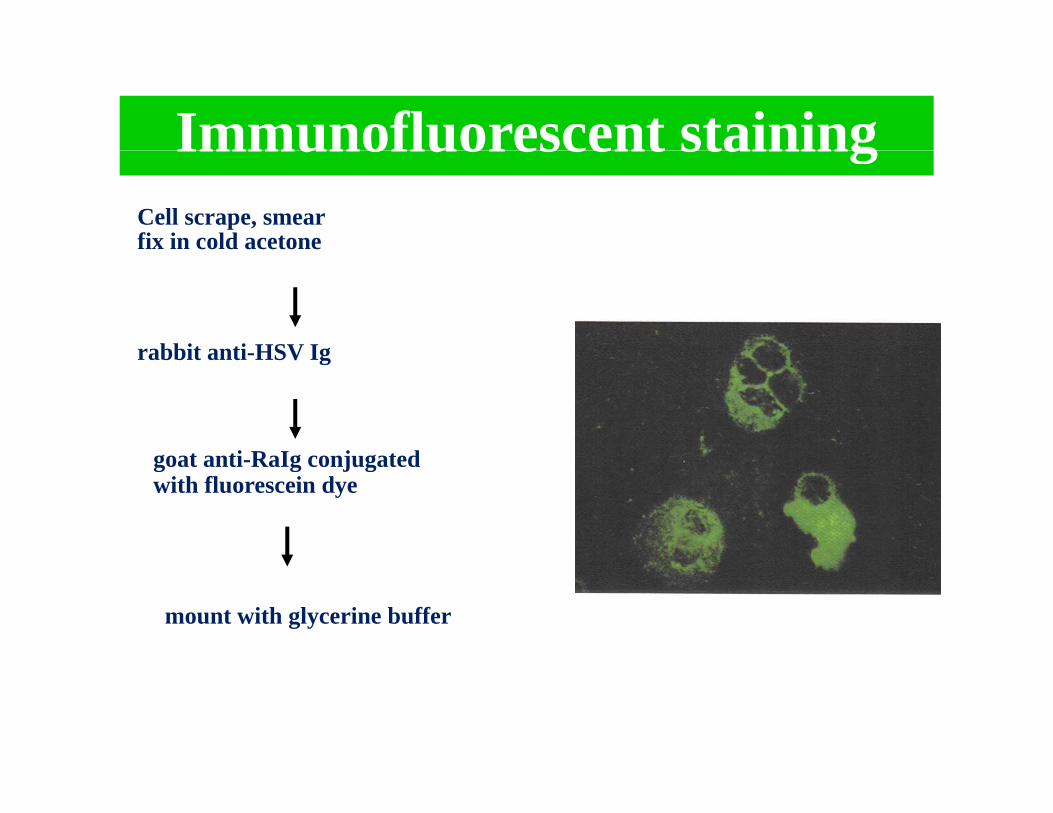
Immunofluorescent stainingImmunofluorescent stainingCell scrape, smear fix in cold acetone
rabbit anti-HSV Ig
goat anti RaIg conjugated
rabbit anti-HSV Ig
goat anti-RaIg conjugated with fluorescein dye
mount with glycerine buffer
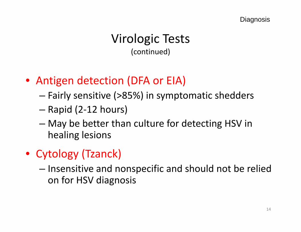
Virologic Tests Diagnosis
o og c ests(continued)
• Antigen detection (DFA or EIA)– Fairly sensitive (>85%) in symptomatic shedders– Rapid (2‐12 hours)– May be better than culture for detecting HSV in healing lesions
• Cytology (Tzanck)– Insensitive and nonspecific and should not be relied on for HSV diagnosis
14
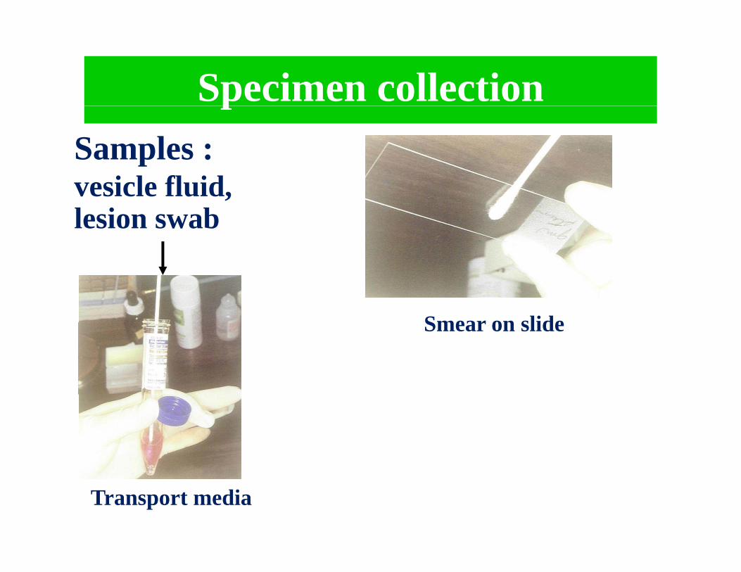
Specimen collectionpSamples :
i l fl idvesicle fluid, lesion swab
Smear on slideSmear on slide
Transport media
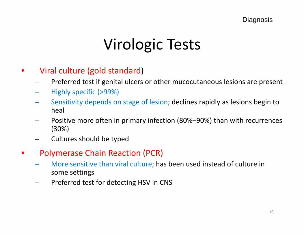
Virologic Tests
Diagnosis
Virologic Tests • Viral culture (gold standard)Viral culture (gold standard)
– Preferred test if genital ulcers or other mucocutaneous lesions are present– Highly specific (>99%) – Sensitivity depends on stage of lesion; declines rapidly as lesions begin toSensitivity depends on stage of lesion; declines rapidly as lesions begin to
heal – Positive more often in primary infection (80%–90%) than with recurrences
(30%) ( )– Cultures should be typed
• Polymerase Chain Reaction (PCR) M iti th i l lt h b d i t d f lt i– More sensitive than viral culture; has been used instead of culture in some settings
– Preferred test for detecting HSV in CNS
16
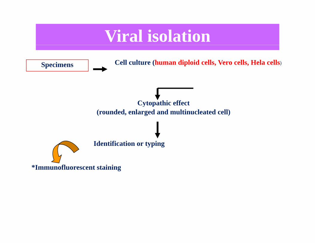
Viral isolationSpecimens Cell culture (human diploid cells, Vero cells, Hela cells)
Cytopathic effect y p(rounded, enlarged and multinucleated cell)
Identification or typing
*Immunofluorescent staining*Immunofluorescent staining

HSV Cytopathic effectHSV Cytopathic effect
Normal cells CPE
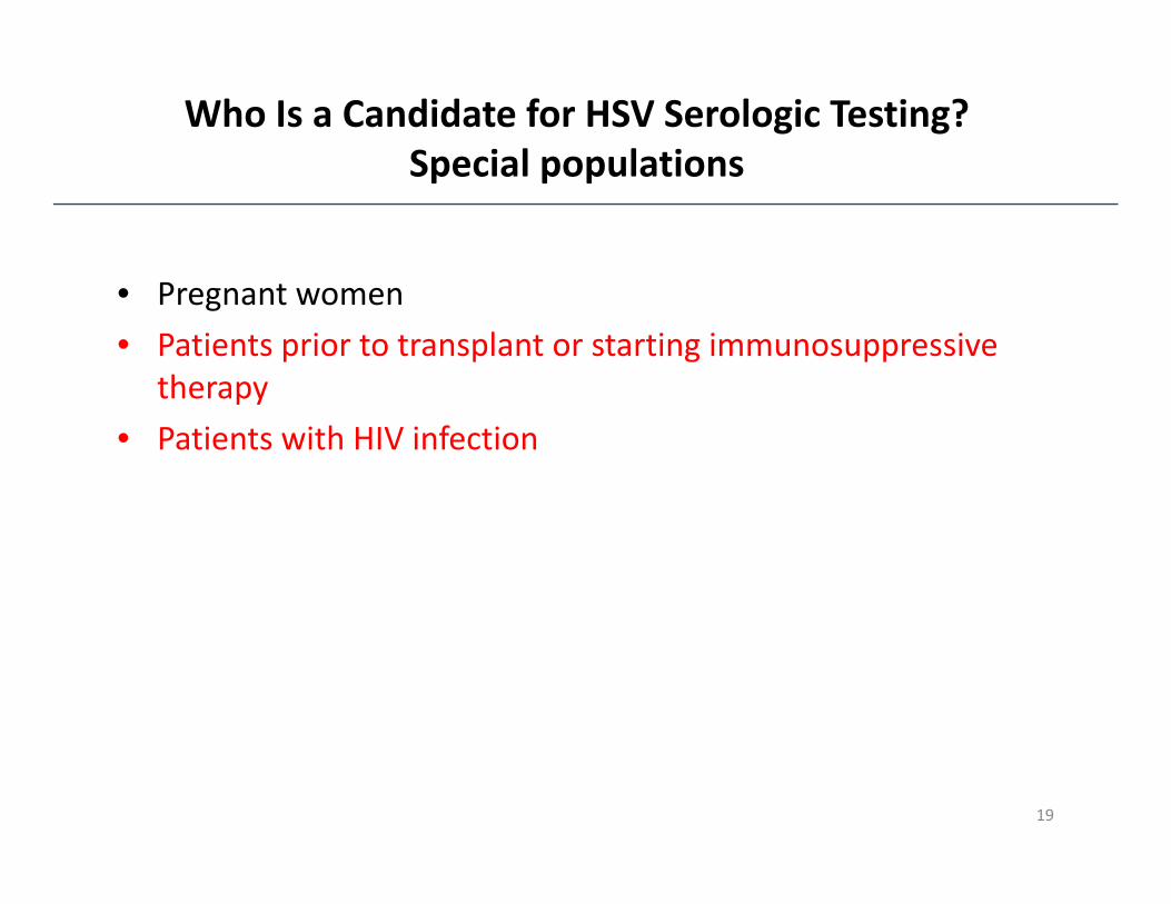
Who Is a Candidate for HSV Serologic Testing?Special populationsSpecial populations
• Pregnant women• Patients prior to transplant or starting immunosuppressive
therapytherapy• Patients with HIV infection
19
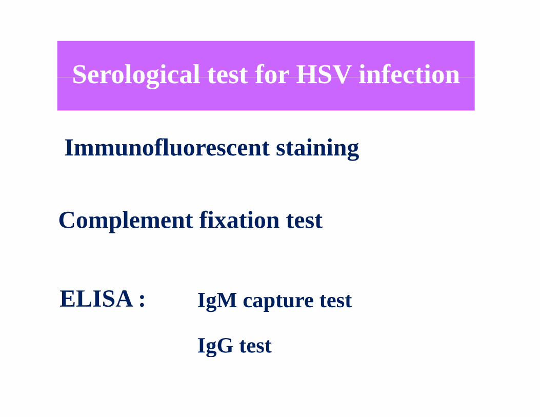
Serological test for HSV infectionSerological test for HSV infection
Immunofluorescent staining
Complement fixation test
I G t t
IgM capture test ELISA :
IgG test
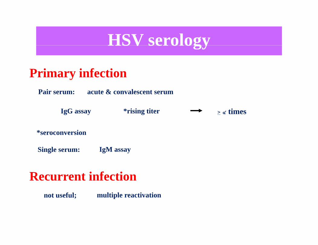
HSV serologyHSV serology
Primary infectionPrimary infection Pair serum: acute & convalescent serum
*rising titer > ๔ times
*seroconversion
IgG assay
IgM assay Single serum:
not useful; multiple reactivation
Recurrent infection
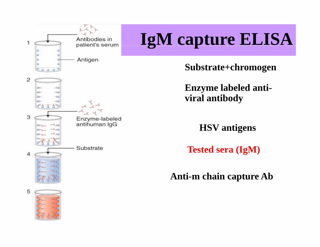
IgM capture ELISAg pSubstrate+chromogen
Enzyme labeled anti-viral antibody
HSV antigens
Tested sera (IgM)
Anti-m chain capture Ab
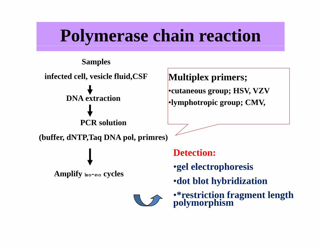
Polymerase chain reactionSamples
infected cell vesicle fluid CSF Multiplex primers;infected cell, vesicle fluid,CSF
DNA extraction
Multiplex primers; •cutaneous group; HSV, VZV•lymphotropic group; CMV,
PCR solution
(buffer, dNTP,Taq DNA pol, primres) (buffer, dNTP,Taq DNA pol, primres)
A lif l
Detection:•gel electrophoresis
Amplify ๒๐-๓๐ cyclesg p
•dot blot hybridization•*restriction fragment length polymorphismpolymorphism
CytomegalovirusCytomegalovirus
• Member of the herpes virus family (EBVMember of the herpes virus family (EBV, varicella‐zoster, herpes simplex)
• Worldwide seroprevalence 30‐100%
• Found in body fluidsBl d li i b ilk– Blood, saliva, urine, breast milk
Types of CMV Infection
• Primary infection (asymptomatic to mononucleosis like syndrome(asymptomatic to mononucleosis like syndrome
in immune competent individuals)• Latent infection (presence of viral genome in• Latent infection (presence of viral genome in mononuclear leukocytes, endothelial cells, and organs in the absence of active replication of infectious virus)
• Reactivation• Reinfection
Laboratory DiagnosisLaboratory Diagnosis1. CMV antigenaemia test - widely used in many European countries.
CMV antigens at the surface of polymorphonuclear leukocytes aredetected by immunoperoxidase or immunofluorescence techniques.A result can be obtained within 4 to 6 hours but the technique isvery tricky.
2. Polymerase chain reaction - becoming the method of choice in afew laboratories, had been reported to carry a higher prognostic, p y g p gvalue for CMV disease than the Detection of Early AntigenFluorescent. Foci (DEAFF) test. Potential problems with sensitivity.
3 Serology not reliable in general but occasionally rises in IgG3. Serology - not reliable in general but occasionally, rises in IgGtitre and the presence of IgM may be seen.
4. viral culture can be insensitive
5. Histopathology

Detection of CMV InfectionDetection of CMV Infection
• Immune status: serology (IgG)Immune status: serology (IgG)• Active infection (viremia)
Hi t l– Histology– Viral culture– Shell vial culture– Antigenemia assay– CMV PCR (qualitative/quantitative)
Laboratory Diagnosisy g`
SerologySerologic tests for antibody to CMV are useful forg y
determining whether a patient had CMV infection in thepast, a determination of great clinical importance for organ
d bl d d d i th t l t l ti fand blood donors, and in the pretransplant evaluation ofprospective transplant recipients.
Antibody tests are not useful in the diagnosis of CMVdisease in the immunocompromised host
Treatment of CMV Infection in Allogeneic Bone M T l P iMarrow Transplant Patients
• Ganciclovir/Foscarnet• Two major treatment approachesTwo major treatment approaches
– Prophylactic treatment – treat all patients at engraftmentengraftment
– Pre‐emptive treatment – monitor patients for viremia and treat when infection detected
• Goal: prevent CMV disease
CMVMonitoringCMV Monitoring
• Patients monitored every 1‐2 weeks for CMVPatients monitored every 1 2 weeks for CMV viremia– Shell vial cultures– Shell vial cultures– Antigenemia assaysCMV PCR– CMV PCR
• Incidence of CMV viremia may vary depending it i t ton monitoring strategy
PCR Based Screening MethodsPCR Based Screening Methods
• PCR is more sensitive than shell vial or antigenemia assays
• Some patients may be pre‐emptively treated unnecessarily using PCR strategies
• Quantitative PCR may be more sensitive than qualitative PCR
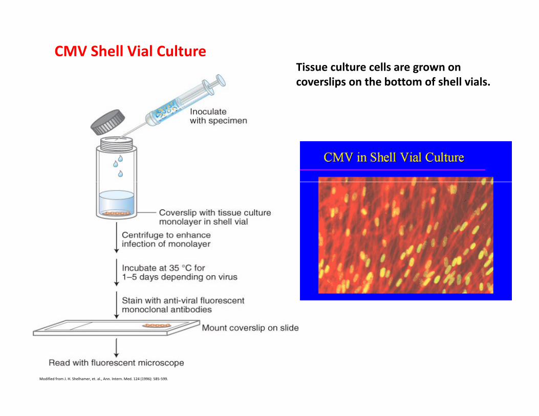
Tissue culture cells are grown on CMV Shell Vial Culture
coverslips on the bottom of shell vials.
Modified from J. H. Shelhamer, et. al., Ann. Intern. Med. 124 (1996): 585‐599.
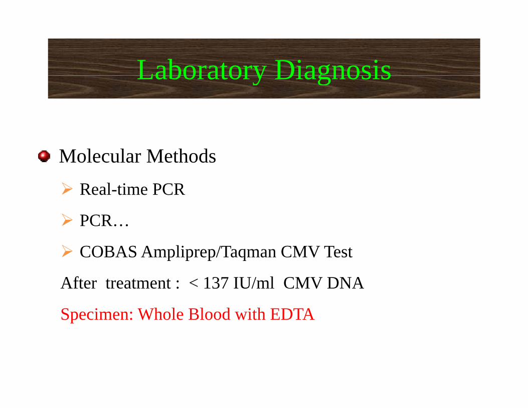
Laboratory DiagnosisLaboratory Diagnosis
Molecular MethodsReal-time PCR
PCR…
COBAS Ampliprep/Taqman CMV Test
After treatment : < 137 IU/ml CMV DNAAfter treatment : < 137 IU/ml CMV DNA
Specimen: Whole Blood with EDTA
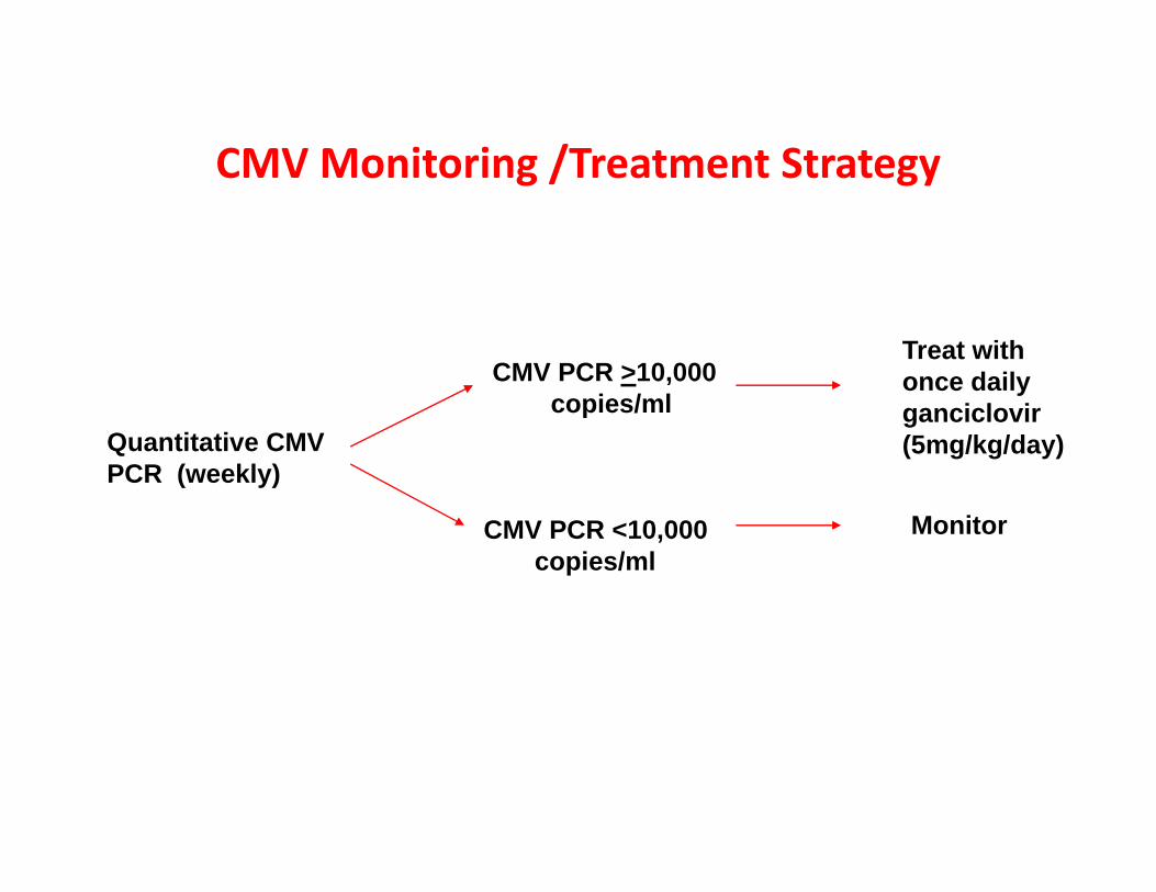
CMVMonitoring /Treatment StrategyCMV Monitoring /Treatment Strategy
CMV PCR >10,000Treat withonce daily
Quantitative CMVPCR (weekly)
copies/mlonce dailyganciclovir(5mg/kg/day)
CMV PCR <10,000copies/ml
Monitor
Patients treated for 21 days
Dose escalation to BID ganciclovir (5mg/kg/BID) for increasing CMV PCR g ( g g ) gon once daily ganciclovir
Successful treatment defined as CMV PCR <2000 copies/ml
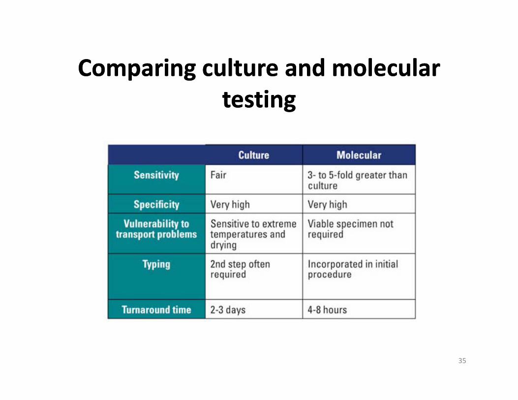
Comparing culture and molecularComparing culture and molecularComparing culture and molecular Comparing culture and molecular testing testing
35
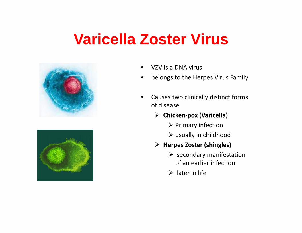
Varicella Zoster VirusVaricella Zoster Virus• VZV is a DNA virus • belongs to the Herpes Virus Family
• Causes two clinically distinct forms of disease.
Chicken‐pox (Varicella) Primary infectionusually in childhood
Herpes Zoster (shingles)secondary manifestation f li i f iof an earlier infectionlater in life
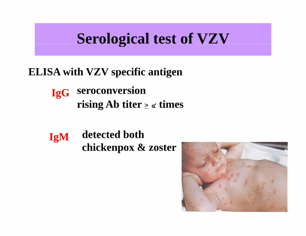
Serological test of VZVSe o og ca test o V V
ELISA with VZV specific antigenELISA with VZV specific antigen
IgG seroconversionrising Ab titer ๔ times
detected both
rising Ab titer > ๔ times
detected bothchickenpox & zoster
IgM

Laboratory diagnosis of VZVLaboratory diagnosis of VZV
S l I f d ll
Direct staining
ballooning cell with Tzanck test
Samples Infected cell scrape
multinucleated cells
gintranuclear inclusion
Tzanck test
fluorescent stainingImmunostaining:

Isolation of VZVIsolation of VZVNasal/throat washing vesicle fluid
Inoculate promptly
Human diploid cell culture
p p y
CPEb ll i l i l d ll
๑-๕ weeks
ballooning,multinucleated cell
Identification: IF
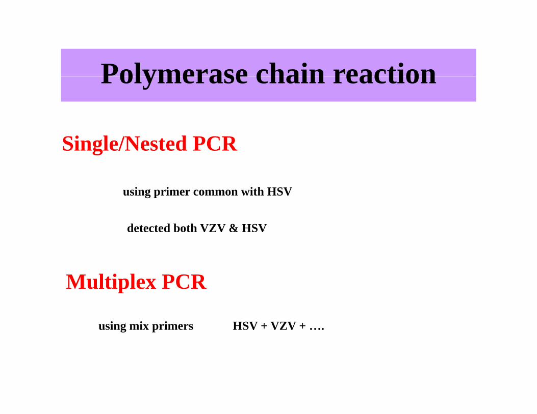
Polymerase chain reactionPolymerase chain reaction
Single/Nested PCR
using primer common with HSV
detected both VZV & HSV
Multiplex PCR
HSV + VZV + ….using mix primers
Multiplex PCR
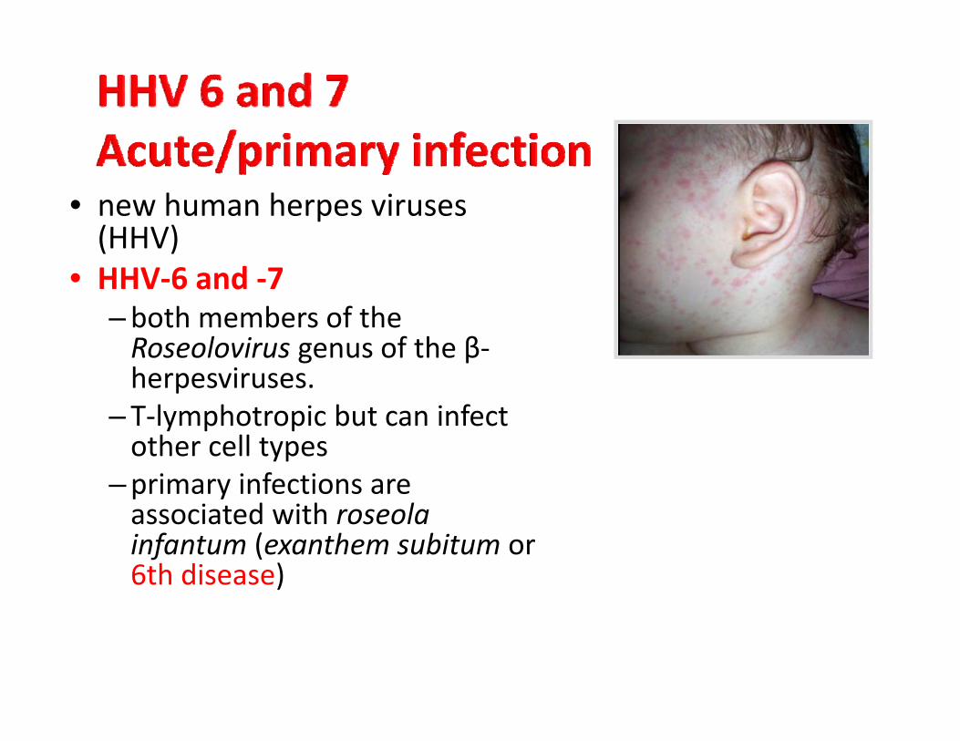
• new human herpes viruses (HHV)(HHV)
• HHV‐6 and ‐7 –both members of theboth members of the Roseolovirus genus of the β‐herpesviruses.
–T‐lymphotropic but can infect–T‐lymphotropic but can infect other cell types
–primary infections are associated with roseolaassociated with roseola infantum (exanthem subitum or 6th disease)
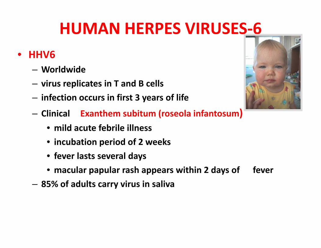
HUMAN HERPES VIRUSES‐6• HHV6
– Worldwide– virus replicates in T and B cells– infection occurs in first 3 years of life
– Clinical Exanthem subitum (roseola infantosum)• mild acute febrile illnessi b ti i d f 2 k• incubation period of 2 weeks
• fever lasts several days• macular papular rash appears within 2 days of fevermacular papular rash appears within 2 days of fever
– 85% of adults carry virus in saliva
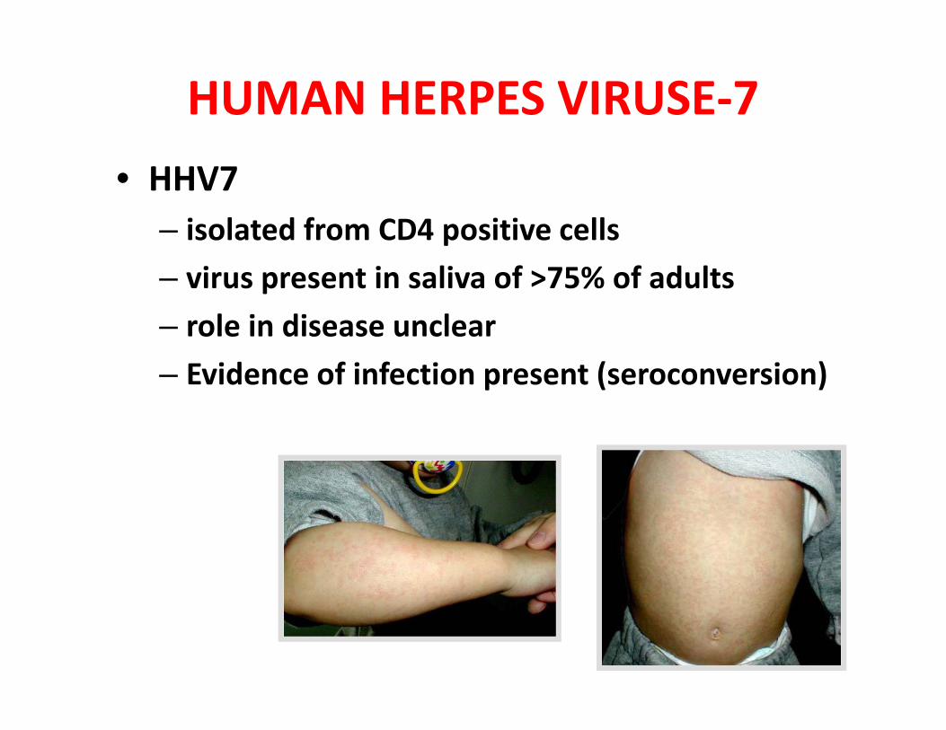
HUMAN HERPES VIRUSE‐7• HHV7
isolated from CD4 positive cells– isolated from CD4 positive cells– virus present in saliva of >75% of adults
l i di l– role in disease unclear– Evidence of infection present (seroconversion)
Human Herpes Virus 6 (HHV6)Human Herpes Virus 6 (HHV6)
• HHV6 associates with febrile convulsions in children under 2 years of age.
• It is a cause of meningitis and encephalitis in immuno‐competent as well as immunocompromised patients.
• In the bone marrow transplant recipient, encephalitis presentation occurs between 10 days to 15 months (median 45 days) after transplantation.
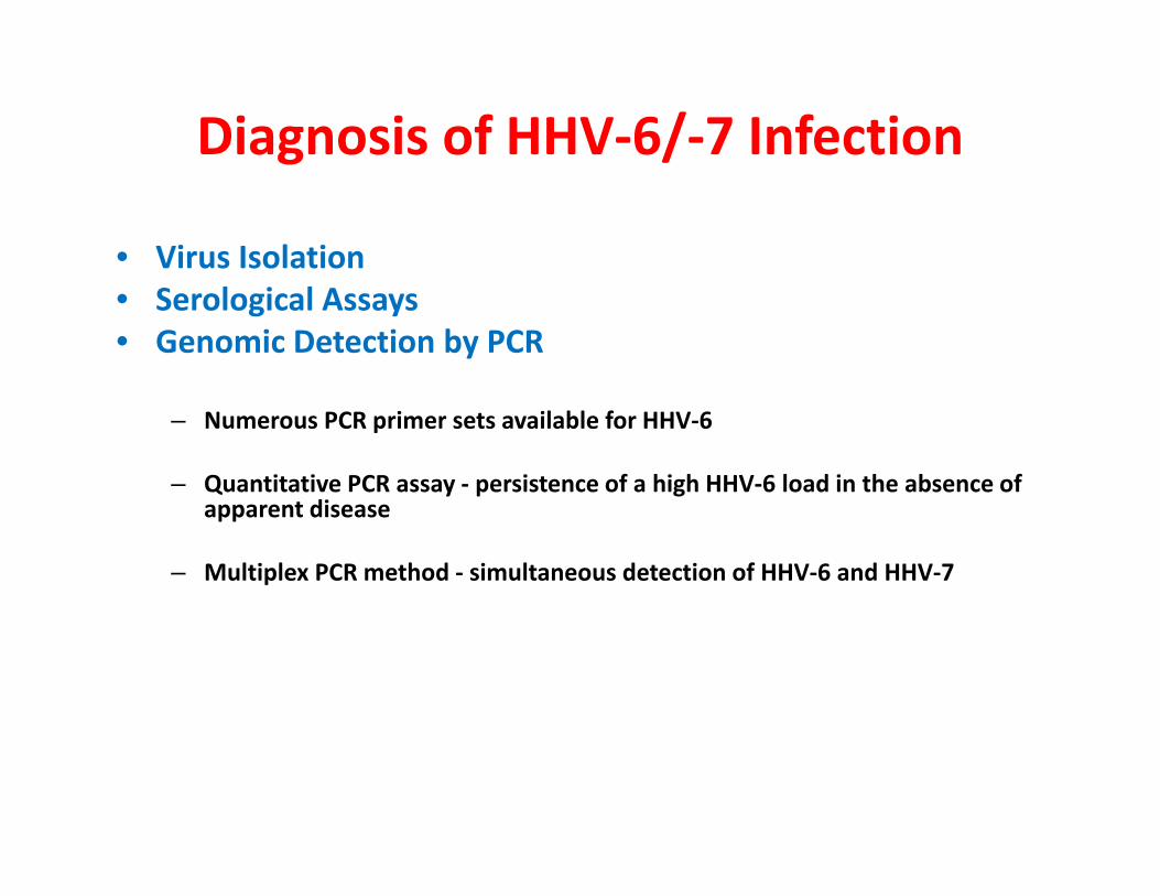
Diagnosis of HHV‐6/‐7 Infection
• Virus Isolation • Serological Assays• Genomic Detection by PCR
– Numerous PCR primer sets available for HHV‐6
– Quantitative PCR assay ‐ persistence of a high HHV‐6 load in the absence of apparent diseaseapparent disease
– Multiplex PCR method ‐ simultaneous detection of HHV‐6 and HHV‐7
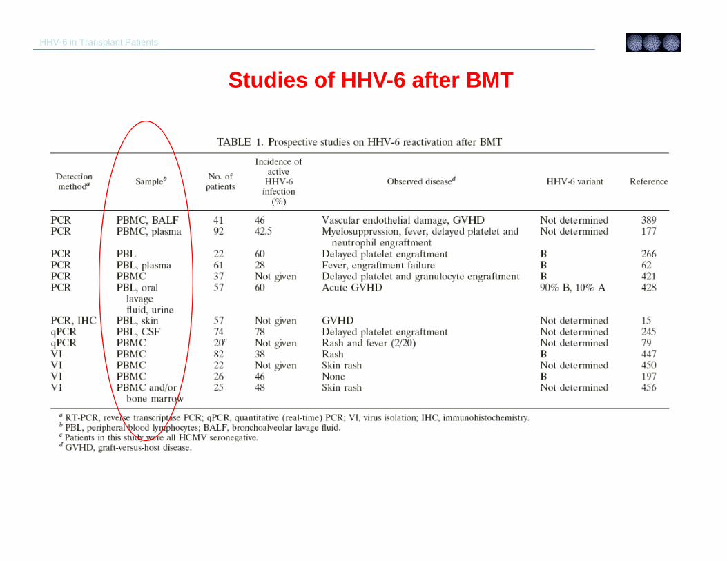
HHV-6 in Transplant Patients
Studies of HHV-6 after BMT
De Bolle, et. al. Clin Microbiol Rev. 2005.Vol 18.
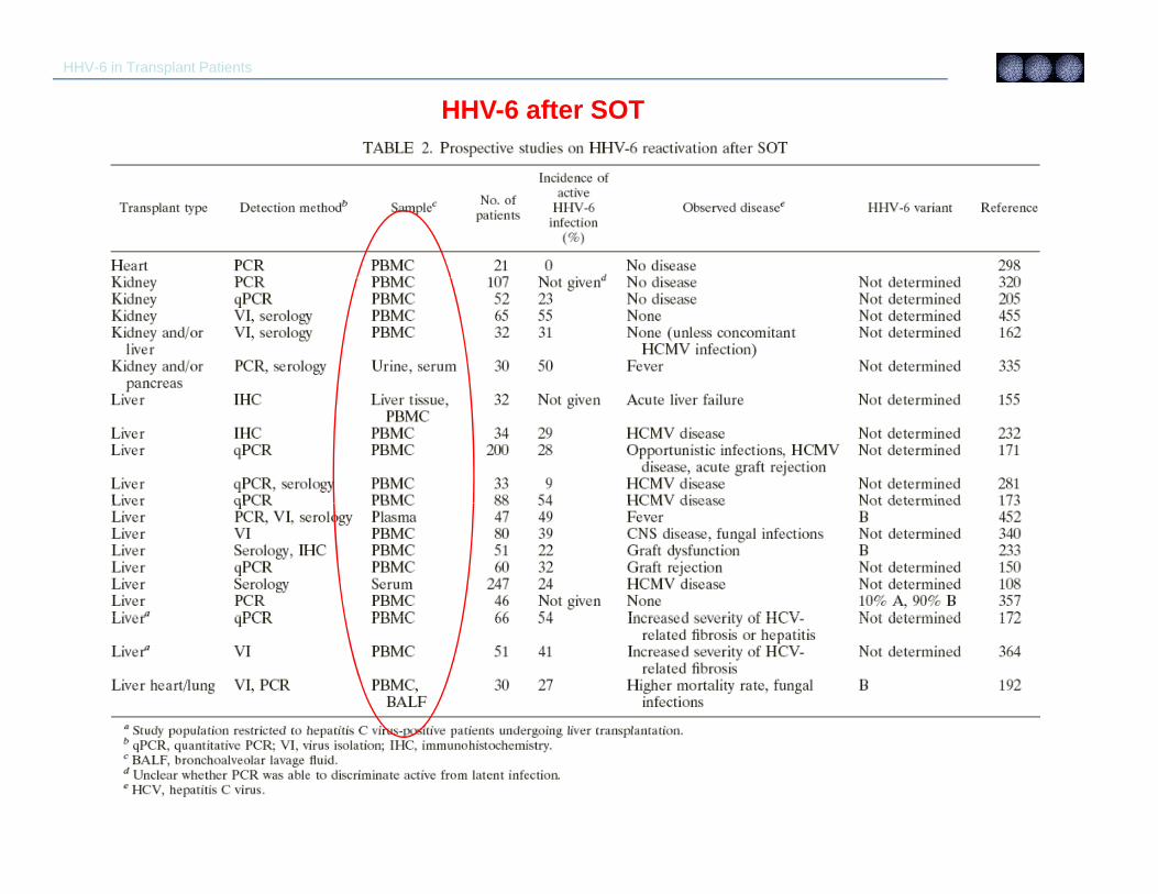
HHV-6 in Transplant Patients
HHV-6 after SOT
De Bolle, et. al. Clin Microbiol Rev. 2005.Vol 18.
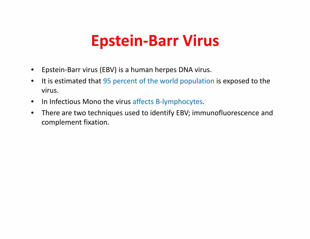
Epstein‐Barr VirusEpstein Barr Virus• Epstein‐Barr virus (EBV) is a human herpes DNA virus.• It is estimated that 95 percent of the world population is exposed to the
virus.• In Infectious Mono the virus affects B‐lymphocytes.y p y• There are two techniques used to identify EBV; immunofluorescence and
complement fixation.
Epstein‐Barr Viral InfectionEpstein Barr Viral Infection• It is a systematic immune complex disease of soluble and
tissue‐fixed antigen involvement characterized by fever, fatigue, chills, headache, myalgia, skin rash, splenomegaly and cervical adenopathy.
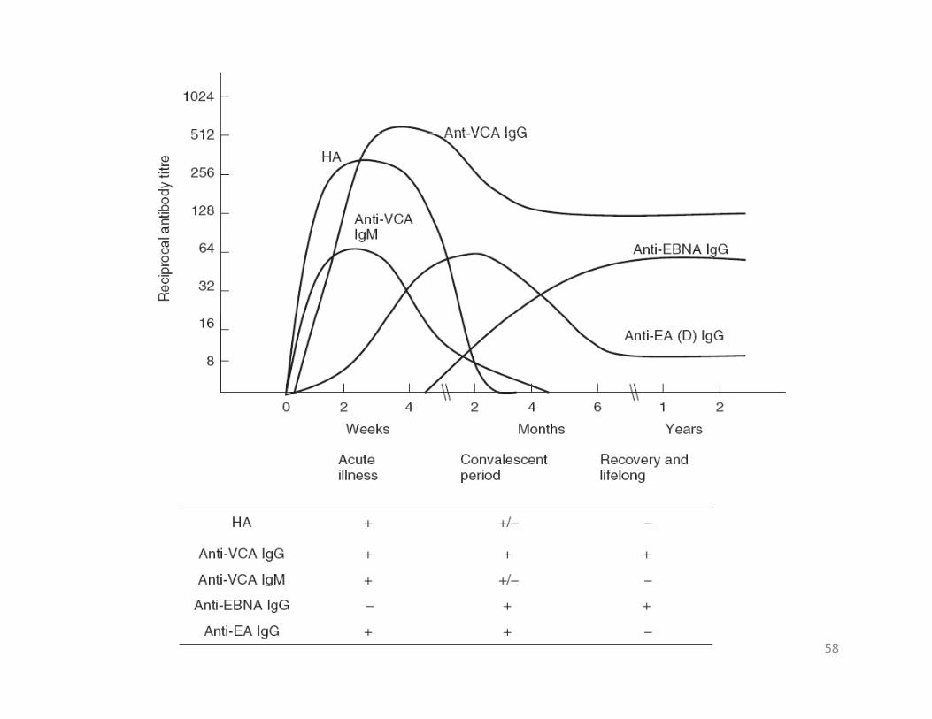
• EBV infected B‐lymphocytes express a variety of “new” antigens encoded by the virus. Infection with EBV results in expression of:expression of: 1. Viral Capsid Antigen (VCA) 2. Early Antigen (EA) 3 Nuclear Antigen (NA)3. Nuclear Antigen (NA) Each antigen expression has corresponding antibody responses.
Epstein‐Barr Virus (VCA)Epstein Barr Virus (VCA)
• Viral capsid antigen (VCA) is produced by infected BViral capsid antigen (VCA) is produced by infected B cells and can be found in the cytoplasm.
• Anti‐VCA IgM is usually detectable early in the g y ycourse of infection, 4 to 7 days after onset of signs and symptoms, but it is low in concentration and disappears within 2 to 4 months.
Epstein‐Barr Virus (EA)Epstein Barr Virus (EA)• Early antigen (EA) is a complex of two components, early antigen‐diffuse
(EA‐D), which is found in both the nucleus and cytoplasm of the B cells, and early antigen‐restricted (EA‐R), which is usually found as a mass only in the cytoplasm.
• Anti‐EA‐D of the IgG type is highly indicative of acute infection, but it is not detectable in 10% to 20% of patients with IM. EA‐D disappears in about 3 months; however, a rise in titer is demonstrated during reactivation of a l i f ilatent EBV infection.
• Anti‐EA‐R IgG is not usually found in young adults during the acute phase. Anti‐EA‐R IgG appears transiently in the later convalescent phase. In
l i EA D d i EA R I G i i di f hgeneral, anti‐EA‐D and anti‐EA‐R IgG are not consistent indicators of the disease stage.
Epstein‐Barr Virus (EBNA)Epstein Barr Virus (EBNA)• Epstein‐Barr nuclear antigen (EBNA) is found in the nucleus of all EBV‐
infected cells. Although the synthesis of NA precedes EA synthesis during the infection of B cells, EBV‐NA does not become available for antibody stimulation until after the incubation period of Infectious Mono, when
d l h d h llactivated T lymphocytes destroy the EBV genome‐carrying B cells. As a result, antibodies to NA are absent or barely detectable during acute IM.
• Anti‐EBNA IgG does not appear until a patient has entered the l i d ib di l l iconvalescent period. EBV‐NA antibodies are almost always present in sera
containing IgG antibodies to VCA of EBV unless the patient is in the early acute phase of IM. Patients with severe immunologic defects or immunosuppressive disease may not have EBV NA antibodies even ifimmunosuppressive disease may not have EBV‐NA antibodies, even if antibodies to VCA are present.
Epstein‐Barr Virus (EBNA)Epstein Barr Virus (EBNA)• Under normal conditions, antibody titers to NA gradually increase through
convalescence and reach a plateau between 3 and 12 months postinfection. The antibody titer remains at a moderate, measurable level indefinitely because of the persistent viral carrier state established f ll ffollowing primary EBV infection.
• Test results of antibodies to EBV‐NA should be evaluated in relationship to patient symptoms, clinical history, and antibody response patterns to EBV‐C d bli h di iVCA and EA to establish a diagnosis.
• The 3 classic criteria for laboratory confirmation 1- lymphocytosis
2- the presence of at least 10% atypical lymphocytes on peripheral smear
3- a positive serologic test for Epstein-Barr virus (EBV).
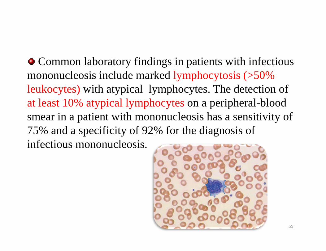
Common laboratory findings in patients with infectious mononucleosis include marked lymphocytosis (>50%mononucleosis include marked lymphocytosis ( 50% leukocytes) with atypical lymphocytes. The detection of at least 10% atypical lymphocytes on a peripheral-blood smear in a patient with mononucleosis has a sensitivity of 75% and a specificity of 92% for the diagnosis of i f ti l iinfectious mononucleosis.
55
• Complete blood count– 40-70%, Leukocytosis
WBC 10 000 20 000 ll )(WBC 10,000-20,000 cells per cm๓)– 80-90% of patients have lymphocytosis,
with greater than 50% lymphocytes Lymphocytosis iswith greater than 50% lymphocytes. Lymphocytosis is greatest during 2-3 weeks of illness and lasts for 2-6 weeks.
– 20-40% of the lymphocytes : atypical lymphocytes > 10% ; Mild h b i– Mild thrombocytopenia
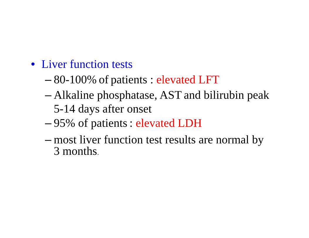
• Liver function tests– 80-100% of patients : elevated LFT
– Alkaline phosphatase, AST and bilirubin peak 5-14 days after onset
– 95% of patients : elevated LDH– most liver function test results are normal by
3 months.
58
تست ها
Anti-
VCA Anti-EA
IgMIgGEA-DEA-RAnti-EBNAهتروفيل بيمارىجدول تغييرات
ھای بيماری مونونوكلئوزسرولوژيک
عفونى حاد+ + +++ - -
سرولوژيک بيماری ھایEBVمرتبط با
+ ± - + - ± دوره نقاهت
قبلي +--+--عفونت + --+-- عفونت قبلي
فعال شدن مجدد --++++ ±
در اثر ضعف ايمنى
+ ++ ±+++ - - لنفوم بوركيت
+ ± +++++ - -كارسينوم
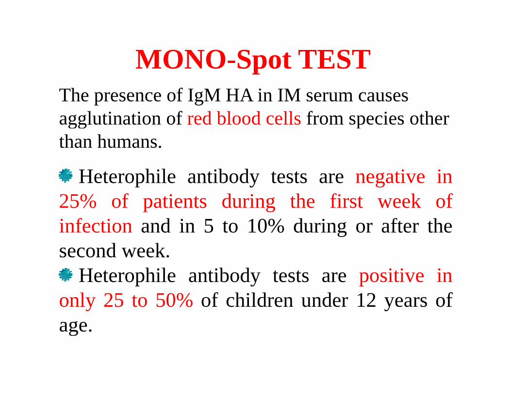
MONO-Spot TESTpThe presence of IgM HA in IM serum causes agglutination of red blood cells from species otheragglutination of red blood cells from species other than humans.
H t hil tib d t t ti iHeterophile antibody tests are negative in25% of patients during the first week ofi f ti d i 5 t 10% d i ft thinfection and in 5 to 10% during or after thesecond week.
H t hil tib d t t iti iHeterophile antibody tests are positive inonly 25 to 50% of children under 12 years ofage.
IM; laboratory diagnosisIM; laboratory diagnosisIM; laboratory diagnosisIM; laboratory diagnosis
– Real‐time PCR• Real‐time PCR is when the amplified DNA is detected as the reaction progresses in real p gtime. Test has 95% sensitivity and 97% specificity for primary EBV infection.
• Is expensive and not commonly used in clinical practice.
• Test can be useful for diagnosis of serologically indeterminate EBV infections.
HUMAN HERPES VIRUSE‐8• HHV8K i i t d H Vi• Kaposi sarcoma‐ associated Herpes Virus (KSHV)
d d i i h li l ll f i– detected in epithelial cells of Kaposi sarcoma– also present in semen– postulated as cause of Kaposi sarcomaDiagnosisS l i l ( S )• Serological Assays (ELISA)
• Genomic Detection by PCR• Indirect IF