Laboratory Tests for Diagnosis of Gastrointestinal and ... · PDF fileTOPICAL REVIEW...
12
TOPICAL REVIEW Laboratory Tests for Diagnosis of Gastrointestinal and Pancreatic Diseases Olivier Dossin, DVM, PhD, Dipl. ECVIM-CA The panel of laboratory tests available for diagnosis of gastrointestinal (GI) diseases in dogs and cats is wide, and, recently, several new tests have been developed. This article will focus on advances in laboratory tests that are available for the general practitioner for diagnosis of GI diseases. Laboratory tests for diagnosis of gastric and intestinal infectious diseases include fecal parasite screening tests, enzyme-linked immunosorbent assays for parvoviral enteritis, and some specific bacterial tests like fluorescent in situ hybridization for identification of specific bacteria attached to the intestinal epithelial cells. Serum concentrations of folate and cobalamin are markers of intestinal absorption, but are also changed in exocrine pancreatic insufficiency and intestinal bacterial overgrowth. Hypocobalaminemia is common in GI and pancreatic disease. Decreased serum trypsin- like immunoreactivity is a very sensitive and specific test for the diagnosis of exocrine pancreatic insufficiency in dogs and cats. Serum pancreatic lipase is currently the most sensitive and specific test to identify pancreatic cell damage and acute pancreatitis. However, serum canine pancreas-specific lipase is less sensitive in canine chronic pancreatitis. Increased serum trypsin-like immunoreactivity is also specific for pancreatic damage but is less sensitive. It is very likely that further studies will help to better specify the role of these new tests in the diagnosis of canine and feline pancreatic diseases. © 2011 Elsevier Inc. All rights reserved. Keywords: laboratory tests, intestinal diseases, pancreas, folate, cobalamin, pancreatic lipase, bacteria G astrointestinal (GI) and pancreatic diseases are fre- quently encountered in canine and feline medicine. The panel of laboratory tests available is wide, and recently sev- eral new tests have been developed to help veterinarians in their workup of such cases. These tests can help characterize infectious agents, functional changes, or organ damage, and are often the first step before identification of a lesion with a biopsy. This review is limited to tests that can be performed on blood or fecal samples. Moreover, the reader is referred to a review on fecal cytology in a previous issue of this journal for further information. 1 Tests for Gastrointestinal Infectious Diseases Fecal Parasite Panel GI parasites are a common cause of chronic GI disorders in dogs and cats. A routine fecal flotation should always be included in the early diagnostic workup of an animal with GI signs to screen for worms and common protozoans such as giardia and coccidia. A detailed review on the diagnostic methods of intestinal protozoans in dogs and cat is beyond the scope of this article, but the reader is referred to a recent issue of this journal devoted to protozoal diseases in small animals (2010, Vol 25, issue 3). Fecal shedding of GI parasites can be intermittent and some clinically normal animals can shed parasites without showing clinical signs. Therefore, a negative test should al- ways be confirmed, ideally by 2 additional fecal panels. A positive diagnostic test does not prove that the parasite is causing disease, but it is usually advised to treat before em- barking on invasive and/or expensive tests (unless some other findings warrant immediate diagnostic workup). Bacteria Characterization of bacterial diarrhea is difficult in dogs and cats, mostly because healthy pets are usually carriers of important pathogenic bacteria such as Clostridium perfrin- gens, Clostridium difficile, Escherichia coli, and Campylo- bacter spp. 2-4 However, it might be important to screen for possible pathogenic bacteria in cases with acute diarrhea, especially because some agents such as Campylobacter, Clos- tridium, or enteropathogenic E. coli have contagious or zoo- notic potential. Clostridium perfringens may induce diarrhea via its cyto- toxic entorotoxin (CPE). This toxin is produced during spo- rulation and released during lysis of vegetative cells. How- From Internal Medicine, Department of Clinical Sciences, National Veteri- nary School, Toulouse, France. Address reprint requests to: Olivier Dossin, DVM, PhD, Dipl. ECVIM-CA, Internal Medicine, Department of Clinical Sciences, National Veterinary School, 23 Chemin des Capelles, 31076 Toulouse Cedex, France. E-mail: [email protected]. © 2011 Elsevier Inc. All rights reserved. 1527-3369/06/0604-0171\.00/0 doi:10.1053/j.tcam.2011.02.005 86
-
Upload
nguyenhuong -
Category
Documents
-
view
232 -
download
4
Transcript of Laboratory Tests for Diagnosis of Gastrointestinal and ... · PDF fileTOPICAL REVIEW...

TOPICAL REVIEW
Laboratory Tests for Diagnosis of Gastrointestinal andPancreatic Diseases
Olivier Dossin, DVM, PhD, Dipl. ECVIM-CAThe panel of laboratory tests available for diagnosis of gastrointestinal (GI) diseases in dogs and cats is wide,and, recently, several new tests have been developed. This article will focus on advances in laboratory tests thatare available for the general practitioner for diagnosis of GI diseases. Laboratory tests for diagnosis of gastricand intestinal infectious diseases include fecal parasite screening tests, enzyme-linked immunosorbent assaysfor parvoviral enteritis, and some specific bacterial tests like fluorescent in situ hybridization for identificationof specific bacteria attached to the intestinal epithelial cells. Serum concentrations of folate and cobalamin aremarkers of intestinal absorption, but are also changed in exocrine pancreatic insufficiency and intestinalbacterial overgrowth. Hypocobalaminemia is common in GI and pancreatic disease. Decreased serum trypsin-like immunoreactivity is a very sensitive and specific test for the diagnosis of exocrine pancreatic insufficiencyin dogs and cats. Serum pancreatic lipase is currently the most sensitive and specific test to identify pancreaticcell damage and acute pancreatitis. However, serum canine pancreas-specific lipase is less sensitive in caninechronic pancreatitis. Increased serum trypsin-like immunoreactivity is also specific for pancreatic damage butis less sensitive. It is very likely that further studies will help to better specify the role of these new tests in thediagnosis of canine and feline pancreatic diseases.© 2011 Elsevier Inc. All rights reserved.
Keywords: laboratory tests, intestinal diseases, pancreas, folate, cobalamin, pancreatic lipase, bacteria
Gastrointestinal (GI) and pancreatic diseases are fre-quently encountered in canine and feline medicine. The
panel of laboratory tests available is wide, and recently sev-eral new tests have been developed to help veterinarians intheir workup of such cases. These tests can help characterizeinfectious agents, functional changes, or organ damage, andare often the first step before identification of a lesion with abiopsy.
This review is limited to tests that can be performed onblood or fecal samples. Moreover, the reader is referred to areview on fecal cytology in a previous issue of this journal forfurther information.1
Tests for Gastrointestinal Infectious Diseases
Fecal Parasite Panel
GI parasites are a common cause of chronic GI disorders indogs and cats. A routine fecal flotation should always beincluded in the early diagnostic workup of an animal with GI
From Internal Medicine, Department of Clinical Sciences, National Veteri-nary School, Toulouse, France.Address reprint requests to: Olivier Dossin, DVM, PhD, Dipl. ECVIM-CA,Internal Medicine, Department of Clinical Sciences, National VeterinarySchool, 23 Chemin des Capelles, 31076 Toulouse Cedex, France. E-mail:[email protected].© 2011 Elsevier Inc. All rights reserved.1527-3369/06/0604-0171\.00/0
doi:10.1053/j.tcam.2011.02.00586
signs to screen for worms and common protozoans such asgiardia and coccidia. A detailed review on the diagnosticmethods of intestinal protozoans in dogs and cat is beyondthe scope of this article, but the reader is referred to a recentissue of this journal devoted to protozoal diseases in smallanimals (2010, Vol 25, issue 3).
Fecal shedding of GI parasites can be intermittent andsome clinically normal animals can shed parasites withoutshowing clinical signs. Therefore, a negative test should al-ways be confirmed, ideally by 2 additional fecal panels. Apositive diagnostic test does not prove that the parasite iscausing disease, but it is usually advised to treat before em-barking on invasive and/or expensive tests (unless some otherfindings warrant immediate diagnostic workup).
Bacteria
Characterization of bacterial diarrhea is difficult in dogsand cats, mostly because healthy pets are usually carriers ofimportant pathogenic bacteria such as Clostridium perfrin-gens, Clostridium difficile, Escherichia coli, and Campylo-bacter spp.2-4 However, it might be important to screen forpossible pathogenic bacteria in cases with acute diarrhea,especially because some agents such as Campylobacter, Clos-tridium, or enteropathogenic E. coli have contagious or zoo-notic potential.
Clostridium perfringens may induce diarrhea via its cyto-toxic entorotoxin (CPE). This toxin is produced during spo-
rulation and released during lysis of vegetative cells. How-
tCpc(a
p
tdpttdhcbtra(
rd
n(f
md
tbtuptflo
cp
Volume 26, Number 2, May 2011 87
ever, detection of endospores on a fecal smear does notcorrelate with presence of CPE nor is it associated with clin-ical signs of diarrhea in dogs.4,5 Therefore, the only usefulests to identify C. perfringens–associated diarrhea are fecalPE enzyme-linked immunosorbent assay (ELISA; reverseassive latex agglutination tests are not reliable) or identifi-ation of toxinogenic strains via polymerase chain reactionPCR).4,6 Fecal culture is not helpful because the bacteria islso isolated from healthy dogs.6
Clostridium difficile is another pathogen associated withdiarrhea. It is especially prevalent with hospital-acquired di-arrhea and secondary to antibiotic treatment.7 Both CPE andC. difficile toxin A measured by ELISA were strongly associ-ated with acute hemorrhagic diarrheal syndrome in dogs.8
Five commercially available ELISAs for toxins A and B of C.difficile were reported to have low sensitivity (up to twothirds of false negatives) on fecal samples but very high sen-sitivity when performed on bacterial isolates.9 However, thespecificities were high (i.e., there are limited numbers of false-positive results).
Campylobacter spp. have been identified in dogs and catswith acute diarrhea,10 but they are also found with similarrevalence in feces of clinically normal animals.2,11,12 There-
fore, a positive culture result for Campylobacter does notprove disease association with the infectious agent. However,the potential risk for infectious or zoonotic disease may war-rant treatment when the pet is exhibiting clinical signs. Iden-tification of spiral- or seagull wing–shaped bacteria in a fecalsmear might suggest the presence of Campylobacter, but thisis not a sensitive nor a specific test. Only half of the dogs withpositive culture had characteristic organisms on the fecalsmear in one study, and other spiral-shaped bacteria such asHelicobacter spp. can be observed on the fecal smear.8 Re-cently, PCR tests for identification of C. jejuni and C. coliwere made available (http://www.vetmed.tamu.edu/gilab);however, other Campylobacter species have been recoveredfrom feline or canine feces.3,11,13
Escherichia coli can be associated with acute diarrhea indogs either as the sole pathogen or in association with intes-tinal viruses or parasites.14 However, because E. coli is amajor component of the normal intestinal flora,15 fecal cul-ures are unhelpful in incriminating E. coli as the cause of theiarrhea. Therefore, when E. coli–associated diarrhea is sus-ected, screening for specific toxins or genes (e.g., Shigaoxin,16 attaching and effacing A gene,14,17 etc.) is manda-ory.15 It was recently reported that granulomatous colitis inogs is associated with the presence of an invasive and ad-erent strain of E. coli in the colonic mucosa.18 A diagnosisan be made by specific labeling of the bacteria in coloniciopsy samples by fluorescence in situ hybridization (FISH);he recommended treatment is fluorinated quinolones. Accu-ate diagnosis is important in these cases because inappropri-te use of antibiotics may induce severe antibiotic resistanceincluding to fluorinated quinolones).19 The FISH test is com-
mercially available at the Simpson Laboratory at Cornell
University (http://www.vet.cornell.edu/labs/simpson/).Salmonellosis seems extremely rare in dogs.10 The isolationate of Salmonella spp. in feces of normal dogs and dogs withiarrhea is not different.2,8 However, recently S. enterica was
isolated in 93% of the fecal samples tested in a breedingfacility where 3 puppies developed fatal clinical salmonello-sis.20 The endemic circulation of the same strain of Salmo-ella in this breeding facility was attributed to the raw meatclassified as unfit for human consumption) that was used toeed the dogs.20
In conclusion, unless specific conditions that warrant asearch for bacterial diarrhea are present (outbreak of di-arrhea in a shelter or veterinary hospital or risk of zoono-sis), routine fecal culture is not recommended in dogs andcats with GI signs. Identification of Clostridium toxins
ay help to diagnose hospital-acquired diarrhea due to C.ifficile.Finally, gastric Helicobacter spp. are normally present in
he stomach of dogs and cats and there is no clear associationetween their presence and disease. Therefore, diagnosticests such as rapid urease tests on gastric biopsy are not veryseful in the clinical management of chronic GI cases. Histo-athological identification of massive colonization of the gas-ric mucosa by spiral-shaped bacteria in association with in-ammatory cells is usually enough to decide whether to treatr not for Helicobacter infection.
Viruses
The main viruses that a clinician may want to identify ina dog or cat with acute diarrhea are canine and felineparvoviruses because they are very resistant and highlycontagious. Several point-of-care tests are available for theidentification of canine parvovirus antigen in the feces(SNAP Parvo, IDEXX, Witness Parvo, Synbiotics). Theirspecificity is very high, but a false positive can occur withrecent vaccination (less than 3 weeks). Their sensitivity isvariable.21 Therefore, if canine parvovirus enteritis is sus-pected, a negative test should not be used to rule out thedisease. Recently, these tests have been validated for thediagnosis of feline parvovirus. The reported positive andnegative predictive values are 100% and 97%, respec-tively, for the 2 above-mentioned tests.22 However, a re-ent modified live parvovirus vaccination may induce aositive test for at least 2 weeks.23 Most of the time, serol-
ogy is difficult to interpret unless the titers are very high orincreasing titers can be proved in paired samples. Conven-tional or quantitative PCR tests for parvovirus and coro-navirus are available in several commercial laboratoriesand can be used to confirm negative results with a rapidtest and confirm strong clinical suspicions.
Fungal agents can be characterized with rectal scrapes orfine-needle aspiration of digestive masses. A urinary antigentest is available to screen for histoplasmosis (http://www.miravistalabs.com), but its diagnostic performances have not
been evaluated yet.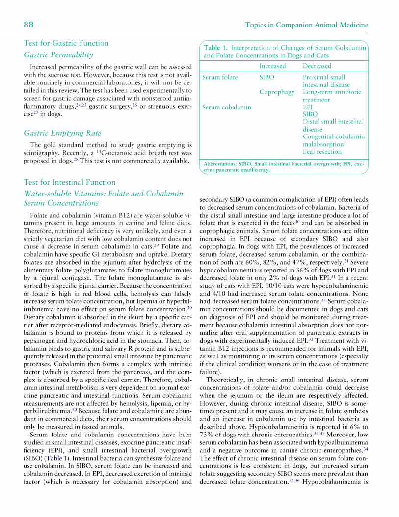
c
hd
mommdtaif
cwHtad7
88 Topics in Companion Animal Medicine
Test for Gastric Function
Gastric Permeability
Increased permeability of the gastric wall can be assessedwith the sucrose test. However, because this test is not avail-able routinely in commercial laboratories, it will not be de-tailed in this review. The test has been used experimentally toscreen for gastric damage associated with nonsteroid antiin-flammatory drugs,24,25 gastric surgery,26 or strenuous exer-ise27 in dogs.
Gastric Emptying Rate
The gold standard method to study gastric emptying isscintigraphy. Recently, a 13C-octanoic acid breath test wasproposed in dogs.28 This test is not commercially available.
Test for Intestinal Function
Water-soluble Vitamins: Folate and CobalaminSerum Concentrations
Folate and cobalamin (vitamin B12) are water-soluble vi-tamins present in large amounts in canine and feline diets.Therefore, nutritional deficiency is very unlikely, and even astrictly vegetarian diet with low cobalamin content does notcause a decrease in serum cobalamin in cats.29 Folate andcobalamin have specific GI metabolism and uptake. Dietaryfolates are absorbed in the jejunum after hydrolysis of thealimentary folate polyglutamates to folate monoglutamatesby a jejunal conjugase. The folate monoglutamate is ab-sorbed by a specific jejunal carrier. Because the concentrationof folate is high in red blood cells, hemolysis can falselyincrease serum folate concentration, but lipemia or hyperbil-irubinemia have no effect on serum folate concentration.30
Dietary cobalamin is absorbed in the ileum by a specific car-rier after receptor-mediated endocytosis. Briefly, dietary co-balamin is bound to proteins from which it is released bypepsinogen and hydrochloric acid in the stomach. Then, co-balamin binds to gastric and salivary R protein and is subse-quently released in the proximal small intestine by pancreaticproteases. Cobalamin then forms a complex with intrinsicfactor (which is excreted from the pancreas), and the com-plex is absorbed by a specific ileal carrier. Therefore, cobal-amin intestinal metabolism is very dependent on normal exo-crine pancreatic and intestinal functions. Serum cobalaminmeasurements are not affected by hemolysis, lipemia, or hy-perbilirubinemia.30 Because folate and cobalamine are abun-dant in commercial diets, their serum concentrations shouldonly be measured in fasted animals.
Serum folate and cobalamin concentrations have beenstudied in small intestinal diseases, exocrine pancreatic insuf-ficiency (EPI), and small intestinal bacterial overgrowth(SIBO) (Table 1). Intestinal bacteria can synthesize folate anduse cobalamin. In SIBO, serum folate can be increased andcobalamin decreased. In EPI, decreased excretion of intrinsic
factor (which is necessary for cobalamin absorption) andsecondary SIBO (a common complication of EPI) often leadsto decreased serum concentrations of cobalamin. Bacteria ofthe distal small intestine and large intestine produce a lot offolate that is excreted in the feces30 and can be absorbed incoprophagic animals. Serum folate concentrations are oftenincreased in EPI because of secondary SIBO and alsocoprophagia. In dogs with EPI, the prevalences of increasedserum folate, decreased serum cobalamin, or the combina-tion of both are 60%, 82%, and 47%, respectively.31 Severeypocobalaminemia is reported in 36% of dogs with EPI andecreased folate in only 2% of dogs with EPI.31 In a recent
study of cats with EPI, 10/10 cats were hypocobalaminemicand 4/10 had increased serum folate concentrations. Nonehad decreased serum folate concentrations.32 Serum cobala-
in concentrations should be documented in dogs and catsn diagnosis of EPI and should be monitored during treat-ent because cobalamin intestinal absorption does not nor-alize after oral supplementation of pancreatic extracts inogs with experimentally induced EPI.33 Treatment with vi-amin B12 injections is recommended for animals with EPI,s well as monitoring of its serum concentrations (especiallyf the clinical condition worsens or in the case of treatmentailure).
Theoretically, in chronic small intestinal disease, serumoncentrations of folate and/or cobalamin could decreasehen the jejunum or the ileum are respectively affected.owever, during chronic intestinal disease, SIBO is some-
imes present and it may cause an increase in folate synthesisnd an increase in cobalamin use by intestinal bacteria asescribed above. Hypocobalaminemia is reported in 6% to3% of dogs with chronic enteropathies.34-37 Moreover, low
serum cobalamin has been associated with hypoalbuminemiaand a negative outcome in canine chronic enteropathies.34
The effect of chronic intestinal disease on serum folate con-centrations is less consistent in dogs, but increased serumfolate suggesting secondary SIBO seems more prevalent than
Table 1. Interpretation of Changes of Serum Cobalaminand Folate Concentrations in Dogs and Cats
Increased Decreased
Serum folate SIBO Proximal smallintestinal disease
Coprophagy Long-term antibiotictreatment
Serum cobalamin EPISIBODistal small intestinaldiseaseCongenital cobalaminmalabsorptionIleal resection
Abbreviations: SIBO, Small intestinal bacterial overgrowth; EPI, exo-crine pancreatic insufficiency.
decreased folate concentration.35,36 Hypocobalaminemia is

csi
e
P
lt
athdTmrp
pma
Volume 26, Number 2, May 2011 89
prevalent (61%) in cats with chronic intestinal diseases (e.g.,GI lymphoma, inflammatory bowel disease [IBD]) and also incats with pancreatitis and liver diseases.38,39 Changes in se-rum folate concentrations are inconsistently reported in fe-line chronic intestinal disease, but decreased folate is morefrequent than increased folate in feline GI and pancreatic andliver diseases.38
The main purpose of cobalamin measurement is to identifydeficiency. Cobalamin is involved in metabolism of sulfur-containing amino acids as well as in lipid and DNA synthesis.Therefore, cobalamin deficiency affects tissues with rapidlydividing cells, especially bone marrow and enterocytes.39 Co-balamin deficiency can lead to severe life-threatening meta-bolic disturbances in dogs40 and cats.39,41-45 However, hypo-obalaminemia is generally very responsive to parenteralupplementation and justifies rapid identification and earlyntervention.39,43 In a recent study, healthy cats over 8 yearsof age had lower serum cobalamin concentrations comparedwith those of younger cats, suggesting that serum cobalaminshould also be monitored in aging, healthy cats.46 In cats withhyperthyroidism, the prevalence of hypocobalaminemia was40.8%, compared with 25% in a control group of euthyroidgeriatric cats.47 Finally, breed-associated hypocobalamine-mia is reported in several dog breeds including Border Col-lies, Giant Schnauzers, Beagles, Australian Shepherds, SharPeis, Staffordshire Bull Terriers, and German Shepherds.48,49
Therefore, close monitoring of serum cobalamin concentra-tions is suggested in sick dogs of these breeds.
Sugar Absorption Tests for Assessment ofPermeability and Absorption
Assessment of intestinal absorption and permeability hasbeen performed with sugar absorption tests in dogs and cats.However, these tests are not commercially available and theirclinical applications are limited.30,50-53 They will not be cov-red in this review.
Fecal �1-Proteinase Inhibitor for Intestinalrotein Loss
�1-Proteinase inhibitor (�1-PI) is a protein that has a mo-lecular mass similar to albumin and can leak through theintestinal wall in cases of protein-losing enteropathy (PLE).Because �1-PI is a protease inhibitor, it is resistant to hydro-ysis and can be recovered in feces. In normal animals, onlyrace amounts of �1-PI are present in the feces.54 ELISAs have
been developed and validated to measure �1-PI in canineserum and feces54 and in feline serum.55 Fecal �1-PI is notffected by long-term nonsteroidal antiinflammatory drugreatment with carprofen or meloxican in dogs.56 Fecal �1-PIas been suggested as an early marker of GI protein loss inogs that are prone to PLE such as Soft-coated Wheatenerriers57-59 and also in dogs with IBD before the develop-ent of hypoalbuminemia.60 Increased fecal �1-PI has been
eported in cats with chronic GI disease and concurrent hy-
ocobalaminemia.61 The test is available exclusively at theGastrointestinal Laboratory at Texas A&M University(http://www.vetmed.tamu.edu/gilab/). The web site shouldbe consulted for specific information about sampling beforesubmission because specific sample preparation is necessary.This test is helpful in monitoring cases before they developovert hypoalbuminemia in situations where PLE is stronglysuspected such as in Soft-coated Wheaten Terriers. In othercases, the suspicion of PLE is mostly based on identifyinghypoalbuminemia and ruling out proteinuria and hepatic in-sufficiency. The test can be helpful, however, in patients withrenal and/or liver disease if concurrent PLE is suspected. Itmight confirm the presence of PLE and help in making adecision to perform intestinal biopsies.
Fecal Occult Blood Tests
Fecal occult blood tests are indicated in conditions whereGI blood loss is suspected (e.g., unexplained microcytic ane-mia), but overt melena or hematochezia are not seen. Theavailable tests are based on guaiac or orthotolodine reac-tions. They are sensitive to amounts of blood that are 20 to50 times less than the amounts required to cause melena.62
Immunologic tests for humans should not be used becausetheir cross-reactivities with canine or feline hemoglobin arenot documented. The sensitivity of fecal occult blood tests isvery high, but false-negative results are possible when onlyvery small amounts of blood are leaking in the GI tract.False-positive results can occur when meat and/or raw fishare a component of the diet.63,64 Therefore, in the face of aositive test, it is recommended to change the diet to a com-ercial fish-based (not raw fish) or soy-protein-based diet
nd to repeat the test after 5 days on the new food.
Tests of Bacterial Overgrowth
Dysbiosis is a general term that covers quantitative and/orqualitative changes in intestinal microbes. Dysbiosis mightcause GI disease such as IBD, but it can also be a consequenceof GI diseases such as EPI. SIBO, that is, an increase in thequantity of small intestinal bacteria, has been considered as apossible cause of chronic GI signs in dogs and cats. Morerecently, the term antibiotic responsive diarrhea (ARD) hasbeen proposed to replace SIBO, which describes a syndromein which animals with chronic diarrhea improve with antibi-otic treatment, relapse after cessation of treatment, and haveno other possible cause to explain the clinical disease.36 How-ever, there is no complete overlap with SIBO. Some dogs mayhave a diagnosis of SIBO associated with other GI conditionsthat are not ARD.36
Direct Bacterial Counts Direct bacterial counts of duodenaljuice are considered the gold standard for diagnosis of SIBO.Historically, SIBO in dogs and cats was defined as bacterialcounts above 105 colony-forming units (cfu)/mL for totalbacteria or above 104 cfu/mL for anaerobes.65 Based on thisdefinition, SIBO was incriminated as a cause of GI disease in
dogs. However, recent studies have shown that the total
l1
cfLcei
eals
rAgdwdT
E
90 Topics in Companion Animal Medicine
number of bacteria is very variable and can exceed thosenumbers from time to time even in healthy dogs or cats.66-68
Therefore, to make a diagnosis of bacterial overgrowth, bac-terial counts of duodenal juice should be above 2.7 � 109
cfu/mL or 1.1 � 109 cfu/mL in dogs and cats, respectively.66
Moreover, aside from quantification of the total number ofbacteria, imbalances between the different species of the in-testinal bacterial populations are probably also important. Itis likely that the use of advanced molecular techniques suchas massive parallel pyrosequencing or FISH will change theanalyses of bacterial populations in chronic intestinal dis-eases such as IBD.69-71 Therefore, except in situations where aspecific pathogen is targeted, it is probably of very low diag-nostic yield to submit duodenal juice for culture in theworkup of an animal with chronic GI disease.
Folates and Cobalamin for Bacterial Overgrowth As men-tioned above, increased serum folate concentrations, de-creased serum cobalamin concentrations, or the combinationof both, may be observed in SIBO. The validation of serumcobalamin and folate concentrations as a test for SIBO isdifficult because there is currently no gold standard for diag-nosis of SIBO. In one seminal study that included 41 dogswith SIBO (diagnosed by duodenal juice culture and totalbacterial counts above 105 cfu/mL or anaerobic bacterialcounts above 104 cfu/mL), high serum folate concentrationsand low serum cobalamin concentrations had good specific-ity (79% and 87%, respectively) but low sensitivity (51%and 24%, respectively).65 The combination of high folate andow cobalamin had a sensitivity of 5% and specificity of00% for SIBO.65 However, in a recent study, 7/9, 5/9, and
3/9 dogs with ARD had increased serum folate concentra-tions, decreased serum cobalamin concentrations, or a com-bination of both, respectively, but similar proportions ofchanges were observed in dogs with IBD.36
Unconjugated Bile Acids Total unconjugated bile acids wereproposed 10 years ago as a sensitive test for SIBO in dogs.72
However, the use of this test has been challenged by a recentstudy showing that increased total unconjugated bile acidswas not correlated with ARD in dogs.36 This test is not com-mercially available.
Tests for Pancreatic Disease
The tests for pancreatic diseases can be divided into tests forexocrine function and tests for pancreatic damage or cytoly-sis.
Test for Exocrine Pancreatic Insufficiency
The TLI test measures trypsinogen and trypsin-like immu-noreactivity in the blood. In a normal pancreas, the physio-logical turnover of pancreatic acinar cells allows leakage ofsmall amounts of trypsinogen into the blood. In an animalwith EPI, the amount of trypsinogen synthesized in pancre-
atic acinar cells is drastically reduced and because trypsino-gen is pancreas specific, the concentration of trypsinogen inthe blood is also severely reduced. The test is species specificand has been validated for use in dogs73 and cats.74 Theanine TLI test is available in several laboratories, but theeline TLI test (fTLI) is only available at the Gastrointestinalaboratory at Texas A&M. Because the test recognizes eitheranine or feline trypsinogen, it is not affected by pancreaticnzyme extracts used for the treatment of EPI. Serum TLI isncreased after feeding in dogs75 and cats,76 and measurementshould be made only after 12 hours of fasting. The test isaffected by lipemia but not hemolysis or hyperbiliru-binemia.30 The sensitivity and specificity of the decrease se-rum TLI for the diagnosis of exocrine pancreatic insufficiencyare almost 100% in dogs73 and are probably also very high incats.32 Other than EPI, the only cause for a decreased TLI isxtreme protein malnutrition. This is because there is andaptative response of the pancreatic enzyme synthesis toow protein diet in dogs with a direct relationship betweenerum TLI and the protein content of the diet.77 As for many
laboratory tests, there is a gray zone for TLI in the diagnosisof EPI (2.5-5.7 �g/L in dogs and 8-12.4 �g/L in cats for theeference range provided by the GI laboratory at Texas&M University). Animals with a TLI measurement in thisray zone may or may not progress to having TLI resultsiagnostic of EPI, and it is recommended to retest themithin 1 to 3 months. In one study that followed up on 44ogs with inconclusive TLI results, 20/44 dogs had a normalLI on the second sample.78 TLI became diagnostic of EPI in
11/44 dogs and remained in the gray zone for 13/44 dogs.These latter dogs underwent gross examination and biopsy ofthe pancreas, which showed a remarkable reduction in theamount of normal exocrine pancreatic tissue, but 8 dogs hadno GI signs and 5 dogs had intermittent GI signs atypical forEPI.78 These dogs were diagnosed with subclinical exocrinepancreatic insufficiency. It is recommended to retest thesedogs on a regular basis because they may develop overt EPIthat requires enzyme supplementation.
Increased serum TLI is discussed in the pancreatic cytolysissection.
The only other test that has been recently studied in canineEPI is fecal elastase-1 measurement. Elastase-1 is a pancreas-specific protease that is resistant to hydrolysis and stableduring intestinal passage. A canine-specific ELISA has beendeveloped and validated for the determination of elastase-1in the stool.79 However, despite a high sensitivity of 100%,the test has a specificity of only 56.5% for the diagnosis ofEPI.80 Therefore, it is probably not useful for the diagnosis of
PI in dogs.
Tests of Pancreatic Acinar Cell Damage
The use of diagnostic tests for the diagnosis of pancreaticdisease and especially pancreatitis has been a matter of con-troversy over the last 10 years. The main tests available aremeasurement of serum amylase and lipase activity, and se-rum pancreatic lipase concentration. The validation of these
tests is complex because it requires assessment of the organ
cccpcou
Ammardeey
n
ca
oiwilr
o
Pmpc
aoalifrrs
1
Volume 26, Number 2, May 2011 91
specificity of the marker as a first step and subsequent fieldvalidation in clinical conditions. The main problem with theclinical validation is that there is no gold standard for thediagnosis of pancreatitis in dogs and cats. Histopathology ofthe pancreas might be considered a gold standard, but it isnot always feasible and its interpretation is problematic. His-topathological lesions consistent with pancreatitis can bevery localized and easily missed if systematic assessment ofthe entire organ is not performed.81 This is obviously impos-sible in field conditions except for postmortem studies. Fur-thermore, histopathological evidence of acute and chronicpancreatic inflammation does not always correlate with clin-ical signs of pancreatitis.81,82 Also, other, less common pan-reatic diseases, such as cysts or neoplasms, may induce pan-reatic acinar cell damage and an increase in bloodoncentration of pancreatic enzymes. Because of lack of aractical gold standard, clinical research often relies on theombination of clinical presentation and results of a myriadf diagnostic tests, with or without histopathology, for theltimate diagnosis of pancreatitis in dogs and cats.
mylase and Lipase Amylase and lipase are not specificarkers of pancreatic disease. For instance, in pancreatecto-ized dogs, a significant amount of serum lipase and amylase
ctivities are conserved.83 Moreover, dogs with EPI have se-um lipase activity within the reference range that is notifferent from that of control dogs.84 Nonpancreatic dis-ases, such as renal failure, hepatic diseases, intestinal dis-ases, and lymphoma, may induce an increase in serum am-lase and/or lipase activities in dogs.85,86 To further
complicate the picture, there is a daily rhythm of serum lipaseand amylase activities in the dog (with a difference betweentrough and peak of 120 U/L and 220 U/L for lipase andamylase, respectively87) that makes the selection of a diag-ostic threshold difficult.In canine pancreatitis, plasma amylase activity is not in-
reased unless lipase is increased. Therefore, measurement ofmylase activity is not considered helpful.88 The sensitivity
and specificity of amylase and lipase in the diagnostic ofpancreatitis in dogs were 40% and 70%, and 66.7% and60%, respectively, in one study.89 In a case series of 70 dogswith fatal acute pancreatitis confirmed in necropsy, 30.8%and 47.6% of the dogs had normal amylase or lipase, respec-tively.90 Moreover, in a recent report in which the diagnosisf acute pancreatitis was based on 5 different experts’ opin-
ons, amylase and lipase did not differ between dogs with andithout pancreatitis.91 In a cohort of 14 dogs with histolog-
cally confirmed chronic pancreatitis, the sensitivity of amy-ase and lipase (3 times above the upper limit of the referenceange) were 14% and 28%, respectively.92 The use of amy-
lase and lipase for the diagnosis of pancreatitis in cats isseldom reported. However, in feline experimental pancreati-tis, serum lipase was increased, whereas serum amylase wasdecreased.93
These findings suggest than serum amylase is of little clin-
ical use in the diagnosis of pancreatitis in dogs and cats.Lipase might be considered useful as a screening test forpancreatitis in dogs, but it should not be used alone to makea definitive diagnosis of pancreatitis. Only elevations above 3to 5 times the upper limit of the reference range of lipasemight be considered suggestive of pancreatic damage86 andnly in an animal with normal serum creatinine.
ancreatic Lipase Pancreatic lipase immunoreactivity (PLI)easurement has been made available after the isolation andurification of pancreas-specific lipase (PL) in dogs94 andats.95 This enzyme is very specific to the pancreas in dogs96
(no immunoreactivity in any of the 37 organ tissues testedexcept the pancreas), but its distribution in feline tissues hasnot been reported. Several tests have been made availablesequentially for the PLI measurement in dogs and cats: theoriginal radioimmunoassay (RIA) based on polyclonal anti-bodies to native purified protein, Spec cPL and Spec fPL,which are commercial quantitative ELISAs based on mono-clonal antibodies and recombinant antigen, and finally,SNAP cPL (IDEXX), a semiquantitative point-of-care ELISAbased on monoclonal antibodies for the measurement of ca-nine PL (cPL). The PLI RIA is not commercially availableanymore. Analytical validation of the Spec cPL test hasshown good precision and reproducibility and no interfer-ence of bilirubin, lipid, and hemoglobin on results.97 TheSNAP cPL has 96% to 100% agreement with the Spec cPL insamples with normal PLI and 88% to 92% agreement insamples with elevated PLI.98 This means that in a case with alow to moderate index of suspicion of pancreatic damage, apositive SNAP cPL result should be confirmed with quanti-tative Spec cPL. As for TLI, PLI tests are species specific. PL isstable for 21 days at temperatures ranging from –80°C to24°C and is not affected by long-term prednisolone treat-ment in dogs99 or by the fat content of the diet.75 However,there is a slight increase in canine PLI (cPLI)75 after eating,nd therefore cPLI and feline PLI (fPLI) should be evaluatednly in fasted animals. In normal dogs, pancreatic fine-needlespiration or surgical biopsy had no effect on cPLI.100 Simi-arly, pancreatic laparoscopic biopsies had no effect on fPLIn healthy cats.101 However, because these studies were per-ormed in healthy animals and the effect of fine-needle aspi-ation or biopsy in an abnormal pancreas is unknown, it isecommended to perform blood sampling for PLI beforeampling of pancreatic tissue.
The effect of renal failure on cPL has been reported in onlyabstract.102 In this study, 3/17 dogs with experimentally
induced renal failure had cPLI values above the referencerange and the median cPLI was significantly higher than themedian cPLI measured in control dogs. However, the mediancPLI in dogs with induced renal failure was not above thereference range.102 Therefore, the effect of renal failure is stillquestionable and further studies on spontaneous cases arenecessary. In a recent study in cats, experimentally inducedrenal failure had no effect on both Spec fPL or fPLI measuredby the original radioimmunoasay.103 cPLI may be increased
in epileptic dogs treated with phenobarbital, potassium bro-
tp
dRprc
wodsSsmec
Spfvc
c
osei
92 Topics in Companion Animal Medicine
mide, or a combination of both.104 The interpretation ofhese findings is open because no histologic evaluation of theancreas was performed in this study.In a recent study among 22 dogs with macroscopic evi-
ence of acute or chronic pancreatitis, serum PL measured byIA or Spec cPL was above the cut-off values suggested forancreatitis in 14/22 dogs and above the upper limit of theeference ranges in 17/22 and 16/22 dogs in RIA and SpecPL, respectively.105 Amylase and lipase activities were above
the reference range in 9/22 and 7/22 dogs, respectively, and 3times above the upper limit of the reference range (suggestedcutoff for pancreatitis) in 4/22 and 3/22 dogs, respectively.TLI was above the reference range in 8/22 dogs.105 This study
as limited to cases with mild or moderate pancreatitis basedn histological evaluation, and it is likely that results could beifferent in more severe cases. A recent multi-institutionaltudy yielded a sensitivity of 93% and specificity of 78% ofpec cPL for the diagnosis of canine acute pancreatitis. In thistudy, the diagnosis of acute pancreatitis was based on infor-ation from medical records and imaging findings that were
valuated by 5 different experts blinded to the results of SpecPL.91 The negative and positive likelihood ratios were 0.029
and 1.3, i.e., Spec cPL within the reference range is better atrejecting the diagnosis than the opposite.91 This means thatpec cPL is currently the best diagnostic test for canine acuteancreatitis. However, as for any diagnostic test, there arealse-positive and false-negative results and it is always ad-isable to make the diagnosis based on the combination oflinical signs, laboratory test results, and imaging studies.
cPL has been evaluated for the diagnosis of histologically
0,00
100,00
200,00
300,00
400,00
500,00
600,00
0 100 200 3
Ser
um c
PLI
(µg/
L)
Figure 1. Elimination of canine pancreatic lipase after intmeasured after intravenous injection of purified canine pancrrapid in a healthy dog.
onfirmed canine chronic pancreatitis with a sensitivity of 26%
r 58% depending on the cutoff.92 This relatively modest sen-itivity is probably explained by a combination of the rapidlimination of PL from the blood (Fig 1) and the high intra-ndividual variability of serum PL measurement.106 fPLI hasbeen evaluated in the diagnosis of pancreatitis in 18 cats withbiopsy-confirmed acute and/or chronic pancreatitis. The sensi-tivity and specificity of fPLI were 67% and 91%, respectively.107
When the analysis was restricted to cats with moderate to severepancreatitis, the sensitivity and specificity were 100%. Overall,fPLI was the best-performing test compared with fTLI and ul-trasonography for the diagnosis of pancreatitis in cats.107
The use of PLI for the follow-up of pancreatitis has beenchallenged recently because in some dogs with clinical pre-sentation compatible with pancreatitis and increased SpeccPL, the Spec cPL may remain increased despite clinical im-provement.108 Moreover, because of high intra-individualvariability in healthy dogs (193.8%), the difference in mea-sured Spec cPL between serial measurements has to be above281 �g/L to be clinically significant.106 It is very likely thatthis variability would be similar or even greater in dogs withpancreatitis. However, despite this very high variability, val-ues measured in healthy dogs remained in the current refer-ence range for Spec cPL. When using Spec PL for the diagno-sis of pancreatitis, a cutoff has been fixed (� 5.4 �g/L in catsand � 400 �g/L in dogs) and there is a gray zone between thecutoff and the upper limit of the reference range (3.5 �g/L incats and 200 �g/L in dogs). For gray-zone results, it is cur-rently recommended to repeat the test after 2 to 3 weeks orearlier if the clinical condition suddenly worsens.
In a recent study, increased Spec cPL was the only factor
400 500 600 700
e (min)
venous administration in a healthy dog. Serum cPLI wastic lipase. This graph shows that elimination of lipase is very
00
Tim
raea
predicting euthanasia or poor response to steroid therapy in
cco1
Tdlcf
sts
fwet
el.
Volume 26, Number 2, May 2011 93
a cohort of dogs with IBD.37 This might suggest that IBD-associated pancreatic damage is a poor prognostic indicator,but no pancreatic histopathology was performed in thestudy. In the same type of study performed in cats with IBD,9/23 cats had fPLI (measured by RIA) above the diagnosticcutoff for pancreatitis. These cats had lower cobalamin andalbumin than cats with normal fPLI, but no correlation wasfound between increased fPLI and outcome.109
TLI as a Test for Pancreatic Damage The TLI test has beenproposed as a test of pancreatic damage because the enzyme isalso specific to the pancreas. Experimentally induced renal fail-ure in cats was associated with an increase in fTLI above thereference range in 13/20 cats,110 suggesting that a false diagnosisof pancreatitis could be made in a patient with renal failure, andalso that a cat with renal failure and EPI may have an fTLIwithin the normal range and therefore give a false-negative re-sult in the diagnosis of EPI. Three independent clinical studies,which included cats with histologically confirmed acute andchronic pancreatitis, reported sensitivities and specificities ofTLI ranging from 28% to 44.4% and from 70% to 90%, re-spectively, depending on the cutoff applied for the diagnosis ofpancreatitis107,111,112 In dogs, sensitivities of canine TLI (cTLI)for the diagnosis of pancreatitis (acute or chronic) ranged from38% to 45.5%.89,105 Two dogs with renal failure, among aontrol group of 28 dogs with nonpancreatic diseases, had in-reased cTLI (specificity of 92%).89 In a recent study on a groupf 14 dogs with chronic pancreatitis, the sensitivity of cTLI was7%.92 Therefore, TLI is not sensitive but fairly specific (if renal
failure is not present) for the diagnosis of pancreatitis in dogs orcats. This lack of sensitivity is probably related to the very rapidelimination of trypsinogen from the blood (at least in dogs).However, it is unlikely that a dog with acute pancreatitis and
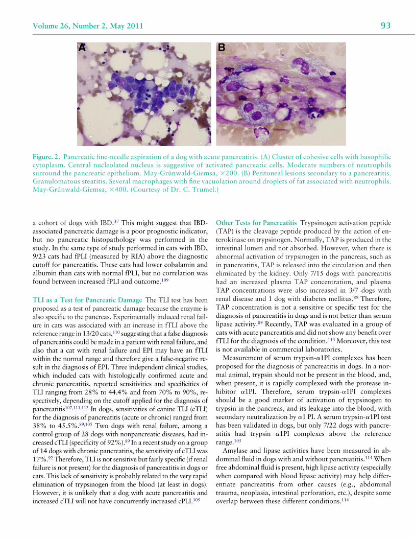
Figure. 2. Pancreatic fine-needle aspiration of a dog with accytoplasm. Central nucleolated nucleus is suggestive of asurround the pancreatic epithelium. May-Grünwald-GiemGranulomatous steatitis. Several macrophages with fine vaMay-Grünwald-Giemsa, �400. (Courtesy of Dr. C. Trum
increased cTLI will not have concurrently increased cPLI.105 o
Other Tests for Pancreatitis Trypsinogen activation peptide(TAP) is the cleavage peptide produced by the action of en-terokinase on trypsinogen. Normally, TAP is produced in theintestinal lumen and not absorbed. However, when there isabnormal activation of trypsinogen in the pancreas, such asin pancreatitis, TAP is released into the circulation and theneliminated by the kidney. Only 7/15 dogs with pancreatitishad an increased plasma TAP concentration, and plasmaTAP concentrations were also increased in 3/7 dogs withrenal disease and 1 dog with diabetes mellitus.89 Therefore,
AP concentration is not a sensitive or specific test for theiagnosis of pancreatitis in dogs and is not better than serumipase activity.89 Recently, TAP was evaluated in a group ofats with acute pancreatitis and did not show any benefit overTLI for the diagnosis of the condition.113 Moreover, this test
is not available in commercial laboratories.Measurement of serum trypsin-�1PI complexes has been
proposed for the diagnosis of pancreatitis in dogs. In a nor-mal animal, trypsin should not be present in the blood, and,when present, it is rapidly complexed with the protease in-hibitor �1PI. Therefore, serum trypsin-�1PI complexeshould be a good marker of activation of trypsinogen torypsin in the pancreas, and its leakage into the blood, withecondary neutralization by �1 PI. A serum trypsin-�1PI test
has been validated in dogs, but only 7/22 dogs with pancre-atitis had trypsin �1PI complexes above the referencerange.105
Amylase and lipase activities have been measured in ab-dominal fluid in dogs with and without pancreatitis.114 Whenree abdominal fluid is present, high lipase activity (especiallyhen compared with blood lipase activity) may help differ-
ntiate pancreatitis from other causes (e.g., abdominalrauma, neoplasia, intestinal perforation, etc.), despite some
e pancreatitis. (A) Cluster of cohesive cells with basophilicvated pancreatic cells. Moderate numbers of neutrophils�200. (B) Peritoneal lesions secondary to a pancreatitis.lation around droplets of fat associated with neutrophils.
)
utctisa,cuo
verlap between these different conditions.114

94 Topics in Companion Animal Medicine
In conclusion, PLI, trypsinogen, and TLI are pancreas spe-cific and, when elevated in the blood, are indicative of pan-creatic damage. They should be envisioned as cytolytic en-zymes, mirroring the approach that has been used fordecades with liver-specific enzymes. They should be consid-ered markers of acinar cell damage in conditions rangingfrom transient increase in plasma membrane permeability tocomplete necrosis (e.g., acute-necrotizing pancreatitis). Theycannot be considered as markers of a specific pancreatic dis-ease, namely pancreatitis. The diagnosis of pancreatitis islargely a diagnosis of exclusion. It should rely on a combina-tion of a strong clinical suspicion, results of laboratory tests,imaging studies, and, whenever possible, cytological (Fig 2)or histopathological evidence of pancreatic inflammation.The clinical significance of mild pancreatic lesions observedin dogs and cats that have no clinical signs of pancreatitis isquestionable. Considering the high prevalence of lesions sug-gestive of chronic pancreatitis in necropsy of dogs (34%)115
and cats (67%) presenting for other diseases, and in appar-ently healthy cats (45%),82 it is very important to keep inmind that the positive predictive value of any test improvessubstantially when performed in an animal that is likely tohave the disease. This means that the performance of thediagnostic tests for pancreatitis will be better when applied toselected cases with a high index of suspicion of the disease.
References
1. Broussard JD: Optimal fecal assessment. Clin Tech SmallAnim Pract 18:218-230, 2003
2. Marks SL, Kather EJ: Bacterial-associated diarrhea in the dog:a critical appraisal. Vet Clin North Am Small Anim Pract33:1029-1060, 2003
3. Rossi M, Hanninen ML, Revez J, et al: Occurrence and specieslevel diagnostics of Campylobacter spp., enteric Helicobacterspp. and Anaerobiospirillum spp. in healthy and diarrheicdogs and cats. Vet Microbiol 129:304-314, 2008
4. Weese JS, Staempfli HR, Prescott JF, et al: The roles of Clos-tridium difficile and enterotoxigenic Clostridium perfringensin diarrhea in dogs. J Vet Intern Med 15:374-378, 2001
5. Marks SL, Melli A, Kass PH, et al: Evaluation of methods todiagnose Clostridium perfringens-associated diarrhea in dogs.J Am Vet Med Assoc 214:357-360, 1999
6. Marks SL, Kather EJ, Kass PH, et al: Genotypic and pheno-typic characterization of Clostridium perfringens and Clos-tridium difficile in diarrheic and healthy dogs. J Vet InternMed 16:533-540, 2002
7. Clooten J, Kruth S, Arroyo L, et al: Prevalence and risk factorsfor Clostridium difficile colonization in dogs and cats hospi-talized in an intensive care unit. Vet Microbiol 129:209-214,2008
8. Cave NJ, Marks SL, Kass PH, et al: Evaluation of a routinediagnostic fecal panel for dogs with diarrhea. J Am Vet MedAssoc 221:52-59, 2002
9. Chouicha N, Marks SL: Evaluation of five enzyme immuno-assays compared with the cytotoxicity assay for diagnosis ofClostridium difficile-associated diarrhea in dogs. J Vet Diagn
Invest 18:182-188, 200610. Sokolow SH, Rand C, Marks SL, et al: Epidemiologic evalu-ation of diarrhea in dogs in an animal shelter. Am J Vet Res66:1018-1024, 2005
11. Sandberg M, Bergsjo B, Hofshagen M, et al: Risk factors forCampylobacter infection in Norwegian cats and dogs. PrevVet Med 55:241-253, 2002
12. Spain CV, Scarlett JM, Wade SE, et al: Prevalence of entericzoonotic agents in cats less than 1 year old in central NewYork State. J Vet Intern Med 15:33-38, 2001
13. Chaban B, Ngeleka M, Hill JE: Detection and quantificationof 14 Campylobacter species in pet dogs reveals an increase inspecies richness in feces of diarrheic animals. BMC Microbiol10:73, 2010
14. Turk J, Maddox C, Fales W, et al: Examination for heat-labile,heat-stable, and Shiga-like toxins and for the eaeA gene inEscherichia coli isolates obtained from dogs dying with diar-rhea: 122 cases (1992-1996). J Am Vet Med Assoc 212:1735-1736, 1998
15. DebRoy C, Maddox CW: Identification of virulence attributesof gastrointestinal Escherichia coli isolates of veterinary sig-nificance. Anim Health Res Rev 2:129-140, 2001
16. Staats JJ, Chengappa MM, DeBey MC, et al: Detection ofEscherichia coli Shiga toxin (stx) and enterotoxin (estA andelt) genes in fecal samples from non-diarrheic and diarrheicgreyhounds. Vet Microbiol 94:303-312, 2003
17. Sancak AA, Rutgers HC, Hart CA, et al: Prevalence of entero-pathic Escherichia coli in dogs with acute and chronic diar-rhoea. Vet Rec 154:101-106, 2004
18. Simpson KW, Dogan B, Rishniw M, et al: Adherent and inva-sive Escherichia coli is associated with granulomatous colitisin boxer dogs. Infect Immun 74:4778-4792, 2006
19. Craven M, Dogan B, Schukken A, et al: Antimicrobial resis-tance impacts clinical outcome of granulomatous colitis inboxer dogs. J Vet Intern Med 24:819-824, 2010
20. Morley PS, Strohmeyer RA, Tankson JD, et al: Evaluation ofthe association between feeding raw meat and Salmonella en-terica infections at a Greyhound breeding facility. J Am VetMed Assoc 228:1524-1532, 2006
21. Schmitz S, Coenen C, Konig M, et al: Comparison of threerapid commercial Canine parvovirus antigen detection testswith electron microscopy and polymerase chain reaction. JVet Diagn Invest 21:344-345, 2009
22. Neuerer FF, Horlacher K, Truyen U, et al: Comparison ofdifferent in-house test systems to detect parvovirus in faeces ofcats. J Feline Med Surg 10:247-251, 2008
23. Patterson EV, Reese MJ, Tucker SJ, et al: Effect of vaccinationon parvovirus antigen testing in kittens. J Am Vet Med Assoc230:359-363, 2007
24. Craven M, Chandler ML, Steiner JM, et al: Acute effects ofcarprofen and meloxicam on canine gastrointestinal permea-bility and mucosal absorptive capacity. J Vet Intern Med 21:917-923, 2007
25. Meddings JB, Kirk D, Olson ME: Noninvasive detection ofnonsteroidal anti-inflammatory drug-induced gastropathy indogs. Am J Vet Res 56:977-981, 1995
26. Mathon DH, Dossin O, Palierne S, et al: A laparoscopic-su-tured gastropexy technique in dogs: mechanical and func-tional evaluation. Vet Surg 38:967-974, 2009
27. Davis M, Willard M, Williamson K, et al: Temporal relation-ship between gastrointestinal protein loss, gastric ulcerationor erosion, and strenuous exercise in racing Alaskan sled dogs.
J Vet Intern Med 20:835-839, 2006
Volume 26, Number 2, May 2011 95
28. McLellan J, Wyse CA, Dickie A, et al: Comparison of thecarbon 13-labeled octanoic acid breath test and ultrasonogra-phy for assessment of gastric emptying of a semisolid meal indogs. Am J Vet Res 65:1557-1562, 2004
29. Wakefield LA, Shofer FS, Michel KE: Evaluation of cats fedvegetarian diets and attitudes of their caregivers. J Am VetMed Assoc 229:70-73, 2006
30. Suchodolski JS, Steiner JM: Laboratory assessment of gastro-intestinal function. Clin Tech Small Anim Pract 18:203-210,2003
31. Batchelor DJ, Noble PJ, Taylor RH, et al: Prognostic factors incanine exocrine pancreatic insufficiency: prolonged survival islikely if clinical remission is achieved. J Vet Intern Med 21:54-60, 2007
32. Thompson KA, Parnell NK, Hohenhaus AE, et al: Feline exo-crine pancreatic insufficiency: 16 cases (1992-2007). J FelineMed Surg 11:935-940, 2009
33. Simpson KW, Morton DB, Batt RM: Effect of exocrine pan-creatic insufficiency on cobalamin absorption in dogs. Am JVet Res 50:1233-1236, 1989
34. Allenspach K, Wieland B, Grone A, et al: Chronic enteropa-thies in dogs: evaluation of risk factors for negative outcome.J Vet Intern Med 21:700-708, 2007
35. Craven M, Simpson JW, Ridyard AE, et al: Canine inflamma-tory bowel disease: retrospective analysis of diagnosis andoutcome in 80 cases (1995-2002). J Small Anim Pract 45:336-342, 2004
36. German AJ, Day MJ, Ruaux CG, et al: Comparison of directand indirect tests for small intestinal bacterial overgrowth andantibiotic-responsive diarrhea in dogs. J Vet Intern Med 17:33-43, 2003
37. Kathrani A, Steiner JM, Suchodolski J, et al: Elevated caninepancreatic lipase immunoreactivity concentration in dogswith inflammatory bowel disease is associated with a negativeoutcome. J Small Anim Pract 50:126-132, 2009
38. Reed N, Gunn-Moore D, Simpson K: Cobalamin, folate andinorganic phosphate abnormalities in ill cats. J Feline MedSurg 9:278-288, 2007
39. Simpson KW, Fyfe J, Cornetta A, et al: Subnormal concentra-tions of serum cobalamin (vitamin B12) in cats with gastroin-testinal disease. J Vet Intern Med 15:26-32, 2001
40. Battersby IA, Giger U, Hall EJ: Hyperammonaemic encepha-lopathy secondary to selective cobalamin deficiency in a juve-nile Border collie. J Small Anim Pract 46:339-344, 2005
41. Packer RA, Cohn LA, Wohlstadter DR, et al: D-lactic acidosissecondary to exocrine pancreatic insufficiency in a cat. J VetIntern Med 19:106-110, 2005
42. Ruaux CG, Steiner JM, Williams DA: Metabolism of aminoacids in cats with severe cobalamin deficiency. Am J Vet Res62:1852-1858, 2001
43. Ruaux CG, Steiner JM, Williams DA: Early biochemical andclinical responses to cobalamin supplementation in cats withsigns of gastrointestinal disease and severe hypocobalamine-mia. J Vet Intern Med 19:155-160, 2005
44. Ruaux CG, Steiner JM, Williams DA: Relationships betweenlow serum cobalamin concentrations and methlymalonic aci-demia in cats. J Vet Intern Med 23:472-475, 2009
45. Salvadori C, Cantile C, De Ambrogi G, et al: Degenerativemyelopathy associated with cobalamin deficiency in a cat. J
Vet Med A Physiol Pathol Clin Med 50:292-296, 200346. Parnell NK, Moore GE, Suchodolski JS, et al: Influence of ageon serum cobalamin and folate concentrations in healthy cats[abstract]. J Vet Intern Med 22:809, 2008
47. Cook AK, Suchodolski JS, Steiner JM, Robertson JE: Theprevalence of hypocobalaminaemia in cats with spontaneoushyperthyroidism. J Small Anim Pract 52:101-106, 2011
48. Grutzner N, Bishop MA, Suchodolski JS, et al: Associationstudy of cobalamin deficiency in the Chinese Shar Pei. J Hered101:211-217, 2010
49. Dandrieux JRS, Noble PJ, Halladay LJ, et al: Breed predispo-sition for severe hypocobalaminemia and relation to folateconcentration in dogs with gastrointestinal disease [abstract].J Vet Intern Med 24:722, 2010
50. Allenspach K, Steiner JM, Shah BN, et al: Evaluation of gas-trointestinal permeability and mucosal absorptive capacity indogs with chronic enteropathy. Am J Vet Res 67:479-483,2006
51. Kobayashi S, Ohno K, Uetsuka K, et al: Measurement of in-testinal mucosal permeability in dogs with lymphocytic-plas-macytic enteritis. J Vet Med Sci 69:745-749, 2007
52. Rodriguez H, Berghoff N, Suchodolski JS, et al: Kinetic anal-ysis of 5 sugar probes in dog serum after orogastric adminis-tration. Can J Vet Res 73:217-223, 2009
53. Rodriguez H, Suchodolski JS, Berghoff N, et al: Developmentand analytic validation of a gas chromatography-mass spec-trometry method for the measurement of sugar probes in ca-nine serum. Am J Vet Res 70:320-329, 2009
54. Melgarejo T, Williams DA, Asem EK: Enzyme-linked immu-nosorbent assay for canine alpha 1-protease inhibitor. Am JVet Res 59:127-130, 1998
55. Fetz K, Ruaux CG, Steiner JM, et al: Purification and partialcharacterization of feline alpha1-proteinase inhibitor (fal-pha1-PI) and the development and validation of a radioimmu-noassay for the measurement of falpha1-PI in serum.Biochimie 86:67-75, 2004
56. Murphy KF, German AJ, Ruaux CG, et al: Fecal alpha1-pro-teinase inhibitor concentration in dogs receiving long-termnonsteroidal anti-inflammatory drug therapy. Vet Clin Pathol32:136-139, 2003
57. Littman MP, Dambach DM, Vaden SL, et al: Familial protein-losing enteropathy and protein-losing nephropathy in SoftCoated Wheaten Terriers: 222 cases (1983-1997). J Vet InternMed 14:68-80, 2000
58. Vaden SL, Hammerberg B, Davenport DJ, et al: Food hyper-sensitivity reactions in Soft Coated Wheaten Terriers withprotein-losing enteropathy or protein-losing nephropathy orboth: gastroscopic food sensitivity testing, dietary provoca-tion, and fecal immunoglobulin E. J Vet Intern Med 14:60-67,2000
59. Vaden SL, Sellon RK, Melgarejo LT, et al: Evaluation of in-testinal permeability and gluten sensitivity in Soft-CoatedWheaten Terriers with familial protein-losing enteropathy,protein-losing nephropathy, or both. Am J Vet Res 61:518-524, 2000
60. Murphy KF, German AJ, Ruaux CG, et al: Fecal alpha1-pro-teinase inhibitor concentration in dogs with chronic gastroin-testinal disease. Vet Clin Pathol 32:67-72, 2003
61. Fetz K, Steiner JM, Ruaux C, et al: Increased fecal �1-protei-nase inhibitor concentrations in cats with gastrointestinal dis-
ease [abstract]. J Vet Intern Med 19:474, 2005
96 Topics in Companion Animal Medicine
62. Gilson SD, Parker BB, Twedt DC: Evaluation of two commer-cial test kits for detection of occult blood in feces of dogs. Am JVet Res 51:1385-1387, 1990
63. Cook AK, Gilson SD, Fischer WD, et al: Effect of diet onresults obtained by use of two commercial test kits for detec-tion of occult blood in feces of dogs. Am J Vet Res 53:1749-1751, 1992
64. Tuffli SP, Gaschen F, Neiger R: Effect of dietary factors on thedetection of fecal occult blood in cats. J Vet Diagn Invest13:177-179, 2001
65. Rutgers HC, Batt RM, Elwood CM, et al: Small intestinalbacterial overgrowth in dogs with chronic intestinal disease.J Am Vet Med Assoc 206:187-193, 1995
66. Johnston KL: Small intestinal bacterial overgrowth. Vet ClinNorth Am Small Anim Pract 29:523-550, 1999
67. Johnston KL, Lamport AI, Ballevre OP, et al: Effects of oraladministration of metronidazole on small intestinal bacteriaand nutrients of cats. Am J Vet Res 61:1106-1112, 2000
68. Johnston KL, Swift NC, Forster-van Hijfte M, et al: Compar-ison of the bacterial flora of the duodenum in healthy cats andcats with signs of gastrointestinal tract disease. J Am Vet MedAssoc 218:48-51, 2001
69. Suchodolski JS: Microbes and gastrointestinal health of dogsand cats. J Anim Sci Nov 12, 2010
70. Suchodolski JS, Xenoulis PG, Paddock CG, et al: Molecularanalysis of the bacterial microbiota in duodenal biopsies fromdogs with idiopathic inflammatory bowel disease. Vet Micro-biol 142:394-400, 2010
71. Xenoulis PG, Palculict B, Allenspach K, et al: Molecular-phy-logenetic characterization of microbial communities imbal-ances in the small intestine of dogs with inflammatory boweldisease. FEMS Microbiol Ecol 66:579-589, 2008
72. Melgarejo T, Williams DA, O’Connell NC, et al: Serum un-conjugated bile acids as a test for intestinal bacterial over-growth in dogs. Dig Dis Sci 45:407-414, 2000
73. Williams DA, Batt RM: Sensitivity and specificity of radioim-munoassay of serum trypsin-like immunoreactivity for the di-agnosis of canine exocrine pancreatic insufficiency. J Am VetMed Assoc 192:195-201, 1988
74. Steiner JM, Medinger TL, Williams DA: Development andvalidation of a radioimmunoassay for feline trypsin-like im-munoreactivity. Am J Vet Res 57:1417-1420, 1996
75. James FE, Mansfield CS, Steiner JM, et al: Pancreatic responsein healthy dogs fed diets of various fat compositions. Am J VetRes 70:614-618, 2009
76. Steiner JM, Williams DA: Influence of feeding on serum felinetrypsin-like immunoreactivity. Am J Vet Res 60:895-897,1999
77. Carro T, Williams DA: Relationship between dietary proteinconcentration and serum trypsin-like immunoreactivity indogs. Am J Vet Res 50:2105-2107, 1989
78. Wiberg ME, Nurmi AK, Westermarck E: Serum trypsinlikeimmunoreactivity measurement for the diagnosis of subclini-cal exocrine pancreatic insufficiency. J Vet Intern Med 13:426-432, 1999
79. Spillmann T, Wittker A, Teigelkamp S, et al: An immunoassayfor canine pancreatic elastase 1 as an indicator for exocrinepancreatic insufficiency in dogs. J Vet Diagn Invest 13:468-474, 2001
80. Spillmann T, Eigenbrodt E, Sziegoleit A: [Determination andclinical relevance of fecal pancreatic elastase in dogs]. Tierar-
ztl Prax Ausg K Klientiere Heimtiere 26:364-368, 199881. Newman S, Steiner J, Woosley K, et al: Localization of pan-creatic inflammation and necrosis in dogs. J Vet Intern Med18:488-493, 2004
82. De Cock HE, Forman MA, Farver TB, et al: Prevalence andhistopathologic characteristics of pancreatitis in cats. VetPathol 44:39-49, 2007
83. Simpson KW, Simpson JW, Lake S, et al: Effect of pancreate-ctomy on plasma activities of amylase, isoamylase, lipase andtrypsin-like immunoreactivity in dogs. Res Vet Sci 51:78-82,1991
84. Steiner JM, Rutz GM, Williams DA: Serum lipase activitiesand pancreatic lipase immunoreactivity concentrations indogs with exocrine pancreatic insufficiency. Am J Vet Res67:84-87, 2006
85. Polzin DJ, Osborne CA, Stevens JB, et al: Serum amylase andlipase activities in dogs with chronic primary renal failure.Am J Vet Res 44:404-410, 1983
86. Steiner JM: Diagnosis of pancreatitis. Vet Clin North AmSmall Anim Pract 33:1181-1195, 2003
87. Piccione G, Giannetto C, Fazio F, et al: Daily rhythm of serumlipase and alpha-amylase activity in fed and fasted dogs. J VetDiagn Invest 20:795-799, 2008
88. Strombeck DR, Farver T, Kaneko JJ: Serum amylase andlipase activities in the diagnosis of pancreatitis in dogs. Am JVet Res 42:1966-1970, 1981
89. Mansfield CS, Jones BR: Plasma and urinary trypsinogen ac-tivation peptide in healthy dogs, dogs with pancreatitis anddogs with other systemic diseases. Aust Vet J 78:416-422,2000
90. Hess RS, Kass PH, Shofer FS, et al: Evaluation of risk factorsfor fatal acute pancreatitis in dogs. J Am Vet Med Assoc 214:46-51, 1999
91. McCord K, Davis J, Leyva F, et al: A multi-institutional studyevaluating diagnostic utility of Spec cPL in the diagnosis ofacute pancreatitis in dogs [abstract]. J Vet Intern Med 23:734,2009
92. Watson PJ, Archer J, Roulois AJ, et al: Observational study of14 cases of chronic pancreatitis in dogs. Vet Rec 167:968-976,2010
93. Kitchell BE, Strombeck DR, Cullen J, et al: Clinical and patho-logic changes in experimentally induced acute pancreatitis incats. Am J Vet Res 47:1170-1173, 1986
94. Steiner JM, Williams DA: Purification of classical pancreaticlipase from dog pancreas. Biochimie 84:1245-1253, 2002
95. Steiner JM, Wilson BG, Williams DA: Purification and partialcharacterization of feline classical pancreatic lipase. CompBiochem Physiol B Biochem Mol Biol 134:151-159, 2003
96. Steiner JM, Berridge BR, Wojcieszyn J, et al: Cellular immu-nolocalization of gastric and pancreatic lipase in various tis-sues obtained from dogs. Am J Vet Res 63:722-727, 2002
97. Huth SP, Relford R, Steiner JM, et al: Analytical validation ofan ELISA for measurement of canine pancreas-specific lipase.Vet Clin Pathol 39:346-353, 2010
98. Beall MJ, Cahill R, Pigeon K, et al: Performance validationand method comparison of an in-clinic enzyme-linked immu-nosorbent assay for the detection of canine pancreatic lipase. JVet Diagn Invest 23:115-119, 2011
99. Steiner JM, Teague SR, Lees GE, et al: Stability of caninepancreatic lipase immunoreactivity concentration in serumsamples and effects of long-term administration of prednisoneto dogs on serum canine pancreatic lipase immunoreactivity
concentrations. Am J Vet Res 70:1001-1005, 2009
Volume 26, Number 2, May 2011 97
100. Cordner AP, Armstrong PJ, Newman SJ, et al: Effect of pan-creatic tissue sampling on serum pancreatic enzyme levels inclinically healthy dogs. J Vet Diagn Invest 22:702-707, 2010
101. Cosford KL, Shmon CL, Myers SL, et al: Prospective evalua-tion of laparoscopic pancreatic biopsies in 11 healthy cats. JVet Intern Med 24:104-113, 2010
102. Steiner JM, Finco DR, Gumminger SR, et al: Serum caninepancreatic lipase immunoreactivity (cPLI) in dogs with exper-imentally induced renal failure [abstract]. J Vet Intern Med15:311, 2001
103. Xenoulis P, Finco DR, Suchodolski JS, et al: Serum fPLI andSPEC fPL concentrations in cats with experimentally inducedchronic renal failure [abstract]. J Vet Intern Med 23:758,2009
104. Steiner JM, Xenoulis PG, Anderson JA, et al: Serum pancre-atic lipase immunoreactivity concentrations in dogs treatedwith potassium bromide and/or phenobarbital. Vet Ther9:37-44, 2008
105. Steiner JM, Newman S, Xenoulis P, et al: Sensitivity of serummarkers for pancreatitis in dogs with macroscopic evidence ofpancreatitis. Vet Ther 9:263-273, 2008
106. Carney PC, Ruaux GC, Suchodolski J, et al: Biological vari-ability of canine pancreatic lipase immunoreactivity and C-re-active protein concentrations in clinically healthy dogs. Paperpresented at: 2010 ACVIM Forum, Anaheim, CA
107. Forman MA, Marks SL, De Cock HE, et al: Evaluation ofserum feline pancreatic lipase immunoreactivity and helical
computed tomography versus conventional testing for the di-agnosis of feline pancreatitis. J Vet Intern Med 18:807-815,2004
108. Prior LM, Forman MA, Shiroma J, et al: Serial evaluation ofcanine pancreatic lipase (SPEC cPL) in dogs with clincal signsof pancreatitis [abstract]. J Vet Intern Med 23:733, 2009
109. Bailey S, Benigni L, Eastwood J, et al: Comparisons betweencats with normal and increased fPLI concentrations in catsdiagnosed with inflammatory bowel disease. J Small AnimPract 51:484-489, 2010
110. Steiner JM, Finco DR, Williams DA: Serum trypsin-like im-munoreactivity (fTLI) in cats with experimentally inducedchronic renal failure [abstract]. J Vet Intern Med 16:819,2002
111. Gerhardt A, Steiner JM, Williams DA, et al: Comparison ofthe sensitivity of different diagnostic tests for pancreatitis incats. J Vet Intern Med 15:329-333, 2001
112. Swift NC, Marks SL, MacLachlan NJ, et al: Evaluation ofserum feline trypsin-like immunoreactivity for the diagnosis ofpancreatitis in cats. J Am Vet Med Assoc 217:37-42, 2000
113. Allen HS, Steiner J, Broussard J, et al: Serum and urine con-centrations of trypsinogen-activation peptide as markers foracute pancreatitis in cats. Can J Vet Res 70:313-316, 2006
114. Guija de Arespacochaga A, Hittmair KM, Schwendenwein I:Comparison of lipase activity in peritoneal fluid of dogs withdifferent pathologies—a complementary diagnostic tool inacute pancreatitis? J Vet Med A Physiol Pathol Clin Med53:119-122, 2006
115. Watson P, Herrtage M: Chronic pancreatitis in dogs. Vet Rec
152:340, 2003