Laboratory Manual VMC 311 -...
74
Laboratory Manual VMC 311 Systematic Veterinary Bacteriology & Mycology
Transcript of Laboratory Manual VMC 311 -...

Laboratory Manual VMC 311 Sys t em at i c V e t e r i n a ry B ac t e r i o l o g y & M yco l o g y

1
LABORATORY MANUAL FOR
VETERINARY MICROBIOLOGY-II (Revised As Per New Syllabus)
CERTIFICATE
Certified that this is a bonafide record of practical work done in the laboratory for the course of Veterinary Systematic Bacteriology & Mycology Course no. VMC-311 (New Syllabus) during the year_________. Name of the student: ________________________________________ Registration No.:___________________________________________ Exam seat No.: ___________________________________________________
Course Teacher
SEMESTER END EXAMINATION Evaluated The Practical Record Submitted For The Annual Board Practical Examination Held On _____________________. Internal Examiner External Examiner

2
INDEX Sr.No.
Practical Page N0. Date Sign
1 Mastitis: Isolation and identification of Staphylococcus spp.
2 Mastitis: Isolation and identification of Streptococcus spp.
3 Diagnosis of Heamorhagic Septicaemia 4 Enteric Infections : Isolation and
identification of Escherichia coli
5 Enteric Infections : Isolation and identification of Salmonella, Klebsiella, Proteus and Shigella
6 Diagnosis of Brucellosis
7 Diagnosis of Tuberculosis
8 Diagnosis of Johne’s Disease
9 Diagnosis of Black Quarter
10 Diagnosis of Enterotoxaemia
11 Diagnosis of Wooden tongue
12 Diagnosis of Anthrax
13 Diagnosis of Glanders
14 Study of genus Aspergillus
15 Diagnosis of Dermatophytosis
16 Isolation & identification of Candida albicans

3
Appendix
COLOUR PLATES
PLATE 1
1.Microscopic picture of Staphylococcus 2.Microscopic picture of Streptococcus 3&4.DNAase activity of Staphylococcus aureus 5.CAMP Test 6.Coagulase Test
PLATE 2
7.Cultural Characteristics of E. coli – MacConkey Agar 8. E. coli on EMB: Characteristic “Metallic sheen’’ 9.Swarming of Proteus mirabilis on an agar plate 10.Salmonella on XLD agar : Colourless colonies with black Centres 11 & 12.Salmonella Plate Test
PLATE 3
13 &14. Brucella Rose Bengal Plate Test 15.Microscopic Picture of Mycobacterium tuberculosis 16. Mycobacterium tuberculosis on L J Medium 17.Clostridium chauvoei –Anaerobic cultivation on Blood Agar 18. Clostridium tetani on FFA
PLATE 4 19. Robertson Cooked Meat Medium: Growth of Saccharolytic Clostridia-Pink Coloration 20. Actinobacillus lignieresi 21. Colony characteristics of Bacillus anthracis 22. Bacillus anthracis in tissue seen in short chains surrounded by a common capsule- Toluidine blue stain 23. Colony characteristics of Candida albicans
24. Germ Tube Test – Candida albicans
PLATE 5 25. Aspergillus flavus 26. Aspergillus flavus- Microscopic 27. Trichophyton rubrum on DTM Agar 28. Pencil shaped macroconidia T. rubrum 29. Microsporum on DTM agar medium 30. Spindle shaped macroconidia Microsporum canis

4
PRACTICAL NO.1
MASTITIS: ISOLATION & IDENTIFICATION OF STAPHYLLOCOCCUS SPP.
Collection of milk sample
The udder of the animal should be thoroughly cleaned with water and antiseptics like 1:1000 solution of potassium permanganate and dried with clean towel. The hands of the person collecting milk should be also cleaned with soap and antiseptics. Disinfect the teats with swab of 70% ethanol. Collect the milk sample from each teat in separate sterilized test tubes. The first 3-4 streams of milk should be discarded and then 5-10 ml of milk sample is collected in tubes. Tubes should be sealed / stoppered properly. Immediately after collection of milk, it should be transported to the laboratory for examination in ice. The examination of the milk sample should be performed immediately after its collection. But if delay is expected in examination, it should be kept in refrigerator at 40C.
Tests for diagnosis of Mastitis
I.California Mastitis Test (CMT)
Aim: To detect the sub clinical mastitis from milk sample of animals
Principle This test is based on increased number of leucocytes and increased alkalinity in milk due to mastitis (Precipitate is formed). CMT Reagent NaOH 1.5 gm Teepol 0.5 ml Bromothymol blue 100ml Distilled water 100ml Filter and use
Procedure Take 0.5 ml of milk from each quarter in plastic peddle cups and add equal quantity of CMT Reagent and mix well by circular movement of peddle on a horizontal plane.
5
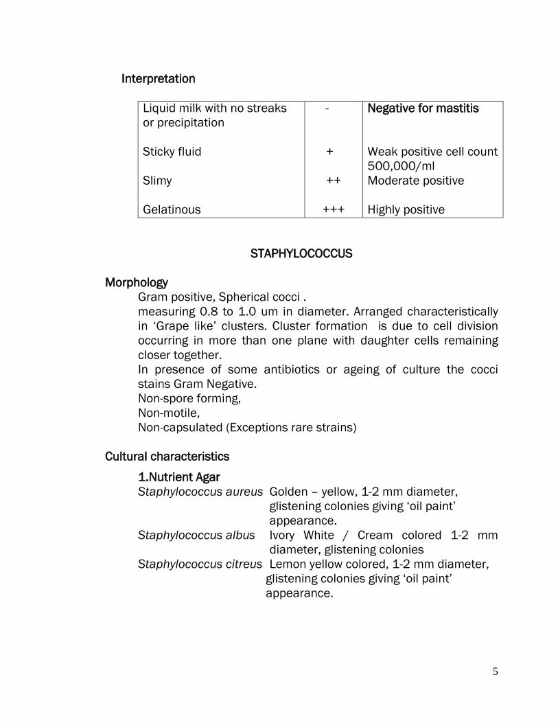
Interpretation
Liquid milk with no streaks or precipitation Sticky fluid Slimy Gelatinous
-
+
++
+++
Negative for mastitis Weak positive cell count 500,000/ml Moderate positive Highly positive
STAPHYLOCOCCUS Morphology
Gram positive, Spherical cocci . measuring 0.8 to 1.0 um in diameter. Arranged characteristically in ‘Grape like’ clusters. Cluster formation is due to cell division occurring in more than one plane with daughter cells remaining closer together. In presence of some antibiotics or ageing of culture the cocci stains Gram Negative. Non-spore forming, Non-motile, Non-capsulated (Exceptions rare strains)
Cultural characteristics
1.Nutrient Agar Staphylococcus aureus Golden – yellow, 1-2 mm diameter,
glistening colonies giving ‘oil paint’ appearance.
Staphylococcus albus Ivory White / Cream colored 1-2 mm diameter, glistening colonies
Staphylococcus citreus Lemon yellow colored, 1-2 mm diameter, glistening colonies giving ‘oil paint’ appearance.

6
Pigment production can be enhanced by incubating at 22OC / addition of 1% Glycerol acetate / incorporation of milk into the medium. Pigmentation is due to carotenoids.
2.Blood agar Colony characteristic similar to those on Nutrient agar, But are surrounded by the zone of alpha, beta - Hemolysis. Hemolysis is marked on bovine, sheep & rabbit blood agar; weak on horse agar.
3.MacConkeys Agar Colonies are smaller in size and impart pink colour due to lactose fermentation.
4.Milk Agar The medium is prepared by mixing 100 ml of sterile nutrient agar containing additional 2% agar and 100ml of sterilized milk. On this medium, colonies appear larger and pigment production is enhanced.
5.Mannitol Salt Agar 1% Mannitol,7.5% sodium chloride & 0.0025% Phenol red is added to the Nutrient agar. This is both selective and Indicator medium. Most strains of Staphylococcus aureus ferments mannitol therefore produce acid, due to which yellow zones surround colonies. Colony characters are similar to that of Nutrient agar.
6. Addition of Potassium tellurite imparts black colour to the colonies.
7.Phenolphthalein Phosphate Agar Indicator medium Prepared by mixing 2ml of 0.6% solution of Sodium phenolphthalein diphosphate, sterilized by filtration and 98 ml of melted nutrient agar and poured into plates. Colony characters are similar to that of Nutrient agar. All strains of Staphylococcus aureus produce phosphatase which liberates phenolphthalein from Sodium phenolphthalein diphosphate .To detect phenolphthalein 0.1 ml of ammonia solution is placed in the lid and the culture plate, is inverted over it. Colonies of Staphylococcus aureus become bright pink in alkaline pH.

7
8. Enrichment Salt medium 10 % sodium chloride may be added to cooked meat broth. Used for primary enrichment.Other bacteria are inhibited at this concentration but halophilic bacteria such as Staphylococci, Vibrios survive.
Bio-chemical Properties Staphylococcus aureus is catalase positive.
(Catalase production may function to inactivate toxic hydrogen peroxide and free radicals formed by myeloperoxidase system within phagocytic cells after ingestion of microorganisms.)
Oxidase negative. It ferments glucose. maltose, lactose, mannitol and sucrose with the production of acid but no gas. Most strains of Staphylococcus aureus ferment mannitol, whereas most strains of Staphylococcus epidermidis, Staphylococcus saprophyticus are Mannitol negative.
Indole Negative, MR Positive, VP Positive Urease Positive, Hydrolyzes Gelatin Reduces nitrates to nitrites (NO3---- NO2) Lipolytic- when grown on media containing egg yolk, produce a dense opacity.
Tests For Identification
1.Catalase Test This test is used to differentiate those bacteria that produce the enzyme catalase. Such as staphylococci, from non-catalse producing bacteria such as Streptococci.
Principle Catalase acts as catalyst in the breakdown of hydrogen peroxide to oxygen and water.
Requirement Hydrogen peroxide - 3% H2O2 (10 volume solution)
Method 1.Pour 2-3 ml of the hydrogen peroxide solution into a test tube. 2.Using sterile wooden stick or glass rod, remove a good growth of the test organisms and immerse it in the hydrogen peroxide solution. 3.Look for immediate bubbling.
8
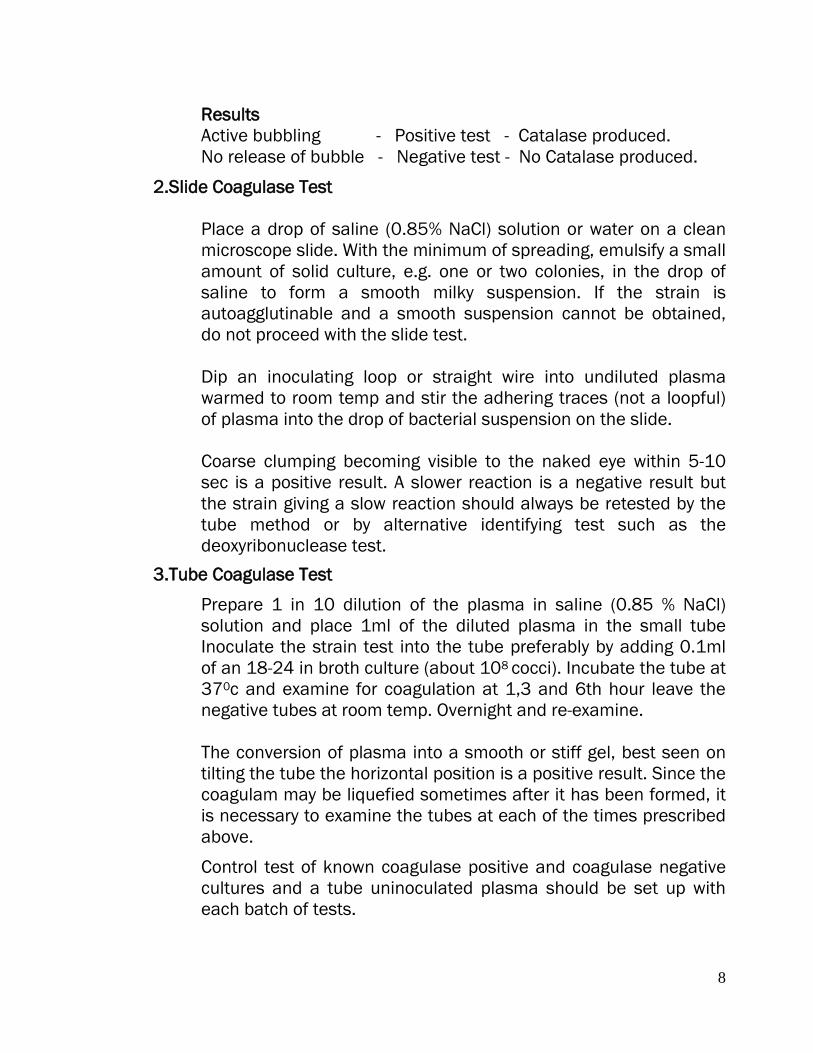
Results Active bubbling - Positive test - Catalase produced. No release of bubble - Negative test - No Catalase produced.
2.Slide Coagulase Test Place a drop of saline (0.85% NaCl) solution or water on a clean microscope slide. With the minimum of spreading, emulsify a small amount of solid culture, e.g. one or two colonies, in the drop of saline to form a smooth milky suspension. If the strain is autoagglutinable and a smooth suspension cannot be obtained, do not proceed with the slide test.
Dip an inoculating loop or straight wire into undiluted plasma warmed to room temp and stir the adhering traces (not a loopful) of plasma into the drop of bacterial suspension on the slide. Coarse clumping becoming visible to the naked eye within 5-10 sec is a positive result. A slower reaction is a negative result but the strain giving a slow reaction should always be retested by the tube method or by alternative identifying test such as the deoxyribonuclease test.
3.Tube Coagulase Test
Prepare 1 in 10 dilution of the plasma in saline (0.85 % NaCl) solution and place 1ml of the diluted plasma in the small tube Inoculate the strain test into the tube preferably by adding 0.1ml of an 18-24 in broth culture (about 108 cocci). Incubate the tube at 370c and examine for coagulation at 1,3 and 6th hour leave the negative tubes at room temp. Overnight and re-examine. The conversion of plasma into a smooth or stiff gel, best seen on tilting the tube the horizontal position is a positive result. Since the coagulam may be liquefied sometimes after it has been formed, it is necessary to examine the tubes at each of the times prescribed above.
Control test of known coagulase positive and coagulase negative cultures and a tube uninoculated plasma should be set up with each batch of tests.
9
4.DNAase Test
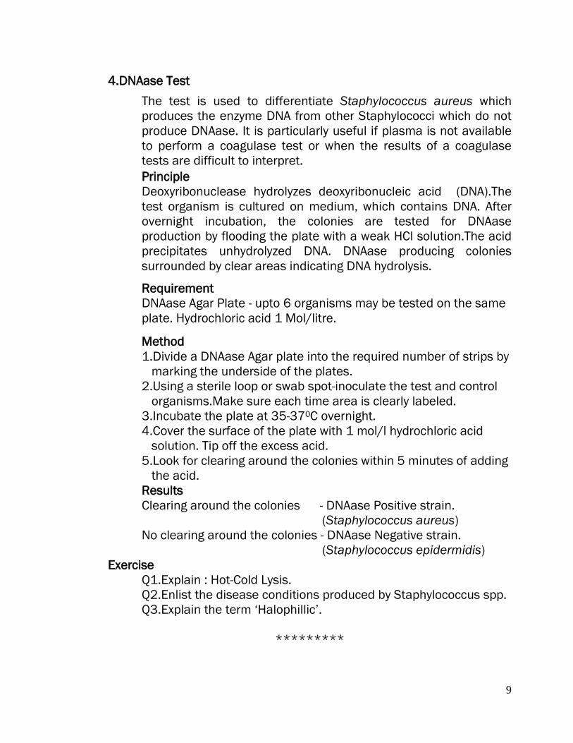
The test is used to differentiate Staphylococcus aureus which produces the enzyme DNA from other Staphylococci which do not produce DNAase. It is particularly useful if plasma is not available to perform a coagulase test or when the results of a coagulase tests are difficult to interpret.
Principle Deoxyribonuclease hydrolyzes deoxyribonucleic acid (DNA).The test organism is cultured on medium, which contains DNA. After overnight incubation, the colonies are tested for DNAase production by flooding the plate with a weak HCl solution.The acid precipitates unhydrolyzed DNA. DNAase producing colonies surrounded by clear areas indicating DNA hydrolysis.
Requirement DNAase Agar Plate - upto 6 organisms may be tested on the same plate. Hydrochloric acid 1 Mol/litre.
Method 1.Divide a DNAase Agar plate into the required number of strips by marking the underside of the plates. 2.Using a sterile loop or swab spot-inoculate the test and control organisms.Make sure each time area is clearly labeled. 3.Incubate the plate at 35-370C overnight. 4.Cover the surface of the plate with 1 mol/l hydrochloric acid solution. Tip off the excess acid. 5.Look for clearing around the colonies within 5 minutes of adding the acid. Results Clearing around the colonies - DNAase Positive strain. (Staphylococcus aureus) No clearing around the colonies - DNAase Negative strain. (Staphylococcus epidermidis)
Exercise Q1.Explain : Hot-Cold Lysis. Q2.Enlist the disease conditions produced by Staphylococcus spp. Q3.Explain the term ‘Halophillic’.
*********

10
PRACTICAL NO.2
MASTITIS: ISOLATION & IDENTIFICATION OF STREPTOCOCCUS SPP.
STREPTOCOCCUS
Morphology
Gram-positive cocci (In older cultures decolorized). Streptococcus pyogenes is a coccus 0.5 um to 1 um in size arranged in chains, chains made up of many diplococci. (Division in one plane.) Chains are shorter in artificial medium. Length of chain is increased by the presence of specific antibodies and is decreased in the absence of abs. Non-motile,Non- sporulating. The majority of the strains when grown under ordinary conditions do not produce capsules. Capsule producing strain - Streptococcus epidemicus.
Colony Characteristics of Streptococci
Nutrient Agar: Colonies are small smooth, glistening, dewdrop, finely granular with age become opaque with raised central position.No pigment is produced.
Blood Agar: Beta-type of hemolysis is seen. After 24 hours - 48 hours incubation . Bouillon is uniformly clouded in early stages of growth, finely granular sediment as the culture ages, which settles on the sides and bottom of the culture. No pellicle is formed.
Crystal violet Blood Agar The addition of low concentrations (1:500000, ie.0.0002%) of crystal violet to blood agar inhibits the growth of some bacteria, notably Staphylococci. Crystal violet blood agar is therefore a selective medium for the isolation of streptococci. Sterile Nutrient agar 90ml Sterile horse blood 10ml Crystal violet in 1:1000 aqueous solution 0.2ml Melt the agar to, cool to 50oC,add the blood and crystal violet and pour plates.
11
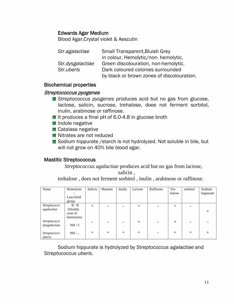
Edwards Agar Medium Blood Agar,Crystal violet & Aesculin Str.agalactiae Small Transparent,Bluish Grey
in colour, Hemolytic/non- hemolytic. Str.dysgalactiae Green discolouration, non-hemolytic. Str.uberis Dark coloured colonies surrounded
by black or brown zones of discolouration.
Biochemical properties
Streptococcus pyogenes Streptococcus pyogenes produces acid but no gas from glucose, lactose, salicin, sucrose, trehalose, does not ferment sorbitol, inulin, arabinose or raffinose.
It produces a final pH of 6.0-4.8 in glucose broth Indole negative Catalase negative Nitrates are not reduced Sodium hippurate /starch is not hydrolyzed. Not soluble in bile, but will not grow on 40% bile blood agar.
Mastitic Streptococcus
Streptococcus agalactiae produces acid but no gas from lactose, salicin ,
trehalose , does not ferment sorbitol , inulin , arabinose or raffinose.
Name Hemolysis / Lancifield group
Salicin Mannite Inulin Lactose Raffinose Tre- halose
sorbitol Sodium hippurate
Streptococci agalactiae Streptococci dysgalactiae Streptococci uberis
B / B (Double zone of hemolysis)
NH / C
NH /…
+ -
+
- -
+
- -
+
+
+
+
- - -
+
+
+
- -
+
+ -
+
Sodium hippurate is hydrolyzed by Streptococcus agalactiae and
Streptococcus uberis.
12
Test For Identification
CAMP test Christie, Atkins and Munch - Peterson first described CAMP test in 1944. Reported that hemolytic activity of Staphylococcal beta lysine on erythrocytes is enhanced by an extracelluar facto 0r produced by group B Streptococci, known as CAMP factor, wherever two reactants overlap in a sheep or bovine blood agar plate.
Make a single streak of the streptococcal test strain perpendicular to the Staphylococcal streak. Leave about 1 cm space between the two inoculation lines. Two streak lines must not touch each other. Incubate the inoculated plate at 37oC for 24 hours in air or in 10 % CO2.
Streptococci produce a positive reaction in the absence of oygen. CAMP factor produced by group B Streptococci enhances the B-lysin produced by Staphylococcus and an increased area of lysis appears at the junction of the two organisms which assume the shape of an arrow head.
Test for diagnosis of mastitis caused by Streptococcus
I.Bromocresol purple test
Add 0.5 ml of sterile 0.5 % aqueous solution of bromocresol purple to 9.5 ml of milk.Incubate 24 hours at 37oC & Read the test
Appearance of canary yellow colonies of bacterial growth along the walls and in the bottom of the tubes is diagnostic of Streptococci agalactiae with clumps.(Fermentation of lactose to acid changes the bromocresol purple to yellow range of indicator.)
Streptococci dysgalactiae and Streptococci uberis though ferments lactose ,but do not form clumps.
Exercise Q1.Explain the following tests :
a. Chloride test b. Strip cup test Q2.Enlist mastitis causing bacteria and fungi. Q3.Write chloride content, pH, Sp.gravity of normal & mastitic milk. Q4.Write the culture characteristic and microscopic examination of the isolates from mastitis cases. Q5. Enlist the disease conditions caused by Streptococcus.
*********

13
PRACTICAL NO. 3 DIAGNOSIS OF HAEMORHAGIC SEPTICAEMIA
Collection Of Material
From sick animals-Blood should be collected at the height of temperature (Septicaemia), fixed blood smears and material from swelling at the neck region (Donot collect the material from the throat region, as the Pasteurella multocida is a normal inhabitant of Upper respiratory tract) . From dead animals on PM, smears from heart blood and liver, heart blood in a sterile bottle, lymph nodes & spleen on ice.
Morphology Small coccoid rod . Measuring 0.25-0.4 u by 0.6-2.6u. Pleomorphic. Gram negative rods with characteristic ‘Bipolar’ appearance in the stained smears. Possess capsule (Hyaluronic acid) when recently isolated from the disease process.On subculturing looses the capsule. Non-sporulating, Non-motile.
Cultural Characteristics Aerobe & facultative anaerobe. Optimum temperature for growth is 370C. PH range is 6-8.5, optimum being 7.2-7.4.
Brain heart infusion agar Fluorescent and iridescent colonies are moderate in size, white, opaque.(Pathogenic) Intermediate colonies vary in appearance in between Fluorescent and blue colonies. Blue forms/R forms Small dew drop like colonies, bluish, rough colonies (with low virulence). Better growth is obtained when blood or serum is added to the medium.
MacConkeys Agar: No growth.
Blood Agar: White opaque or dew drop like colonies as that of rain Heart Infusion agar medium, No Hemolysis.
Biochemical Properties
14
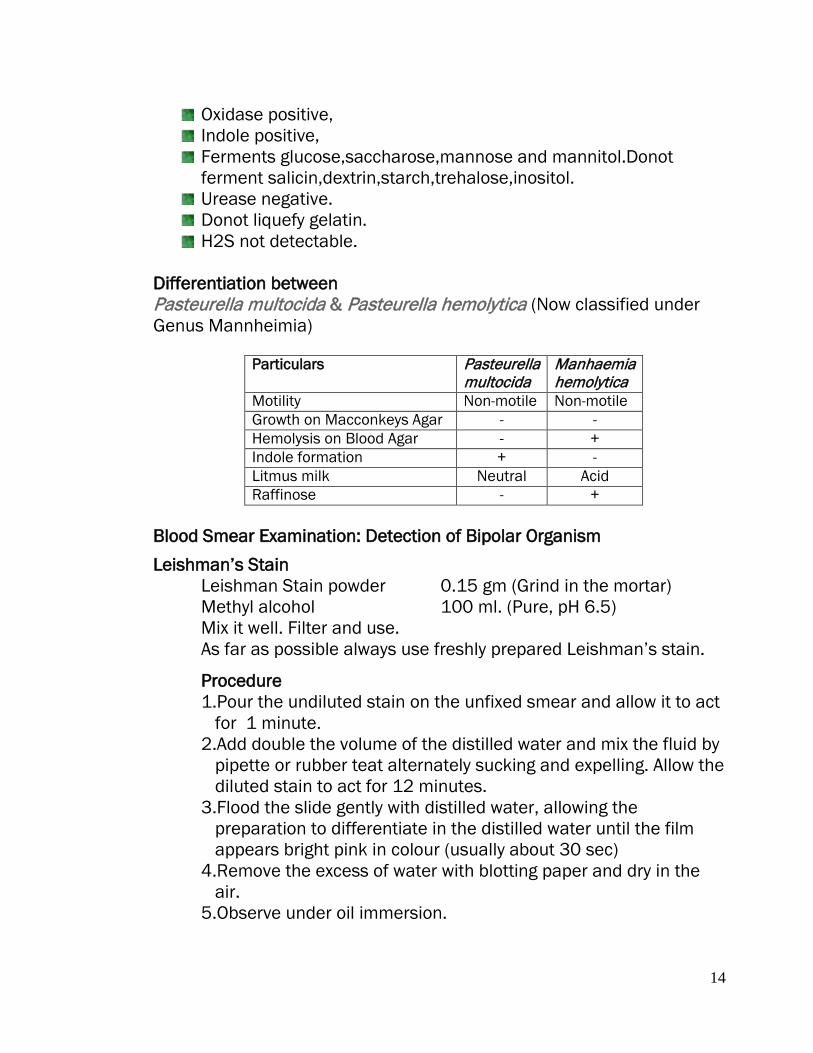
Oxidase positive, Indole positive, Ferments glucose,saccharose,mannose and mannitol.Donot ferment salicin,dextrin,starch,trehalose,inositol.
Urease negative. Donot liquefy gelatin. H2S not detectable.
Differentiation between Pasteurella multocida & Pasteurella hemolytica (Now classified under Genus Mannheimia)
Particulars Pasteurella multocida
Manhaemia hemolytica
Motility Non-motile Non-motile Growth on Macconkeys Agar - - Hemolysis on Blood Agar - + Indole formation + - Litmus milk Neutral Acid Raffinose - +
Blood Smear Examination: Detection of Bipolar Organism
Leishman’s Stain Leishman Stain powder 0.15 gm (Grind in the mortar) Methyl alcohol 100 ml. (Pure, pH 6.5) Mix it well. Filter and use. As far as possible always use freshly prepared Leishman’s stain.
Procedure 1.Pour the undiluted stain on the unfixed smear and allow it to act for 1 minute. 2.Add double the volume of the distilled water and mix the fluid by pipette or rubber teat alternately sucking and expelling. Allow the diluted stain to act for 12 minutes. 3.Flood the slide gently with distilled water, allowing the preparation to differentiate in the distilled water until the film appears bright pink in colour (usually about 30 sec) 4.Remove the excess of water with blotting paper and dry in the air. 5.Observe under oil immersion.
15
Interpretation
Pasteurella multocida appears to be ‘bipolar’ rods with hallow space surrounding the bacilli indicating the presence of capsule.
Exercise
Q1.Justify - Pasteurella multocida appears to be ‘Bipolar’ . Q2.Differentiate between Pasteurella multocida and Escherichia coli.
*********

16
PLATE 1
1.Microscopic picture of Staphylococcus 2.Microscopic picture of Streptococcus
3.DNAase activity of Staphylococcus aureus DNAase Positive Test
4.Negative Test- Note the clearing around the colonies
5.CAMP Test: The vertical streak is a beta -hemolysin producing strain of Staph aureus, & at right angles to it is (3) S. agalactiae *Note the large area of complete lysis where the extracellular compound of S. agalactiae encounters the beta-lysin of S. aureus.
6.Coagulase Test
17
PRACTICAL NO. 4
ENTERIC INFECTIONS ISOLATION AND IDENTIFICATION OF ESCHERICHIA COLI
Escherichia coli
Morphology
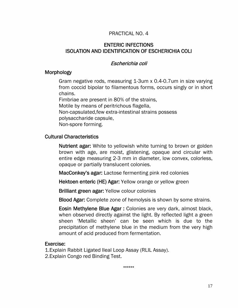
Gram negative rods, measuring 1-3um x 0.4-0.7um in size varying from coccid bipolar to filamentous forms, occurs singly or in short chains. Fimbriae are present in 80% of the strains, Motile by means of peritrichous flagella, Non-capsulated,few extra-intestinal strains possess polysaccharide capsule, Non-spore forming.
Cultural Characteristics
Nutrient agar: White to yellowish white turning to brown or golden brown with age, are moist, glistening, opaque and circular with entire edge measuring 2-3 mm in diameter, low convex, colorless, opaque or partially translucent colonies.
MacConkey’s agar: Lactose fermenting pink red colonies
Hektoen enteric (HE) Agar: Yellow orange or yellow green
Brilliant green agar: Yellow colour colonies
Blood Agar: Complete zone of hemolysis is shown by some strains.
Eosin Methylene Blue Agar : Colonies are very dark, almost black, when observed directly against the light. By reflected light a green sheen ‘Metallic sheen’ can be seen which is due to the precipitation of methylene blue in the medium from the very high amount of acid produced from fermentation.
Exercise: 1.Explain Rabbit Ligated Ileal Loop Assay (RLIL Assay). 2.Explain Congo red Binding Test.
******

18
PRACTICAL NO. 5
ENTERIC INFECTIONS ISOLATION AND IDENTIFICATION OF SALMONELLA, KLEBSIELLA,
PROTEUS & SHIGELLA
Salmonella Pullorum /Salmonella Gallinarum Collection of Material
Salmonellosis: Intestinal swab, heart blood, bile, liver, spleen etc. in a sterile container on ice.
Morphology
Gram-negative short plump rods, measuring 0.4-0.6um x 0.8-1.6um in size. Occur singly or in short chains Non-motile, non-spore forming Non-capsulated,Long filamentous forms occur.
Cultural Characteristics
Nutrient agar: Grayish white, smooth, glistening, opalescent, entire colonies. Variants with rough, dry & irregular edges, mucoid colonies are also encountered. MacConkey’s agar: Colorless or pale yellow, non-lactose fermenting colonies.
Hektoen enteric (HE) Agar: Blue green with black centers. Brilliant green agar: Whitish pink colour colonies.
XLD Agar: Colorless colonies with black centres.
Salmonella –Shigella Agar: Colorless colonies with black centres. Eosin Methylene Blue Agar: Non-lactose-fermenting colonies produce no acid from fermentation, so the lighter-colored alkaline reaction is seen.
19
DIAGNOSIS OF SALMONELLOSIS Plate agglutination test
Plate agglutination test can be employed for the diagnosis of Fowl typhoid or pullorum disease, by the detection of antibodies in suspected sera / blood. The whole blood test provides a rapid test for Fowl Typhoid & Pullorum disease that can be used on the farm. The sensitivity of the whole blood test is low and in inexperienced hands false positive and false negative results may be recorded. The test is useful when large no of samples / birds are to be tested, i.e., field level.
Material Required
Salmonella polyvalent colored antigen, Suspected sera / blood, Glass plate.
Method
1.Serum 0.02 ml is mixed with polyvalent crystal violet stained antigen (0.02ml). 2.The plate/slide is rocked gently for 2 minutes, after which the test is read.
Interpretation Positive Test: Agglutination / Clump formation within 2 minutes. Negative Test: No agglutination (No clump formation) The test components are stored at 4OC and must have reached room temperature before being used. Due to transient, non-specific reactions, positive/suspicious sera should be retested after heat inactivation at 56OC for 30 min.
Klebsiella pneumoniae
Collection of Material Spleen,lung,blood,nasal discharge..
20
Morphology Gram-negative rods, Non-motile, Possess well-defined polysaccharide capsule, Measuring 0.9um-1.7 um by1.8um-3.7um in length with parallel or bulging sides and slightly pointed or rounded ends. Arranged either in pairs or singly. Coccid form most prevalent in older cultures Non-spore forming.
Cultural Characteristics
Nutrient agar: Large, raised and round colonies. Yellowish in color, moist glistening and viscid colonies and mucoid appearance because of loose slime accumulation. Gelatin stab: ‘Nail-head’ type of growth.
MacConkey’s agar: Lactose fermenting, large, mucoid, red colonies with entire edge.
Christensen’s medium (Urease test): Urease positive
Eosin Methylene Blue Agar: colonies are less dark. Often a dark center is seen surrounded by a wide, light-colored, mucoid rim – resulting in a ‘Fish-eye’ type of colony (Black centered colonies).
Proteus mirabilis / Proteus vulgaris
Collection of Material Faecal sample, urine. Morphology
Gram-negative plump rods measuring 0.5um by 1-3um in size, In young cultures most of them are long, filamentous and curved. Motile by means of peritrichous flagella Non-spore forming, Non-capsulated
Cultural Characteristics
Nutrient agar:Transparent, thin film like swarming growth on nutrient agar is seen.Emits characteristic “fishy / seminal smell”
21
MacConkey’s agar: Non-lactose fermenting colorless / pale yellow colonies
Christensen’s medium (Urease test): Urease positive
Eosin Methylene Blue Agar: Non-lactose-fermenting colonies produce no acid from fermentation, so the lighter-colored alkaline reaction is seen.
Shigella dysenteriae
Collection of Material Faecal sample.
Morphology
Gram-negative short plump rods, Measuring 1.0um-3.0um by 0.4um - 0.6 um. Occurs singly Non-motile,Non-capsulated, Non-spore forming.
Cultural Characteristics
Nutrient agar: Rough, irregular, mucoid colonies, 3-5mm in diameter. Grayish or colorless colonies and translucent Colonies are raised, opaque and hemispherical. Surface lobulated gives the appearance of radially striated colonies in transmitted light. On ageing smooth border develops from the edge of the colony.
MacConkey’s agar: Non-lactose fermenting colonies, colorless / pale yellow color colonies.
Hektoen enteric (HE) Agar: Green colonies.
XLD Agar: Red colonies without black centers. Salmonella – Shigella Agar: Colorless colonies without black centers.
Eosin Methylene Blue Agar:Non-lactose-fermenting colonies produce no acid from fermentation, so the lighter-colored alkaline reaction is seen.

22
Exercise Q1.Enlist the lactose fermenting & non-lactose fermenting genera of the family Enterobacteriaceae.
Q2.Give the flowchart for the isolation & Identification of Salmonella spp. from the clinical material.
Q3.Write biochemical reactions of the following enteric bacteria.
Biochemical Tests
E.coli
Sal. Pullorum
Sal. Gallinarum
Proteus vulgaris
Proteus mirabilis
Shigella Kleb. Pneu.
Indole Methyl Red Voges Prouskeur Citrate Nitrate Reduction Catalase H2S Motility Urease Malonate Utilization
Phenylalanine Deaminase
Oxidase Arginine Ornithine TSI Slant TSI Butt Sugar Fermentations Tests Lactose Glucose Sucrose Maltose Arabinose Starch Dextrin Inositol Sugar Fermentation: A - Acid; G - Gas; AG - Acid & Gas
*********
23
PLATE 2
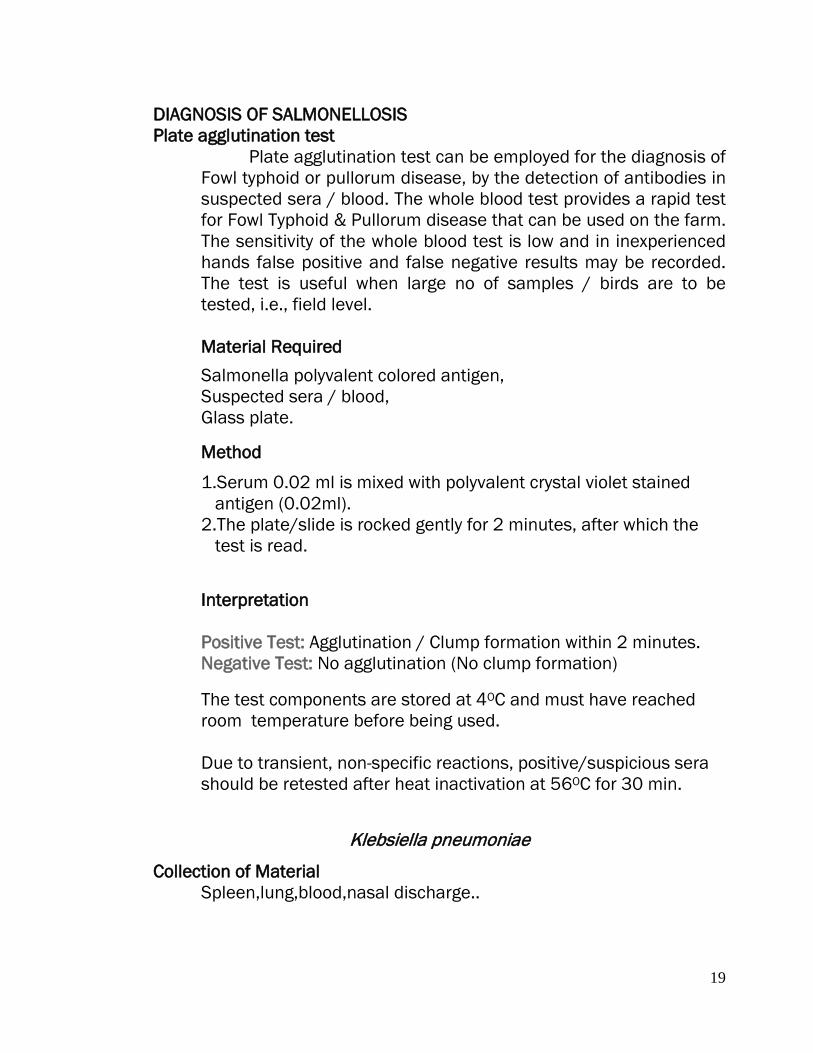
7.E. coli on MacConkeys Agar Lactose Fermenting Pink Red Colonies
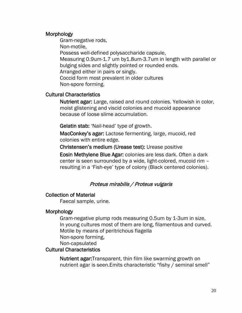
8.E. coli on EMB: Characteristic “Metallic sheen’’
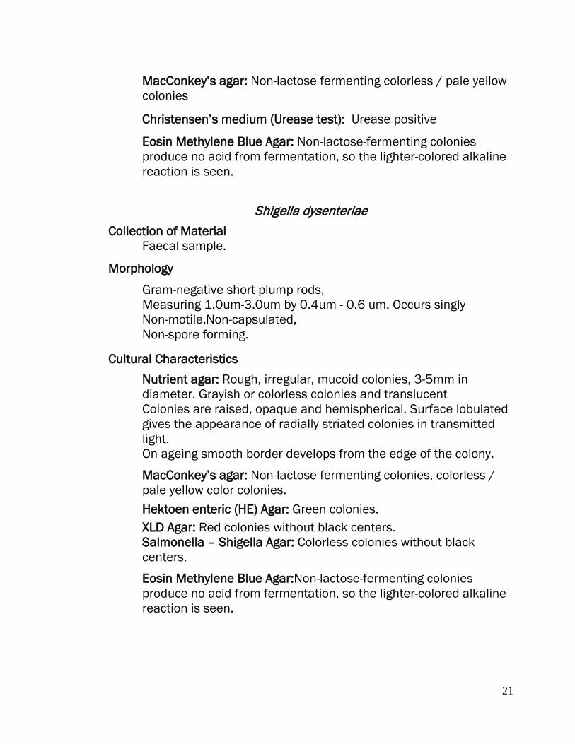
9.Swarming of Proteus mirabilis on an agar plate showing - typical growth rings.
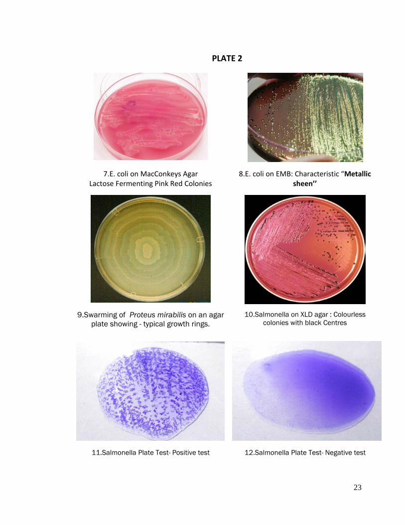
10.Salmonella on XLD agar : Colourless colonies with black Centres
11.Salmonella Plate Test- Positive test
12.Salmonella Plate Test- Negative test
24
PRACTICAL NO. 6
DIAGNOSIS OF BRUCELLOSIS
Isolation & Identification
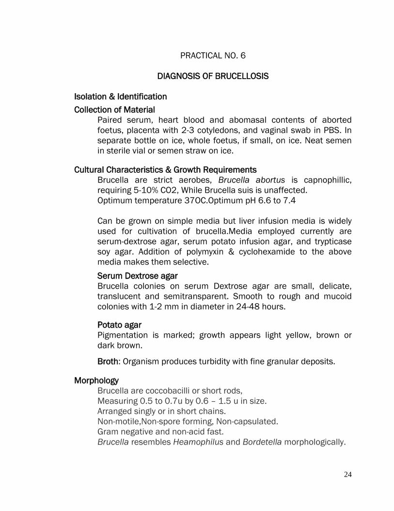
Collection of Material Paired serum, heart blood and abomasal contents of aborted foetus, placenta with 2-3 cotyledons, and vaginal swab in PBS. In separate bottle on ice, whole foetus, if small, on ice. Neat semen in sterile vial or semen straw on ice.
Cultural Characteristics & Growth Requirements
Brucella are strict aerobes, Brucella abortus is capnophillic, requiring 5-10% CO2, While Brucella suis is unaffected. Optimum temperature 37OC.Optimum pH 6.6 to 7.4
Can be grown on simple media but liver infusion media is widely used for cultivation of brucella.Media employed currently are serum-dextrose agar, serum potato infusion agar, and trypticase soy agar. Addition of polymyxin & cyclohexamide to the above media makes them selective.
Serum Dextrose agar Brucella colonies on serum Dextrose agar are small, delicate, translucent and semitransparent. Smooth to rough and mucoid colonies with 1-2 mm in diameter in 24-48 hours.
Potato agar Pigmentation is marked; growth appears light yellow, brown or dark brown.
Broth: Organism produces turbidity with fine granular deposits. Morphology Brucella are coccobacilli or short rods,
Measuring 0.5 to 0.7u by 0.6 – 1.5 u in size. Arranged singly or in short chains. Non-motile,Non-spore forming, Non-capsulated. Gram negative and non-acid fast. Brucella resembles Heamophilus and Bordetella morphologically.

25
Biochemical Properties Do not ferment carbohydrates, Catalase positive, Oxidase positive (except Brucella ovis & Brucella neotomae) Urease positive Nitrates are reduced to nitrites, Indole negative, MR & VP negative & Citrate is not utilized.
Serological tests for diagnosis
Agglutination Test
Principle: When a particulate antigen is mixed with the specific antibody in the presence of electrolytes at suitable temperature and pH ,clumps are formed at the bottom of the tube i.e., agglutination reaction is seen. STAT and Rose Bengal Plate tests are based on the principle of agglutination reaction.
1.Rose Bengal Plate Test
The test is used for preliminary screening and is of qualitative test.
Material required Brucella colored antigen, test serum, positive serum, Negative serum, and slide or gooch plate.
Procedure 1.place one drop of suspected serum on the slide/plate. 2Place a drop of colored antigen next to the serum sample. 3.Mix the reagents with the toothpick and rotate the slide/plate gently. 4.After few minutes, in case of positive reaction, clumping takes place. 5.Confirm the reaction with suitable controls.

26
2.Standard Tube Agglutination Test (STAT)
Material Required Brucella plain antigen, test serum positive serum, Negative serum, Carbol saline (Normal saline with 0.5%carbolic acid), Agglutination tubes 8 Nos., Rack for holding test tubes.
Procedure 1.Place 8 agglutination tubes in a rack, number serially 1 to 8. 2.Add 0.9 ml of diluent to the first test tube and 0.5 ml to rest of the tubes. 3.Add 0.1ml of serum to the first test tube mix it well and transfer 0.5 ml to the 2nd tube. 4.Mix thoroughly and transfer 0.5ml to the third tube and continue this process up to 7th tube. Keep 8th tube as antigen control. 5.Discard 0.5 ml i.e., diluted serum from 7th tube. 6.Add 0.5 ml of Brucella plain antigen to all the tubes. Mix and incubate at 37 OC for 24 hours. 7.Keep positive control. Interpretation If agglutination occurs, the clumps of antigen and antibody complexes will settle down leaving the clean supernatant. In case of no agglutination, the turbid suspension remains the same.
Exercise:
Q1.What is the right time to collect the serum sample for the sero-diagnosis of Brucellosis? Q2.How will you interpret the STAT titres for brucellosis in case of cattle, goat, sheep and human?
*********
27
PRACTICAL NO. 07
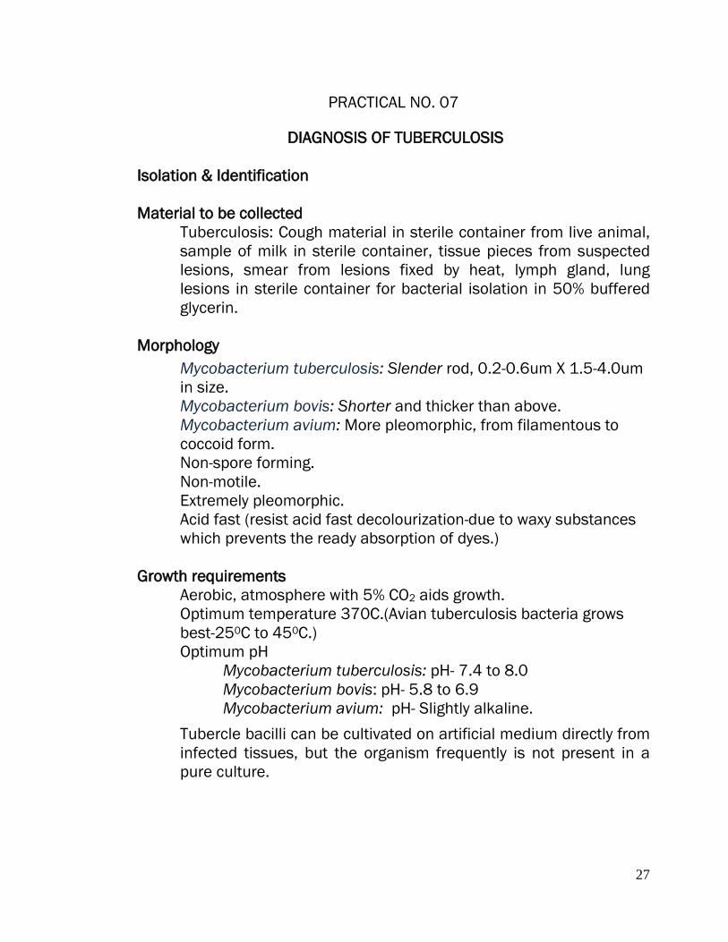
DIAGNOSIS OF TUBERCULOSIS Isolation & Identification Material to be collected
Tuberculosis: Cough material in sterile container from live animal, sample of milk in sterile container, tissue pieces from suspected lesions, smear from lesions fixed by heat, lymph gland, lung lesions in sterile container for bacterial isolation in 50% buffered glycerin.
Morphology
Mycobacterium tuberculosis: Slender rod, 0.2-0.6um X 1.5-4.0um in size. Mycobacterium bovis: Shorter and thicker than above. Mycobacterium avium: More pleomorphic, from filamentous to coccoid form. Non-spore forming. Non-motile. Extremely pleomorphic. Acid fast (resist acid fast decolourization-due to waxy substances which prevents the ready absorption of dyes.)
Growth requirements
Aerobic, atmosphere with 5% CO2 aids growth. Optimum temperature 370C.(Avian tuberculosis bacteria grows best-250C to 450C.) Optimum pH
Mycobacterium tuberculosis: pH- 7.4 to 8.0 Mycobacterium bovis: pH- 5.8 to 6.9 Mycobacterium avium: pH- Slightly alkaline.
Tubercle bacilli can be cultivated on artificial medium directly from infected tissues, but the organism frequently is not present in a pure culture.

28
Petroff’s Method To overcome contamination, while isolating the mycobacterium following procedure can be followed to overcome the contamination.
1. Take 3% solution of NaOH + tissue/exudates(suspected)-equal
volume. 2. Shake the tube vigorously. 3. Stand for 30 mins. 4. Neutralize by 3N Hcl. 5. Centrifuged. 6. Discard the supernatant. 7. Collect the sediment and is ‘seeded’ over the surface of slants
of suitable culture medium Cultural characteristics
Stonebrinks Medium Suitable for isolation of bovine strains of the tubercle bacillus.(No Glycerol-No inhibition of bovine type).
Lowenstein-Jensen medium
Mycobacterium tuberculosis: Thicker, wrinkled, cream buff, white heaped up,crumb like.
Mycobacterium bovis: Small, shining, grey and later coalesce to form buff white with fine granular surface.
Mycobacterium avium: Moist creamy, smooth, no surface pellicle.
[Mycobacterium tuberculosis: Growth is inhibited on addition of glycerin . Mycobacterium bovis & Mycobacterium avium: Glycerin aids growth and prevents desiccation.]
Biochemical properties
Catalase positive. H2S positive (slightly). MR, VP-Negative. Slight acid reaction-In glucose, maltose, trehalose & glycerol.

29
Smear Examination The acid-fast bacilli are demonstrated in tuberculosis and Johne’s disease in animals.
Ziehl-Neelsen staining method 1. Films are made dried and fixed by flaming. 2. Cover the slide with filtered carbol fuchsin and heat until steam
rises. Allow the preparation to stain for 5 min, heat gently .The stain must not be allowed to evaporate and dry on the slide. If necessary pour on more carbol fuchsin to keep the whole slide covered. (The slide may be heated with a torch prepared by twisting a small piece of cotton wool on to the tip of an inoculating wire and soaking it in methylated spirit before lighting. when steam rises from the slide, remove and extinguish the torch. after about 1 minute recharge the torch with spirit, relight it and again heat the slide until the steam rises, continue this way for 5 min.
3. Wash with water. 4. Cover the slide with 20% sulphuric acid. Keep for 1` min. wash
with water. 5. Treat with 95% alcohol for 2 min 6. Wash with water 7. Counterstained with Loeffler’s methylene blue. Or dilute
malachite green for 15-20 sec. 8. Wash, blot dry and mount.
Interpretation Acid-fast bacilli take pink red colour against blue background.
Exercise
Q1.Explain: a. Tuberculin test.
*********

30
PRACTICAL NO. 08
DIAGNOSIS OF JOHNE’S DISEASE Collection Of Material
Rectal pinch smears, bowl washings. In dead animals terminal portion of ileum with ileo-caecal valve, mesenteric lymph gland. Films are made dried and fixed by flaming. Stained with Ziehl-Neelsen staining method.
Isolation & Identification Herrold's egg yolk medium with mycobactin Modified Dubos's medium Middlebrook 7H9, 7H10 and 7H11 Löwenstein-Jensen medium with mycobactin
Rectal Pinch Smear Examination Procedure 1.Insert hand per rectally and pinch out the mucous membrane of the rectum. 2.Wash the mucous membrane with sterile saline. 3.Place the mucous membrane on the slide and munch with another slide. 4.Dry the smear and stain with Ziehl-Neelsen staining method.
Ziehl-Neelsen stain 1 Films are made dried and fixed by flaming. 2 Cover the slide with filtered carbol fuchsin and heat until steam rises. Allow the preparation to stain for 5 min, heat gently .The stain must not be allowed to evaporate and dry on the slide. If necessary pour on more carbol fuchsin to keep the whole slide covered.(The slide may be heated with a torch prepared by twisting a small piece of cotton wool on to the tip of an inoculating wire and soaking it in methylated spirit before lighting. when steam rises from the slide, remove and extinguish the torch. after about 1 minute recharge the torch with spirit, relight it and again heat the slide until the steam rises, continue this way for 5 min.) 3.Wash with water.
31
4.Cover the slide with 20% sulphuric acid. Keep for 1` min. wash with water. 5.Treat with 95% alcohol for 2 min. 6.Wash with water & Counter stain with Loeffler’s methylene blue. or dilute malachite green for 15-20 sec. 7.Wash, blot dry and mount. Interpretation Acid-fast bacilli take pink red colour against blue background.
Exercise
Q1Write the colony characteristic and microscopic picture of Mycobacterium avium subspecies paratuberculosis. Q2.Explain:
a. Johnin test. *********

32
PLATE 3
13.RBPT- Brucella positive test 14.RBPT- Brucella Negative test
15. Microscopic Picture of Mycobacterium tuberculosis
16.Mycobacterium tuberculosis on L J Medium
www.pathport.vbi.vt.edu
17. Clostridium chauvoei Anaerobic cultivation on Blood Agar
18. Clostridium tetani on FFA

33
PRACTICAL NO. 09
DIAGNOSIS OF BLACK QUARTER
ANAEROBIC CULTIVATION METHODS
Collection Of Material Piece of muscle from emphysematic area of BQ affected animal.
A variety of anaerobic culture methods are available for the culture of anaerobic organisms on the laboratory.
I.Exclusion of oxygen from the medium is the simplest method. And is effected by growing the organisms in freshly steamed liquid medium or deep stab in nutrient agar with 0.5% glucose / 1% ascorbic acid / 0.1% Cysteine / 0.1% sodium thioglycollate or particles of meat in cooked meat broth e.g. Robertson cooked meat medium. With minimal shaking and solidifying rapidly by placing the tubes in cold water.
Preparation of Robertson’s Cooked Meat Medium 1.Procure 500 gm bullock heart, mince it (shredded into small freckles) and add 1N NaOH 1.5 ml. Also add 500ml-distilled water. 2.Simmer the above for 20 minutes in boiling water. 3.Drain off the liquid. (Collect the liquid in another sterile container-, which can be used later for the preparation of Peptone Infusion Broth) 4. Minced boiled (cooked) meat is placed in sterile test tubes. 5.For the preparation of Peptone Infusion broth use the drained liquid ( as above) i.e.,500ml,add peptone 2.56 gm and Nacl 1.25 gm. 6.Steam for 20 mins (100OC) and cool immediately. 7.Add 1ml of pure HCl and filter. 8.Adjust pH 8.2 and steam for 30 mins at 100OC. 9.Adjust pH 7.8. (Peptone Infusion Broth). 10.Add the peptone infusion broth to the tubes with minced cooked meat (as above) in such a way that the level of Peptone infusion broth should be 2.5 cm above the level of cooked meat. 11.Adjust the pH 7.8 and autoclave.
34
II. Anaerobic jars (McIntosh Fildes’ anaerobic jar)
Material Required McIntosh Fildes’ anaerobic jar, Catalyst (Asbestos/Palladium), Resazurin indicator,10ml tap water.
Method
1.Clean the McIntosh Fildes’ anaerobic jar with spirit. 2.Pick up the stock culture of Clostridium maintained in the Robertson’s cooked meat media and draw approximately 0.1ml 0f inoculum in sterilized Pasteur pipette. 3.Discharge the inoculum at a corner of blood agar plate and spread by inoculating loop following quadrant pattern. 4.Place the blood agar plate along with chargedcatalyst. (Charging is done by pre-heating at 100 OC and risazurin indicator in the jar. 5.Cut open the ‘ Gas-Pack ’ at the corner with a scissor. Put 10 ml of tap water in the pack and immediately place in upright position in the jar. And tighten the lid. 6.Place the jar in incubator at 37OC for 48-72 hrs. (Maintenance of anaerobic condition in the jar will be shown by risazurin indicator turning white from its original pink colour . 7.Open the jar after 72 hrs and examine the plate for Clostridial growth. 8.Prepare the smear of a suggestive colony, stain with Grams method and observe under microscope.
Exercise Q1.Explain the principle of ‘ Gas-Pack ’ system. Q2.Write the morphological features & colony characteristics of Cl.tetani, Cl.chauvoei & Cl/welchi Q3.Explain: Nagler’s reaction. Q4.Write the procedure of inoculating the suspected material for the isolation of Clostridia using Robertson’s cooked meat media.
*********

35
PRACTICAL NO. 10
DIAGNOSIS OF ENTEROTOXAEMIA
MOUSE INOCULATION TEST
Collection Of Material Enterotoxaemia: Contents of small intestine on ice, kidney and urine.
Mouse Inoculation Test
Method 1.Mix contents of small intestine i.e., 2gm with 2 ml of antitoxin of Clostridium welchii type D. 2.Inject 1ml of above mixture intramuscularly in mice. (Group A) 3.For Group B, mix contents of small intestine i.e., 2gm with 2 ml of distilled water and inject 1ml.intramuscularly in mice. 4.After 24 –48 hours take the results.
Interpretation If Group A survives and Group B dies, it indicates the presence of toxin of Clostridium welchii type D.
Exercise
Q1.Enlist the toxins produced by Clostridium welchii. Explain their activity and disease produced by the different types. Q2.Write the lethal dose of toxin produced by Clostridium welchii For Guinea pig.
*********

36
PRACTICAL NO. 11
DIAGNOSIS OF WOODEN TONGUE
Wooden tongue is a well-defined disease of the soft tissues of the mouth region in adult cattle. It is caused by Actinobacillus lignieresii, part of the normal bacterial flora of the upper digestive tract. The bacteria usually invade the skin through a wound or minor trauma caused by sticks or straw or barley awns.
Collection of Material Wooden Tongue: Smears from pus lesions, pus in vials on ice from affected materials.
Morphology:
Actinobacillus lignieresii : Small, Gram Negative rods, coccobacilli, 1.5ux04 u in size with grey or white granules ~1mm (In direct smear examination), small granules can be observed at the poles of the bacteria (Examine granules under a coverslip in 10% NaOH, prepare smear & stain). Pleomorphism, arranged in pairs or singly. Long filamentous forms can be observed if the medium contains glucose or maltose.
Non-motile Cultural Characteristics:
Facultatively anaerobic, 37OC optimum temperature.
Serum Agar or Blood Agar: Small, translucent, smooth glistening colonies on 24-48 Hrs incubation. No hemolysis on blood agar.
MacConkey Agar: Grows and is lactose fermenter.
Biochemical Tests:
Catalase Positive Ornithine decarboxylase negative, Ttryptophanase Negative, Urease Positive, Methyl red Negative, Voges-Proskauer Variable,
37
Hydrogen Sulfide Positive, Ferments Glucose,Lactose,Maltose , Mannitol & Sucrose . Donot ferment Salicin,Rhamnose,Trehalose, Inulin ,Starch.
Exercise
Q1. Differentiate between Actinobacillu lignieresii & Actinomyces bovis. Q2.Enlist other species of Actinobacillus and disease conditions produced.
*****
38
PRACTICAL NO. 12
DIAGNOSIS OF ANTHRAX
Isolation & Identification Collection of Material
Anthrax: Flame fixed blood smears of cattle and sheep. From subcutaneous swelling in horses, swine and dogs. Swab of blood from ear vein for cultural examination from dead animals. A small piece from tip of ear or muzzle (0.5 cm approx) in saline or without any preservatives in sterile glass test tubes or bottle on ice duly sealed. It is not advisable to open the carcass suspected for a Anthrax in field. If opened, it should be properly disposed off by burning. All natural orifices of dead animal as well as bleeding surfaces may be sealed with cotton soaked in carbolic acid.
Morphology
Largest pathogenic Bacteria, 1-1.5 X4-8 u in size. Rod shaped bacilli with truncated ends , Arranged in chain ‘Bamboo’ like appearance Gram Positive Non-motile Capsulated(D-glutamic acid-polypeptide) McFadyeans reaction Spore forming
Cultural Characteristics PLET medium is prepared by using heart-infusion agar base with
the addition of 0.25-0.3 g/litre EDTA and 0.04 g/litre thallous acetate. (NOTE: Thallous acetate is poisonous and should be handled with care.) The mixture is autoclaved and uniformly cooled to 50°C before adding the polymyxin at 30,000 units/litre & lysozyme at 300,000 units/litre. After mixing thoroughly, the agar is dispensed into Petri dishes.
39
Blood Smear Examination Demonstration of encapsulated B. anthracis in smears of blood or tissues from fresh anthrax-infected carcasses and growth of the organism on blood agar plates is relatively uncomplicated and within the capability of most bacteriology laboratories
Capsule visualization
Encapsulated B. anthracis present in tissues should be looked for in smears of these specimens that have been dried, fixed and stained with polychrome methylene blue (Mac'Fadyean's reaction). The capsule is not present on B. anthracis grown aerobically on nutrient agar or in nutrient broths, but can be seen when the virulent bacterium is cultured for a few hours in a few milliliters of blood (defribrinated horse blood seems to work best). Alternatively, the capsule is produced when the virulent B. anthracis is cultured on nutrient agar containing 0.7% sodium bicarbonate and incubated in the presence of CO2 (20% is optimal, but a candle jar works well). The agar is prepared by reconstituting enough nutrient agar base powder for 100 ml of agar in 90 ml of water. Autoclave and cool to 50°C in a water bath. Add 10 ml of a filter-sterilized (0.22-0.45 µm filter) 7% solution of sodium bicarbonate. Mix and pour into Petri dishes. The encapsulated B. anthracis will form mucoid colonies and the capsule can be visualised by making thin smears on microscope slides, fixing and staining with polychrome methylene blue as before. Preparation of Polychrome methylene blue (Mac'Fadyean's stain)
0.3 g of methylene blue is dissolved in 30 ml of 95% ethanol; 100 ml of 0.01% potassium hydroxide (KOH) is mixed with the methylene blue solution. Ideally, this should be allowed to stand exposed to the air, with occasional shaking, for at least 1 year to oxidise and mature.
[Addition of 1% Potassium carbonate ripens the stain quickly]

40
Polychrome methylene blue staining
1.Prepare a thin, small smear from small drops of blood or tissue f fluid. 2.After fixing and drying, a small (approximately 20 µl) drop of stain is placed on the smear and spread over it with an inoculating loop. 3.After 1 minute the stain is washed with water into a hypochlorite solution (10,000 ppm available chlorine). 4.The slide is blotted, air-dried and observed initially using the x10 objective lens under which the short chains appear like short hairs; once found, these can be observed under oil immersion (x1000). 5.The capsule stains pink whereas the bacillus cells stain dark blue. The cells are found in pairs or short chains and are often square- ended. The Gram & regular Giemsa stains do not reveal the capsule.
Exercise
Q1. How will you differentiate between Bacillus anthracis and Bacillus cereus.
Q2.Explain: Ascoli’s test. *********

41
PRACTICAL NO.13
DIAGNOSIS OF GLANDERS
Collection of clinical material: Nasal muco purulent discharge, pus from the lesions. Morphology Burkholderia mallei : The organisms are fairly numerous in smears from fresh lesions, but in older lesions they are scanty.
Extracellular, Gram-negative rods , rounded ends, 2–5 μm long and 0.3–0.8 μm wide with granular inclusions of various size.
Nonmotile,
Do not have a readily visible capsule, under the light microscope, or The presence of a capsule-like cover has been established by electron microscopy.
Non-spore forming.
In older cultures, there is much pleomorphism. Branching filaments form on the surface of broth cultures. Cultural Characteristics & Growth Requirements: Aerobic & facultatively anaerobic only in the presence of nitrate.
Grows well, but slowly, on ordinary culture media, 72-hour incubation of cultures is recommended; glycerol enrichment is particularly useful.
Glycerol agar: Confluent, slightly cream-coloured , smooth, moist, and viscuous growth.With continued incubation, the growth thickens and becomes dark brown and tough.
Grows well on glycerol potato agar and in glycerol broth, on which a slimy pellicle forms.
Nutrient agar: Growth is much less luxuriant, and Growth is poor on gelatin.
In samples not obtained under sterile conditions B. mallei is regularly overgrown by other bacteria.

42
Biochemical Tests: Litmus milk is slightly acidified by B. mallei, and coagulation may occur after long incubation.
Reduces nitrates to nitrites. Glucose is the only carbohydrate that is fermented (slowly and inconstantly), other workers have shown that if an appropriate medium and indicator are used, glucose and other carbohydrates, such as arabinose, fructose, galactose and mannose, are consistently fermented by B. mallei .
Indole is not produced, Horse blood is not haemolysed No diffusible pigments are produced in cultures.
In contaminated samples, supplementation of media with substances that inhibit the growth of Gram-positive organisms (e.g. crystal violet, proflavine) has proven to be of use, as has pretreatment with penicillin (1000 units/ml for 3 hours at 37°C).
Selective medium developed is composed of Polymyxin E (1000 units), Bacitracin (250 units), and Actidione (0.25 mg) incorporated into nutrient agar (100 ml) containing glycerine (4%), donkey or horse serum (10%), and ovine haemoglobin or tryptone agar (0.1%).
Resistance & Physical Properties: Little resistance to drying, heat, light or chemicals, so that survival beyond 2 weeks is unlikely . Under favourable conditions, however, it can probably survive a few months. Burkholderia mallei can remain viable in tap water for at least 1 month.
For disinfection, benzalkonium chloride or ‘roccal’ (1/2,000), sodium hypochlorite (500 ppm available chlorine), iodine, mercuric chloride in alcohol, and potassium permanganate have been shown to be highly effective against B. mallei .
Phenolic disinfectants are less effective. Strauss’s Test
Suspected material is inoculated intraperitoneally into a male guinea-pig. As this technique has a sensitivity of only 20%, the inoculation of at least five animals is recommended.

43
Positive material will cause a severe localised peritonitis and orchitis (the Strauss reaction). The cheesy mass will protrude out through the scrotum .
The number of organisms and their virulence determines the severity of the lesions.
Other Tests
Polymerase Chain Reaction & Real Time PCR can be used in the diagnosis of glanders in horses.
Enzyme-linked immunosorbent assays Both plate and membrane (blot) enzyme-linked immunosorbent assays (ELISAs) have been reported for the serodiagnosis of glanders.
The mallein test The mallein purified protein derivative (PPD), which is available commercially, is a solution of water-soluble protein fractions of heat-treated B. mallei. The test depends on infected horses being hypersensitive to mallein. Advanced clinical cases in horses and acute cases in donkeys and mules may give inconclusive results requiring additional methods of diagnosis to be employed. The intradermo-palpebral test This is the most sensitive, reliable and specific test for detecting infected perissodactyls or odd-toed ungulates, and has largely displaced the ophthalmic and subcutaneous tests:
0.1 ml of concentrated mallein PPD is injected intradermally into the lower eyelid and the test is read at 24 and 48 hours.
A positive reaction is characterised by marked oedematous swelling of the eyelid, and there may be a purulent discharge from the inner canthus or conjunctiva. This is usually accompanied by a rise in temperature.
Negative response, there is usually no reaction or only a little swelling of the lower lid.
Exercise Q1. What Is Glanders & Farcy Act.
Q2. What is notifiable disease.Enlist the notifiable diseases in animals/birds.
*******
44
PLATE 4
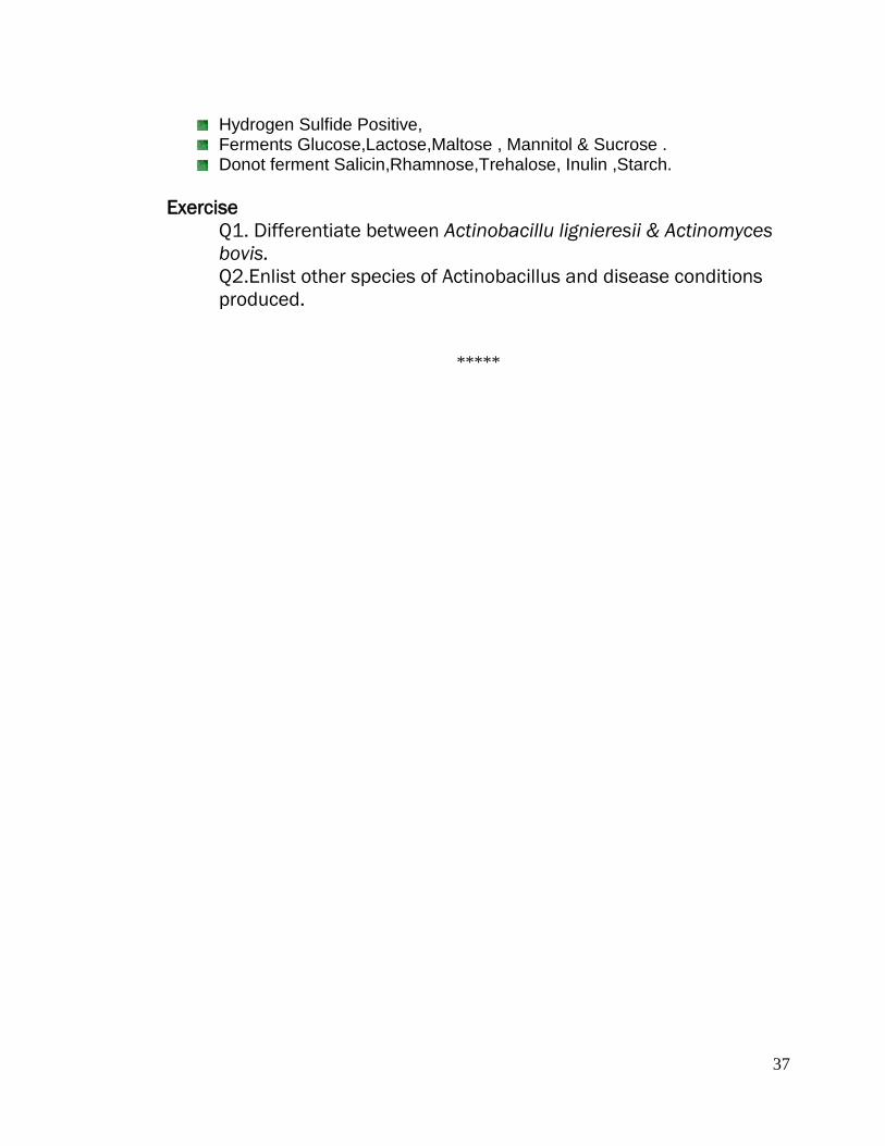
19.Robertson Cooked Meat Medium: Note the growth of Saccharolytic
Clostridia- Pink Coloration
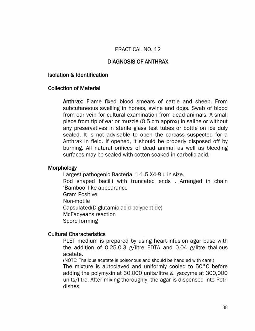
20.Actinobacillus lignieresii
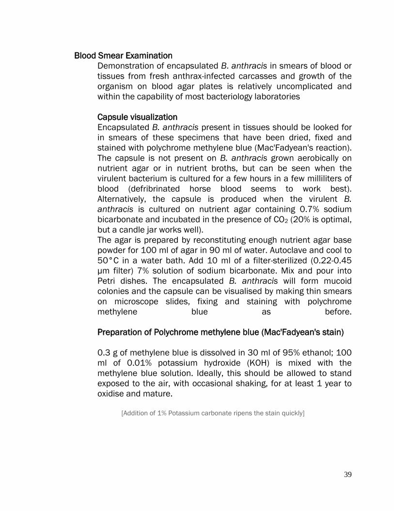
21.Colony characteristics of Bacillus anthracis
22.Bacillus anthracis. in tissue seen in short chains surrounded by a common
capsule- Toluidine blue stain
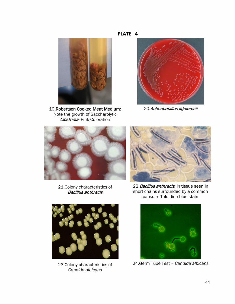
23.Colony characteristics of Candida albicans
24.Germ Tube Test – Candida albicans
45
PRACTICAL NO.14
STUDY OF GENUS ASPERGILLUS Genus Aspergillus
There are near about 300 species worldwide in distribution. Aspergilli are ubiquitous in nature. Important Aspergillus spp. From veterinary point of view are enlisted below:
Aspergillus flavus Aspergillus niger Aspergillus fumigatus Aspergillus ochracious.
Isolation of Fungi on Sabouraud’s Agar
Sabouraud’s Agar medium is suitable for the growth of Aspergillus species.
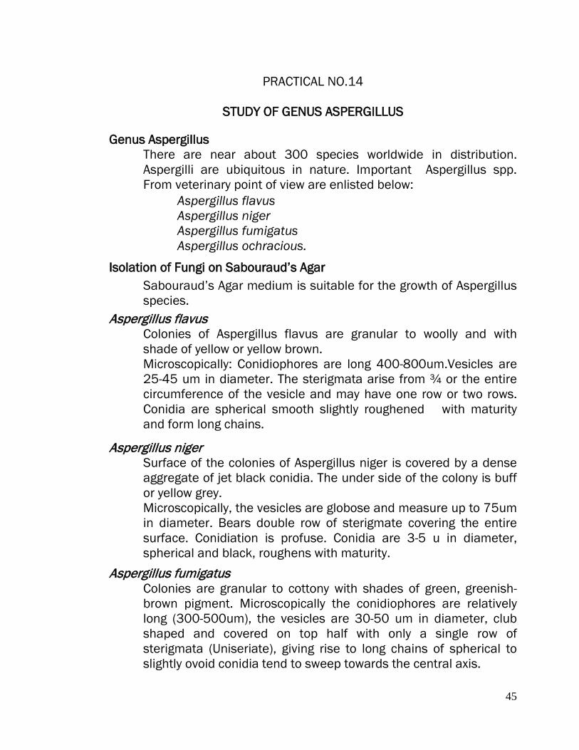
Aspergillus flavus Colonies of Aspergillus flavus are granular to woolly and with shade of yellow or yellow brown. Microscopically: Conidiophores are long 400-800um.Vesicles are 25-45 um in diameter. The sterigmata arise from ¾ or the entire circumference of the vesicle and may have one row or two rows. Conidia are spherical smooth slightly roughened with maturity and form long chains.
Aspergillus niger Surface of the colonies of Aspergillus niger is covered by a dense aggregate of jet black conidia. The under side of the colony is buff or yellow grey. Microscopically, the vesicles are globose and measure up to 75um in diameter. Bears double row of sterigmate covering the entire surface. Conidiation is profuse. Conidia are 3-5 u in diameter, spherical and black, roughens with maturity.
Aspergillus fumigatus Colonies are granular to cottony with shades of green, greenish-brown pigment. Microscopically the conidiophores are relatively long (300-500um), the vesicles are 30-50 um in diameter, club shaped and covered on top half with only a single row of sterigmata (Uniseriate), giving rise to long chains of spherical to slightly ovoid conidia tend to sweep towards the central axis.

46
Staining Of Fungi In Wet Mounts With Lactophenol Blue Stain solution Phenol crystals 20g Lactic acid 20ml Glycerol 40ml Distilled water 20ml Cotton blue or methyl blue 0.075g Dissolve the phenol crystals in the liquids by gentle warning and then add the dye.
Needle mount method
1.Place one drop of 95% alcohol with needles or straight wires. When it is satisfactorily spread, let most of the alcohol evaporate and then add a drop of stain. 2.Apply cover slip, avoiding bubbles and exert gentle pressure if the fungus fragments do not lie flat. 3.Remove any excess stain round the cover slip with the edge of a piece of blotting paper. Let the stain penetrate. For permanent preparations, seal the edges with nail varnish or cellulose lacquer.
Exercise
Q1. Write the composition of Sabouraud’s Agar medium. Q2. Describe the disease conditions produced by Aspergillus species. Q3.Explain the morphological & culture characteristics of Aspergillus ochracious.
*********

47
PRACTICAL NO.15
DIAGNOSIS OF DERMATOPHYTOSIS Dermatophytosis or Dermatomycosis relates to the fungal infection caused by three related genera of Fungi Imperfecti namely Trichophyton, Microsporum & Epidermophyton. Genus Trichophyton
Trichophyton rubrum Trichophyton verrucosum Trichophyton mentagrophytes Trichophyton violaceum Trichophyton schoenleinii Trichophyton simii
Genus Microsporum Microsporum canis Microsporum audouinii Microsporum distortum Microsporum equinum Microsporum gypseum
Genus Epidermophyton Epidermophyton floccosum
Diagnosis
1. Skin Scrapping Examination Hydrolysation and partial digestion of keratin by 10-30% Potassium hydroxide can be hastened by gently heating the slide under low flame. Arrangement of arthrospores in infected hair: Staining of fungi In wet mounts with Lactophenol Blue Stain solution
Phenol crystals 20g Lactic acid 20ml Glycerol 40ml Distilled water 20ml Cotton blue or methyl blue 0.075g Dissolve the phenol crystals in the liquids by gentle warning and then add the dye.
48
Staining Method 1.Take the scrapping and along with one drop of 95% alcohol place with needles or straight wires on the slide. When it is satisfactorily spread, let most of the alcohol evaporate and then add a drop of stain. 2.Apply cover slip, avoiding bubbles and exert gentle pressure if the fungus fragments do not lie flat.
3.Remove any excess stain round the cover slip with the edge of a piece of blotting paper. Let the stain penetrate. For permanent preparations, seal the edges with nail varnish or cellulose lacquer. 2.Wood’s Lamp When viewed under the filtered UV rays of Wood’s Lamp the Hairs infected with M. canis and certain other species of fungi fluoresce with yellow-green coloration while the species of Trichophyton mentioned below either do not show any kind of fluorescence or very poor fluorescence is seen with some species. Whereas Trichophyton simii shows bright yellow –green fluorescence when exposed to the Woods lamp .
M.canis Bright yellow-green fluorescence M audouinii Bright yellow-green fluorescence M.distortum Bright yellow-green fluorescence M.equinum Bright yellow-green fluorescence M.gypseum Weak fluorescence T rubrum No fluorescence T verrucosum No fluorescence T mentagrophytes No fluorescence T.violaceum No fluorescence T.Schoenleinii Very poor fluorescence/None T.simii Bright yellow-green fluorescence
As many species donot fluoresce on the exposure to the Wood’s UV Lamp, Negative results with Wood’s Lamp donot rule out the presence of ringworm infection. 3.Isolation & Identification Sample collection: Sample taken for the laboratory diagnosis should include the material from all parts of the lesion. Preliminarily cleanse the
49
affected part/lesion with 70% ethanol. It helps in reducing the bacterial contamination. Scales and crusts should be scrapped with the blunt scalpel and hairs should be plucked from the lesions ( never cut).Collect the scales, crust/hairs in a paper envelope and never collect the material in a bottle with closed cap.
Cultural & morphological Characters:
Dermatophytes Test Medium supplemented with cyclohexamide or Sabraoud’s Dextrose Agar Medium is used for isolation and identification.
Trichophyton rubrum
Colony Characteristics
Colonies are flat to slightly raised, white to cream, suede-like with a pinkish-red reverse
Microscopic Most cultures have numerous clavate to pyriform microconidia and moderate numbers of smooth, thin walled multiseptate, slender cylindrical macroconidia
Trichophyton verrucosum
Colony Characteristics
Colonies are slow growing, small, buttonor-disk-shaped, white to cream coloured,with a suede-like to velvety surface, a raised centre, and flat periphery with some submerged growth. Reverse pigment may vary from non-pigmented to yellow.
Microscopic
All strains produce typical chains of chlamydoconidia, often referred to as "chains of pearls", when grown in brain heart infusion broth containing para-aminobenzoic acid (P.A.B.) and agar at 37oC.
Key features include culture characteristics and requirements for thiamine and inositol, large ectothrix invasion of hair, clinical lesions and history.
Trichophyton mentagrophytes
Colony Characteristics
Colonies are generally flat, white to cream in colour, with a powdery to granular surface. Some cultures show central folding or develop
50
raised central tufts or pleomorphic suede-like to downy areas. Reverse pigmentation is usually a yellow-brown to reddish-brown colour.
Microscopic Numerous single-celled, spherical to subspherical microconidia are formed, often in dense clusters. Varying numbers of spherical chlamydoconidia, spiral hyphae and smooth, thin-walled, clavate shaped, multicelled macroconidia may also be present.
Microsporum canis
Colony Characteristics
Colonies are flat, white to cream-coloured, with a dense cottony surface and usually have a bright golden reverse pigment, but nonpigmented strains may occur.
Microscopic Macroconidia are typically spindle-shaped with 5-15 cells, verrucose, thick-walled and often have a terminal knob.
Microsporum gypseum
Colony Characteristics
Colonies are usually flat, suede-like to granular, with a deep cream to tawny-buff to pale cinnamon coloured surface and a yellowbrown reverse pigment.
Microscopic Macroconidia are ellipsoidal, thin-walled, verrucose and 4-6 celled. Large spored ectothrix hair invasion.
Epidermophyton floccosum
Colony Characteristics
Slow growing, greenish-brown or khaki suede-like surface, often raised and folded in the centre. Deep yellowish-brown reverse.
Microscopic Smooth, thin-walled, club-shaped macroconidia, often in clusters. No microconidia are formed.Macroconidia rapidly undergo transformation to large “balloon” chlamydoconidia
Exercise: 1. Write the composition of Dermatophyte test medium & colony
characteristics of Trichophyton and Microsporum species.
*****

51
PRACTICAL NO.16
ISOLATION & IDENTIFICATION OF CANDIDA ALBICANS Candida albicans occurs naturally as a commensal of mucous membranes and in the digestive tract of humans and animals. It accounts for up to 70% of Candida species isolated from sites of infection and has been reported as a causative agent of all types of candidiasis i.e., Thrush in poultry and mastitis caused by Candida albicans. Collection of clinical material: Milk from mastitis caused by Candida albicans in a sterile milk sampling bottle.
Colony characteristics: Corn Meal Agar / Sabouraud's dextrose agar : colonies are white to cream colored, smooth, glabrous and yeast-like in appearance. Morphology: Microscopic morphology shows spherical to subspherical budding yeast-like cells or blastoconidia, 2.0-7.0 x 3.0-8.5 um in size. Staining: Apply either Gram’s staining /Simple staining method. Biochemical /Physiological Tests:
Germ Tube test is Positive within 3 hours Hydrolysis of Urea is Negative Growth on Cycloheximide medium is Positive Growth at 37OC is Positive
Fermentation Reactions: Fermentation means the production of gas and is independent of pH changes.
Positive: Glucose; Maltose. Variable: Galactose; Trehalose. Negative: Sucrose (some strains positive); Lactose.
52
Assimilation Tests:
Positive: Glucose; Maltose; Galactose; Trehalose; Sucrose (some negative);D-Xylose; Soluble Starch; D-Mannitol; D-Glucitol (Delayed). Variable: Melezitose; Glycerol; Succinic acid; L-Arabinose; L-Sorbose; D-Ribose (some positive); Citric acid; DL-Lactic acid. Negative: Potassium nitrate; Lactose; Ribitol (some positive); Raffinose; Cellobiose; Melibiose; Erythritol; Inositol; L-Rhamnose; D-Arabinose; Galactitol; Salicin.
Exercise:
Q1. Explain Germ tube test.
******

53
PLATE 5
25. Aspergillus flavus
26.Aspergillus flavus- Microscopic
27.Trichophyton rubrum on DTM Agar 28. Pencil shaped macroconidia T. rubrum
29.Microsporum on DTM agar medium
30.Spindle shaped macroconidia Microsporum canis

54
APPENDIX
A. BIOCHEMICAL TESTS
INDOLE TEST
Some bacteria have the ability to break down tryptophan for nutritional needs using the enzyme tryptophanase. When tryptophan is broken down, the presence of indole can be detected by a colorimetric reaction with Kovac’s Reagent (p-dimethyl–aminobenzaldehyde).
Kovac’s Reagent Amyl alcohol or iso-amyl alcohol 150ml p-Dimethy-bezaldehyde 10g Conc. Hydrochloric acid 50ml Dissolve the aldehyde in the alcohol and slowly add the acid. Prepare in small quantities and store in the refrigerator. Shake gently before use .
Method 1. Inoculate one tube of peptone water with bacterial isolate under test 2. Incubate at 37 OC for 48 h (Sometimes 96 hrS at 37 OC). 3. Add 0.5 ml Kovac’s reagent and shake gently. Interpretation Red colour ring in the alcohol layer indicates a positive reaction. Yellow colour ring (colour of Kovac’s reagent) indicates negative test.
Tryptophanase Tryptophan --------------------- Indole + Pyruvic Acid + Ammonia
55
METHYL RED TEST Detects the production of acid due to fermentation of glucose.
MR-VP Medium (Glucose phosphate peptone water) Peptone 5gm Di-potassium hydrogen phosphateK2HPO4 5g Distilled Water 1000ml Glucose 10% solution (sterilized separately) 50ml Dissolve the peptone and phosphate, adjust the pH to 7.6,filter, dispense in 5 ml amounts and sterilize at 121 OC for 15 minutes. Sterilize the glucose solution by filtration and add 0.25ml to each tube. (Final concentration 0.5 %)
Methyl red indicator solution Methyl red 0.1gm Ethanol 300ml Distilled water 200ml
Method: 1. Inoculate the MR-VP medium lightly from a young agar slope of bacterial isolate under test. 2. Incubate at 37 OC for 48 h. 3. Add 4 -5 drops of methyl red reagent. 4. Mix and read immediately. Interpretation Bright red colour indicates positive test and negative are yellow.
VOGES-PROUSKER TEST
Detects the production of acetoin (acetyl methyl carbinol) which is produced by the fermentation of CHO by many bacteria. MR-VP Medium (Glucose phosphate peptone water) Peptone 5gm Di-potassium hydrogen phosphateK2HPO4 5g Distilled Water 1000ml Glucose 10% solution (sterilized separately) 50ml
56
Dissolve the peptone and phosphate, adjust the pH to 7.6,filter, dispense in 5 ml amounts and sterilize at 121 OC for 15 minutes. Sterilize the glucose solution by filtration and add 0.25ml to each tube. (Final concentration 0.5 %)
Method 1. Inoculate the MR-VP medium lightly from a young agar slope of bacterial isolate under test. 2.Incubate at 37 OC for 48 h. 3.Add 1ml of potassium hydroxide and 3ml of 5% solution of a- napthol in absolute alcohol. Interperetation A positive reaction is indicated by the development of pink colour in 2-5 minutes and crimson in 30 minutes.
[Generally Members of Family Enterobacteraceae are either MR positive and VP negative or MR Negative and VP Positive] CITRATE UTILIZATION TEST
Test detects the ability of an organism to utilize citrate as the sole source of carbon and energy for growth and ammonium salt as the sole source of nitrogen. Koser’s Liquid citrate medium or Simmon’s citrate agar may be used. Koser’s Medium Sodium chloride 5.0g Magnesium sulphate 0.2g Ammonium di-hydrogen phosphate 1.0g Potassium di-hydrogen phosphate 1.0g Sodium citrate 5.0g Distilled water 1000ml The pH should be 6.8. The medium is dispensed &sterilized by autoclaving at 121 OC for 15 min.
57
Simmon’s Medium (Modification of Koser’s medium with agar and indicator added.) Koser’s medium 1000ml Agar 20g Bromothymol blue (0.2%) 40ml Dispense autoclave at 121 OC for 15 min and allow to set as slopes. Method 1.Inoculate the suspension of the organism to be tested. 2. Incubate for 96 hours at 37 OC 3.Read the results as follows Interpretation Koser’s medium Positive = Turbidity i.e., Growth Negative = No turbidity
Simmon’s citrate medium Positive = Blue colour and streak of growth Negative = Original green colour and no growth.
Dead Organisms can act as a source of carbon and may produce false positive test . Oxidase Test
The oxidase test is used to determine if an organism possesses the cytochrome oxidase enzyme.The test is used as an aid for the differentiation of Neisseria, Moraxella, Campylobacter and Pasteurella species (oxidase-positive).
Principle Oxidase positive bacteria possess cytochrome oxidase or indophenol oxidase (an iron containing haemoprotein).Both catalyse the transport of electrons from donor compounds (NADH) to electron acceptors (usually oxygen).The test reagent, N, N, N’, N’-tetra-methyl-p-phenylenediamine dihydrochloride acts as an artificial electron acceptor for the enzyme oxidase. The oxidised reagent forms the coloured compound indophenol blue.
58
Reagent 1% N, N, N’, N’-tetramethyl-p-phenylenediamine dihydrochloride in distilled water or impregnated oxidase test strips [The test solution auto-oxidises rapidly- use a fresh solution or add 1% ascorbic acid to retard oxidation. Do not use if the solution is blue.] Method Direct Plate Method (do not use on colonies intended for sub-culture) Add 2 drops of reagent to suspect colonies on an agar plate. Do not flood the plate. Examine for blue colour within 10 seconds.
Filter Paper Method Soak a piece of filter paper in the reagent solution. Scrape some fresh growth from the plate with a disposable loop or stick and rub onto the filter paper or touch a colony with edge of paper. Examine for blue colour within 10 seconds. Interpretation Positive result : development of a blue colour indicates oxidase production Negative result : No blue colour
Do not use nichrome inoculating loops or wires. False positive reactions may occur due to surface oxidation products formed during flame sterilisation .
Nitrate Reduction Test
This test detects the production of enzyme nitrate reductase which redues nitrate to nitrite
e.g.,Enteropbacteriaceae family members are positive for the test. Medium Potassium Nitrate (KNO3) 0.2 G Peptone 0.5 G Distilled Water 1 L Tube in 5 ml amount and autoclave 121 OC for 15 minutes.

59
Reagents Reagent A Disssolve 8.0 G of Sulphanilic acid in 1 L of 5N acetic acid. Reagent B Dissolve 5.0G of alpha –Napthylamine in 1L of 5N acetic acid. Immediately before use mix equal volumes of solution A & B.
Method Inoculate test organisms in 5ml medium containing potassium nitrate,peptone and distilled water.
Incubate at 37OC for 96 Hrs.
[Ad 0.1 ml test reagent which consists of equal volumes of 0.8% Sulphanilic acid and 0.5 % alpha napthylamine in 5N acetic acid mixed just before use.]
Interpretation
A red colour develops within few minutes indicating the presence of nitrite and indicating the ability of test organism to reduce nitrate to nitrites.
Medium + Nitrate Sulphanilic acid 0.02 % Potassium ---------------- Nitrite ----------------- Diazo Red Dye nitrate & 0.55 Reductase Alpha Naphtylamine peptone
If no colour develops this may indicate that either nitrate has not been reduced or that nitrate has been reduced beyond nitrite to nitrogen gas, nitric oxide or nitrous oxide, which the reagents will not be able to detect. To detect this add Zinc dust to the test. Metallic zinc reduces nitrate to nitrite, and red colour develops following addition of zinc dust means that the organism was unable to reduce the nitrate to nitrite.
60
Sugar Fermentation Test
Used to differentiate bacteria on the basis of CHO fermentation abilities.
Ability of an organism to ferment a specific carbohydrate added in a basal medium results in the production of acid or acid and gas.This ability has been used to characterize a specific species of bacteria which helps in differentiation between genera and aid in the differentiation between genera and species as well.
Principle When CHO is added to a culture medium.On incubation,it is fermented by microorganisms,the acid (or acid and gas) produced lowers the pH and the indicator in the basal medium changes the colour e.g., Phenol red changes from red to orange to yellow and the gas produced if any, collects in the Durhams tube.
Media and Reagents Sugars are used as 1% solutions in peptone water to test fermentative reactions of bacteria. Beef extract is also added to the medium. Small inverted tube ( Durhams tube) is placed in the medium to detect the formation of gas and one of the indicators such as Phenol Red, Andrades Indicator etc., as shown in the table given below is added to detect formation of acid.
Interpretation A positive result for acid is yellow after indicator is added (indicating sugar fermentation) A positive result for gas is a bubble in the Durhams tube. A completely negative result has no color change or reddish color & no bubble.
Sugars used Pentoses Arabinose, Rhamnose, Xylose. Hexoses Glucose(Dextrose), Fructose(Laevulose),
Galactose, Mannose, Trehalose. Disaccharides Sucrose(Saccharose), Lactose, Maltose,
Trehalose. Trisaccharides Raffinose. Polysaccharides Starch,Dextrin,Inulin,Glycogen.

61
Glucosides Salicin,Aesculin Alcohols Glycerol, Erythritol, Adonitol, Dulcitol, Mannitol,
Sorbitol, Inositol.
Reactions of Indicators at different pH ranges
Indicators Conc. used in the medium Colour Change pH Range
Andrade Phenol Red Bromothymol Blue Bromocresol Purple
1 N NaOH in 0.5% acid fuchsin (until colour becomes yellow) 5% of 0.2% Solution 1% of 0.2% solution 1% of 0.4% solution
Pink –-Yellow Yellow –-Red Yellow–-Blue Yellow–-Blue
5.0-8.0 6.8-8.4 6.0-7.6 5.2-6.8
Phenylalanine Deaminase Test
This test indicates the ability of an organism to deaminate phenylalanine with the production of phenylpyruvic acid which will react with ferric acid to give a green colour.
Medium Yeast extract 3g DL-Phenylalanine 2g Na2HPO4 1g Sodium Chloride 5g Agar 12g Distilled Water 1L Adjust the pH to 7.4, distribute and sterilize by autoclaving at 121OC for 15 minutes. Allow to to solidify in tubes as long slopes.
Method Inoculate with a fairly heavy inoculum. Incubate for 4 Hrs or if desired for up to 24 Hrs at 37OC.Allow few drops of a 10% solution of ferric chloride to run down over the growth on the slope. If the test is positive, a green colour will develop in the fluid and in the slope.
This broth contains 3 essential ingredients: 0.5%-1.0% of the carbohydrate to be tested (e.g. lactose or glucose), nutrient broth, and the pH indicator phenol red. The nutrient broth, which is a light red color, supports the growth of most organisms whether they are able to ferment the sugar or not.
62
The test organism is inoculated into a broth containing the test sugar and incubated. A bright yellow color indicates the production of enough acid products from fermentation of the sugar to drop the pH to 6.9 or less. Production of gas is determined with a Durham tube ,a small inverted vial filled with the carbohydrate fermentation broth. If gas is produced during fermentation of the sugar, it is trapped at the top of the Durham tube and appears as a bubble. Slow fermenters may take a week or more to cause color changes detectable by the human eye. Interpretation Positive (yellow color or yellow color with gas bubble) and negative results (red color, no gas bubble) .
B. Collection, Preservation and Dispatch of Material For Laboratory Examination. Most of the infectious diseases cannot be diagnosed by the clinical symptoms alone .The help of a bacteriological laboratory is absolutely necessary for correct diagnosis. As the clinicians always do not find such laboratories at hand, the specimens have to be sending either through special messengers or by post parcels so as to reach the laboratory within a short period of time. The specimens should be received in the laboratory with minimum or no alterations. Materials for the bacteriological isolation should be placed in convenient sized watertight glass or plastic containers. Plastic bags of screw capped plastic tubes and jars are now available which are unbreakable and very handy for the dispatch of the material by post parcel. Refrigeration can be accomplished by placing the water tight specimen containers in ice and saw dust in a larger tin or container which itself should be water proof to avoid any leakage when ice melts. Each specimen contained in the same package should be properly labeled. From postmortem cases information mentioning the name of the owner of the dead animal, identification of the animal, date and time of death, time of postmortem, disease suspected, methods of preservation of specimen, type of examination desired, history, clinical symptoms and treatment, if any before death should be accompanied along with the material. A brief note on postmortem findings should also be send along with the specimens.
63
For diagnostic purposes various types of specimens may be collected from living or postmortem animal .The type of specimen for bacteriological examination may be collected as follows: Organs and tissues: Specimens from postmortem animals should be collected as early as possible after the death and opening the body of the animal. Collections should always be made with the help of sterile instruments and equipments. With aseptic precautions and immediately send to the laboratory in leak proof sterile containers under refrigerated conditions. Blood: For the collection of whole bloods it should be citrated to prevent clotting. During life time blood from large animals is usually collected from the jugular vein. From post mortem cases blood is drawn from the right auricle or right ventricle of the heart with the help of syringe or a sterile Pasteur pipette after searing the surface with hot spatula. Blood can be collected in a leak proof screw capped vial and dispatched in ice and saw dust. If blood is drawn by Pasteur pipette, the same may be sealed over flame and dispatched as it is. Serum is frequently required for serological reactions. Blood is collected in sterile test tubes, which is allowed to clot in a cold place. Serum is separated with the help of Pasteur pipette and transferred into another sterile screw capped tubes. Merthiolate in 1:10000 strength may be added as a preservative. Pus, sputum, nasal discharge, throat and uterine discharges: These specimens are usually collected directly from the sterile cotton swabs, sealed in screw capped test tubes and dispatched under refrigeration. Exudates, transudates & other body fluids Should be collected with the help of sterile pipettes or syringes immediately after the body is opened and dispatched in leak proof screw capped container under refrigeration conditions.
64
Blood Exudates and tissue smears Thin smears are prepared from blood; body exudates and external cut surfaces of various organs and properly marked for identification. Smears are dried in air, fixed in methyl alcohol and packed in non-absorbent papers with matchsticks between two adjacent slides to keep the smears apart from each other. Pieces of intestines and their contents The two ends of the representative portions of intestine should be tied with threads and separated from the rest of the organ. The pieces are packed in a wide mouth screw capped bottles and dispatched under refrigeration condition. Few drops of chloroform should be added as a preservative. Milk The udder should be washed with some antiseptic lotion. Discard first strippings before collecting15-20 ml milk from infected quarter and other quarters. Separate samples should be collected in separate screw capped test tubes and dispatched under refrigeration. Urine Urine samples should be collected with the help of a sterile catheter and dispatched in a screw caped tubes. Boric acid may be added as a preservative. Hairs and scrapings from the skin From dermatomycotic cases material may be collected after sterilizing the part with 70% alcohol. Scrapings should be collected from the margins of the lesions with the help of a blunt scalpel. Few hairs should also be pulled out and included in the specimen, which is packed in nonabsorbent paper and dispatched to the laboratory.

65
Collection Of Material In Bacterial Diseases 1.Actinomycosis & Actinobacillosis: Smears from pus lesions, pus in vials on ice from affected materials. 2. Anthrax: Flame fixed blood smears of cattle and sheep. From subcutaneous swelling in horses, swine and dogs. Swab of blood from ear vein for cultural examination from dead animals. A small piece from tip of ear or muzzle (0.5 cm approx) in saline or without any preservatives in sterile glass test tubes or bottle on ice duly sealed. It is not advisable to open the carcass suspected for a Anthrax in field. If opened, it should be properly disposed off by burning. All natural orifices of dead animal as well as bleeding surfaces may be sealed with cotton soaked in carbolic acid. 3.Bacillary White Diarrhea /Pullorum Disease (Poultry): Heart blood, faecal samples and tissues from liver, spleen an kidneys under refrigeration. 4. Black quarter: Impression smears from affected muscle tissues; exudates from lesion; pieces of affected muscles on ice. 5.Botulism: Intestinal contents and suspected food for demonstration of toxins under refrigeration. 6. Bovine genital campylobacteriosis: In males: preputial mucus / smegma / secretions/washing, smear. In females: vaginal mucus / lavage, aborted fetus, placenta, stomach contents, lungs, liver etc. 7. CCPP/ CBPP/Mycoplasmosis / Coryza : Swab from lesions nose and vagina in PBS on ice, paired serum. 8. Brucellosis: Paired serum, heart blood and abomasal contents of aborted fetus, placenta with 2-3 cotyledons, vaginal swabs in PBS, in separate bottle on ice, whole fetus, if small, on ice . Neat semen in sterile vial or semen straw on ice.
66
9. Chalmydosis / Psittacosis: Nasal swab, lung pieces in sterile container on ice, fixed impression smear from liver, lung and fetus. Sterile paired sera. 10.Contagious equine metritis: Urogenital swabs from mare & stallion, paired serum samples. 11.Cholerae/Shigellosis in human beings: Faecal swabs under refrigeration. 12. Chronic respiratory disease (CRD) in poultry: Swab from lesions nose and vagina in PBS on ice, paired serum. 13.Colisepticaemia in chick due to E.coli: Heart blood from live chicks or freshly dead or sick chicks, under refrigeration. 14. Coryza (Haemophilus paragallinarum): Swab from lesions nose and vagina in PBS on ice,paired serum. 15. Enterotoxaemia: Contents of small intestine with or without chloroform separately on ice, kidney, and urine. 16.Erysipelas in swine: Skin discharges or swabs and scrapings from ‘Diamond skin disease’ under refrigeration. 17.Fowl typhoid/Paratyphoid / Salmonellosis: Intestinal swab, heart blood, bile, liver, Spleen etc. In sterile container on ice. 18. Glanders: Exudate from skin and lung lesions in vials on ice Impression smears from exudates duly fixed. 19. Johne`s disease: Rectal pinch smears, bowl washings (at least 10gm preserved in 10%neutral formol saline solution-For rectal pinch smear examination and not for the bacterial isolation). 20. Lamb dysentery: Contents of small intestine with or without chloroform separately on ice, kidney, urine.
67
21. Leptospirosis: Blood, serum, milk, 20ml urine in sterile vials by adding 1drop of formalin (for microscopic examination). 22. Listeriosis: Aborted fetus, brain, placenta and all internal organs in sterile vials on ice. 23..Necrobacillosis /Foot rot disease in sheep: Affected tissues under refrigeration. 24. Pasteurellosis/Haemorrhagic septicemia/Fowl cholera: From sick animals fixed smears from blood and throat swelling and from dead animals, smears from heart blood and liver, heart blood in a sterile pipette / bottle, lymph nodes and spleen on ice. 25.Tetanus: Deep swab from the local tissues of affected part under refrigeration 26.Tuberculosis: Cough material in sterile container from live animal, sample of milk in sterile container,smear from lesions fixed by heat, lymph gland, lung lesions in sterile container for bacterial isolation in 50% buffered glycerin. 27.Ulcerative lymphangitis/Caseous lymphadenitis: Smears and swabs from lesions under refrigeration. 28.White scours in calves: Faecal swabs from live animal and mesenteric lymph gland, intestinal tissues and its contents under refrigeration from the postmortem. Collection Of Material From Mycotic infections 1.Aspergillosis: Affected tissue, nodular lesions from the lungs in case of Brooder’s Pneumonia, under refrigeration. 2.Blastomycosis/Candidiasis/Coccidiodomycosis: Affected tissue under refrigeration. 3.Cryptococcosis: Affected tissue under refrigeration and brain tissues should be sent in Zenker’s fluid.

68
4.Histoplasmosis: Smears and swabs from lesions and affected tissues under refrigeration. 5.Rhionosporiodosis: Affected tissues (Polyps) fresh. 6.Ringworm: Skin scabs or dead tissue (keratinised tissue), hairs, nails etc to be dispatched in an envelope.
*********
C. Commonly used Media Nutrient Broth Peptic digest of animal tissue 5gm Sodium chloride 5gm Beef extract 1.5 gm Yeast Extract 1.5 gm Distilled water 1000ml pH 7.4 Mix the contents boil to dissolve. Autoclave at 15lb pressure at 121OCfor 20 minutes. Dispense in sterile test tubes and use. Nutrient Agar Peptone 10gm Sodium chloride 5gm Beef extract 4gm Agar 20 gm Distilled water 1000ml pH 7.4 Mix the contents boil to dissolve. Autoclave at 15lb pressure at 121OCfor 20 minutes. Dispense in sterile petridish and use.
69
MacConkey’s Agar
Peptone 5gm Bile salt (Sodium taurocholate) 5gm Sodium chloride 5gm Neutral Red (2% in 50% ethanol) 3.5ml Crystal violet 0.001gm Lactose 10gm Agar 20gm Distilled water 1000ml pH 7.4 Mix the contents boil to dissolve. Autoclave at 15lb pressure at 121OCfor 20 minutes. Dispense in sterile petridish and use. Brilliant Green Agar
Yeast Extract 3gm Peptone 10gm Sodium chloride 5gm Lactose 10gm Sucrose 10gm Phenol Red 0.08gm Brilliant Green 0.125gm Agar 20gm Distilled water 1000ml pH 7.4 Mix the contents boil to dissolve. Autoclave at 15lb pressure at 115OCfor 15 minutes. Dispense in sterile petridish and use. Eosin Methylene Blue Agar
Peptone 10gm Sucrose 5gm Lactose 5gm Di-potassium phosphate 2gm Eosin Y 0.4gm Methylene Blue 0.065gm Agar 13.5gm Distilled water 1000ml pH 7.2

70
Mix the contents boil to dissolve the medium completely. Cool to 50OC and shake the medium in order to oxidize the methylene blue (i.e., to restore it’s blue colour) and to suspend the flocculent precipitate, which is an essential part of the medium. Dispense and sterilize by autoclaving at 15lb pressure at 121OCfor 15 minutes. Blood Agar Prepare nutrient agar. Cool it to 40-50OC. And add 5-10% defibrinated blood collected in sterilized flask.
Lowenstein Jensen Medium
Recommended for the isolation of human type of the tubercle bacillus whose growth is ehnaced by glycerol. Suitable for drug sensitivity tests.
Mineral Salts Potassium dihydrogen phosphate,KH2PO4 anhydrous 2.4g Magnesium sulphate MgSO4 0.24g Magnesium citrate Mg(C6H5O7).14H20 0.6g Asparagine 3.6g Glycerol 12ml Distilled Water 600ml
Dissolve the salts and autoclave at 121OC for 30 min. This solution keeps indefinitely.
Malachite Green solution 2% (Malachite Green suppresses the growth of organisms other than mycobacteria) Preparation of Complete Medium Mineral salt solution 600ml Malachite Greeen Solution 20ml Beaten egg (20-22 hens eggs depending on size) 1 Ltr
Prepare the beaten egg, mix and dispense the medium with sterile precautions and inspissate it.

71
Stonebrinks Medium Suitable for the isolation of Bovine strains of tubercle bacillus. It does not contain glycerol, hence no inhibitory effect on the growth of bovine types. Not suitable for drug sensitivity.
Mineral Salts Potassium hydrogen phosphate,KH2PO4 anhydrous 7G Disodium hydrogen phosphate,Na2HPO4.2H2O 4G Sodium Pyruvate 12.5G Distilled Water 1 Ltr
Dissolve the salts and autoclave at 121OC for 30 min. This solution keeps indefinitely. Malachite Green solution 2% (Malachite Green suppresses the growth of organisms other than mycobacteria)
Preparation of Complete Medium Mineral salt solution 1 Ltr Malachite Greeen Solution 40ml Beaten egg 2 Ltr
Prepare the beaten egg, mix and dispense the medium with sterile precautions and inspissate it.
Fletchers Semi Solid Agar Medium for Leptospires
Distilled Water 5-7ml Nutrient Agar (2.5%) 0.5ml Rabbit Serum 1.0ml Sterilize the above ingradients separately and mix in tubes. Suitable for growth and maintenance of Leptospires for several months. Other media used for the isolation of Leptospires include Korthof’s Medium.Dinger’s Medium,Modified Stuart’s Medium.
--------------------------------------------------------------------------------------------------------------------------

72
COVASU
Ashok V Bhonsle & A.G. Karpe
07721025073 ph
02385 258756 fax
Systematic Bacter iology & Mycology
Pract ical
Manual
Department of Veterinary Microbiology
College of Veterinary & Animal Sciences, Udgir
MAHARASHTRA ANIMAL & FISHERY SCIENCES UNIVRESITY,
NAGPUR