Allergy Skin Testing Techniques - World Allergy Organization
49МЕТОДИ ДІАГНОСТИКИ
АСТМА ТА АЛЕРГІЯ, № 4 • 2013
Due to the success of pharmacology and the creation of
many new drugs, the use and abuse of drugs is increasing [1,
2]. Increase of allergy in the population, due to the increase in
the amount of medication prescribed to patients, as well as the
reactivity of the body, leading to an increase in complications
of therapy, including allergic reactions to drugs [3]. Now 6 %
of patients who are hospitalized, marked certain adverse reac-
tions to medicines. Thus true allergic reactions to drugs are
found in 66 % of patients, and the reaction pseudoallergy – 34
% from the indicated number of patients [3].
Most drugs – are simple organic compounds with low
molecular weight (less than 1000 D) [4]. Often the primary
medicament is not immunogenic because it does not bind to
protein sufficiently stable covalent bond [5]. The immuno-
genic potential of such drugs is often defined by one or more
metabolites that bind hapten in the form of various body
proteins. By increasing the molecular weight and structural
complexity of the molecule ‘s ability to induce an immune
response in the drug increases. Macromolecular drugs (heter-
ologous serum, streptokinase, insulin) are full allergens.
Allergic reactions to proteins and peptides are often mediated
by IgE – antibodies or immune complexes. However, the
mechanism of allergic reactions can also be mixed [6,7].
Besides the specific chemical structure is responsible for
cross- sensitivity, which in some cases may be associated with
elements of the kernel, while others can only be determined
specificity of the side chains [8]. For example, consider that
the frequency of cross-reactions to cephalosporins in penicil-
lin allergy is not very high [9], since the nuclei of both differ-
ent. However, cases of cross hypersensitivity to these antibiot-
ics are described in the literature [10], which depended on the
structure of the side chains. These facts should be considered
when prescribing [11].
Skin tests for the diagnosis of drug allergy immediate type
can not always be used to conduct them there are many con-
traindications and reliability of these tests is relatively low due
to the fact that the cause of the allergic reaction is often not
the initial drug and its metabolites [4 12]. Therefore, for the
diagnosis of drug allergy skin tests with laboratory tests are
used [13].
It is absolutely reliable tests for the diagnosis of drug allergy
in vitro does not exist, so the researchers are improving exist-
ing practices and creating new tests. In connection with this
drug for the diagnosis of allergy tests are developed, which are
based on activated cells allergens [14]. The advantage of that
tests are that no use of specific IgE and IgG antibodies to the
study medication. Restriction informativeness of these tests is
the same type as the test results in allergic and in response to
pseudoallergy preparatat. Earlier the same reactivity was
shown in tests damaging leukocytes and stimulation of hista-
mine release by basophils. An example of such a test is a direct
test of basophil degranulation.
Direct basophil degranulation test (BDT) allows determining
the antibody bound to the leukocytes. BDTs based on baso-
phil degranulation allergy patients sensitized antibody class
IgE, formed under the influence of specific allergen [15].
Basophil degranulation test is performed as follows. Fasting
patient receive 10-15 ml (1 ml - 1 allergen) blood from a vein
into a tube with heparin (20 IU/ml). Blood is allowed to stand
for 30-45 min. Plasma was aspirated and centrifuged leuko-
cyte 3-5 min at 500 rev/min. The supernatant was removed
and leukocyte pellet resuspended in 0.5-1 ml of saline (at a
Laboratory diagnosis of drug allergy. Part 2. Methodology for determining
the activation of cells to drugs allergens
Key words: in vitrodiagnostics drug allergy, basophil activation test, the test antigen stimulation of cells, lymphocyte transformation test, test definition upregulation receptor CD69.
UDC 616.517-07-085
1V.D. Babacan 2L.V. Kuznetsova, 1P.G. Kravchun, 1N.G. Ryndina1Kharkiv’s National Medical University, Kharkiv2National Academy of Postgraduate Education named by P.L. Shupyk, Kiev
© V.D. Babacan, L.V. Kuznetsova, P.G. Kravchun, N.G. Ryndina, 2013
50 МЕТОДИ ДІАГНОСТИКИ
АСТМА ТА АЛЕРГІЯ, № 4 • 2013
concentration of 5-10 million cells per 1 ml). In wells plate for
immunological studies contribute to 0.05 ml of a suspension
of leukocytes in test wells - equal volume of allergens in con-
trol - the solvent. Incubate 15 min at 37 ° C and added to each
well 0.05 ml of a solution of toluidine blue. 0.025 % solution
of toluidine blue stain metahromatic granules of basophils.
This solution was prepared by 30 % ethanol, adjust pH to 3.2-
3.4 by addition of glacial acetic acid (approx. 3-4 µl of 10 ml
of paint). Then the suspension is filled Goryaev’s chamber
and count the number of stained basophils. In the control
group must contain at least 30 cells. The reaction is consid-
ered positive if the number of stained cells in the experiment
was reduced by 30 % or more relative to control. If there are
50 or more basophils control test is positive when reducing the
number of basophils in a test sample by 20 % or more as com-
pared with the control. Drugs used in concentrations, pro-
caine 2.5 mg/ml, analgin 0.5 mg/ml vitamin B1 1 mg/ml.
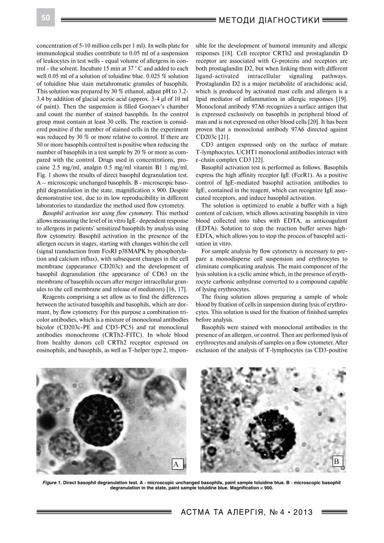
Fig. 1 shows the results of direct basophil degranulation test.
A – microscopic unchanged basophils. B - microscopic baso-
phil degranulation in the state, magnification × 900. Despite
demonstrative test, due to its low reproducibility in different
laboratories to standardize the method used flow cytometry.
Basophil activation test using flow cytometry. This method
allows measuring the level of in vitro IgE- dependent response
to allergens in patients’ sensitized basophils by analysis using
flow cytometry. Basophil activation in the presence of the
allergen occurs in stages, starting with changes within the cell
(signal transduction from FcsRI p38MAPK by phosphoryla-
tion and calcium influx), with subsequent changes in the cell
membrane (appearance CD203c) and the development of
basophil degranulation (the appearance of CD63 on the
membrane of basophils occurs after merger intracellular gran-
ules to the cell membrane and release of mediators) [16, 17].
Reagents comprising a set allow us to find the differences
between the activated basophils and basophils, which are dor-
mant, by flow cytometry. For this purpose a combination tri-
color antibodies, which is a mixture of monoclonal antibodies
bicolor (CD203c-PE and CD3-PC5) and rat monoclonal
antibodies monochrome (CRTh2-FITC). In whole blood
from healthy donors cell CRTh2 receptor expressed on
eosinophils, and basophils, as well as T-helper type 2, respon-
sible for the development of humoral immunity and allergic
responses [18]. Cell receptor CRTh2 and prostaglandin D
receptor are associated with G-proteins and receptors are
both prostaglandin D2, but when linking them with different
ligand-activated intracellular signaling pathways.
Prostaglandin D2 is a major metabolite of arachidonic acid,
which is produced by activated mast cells and allergen is a
lipid mediator of inflammation in allergic responses [19].
Monoclonal antibody 97A6 recognizes a surface antigen that
is expressed exclusively on basophils in peripheral blood of
man and is not expressed on other blood cells [20]. It has been
proven that a monoclonal antibody 97A6 directed against
CD203c [21].
CD3 antigen expressed only on the surface of mature
T-lymphocytes. UCHT1 monoclonal antibodies interact with
ε-chain complex CD3 [22].
Basophil activation test is performed as follows. Basophils
express the high affinity receptor IgE (FcεR1). As a positive
control of IgE-mediated basophil activation antibodies to
IgE, contained in the reagent, which can recognize IgE asso-
ciated receptors, and induce basophil activation.
The solution is optimized to enable a buffer with a high
content of calcium, which allows activating basophils in vitro
blood collected into tubes with EDTA, as anticoagulant
(EDTA). Solution to stop the reaction buffer serves high-
EDTA, which allows you to stop the process of basophil acti-
vation in vitro.
For sample analysis by flow cytometry is necessary to pre-
pare a monodisperse cell suspension and erythrocytes to
eliminate complicating analysis. The main component of the
lysis solution is a cyclic amine which, in the presence of eryth-
rocyte carbonic anhydrase converted to a compound capable
of lysing erythrocytes.
The fixing solution allows preparing a sample of whole
blood by fixation of cells in suspension during lysis of erythro-
cytes. This solution is used for the fixation of finished samples
before analysis.
Basophils were stained with monoclonal antibodies in the
presence of an allergen, or control. Then are performed lysis of
erythrocytes and analysis of samples on a flow cytometer. After
exclusion of the analysis of T-lymphocytes (as CD3-positive
A B
Figure 1. Direct basophil degranulation test. A - microscopic unchanged basophils, paint sample toluidine blue. B - microscopic basophil degranulation in the state, paint sample toluidine blue. Magnification × 900.
51МЕТОДИ ДІАГНОСТИКИ
АСТМА ТА АЛЕРГІЯ, № 4 • 2013
cells) is being analyzed by the expression of basophils CRTh2
and CD203c. Members basophils (basophils, which are dor-
mant) show the phenotype CRTh2posCD203c dimCD3neg,
whereas in vitro activated basophils have phenotype
CRTh2posCD203c brightCD3neg.
When another modification of the method determines the
number of activated basophils in the level of expression of
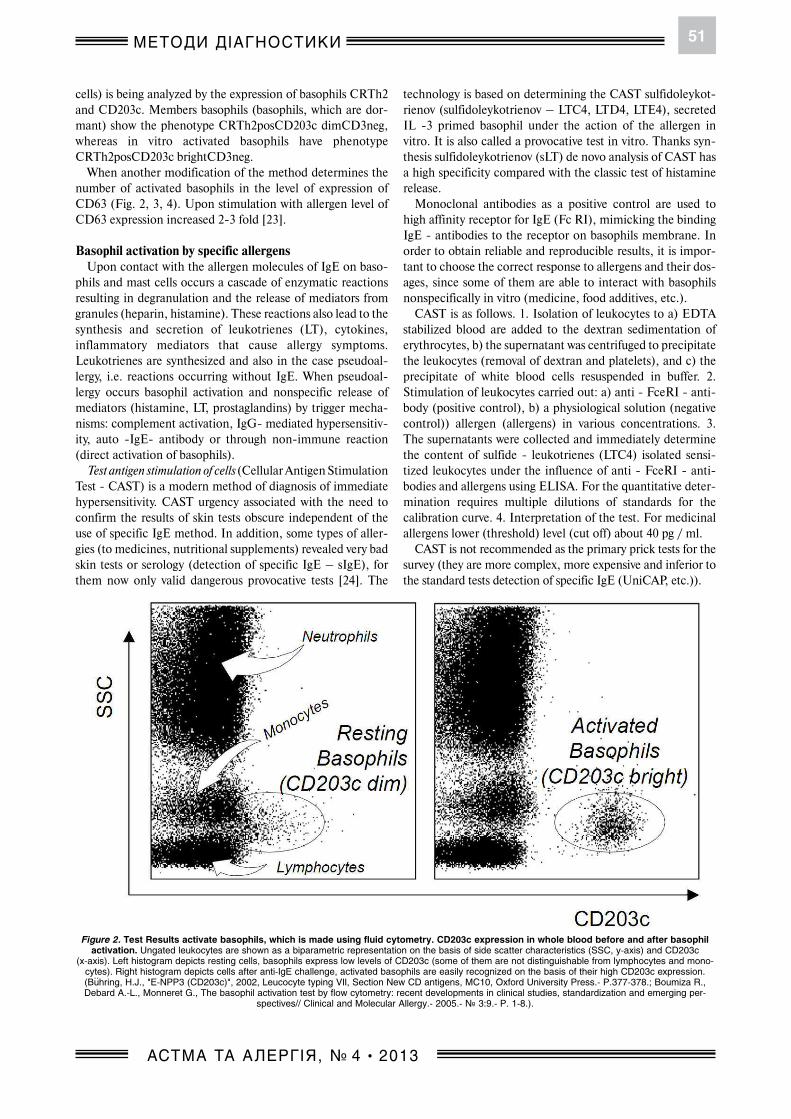
CD63 (Fig. 2, 3, 4). Upon stimulation with allergen level of
CD63 expression increased 2-3 fold [23].
Basophil activation by specific allergens
Upon contact with the allergen molecules of IgE on baso-
phils and mast cells occurs a cascade of enzymatic reactions
resulting in degranulation and the release of mediators from
granules (heparin, histamine). These reactions also lead to the
synthesis and secretion of leukotrienes (LT), cytokines,
inflammatory mediators that cause allergy symptoms.
Leukotrienes are synthesized and also in the case pseudoal-
lergy, i.e. reactions occurring without IgE. When pseudoal-
lergy occurs basophil activation and nonspecific release of
mediators (histamine, LT, prostaglandins) by trigger mecha-
nisms: complement activation, IgG- mediated hypersensitiv-
ity, auto -IgE- antibody or through non-immune reaction
(direct activation of basophils).
Test antigen stimulation of cells (Cellular Antigen Stimulation
Test - CAST) is a modern method of diagnosis of immediate
hypersensitivity. CAST urgency associated with the need to
confirm the results of skin tests obscure independent of the
use of specific IgE method. In addition, some types of aller-
gies (to medicines, nutritional supplements) revealed very bad
skin tests or serology (detection of specific IgE – sIgE), for
them now only valid dangerous provocative tests [24]. The
technology is based on determining the CAST sulfidoleykot-
rienov (sulfidoleykotrienov – LTC4, LTD4, LTE4), secreted
IL -3 primed basophil under the action of the allergen in
vitro. It is also called a provocative test in vitro. Thanks syn-
thesis sulfidoleykotrienov (sLT) de novo analysis of CAST has
a high specificity compared with the classic test of histamine
release.
Monoclonal antibodies as a positive control are used to
high affinity receptor for IgE (Fc�RI), mimicking the binding
IgE - antibodies to the receptor on basophils membrane. In
order to obtain reliable and reproducible results, it is impor-
tant to choose the correct response to allergens and their dos-
ages, since some of them are able to interact with basophils
nonspecifically in vitro (medicine, food additives, etc.).
CAST is as follows. 1. Isolation of leukocytes to a) EDTA
stabilized blood are added to the dextran sedimentation of
erythrocytes, b) the supernatant was centrifuged to precipitate
the leukocytes (removal of dextran and platelets), and c) the
precipitate of white blood cells resuspended in buffer. 2.
Stimulation of leukocytes carried out: a) anti - FceRI - anti-
body (positive control), b) a physiological solution (negative
control)) allergen (allergens) in various concentrations. 3.
The supernatants were collected and immediately determine
the content of sulfide - leukotrienes (LTC4) isolated sensi-
tized leukocytes under the influence of anti - FceRI - anti-
bodies and allergens using ELISA. For the quantitative deter-
mination requires multiple dilutions of standards for the
calibration curve. 4. Interpretation of the test. For medicinal
allergens lower (threshold) level (cut off) about 40 pg / ml.
CAST is not recommended as the primary prick tests for the
survey (they are more complex, more expensive and inferior to
the standard tests detection of specific IgE (UniCAP, etc.)).
Figure 2. Test Results activate basophils, which is made using fluid cytometry. CD203c expression in whole blood before and after basophil activation. Ungated leukocytes are shown as a biparametric representation on the basis of side scatter characteristics (SSC, y-axis) and CD203c
(x-axis). Left histogram depicts resting cells, basophils express low levels of CD203c (some of them are not distinguishable from lymphocytes and mono-cytes). Right histogram depicts cells after anti-IgE challenge, activated basophils are easily recognized on the basis of their high CD203c expression. (Bu
..hring, H.J., "E-NPP3 (CD203c)", 2002, Leucocyte typing VII, Section New CD antigens, MC10, Oxford University Press.- P.377-378.; Boumiza R.,
Debard A.-L., Monneret G., The basophil activation test by flow cytometry: recent developments in clinical studies, standardization and emerging per-spectives// Clinical and Molecular Allergy.- 2005.- № 3:9.- P. 1-8.).
52 МЕТОДИ ДІАГНОСТИКИ
АСТМА ТА АЛЕРГІЯ, № 4 • 2013
Cytometric test version of antigenic stimulation of cells -
FLOW – CAST/FAST. Stages separate lymphocytes and
allergen challenge for both options, and enzyme-linked
immunosorbent cytometric identical. But instead of the third
stage sLT FLOW - CAST determine the number of activated
basophils expressing surface antigen CD63 (gp53) in response
to stimulation with an allergen. The main indications for use
CAST/FAST: suspicion of immediate hypersensitivity to
mediator type and the absence of specific IgE, the presence of
hypersensitivity to food additives and drug allergens suspicion
of pseudoallergic mechanism of clinical manifestations, diag-
nosis of asthma and aspirinlike classes.
Beta-lactam antibiotics (penicillins, amoxicillin, ampicil-
lin, cephalosporins) hitting a body formed hapten conjugates
type transport protein that can stimulate the immune system.
Using a combination of tests in vivo and in vitro can effec-
tively and objectively diagnose clinical immediate-type hyper-
sensitivity reactions to the beta-lactam antibiotics.
Benzylpenicillin and its metabolites identified as the major
antigens. It is known that the determination of allergen-spe-
cific IgE has limited value for these allergens, since they dis-
appear for several weeks or months after the last administra-
tion of drug. Furthermore, 25% of patients with allergy to
beta - lactam antibiotics have negative skin tests to determine
the allergen-specific IgE. One third of these patients may be
identified only by allergies CAST / FAST – one of the few
tests in vitro, can detect pseudoallergic response characteris-
tic of many medicinal compounds.
To assess the controlled stimulation of immune system cells
allergens penicillin group using the CAST/FAST was a complex
benzylpenicillin - polylysine (PPL), and to assess the allergenic-
ity of penicillin metabolites was selected minor determinant
mixture (MDM). Thus, when included in the diagnostic algo-
rithm CAST/FAST in many cases it is possible to avoid the pro-
vocative test in vivo. The sensitivity of this method is 70 %, a
specificity of – 100 %. CAST/FAST besides allergy to benzyl-
penicillin can detect an increased sensitivity to amoxicillin.
Aspirin and other NSAIDs often lead to the development of
allergic reactions when they are repeated administration.
Determination of allergen-specific IgE is ineffective, since
the sensitivity to NSAIDs has not - IgE- mediated. Found
that classical cell allergotesty have negative results for the
detection of hypersensitivity to aspirin. At the same time,
studies have shown that aspirin / NSAIDs can induce release
of basophil degranulation and sLT. CAST/FAST is developed
for aspirin, diclofenac and naproxen. Sensitivity of the CAST
for aspirin reaches 63 % and specificity - 93 %, for diclofenac
sensitivity CAST – 53 %, for naproxen sensitivity CAST – 89
%, for metamizol (dipyrone) sensitivity CAST – 76 %, when
combined with FLOW - CAST (CAST/FAST) –- 77 %.
Number of reports of anaphylactic reactions during anes-
thesia muscle relaxants are increasing in recent years. In this
connection for relaxants CAST, sensitivity is 54-90 % and
specificity - 100 %. In conjunction with skin tests sensitivity is
80 %, which reduces the risk to the patient and the need to
provocative tests in vivo [23].
Reaction blastotransformation of lymphocytes (RBTL test).
During RBTL test drug allergy can be identified in 60-70% of
cases in the presence of immediate-type hypersensitivity [25].
When performing this technique it is shown that both drugs
can induce CD4 + Th1 and / or Th2, and CD8 + T - cell
response [26, 27]. However, the relatively low frequency of
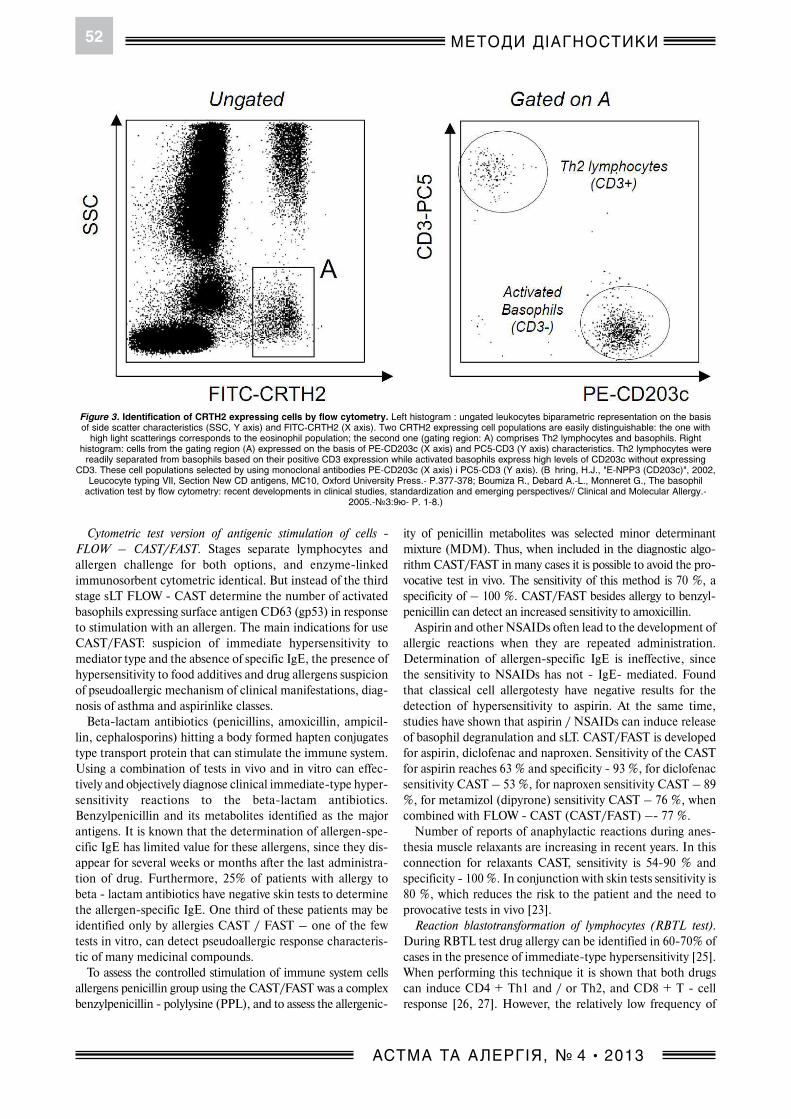
Figure 3. Identification of CRTH2 expressing cells by flow cytometry. Left histogram : ungated leukocytes biparametric representation on the basis of side scatter characteristics (SSC, Y axis) and FITC-CRTH2 (X axis). Two CRTH2 expressing cell populations are easily distinguishable: the one with
high light scatterings corresponds to the eosinophil population; the second one (gating region: A) comprises Th2 lymphocytes and basophils. Right histogram: cells from the gating region (A) expressed on the basis of PE-CD203c (X axis) and PC5-CD3 (Y axis) characteristics. Th2 lymphocytes were
readily separated from basophils based on their positive CD3 expression while activated basophils express high levels of CD203c without expressing CD3. These cell populations selected by using monoclonal antibodies PE-CD203c (X axis) і PC5-CD3 (Y axis). (B�hring, H.J., "E-NPP3 (CD203c)", 2002,
Leucocyte typing VII, Section New CD antigens, MC10, Oxford University Press.- P.377-378; Boumiza R., Debard A.-L., Monneret G., The basophil activation test by flow cytometry: recent developments in clinical studies, standardization and emerging perspectives// Clinical and Molecular Allergy.-
2005.-№3:9ю- P. 1-8.)
53МЕТОДИ ДІАГНОСТИКИ
АСТМА ТА АЛЕРГІЯ, № 4 • 2013
positive reactions, high duration setting reaction (keeping in
3-4 Denya) make this method of little use for the diagnosis of
drug allergy [28].
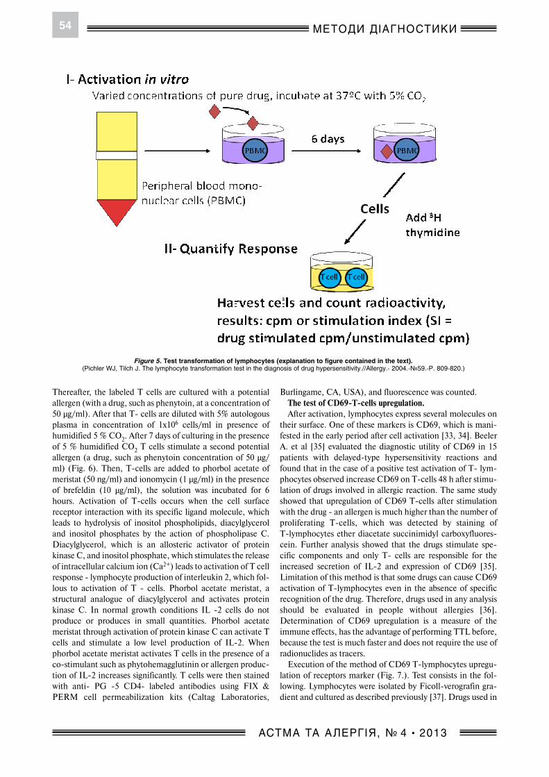
Test transformation of lymphocyts (TTL) allows to identify in
vitro delayed type allergy to medicines sensitization of
T-lymphocytes [29, 30]. Phenol-verografin gradient is used
for isolating lymphocytes in the TTL technique. By
T-lymphocytes added solutions suspected drugs in increasing
concentrations. Incubation of lymphocytes with the solution
of drug continue for six days. Sensitization of T-cells show
increased proliferation by T-cells under the influence of the
allergens according to the degree of blast transformation to
increase the inclusion of 3H-thymidine incorporation into
DNA (Fig. 5) [31]. The disadvantage of TTL technique is the
need to use radionuclides (3H - thymidine).
Test transformation of lymphocyts breeding succinimidyl
ester carboxyfluorescein diacetate (CFSE).
The technique is based on conducting flow cytometry with
coloring proliferating T- lymphocytes using non-radioactive
labels – succinimidyl ester carboxyfluorescein diacetate
(CFSE) [32]. CFSE penetrates the T-cells dye that is able to
bind the amino group of cytoplasmic proteins. During cell
division CFSE labeled proteins are distributed equally
between the daughter cells, thereby doubling the fluorescence
intensity of normal T cells. At the same time, fluorescence
antigen specific T-cell decreases.
Lymphocyte transformation test principle for breeding (CFSE).
Ficoll-verografin gradient is used for isolation T-cells. After
that T-cells are incubated with CFSE (at a concentration of 5
µM) for 10 min at 37°C and washed with excess paint.
��
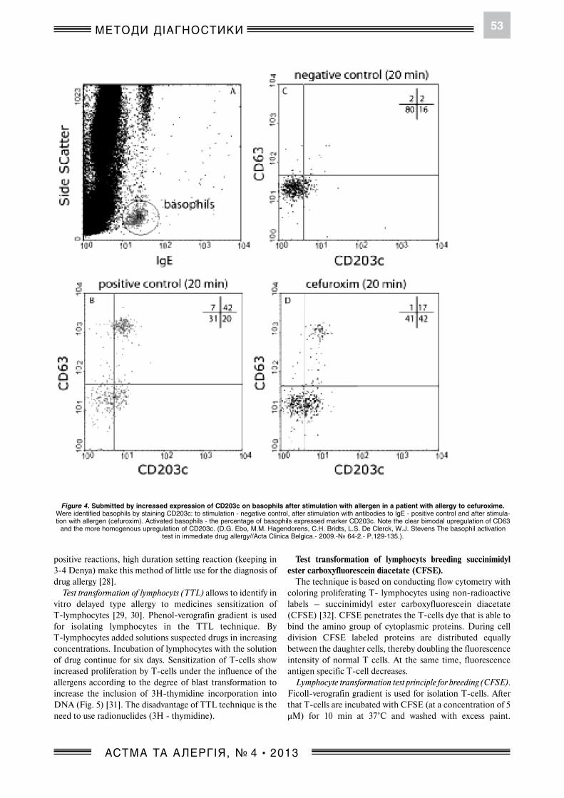
Figure 4. Submitted by increased expression of CD203c on basophils after stimulation with allergen in a patient with allergy to cefuroxime. Were identified basophils by staining CD203c: to stimulation - negative control, after stimulation with antibodies to IgE - positive control and after stimula-tion with allergen (cefuroxim). Activated basophils - the percentage of basophils expressed marker CD203c. Note the clear bimodal upregulation of CD63
and the more homogenous upregulation of CD203c. (D.G. Ebo, M.M. Hagendorens, C.H. Bridts, L.S. De Clerck, W.J. Stevens The basophil activation test in immediate drug allergy//Acta Clinica Belgica.- 2009.-№ 64-2.- P.129-135.).
54 МЕТОДИ ДІАГНОСТИКИ
АСТМА ТА АЛЕРГІЯ, № 4 • 2013
Thereafter, the labeled T cells are cultured with a potential
allergen (with a drug, such as phenytoin, at a concentration of
50 µg/ml). After that T- cells are diluted with 5% autologous
plasma in concentration of 1x10⁶ cells/ml in presence of
humidified 5 % CO2. After 7 days of culturing in the presence
of 5 % humidified CO2 T cells stimulate a second potential
allergen (a drug, such as phenytoin concentration of 50 µg/
ml) (Fig. 6). Then, T-cells are added to phorbol acetate of
meristat (50 ng/ml) and ionomycin (1 µg/ml) in the presence
of brefeldin (10 µg/ml), the solution was incubated for 6
hours. Activation of T-cells occurs when the cell surface
receptor interaction with its specific ligand molecule, which
leads to hydrolysis of inositol phospholipids, diacylglycerol
and inositol phosphates by the action of phospholipase C.
Diacylglycerol, which is an allosteric activator of protein
kinase C, and inositol phosphate, which stimulates the release
of intracellular calcium ion (Ca2+) leads to activation of T cell
response - lymphocyte production of interleukin 2, which fol-
lous to activation of T - cells. Phorbol acetate meristat, a
structural analogue of diacylglycerol and activates protein
kinase C. In normal growth conditions IL -2 cells do not
produce or produces in small quantities. Phorbol acetate
meristat through activation of protein kinase C can activate T
cells and stimulate a low level production of IL-2. When
phorbol acetate meristat activates T cells in the presence of a
co-stimulant such as phytohemagglutinin or allergen produc-
tion of IL-2 increases significantly. T cells were then stained
with anti- PG -5 CD4- labeled antibodies using FIX &
PERM cell permeabilization kits (Caltag Laboratories,
Burlingame, CA, USA), and fluorescence was counted.
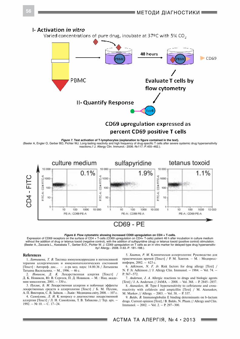
The test of CD69-T-cells upregulation.
After activation, lymphocytes express several molecules on
their surface. One of these markers is CD69, which is mani-
fested in the early period after cell activation [33, 34]. Beeler
A. et al [35] evaluated the diagnostic utility of CD69 in 15
patients with delayed-type hypersensitivity reactions and
found that in the case of a positive test activation of T- lym-
phocytes observed increase CD69 on T-cells 48 h after stimu-
lation of drugs involved in allergic reaction. The same study
showed that upregulation of CD69 T-cells after stimulation
with the drug - an allergen is much higher than the number of
proliferating T-cells, which was detected by staining of
T-lymphocytes ether diacetate succinimidyl carboxyfluores-
cein. Further analysis showed that the drugs stimulate spe-
cific components and only T- cells are responsible for the
increased secretion of IL-2 and expression of CD69 [35].
Limitation of this method is that some drugs can cause CD69
activation of T-lymphocytes even in the absence of specific
recognition of the drug. Therefore, drugs used in any analysis
should be evaluated in people without allergies [36].
Determination of CD69 upregulation is a measure of the
immune effects, has the advantage of performing TTL before,
because the test is much faster and does not require the use of
radionuclides as tracers.
Execution of the method of CD69 T-lymphocytes upregu-
lation of receptors marker (Fig. 7.). Test consists in the fol-
lowing. Lymphocytes were isolated by Ficoll-verografin gra-
dient and cultured as described previously [37]. Drugs used in
Figure 5. Test transformation of lymphocytes (explanation to figure contained in the text). (Pichler WJ, Tilch J. The lymphocyte transformation test in the diagnosis of drug hypersensitivity.//Allergy.- 2004.-№59.-P. 809-820.)
55МЕТОДИ ДІАГНОСТИКИ
АСТМА ТА АЛЕРГІЯ, № 4 • 2013
non-toxic concentrations of solutions prepared immediately
before use. Drugs used in the following concentrations: 100,
200, 500 µg/ml amoxicillin; 1, 10, 100 µg/ml vancomycin; 1,
10, 100 µg/ml carbamazepine 1, 10, 100 µg/ml sulfapyridine;
1, 10, 100 µg/ml cefuroxime; 50, 100, 200 µg/ml sulmetok-
sazola; 10, 50, 100 µg/ml of phenytoin; 1, 10, 50 µg/ml of
clavulanic acid; 1, 10, 100 µg/ml levofloxacin et al [38].
Freshly isolated T - lymphocytes (2 × 10⁵) were cultured in
three duplicate 96 well plates with a U - shaped bottomed tis-
sue culture in the presence of the indicated concentrations of
the drug as well as tetanus toxoid or interleukin 2 - a positive
control. The appearance of the surface marker CD69 on CD4
+/CD8 + T-lymphocytes measured by flow cytometry in the
presence of human monoclonal antibodies (PE - CD69,
APC-CD3, FITC-CD-8, CD-4 PERCP-I, PE - IgG1) and
in isotypic negative control. Data are expressed as cytometric
normalized mean fluorescence intensity. The stimulation
index was determined as the ratio of normalized average fluo-
rescence intensity of cells cultured in the presence of antigen
separated by the normalized mean fluorescence intensity of
cells cultured in the absence of antigen [38].
Before painting T - peripheral blood lymphocytes stimu-
lated with solutions of drugs or antigens for 24 h, parallel
inkubates unstimulated T-lymphocytes - negative control.
Monensin was added to a concentration of 6 µg/ml in the
last 8 h of incubation, before the determination. Cells were
washed 2 times with ice-cold phosphate-buffered saline
containing 0.5 % bovine serum albumin (BSA) and 0.1 %
sodium azide and color within 20 min of human monoclonal
antibodies (PE - CD4 mAb, PERCP - CD3 mAb, FITC -
CD69 nAb). Cells were fixed with a solution of Cytofix/
Cytoperm for 20 minutes, resuspended in Per/Wosh solution
containing a drug and incubated for 30 min in the dark. As a
control, isotype FITC- and alofikotsianin - conjugated IgG1
and IgG2a. After washing twice with Perm/Wash cells were
resuspended in phosphate buffered saline containing 0.5 %
BSA and 0.1% sodium azide and analyzed as described
above [38].
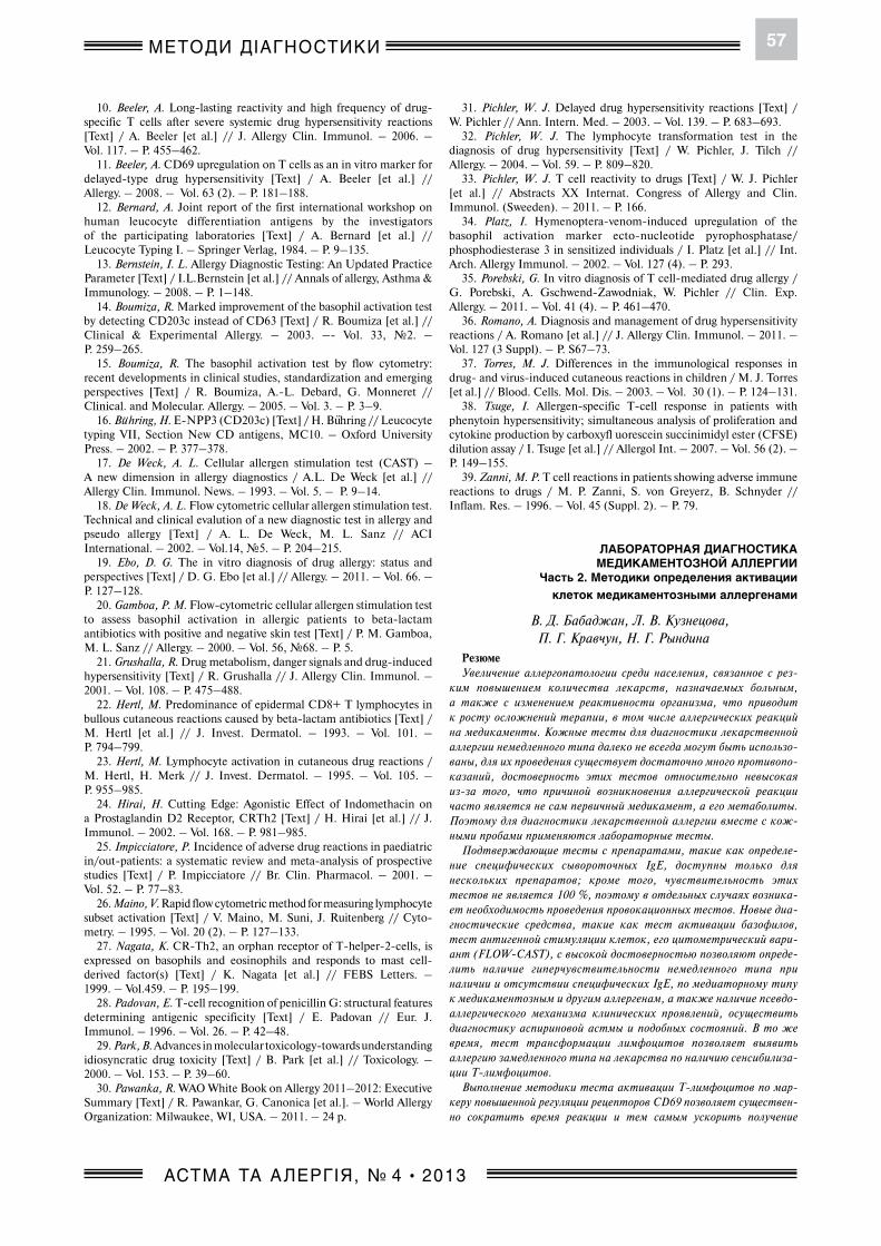
Example receptor CD69 upregulation on the surface of
CD4 + T-cells in a patient with an allergy to sulfapyridine
shown in Fig. 8. During exposure, sulfapyridine and tetanus
toxoid 1.9 % and 1.1 % of CD4 + cells showed an increased
amount of SD69 + receptor, and only 0.1 % of these cells
were incubated with corresponding receptors in a medium
without the addition of drugs. After 6 and 12 hours after
antigen challenge upregulation of receptors on the surface of
CD69+ CD4+ as T - cells, and CD8+ T-cells, were detected.
Increasing the incubation period (36 or 72 hours) was
accompanied by the appearance receptor CD69+, however
incubation for 18 h minimum for a detection of an elevated
level of receptor CD69 + [38]. Elevated levels of CD69+ were
not detected in the control without the addition of the drug.
For practical purposes, we can recommend 48 hours of
incubation of CD4+ T-cells with the addition of the drug
(Fig. 8.).
The diagnosis of hypersensitivity to drugs usually depends
on history of the disease, and the results of skin tests per-
formed in the laboratory confirmatory tests with drugs, such
as the determination of serum specific IgE, which are avail-
able for only a few drugs. The sensitivity of these tests is not
100 %, so in some cases a need for provocation testing. New
diagnostic tools, such as the BAT test antigen stimulation of
cells and lymphocyte transformation test, developed a few
years ago, is now thoroughly tested in leading immunological
centers around the world. Their use can lead to increased
performance of diagnostic tests, improving the accuracy of
diagnosis of drug allergy, thereby reducing the need for provo-
cation tests.
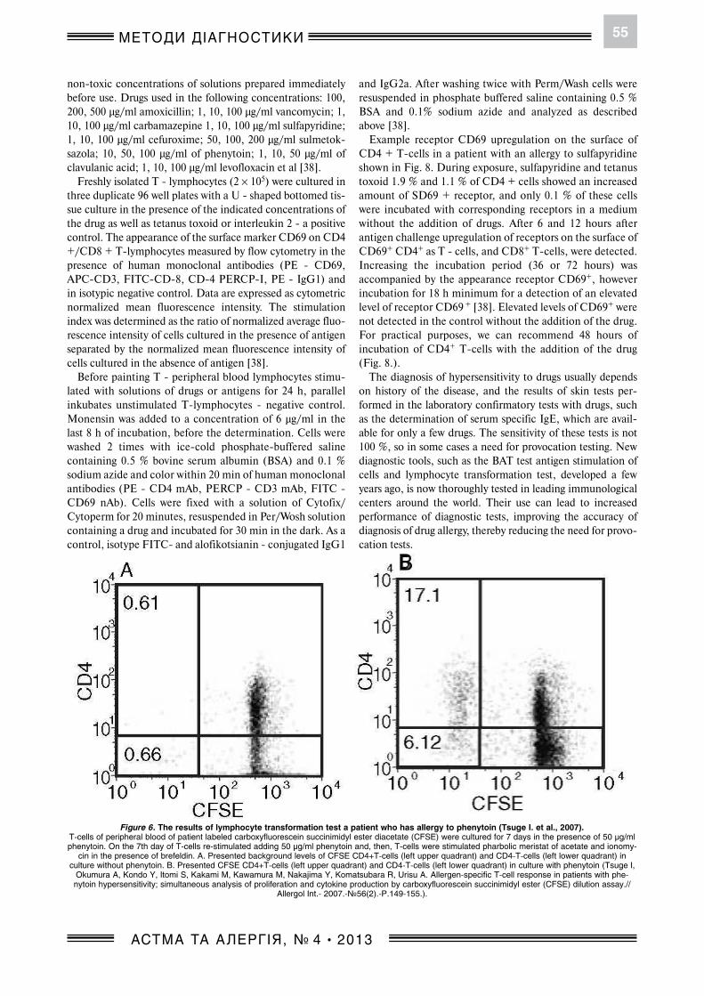
Figure 6. The results of lymphocyte transformation test a patient who has allergy to phenytoin (Tsuge I. et al., 2007).
T-cells of peripheral blood of patient labeled carboxyfluorescein succinimidyl ester diacetate (CFSE) were cultured for 7 days in the presence of 50 µg/ml phenytoin. On the 7th day of T-cells re-stimulated adding 50 µg/ml phenytoin and, then, Т-cells were stimulated pharbolic meristat of acetate and ionomy-
cin in the presence of brefeldin. А. Presented background levels of CFSE CD4+Т-cells (left upper quadrant) and CD4-Т-cells (left lower quadrant) in culture without phenytoin. В. Presented CFSE CD4+Т-cells (left upper quadrant) and CD4-T-cells (left lower quadrant) in culture with phenytoin (Tsuge I,
Okumura A, Kondo Y, Itomi S, Kakami M, Kawamura M, Nakajima Y, Komatsubara R, Urisu A. Allergen-specific T-cell response in patients with phe-nytoin hypersensitivity; simultaneous analysis of proliferation and cytokine production by carboxyfluorescein succinimidyl ester (CFSE) dilution assay.//
Allergol Int.- 2007.-№56(2).-P.149-155.).
56 МЕТОДИ ДІАГНОСТИКИ
АСТМА ТА АЛЕРГІЯ, № 4 • 2013
Figure 7. Test activation of T-lymphocytes (explanation to figure contained in the text). (Beeler A, Engler O, Gerber BO, Pichler WJ. Long-lasting reactivity and high frequency of drug-specific T cells after severe systemic drug hypersensitivity
reactions.// J. Allergy Clin. Immunol.- 2006.-№117.-P.455–462.).
�
Figure 8. Flow cytometric showing increased CD69 upregulation on CD4 + T-cells. Expression of CD69 receptors on the surface of CD4 + T-cells (CD69 upregulation on CD4+ T-cells) patient 48 h after incubation in culture medium
without the addition of drug or tetanus toxoid (negative control), with the addition of sulfapyridine (drug) or tetanus toxoid (positive control) stimulation. (Beeler A., Zaccaria L., Kawabata T., Gerber B.O., Pichler W. J. CD69 upregulation on T cells as an in vitro marker for delayed-type drug hypersensitiv-
ity// Allergy.- 2008.-V.63.-P. 181–188.).
References1. Латышева, Т. В. Тактика иммунокоррекции в интенсивной
терапии аллергических и иммунопатологических состояний [Текст] : Автореф. дис… – д-ра мед. наук: 14.00.36 / Латышева Татьяна Васильевна. – М., 1996. – 46 с.
2. Новиков, Д. К. Лекарственная алергия [Текст] / Д. К. Новиков, Ю. В. Сергеев, П. Д. Новиков. – М. : Нац. акаде-мия микологии, 2001. – 330 с.
3. Пухлик, Б. М. Лекарственная аллергия и побочные эффекты лекарственных средств в аллергологии [Текст] / Б. М. Пухлик, А. П. Викторов, С. В. Зайков. – Львів : Медицина світу, 2008. – 107 с.
4. Самойлова, Л. Н. К вопросу о диагностике лекарственной аллергии [Текст] / Л. Н. Самойлова, Т. В. Табакова // Тер. арх. – 1992. – № 10. – С. 17–24.
5. Хаитов, Р. М. Клиническая аллергология: Руководство для
практических врачей [Текст] / Р. М. Хаитов. – М. : Медпресс-
информ, 2002. – 623 с.
6. Adkinson, N. F. Jr. Risk factors for drug allergy [Text] /
N. F. Jr. Adkinson // J. Allergy Clin. Immunol. – 1984. – Vol. 74. –
P. 567–572.
7. Anderson, J. A. Allergic reactions to drugs and biologic agents
[Text] / J. A. Anderson // JAMA. – 2008. – Vol. 368. – P. 2845–2857.
8. Atanaskov, M. Type-I hypersensitivity to ceftriaxone and cross-
reactivity with cefalexin and ampicillin [Text] / M. Atanaskov,
M. Markov // Allergy. – 2003. – Vol. 58. – P. 537.
9. Baldo, B. Immunoglobulin E binding determinants on b-lactam
drugs. Current opinion [Text] / B. Baldo, N. Pham // Allergy and Clin.
Immunol. – 2002. – Vol. 2. – P. 297–300.
57МЕТОДИ ДІАГНОСТИКИ
АСТМА ТА АЛЕРГІЯ, № 4 • 2013
10. Beeler, A. Long-lasting reactivity and high frequency of drug-
specific T cells after severe systemic drug hypersensitivity reactions
[Text] / A. Beeler [et al.] // J. Allergy Clin. Immunol. – 2006. –
Vol. 117. – P. 455–462.
11. Beeler, A. CD69 upregulation on T cells as an in vitro marker for
delayed-type drug hypersensitivity [Text] / A. Beeler [et al.] //
Allergy. – 2008. – Vol. 63 (2). – P. 181–188.
12. Bernard, A. Joint report of the first international workshop on
human leucocyte differentiation antigens by the investigators
of the participating laboratories [Text] / A. Bernard [et al.] //
Leucocyte Typing I. – Springer Verlag, 1984. – P. 9–135.
13. Bernstein, I. L. Allergy Diagnostic Testing: An Updated Practice
Parameter [Text] / I.L.Bernstein [et al.] // Annals of allergy, Asthma &
Immunology. – 2008. – P. 1–148.
14. Boumiza, R. Marked improvement of the basophil activation test
by detecting CD203c instead of CD63 [Text] / R. Boumiza [et al.] //
Clinical & Experimental Allergy. – 2003. –- Vol. 33, №2. –
P. 259–265.
15. Boumiza, R. The basophil activation test by flow cytometry:
recent developments in clinical studies, standardization and emerging
perspectives [Text] / R. Boumiza, A.-L. Debard, G. Monneret //
Clinical. and Molecular. Allergy. – 2005. – Vol. 3. – P. 3–9.
16. Bu..hring, H. E-NPP3 (CD203c) [Text] / H. Bu
..hring // Leucocyte
typing VII, Section New CD antigens, MC10. – Oxford University
Press. – 2002. – P. 377–378.
17. De Weck, A. L. Cellular allergen stimulation test (CAST) –
A new dimension in allergy diagnostics / A.L. De Weck [et al.] //
Allergy Clin. Immunol. News. – 1993. – Vol. 5. – P. 9–14.
18. De Weck, A. L. Flow cytometric cellular allergen stimulation test.
Technical and clinical evalution of a new diagnostic test in allergy and
pseudo allergy [Text] / A. L. De Weck, M. L. Sanz // ACI
International. – 2002. – Vol.14, №5. – P. 204–215.
19. Ebo, D. G. The in vitro diagnosis of drug allergy: status and
perspectives [Text] / D. G. Ebo [et al.] // Allergy. – 2011. – Vol. 66. –
P. 127–128.
20. Gamboa, P. M. Flow-cytometric cellular allergen stimulation test
to assess basophil activation in allergic patients to beta-lactam
antibiotics with positive and negative skin test [Text] / P. M. Gamboa,
M. L. Sanz // Allergy. – 2000. – Vol. 56, №68. – P. 5.
21. Grushalla, R. Drug metabolism, danger signals and drug-induced
hypersensitivity [Text] / R. Grushalla // J. Allergy Clin. Immunol. –
2001. – Vol. 108. – Р. 475–488.
22. Hertl, M. Predominance of epidermal CD8+ T lymphocytes in
bullous cutaneous reactions caused by beta-lactam antibiotics [Text] /
M. Hertl [et al.] // J. Invest. Dermatol. – 1993. – Vol. 101. –
P. 794–799.
23. Hertl, M. Lymphocyte activation in cutaneous drug reactions /
M. Hertl, H. Merk // J. Invest. Dermatol. – 1995. – Vol. 105. –
P. 955–985.
24. Hirai, H. Cutting Edge: Agonistic Effect of Indomethacin on
a Prostaglandin D2 Receptor, CRTh2 [Text] / H. Hirai [et al.] // J.
Immunol. – 2002. – Vol. 168. – P. 981–985.
25. Impicciatore, P. Incidence of adverse drug reactions in paediatric
in/out-patients: a systematic review and meta-analysis of prospective
studies [Text] / P. Impicciatore // Br. Clin. Pharmacol. – 2001. –
Vol. 52. – P. 77–83.
26. Maino, V. Rapid flow cytometric method for measuring lymphocyte
subset activation [Text] / V. Maino, M. Suni, J. Ruitenberg // Cyto-
metry. – 1995. – Vol. 20 (2). – P. 127–133.
27. Nagata, K. CR-Th2, an orphan receptor of T-helper-2-cells, is
expressed on basophils and eosinophils and responds to mast cell-
derived factor(s) [Text] / K. Nagata [et al.] // FEBS Letters. –
1999. – Vol.459. – P. 195–199.
28. Padovan, E. T-cell recognition of penicillin G: structural features
determining antigenic specificity [Text] / E. Padovan // Eur. J.
Immunol. – 1996. – Vol. 26. – Р. 42–48.
29. Park, B. Advances in molecular toxicology-towards understanding
idiosyncratic drug toxicity [Text] / B. Park [et al.] // Toxicology. –
2000. – Vol. 153. – P. 39–60.
30. Pawanka, R. WAO White Book on Allergy 2011–2012: Executive
Summary [Text] / R. Pawankar, G. Canonica [et al.]. – World Allergy
Organization: Milwaukee, WI, USA. – 2011. – 24 p.
31. Pichler, W. J. Delayed drug hypersensitivity reactions [Text] /
W. Pichler // Ann. Intern. Med. – 2003. – Vol. 139. – P. 683–693.
32. Pichler, W. J. The lymphocyte transformation test in the
diagnosis of drug hypersensitivity [Text] / W. Pichler, J. Tilch //
Allergy. – 2004. – Vol. 59. – P. 809–820.
33. Pichler, W. J. T cell reactivity to drugs [Text] / W. J. Pichler
[et al.] // Abstracts XX Internat. Congress of Allergy and Clin.
Immunol. (Sweeden). – 2011. – P. 166.
34. Platz, I. Hymenoptera-venom-induced upregulation of the
basophil activation marker ecto-nucleotide pyrophosphatase/
phosphodiesterase 3 in sensitized individuals / I. Platz [et al.] // Int.
Arch. Allergy Immunol. – 2002. – Vol. 127 (4). – P. 293.
35. Porebski, G. In vitro diagnosis of T cell-mediated drug allergy /
G. Porebski, A. Gschwend-Zawodniak, W. Pichler // Clin. Exp.
Allergy. – 2011. – Vol. 41 (4). – P. 461–470.
36. Romano, A. Diagnosis and management of drug hypersensitivity
reactions / A. Romano [et al.] // J. Allergy Clin. Immunol. – 2011. –
Vol. 127 (3 Suppl). – P. S67–73.
37. Torres, M. J. Differences in the immunological responses in
drug- and virus-induced cutaneous reactions in children / M. J. Torres
[et al.] // Blood. Cells. Mol. Dis. – 2003. – Vol. 30 (1). – P. 124–131.
38. Tsuge, I. Allergen-specific T-cell response in patients with
phenytoin hypersensitivity; simultaneous analysis of proliferation and
cytokine production by carboxyfl uorescein succinimidyl ester (CFSE)
dilution assay / I. Tsuge [et al.] // Allergol Int. – 2007. – Vol. 56 (2). –
P. 149–155.
39. Zanni, M. P. T cell reactions in patients showing adverse immune
reactions to drugs / M. P. Zanni, S. von Greyerz, B. Schnyder //
Inflam. Res. – 1996. – Vol. 45 (Suppl. 2). – P. 79.
ЛАБОРАТОРНАЯ ДИАГНОСТИКА МЕДИКАМЕНТОЗНОЙ АЛЛЕРГИИ
Часть 2. Методики определения активации
клеток медикаментозными аллергенами
В. Д. Бабаджан, Л. В. Кузнецова,
П. Г. Кравчун, Н. Г. Рындина
Резюме
Увеличение аллергопатологии среди населения, связанное с рез-
ким повышением количества лекарств, назначаемых больным,
а также с изменением реактивности организма, что приводит
к росту осложнений терапии, в том числе аллергических реакций
на медикаменты. Кожные тесты для диагностики лекарственной
аллергии немедленного типа далеко не всегда могут быть использо-
ваны, для их проведения существует достаточно много противопо-
казаний, достоверность этих тестов относительно невысокая
из-за того, что причиной возникновения аллергической реакции
часто является не сам первичный медикамент, а его метаболиты.
Поэтому для диагностики лекарственной аллергии вместе с кож-
ными пробами применяются лабораторные тесты.
Подтверждающие тесты с препаратами, такие как определе-
ние специфических сывороточных IgE, доступны только для
нескольких препаратов; кроме того, чувствительность этих
тестов не является 100 %, поэтому в отдельных случаях возника-
ет необходимость проведения провокационных тестов. Новые диа-
гностические средства, такие как тест активации базофилов,
тест антигенной стимуляции клеток, его цитометрический вари-
ант (FLOW-CAST), с высокой достоверностью позволяют опреде-
лить наличие гиперчувствительности немедленного типа при
наличии и отсутствии специфических IgE, по медиаторному типу
к медикаментозным и другим аллергенам, а также наличие псевдо-
аллергического механизма клинических проявлений, осуществить
диагностику аспириновой астмы и подобных состояний. В то же
время, тест трансформации лимфоцитов позволяет выявить
аллергию замедленного типа на лекарства по наличию сенсибилиза-
ции Т-лимфоцитов.
Выполнение методики теста активации Т-лимфоцитов по мар-
керу повышенной регуляции рецепторов CD69 позволяет существен-
но сократить время реакции и тем самым ускорить получение
58 МЕТОДИ ДІАГНОСТИКИ
АСТМА ТА АЛЕРГІЯ, № 4 • 2013
результатов исследования. Таким образом, использование in vitro
методик определения активации клеток медикаментозными аллер-
генами приведет к увеличению результативности диагностических
обследований, повышению точности диагностики лекарственной
аллергии, тем самым уменьшив необходимость проведения провока-
ционных тестов.
Ключевые слова: in vitro диагностика лекарственной аллергии,
тест активации базофилов, тест антигенной стимуляции клеток,
тест трансформации лимфоцитов, тест определения повышенной
регуляции рецепторов CD69.
Научно-практический журнал «Астма и аллергия», 2013, № 4.
В. Д. Бабаджан
Харьковский национальный медицинский университет, профессор
61001, Харьков, просп. Ленина, 4
тел.: +380(57)706-30-17, моб.:+ 380(67)573-23-38
e-mail: [email protected]
LABORATORY DIAGNOSIS OF DRUG ALLERGYPart 2. Methods used for determination of drug allergens
triggered cell activation
V. D. Babadzhan., L. V. Kuznetsova,
P. G. Kravchun, N. G. RyndinaSummary
Increase of allergy in the population, associated with a significant
increase in the amount of medication prescribed to patients, as well as the
immune reactivity changes, which leads to increased complications of
therapy, including allergic reactions to medications. Skin tests to diagnose
immediate type allergy drug can not always be used. There are many
contraindications to carry them out. The reliability of these tests is
relatively low due to the fact that the cause of allergic reaction is often not
the initial drug and its metabolites. Therefore, skin tests with laboratory
tests are usually used for diagnostic of drug allergy.
Confirmatory tests with drugs, such as the definition of specific serum
IgE, only available for certain drugs. The sensitivity of these tests is not
100 %, so in some cases there is a need in provocation tests. New diagnostic
agents such as basophil activation test, cellular antigen stimulation test and
its flow cytometric version (FLOW-CAST) with high reliability can
determine the presence of immediate hypersensitivity, the presence or
absence of specific IgE, according to the type of neurotransmitter to medical
and other allergens, as well as availability pseudoallergic mechanism
of clinical manifestations, diagnosis of aspirin-induced asthma and similar
conditions. At the same time, the lymphocyte transformation test reveals the
delayed type allergies to medicines sensitization of T-lymphocytes.
Performing the test by determining of CD69 upregulation, as the
T-lymphocyte activation marker, significantly reduces the response time
and thereby accelerate results of the study. Thus, the use of in vitro methods
for determination of drug allergens triggered cell activation will increase the
effectiveness of diagnostic tests, improve the accuracy of diagnosis of drug
allergy, thereby reducing the need in provocation tests.
Key words: in vitro-diagnostics drug allergy, basophil activation test,
cellular antigen stimulation test, lymphocyte transformation test, CD69
receptors upregulation test.
Theoretical and practical J. «Asthma and Allergy», 2013, 4.
V. D. Babadzan
Kharkiv National Medical Universit, professor
61001, Kharkiv, ave. Lenin, 4
tel.: +380(57)706-30-17, mob.: + 380(67)573-23-38
e-mail: [email protected]