Laboratory diagnosis gram positive and gram negative bacilli
36
Laboratory diagnosis of Gram positive and Gram negative bacilli
-
Upload
dana-sinziana-brehar-cioflec -
Category
Education
-
view
340 -
download
4
Transcript of Laboratory diagnosis gram positive and gram negative bacilli

Laboratory diagnosis of Gram positive and Gram negative bacilli

Gram positive and Gram negative bacilli
Genera:• Corynebacterium• Bacillus • Listeria
Family EnterobacteriaceaeGenera:A. Highly pathogenic:
• Yersinia• Salmonella• Shigella
A. Facultatively pathogenic:• E.coli• Klebsiella• Proteus• Enterobacter• Serratia• Citrobacter

Genus Corynebacterium Species Corynebacterium diphtheriae
- high pathogenicity- Clinical significance: diphtheria = disease produced by
the diphtheric toxine
- → symptoms at the entry gate: sore throat, adherent membrane (pseudomembrane) on tonsils, pharynx,
nasal cavity - → general toxic symptoms: fever (hematogenic difusion
of diphtheric toxin)
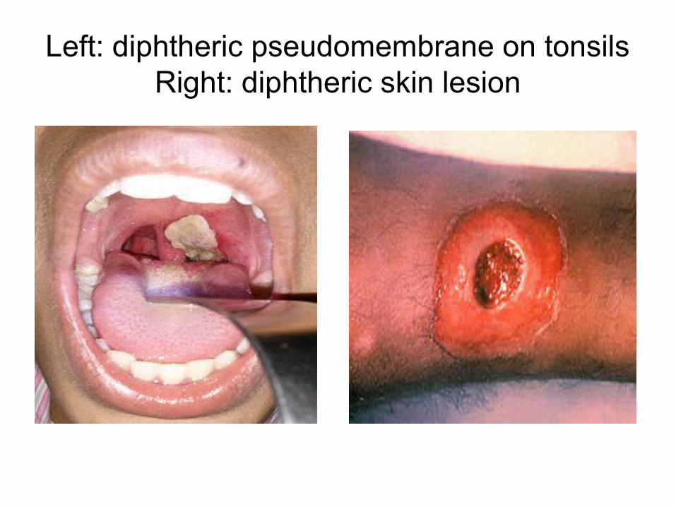
Left: diphtheric pseudomembrane on tonsilsRight: diphtheric skin lesion

Species Corynebacterium diphtheriaeLaboratory diagnosis
• Colection of specimens: throat / nasal / wound swab• Microscopy:
– Gram positive bacilli, aspect of ”Chinese letters” / capital letters– low value (C.diphtheriae – similar to other comensal
corynebacteria in the throat)
• Cultivation:– Blood agar – Selective media with tellurite (Tinsdale, Gundel-Tietz)– Highly selective Loffler medium

Corynebacterium diphtheriae: encurved rods, ”swolen” ends
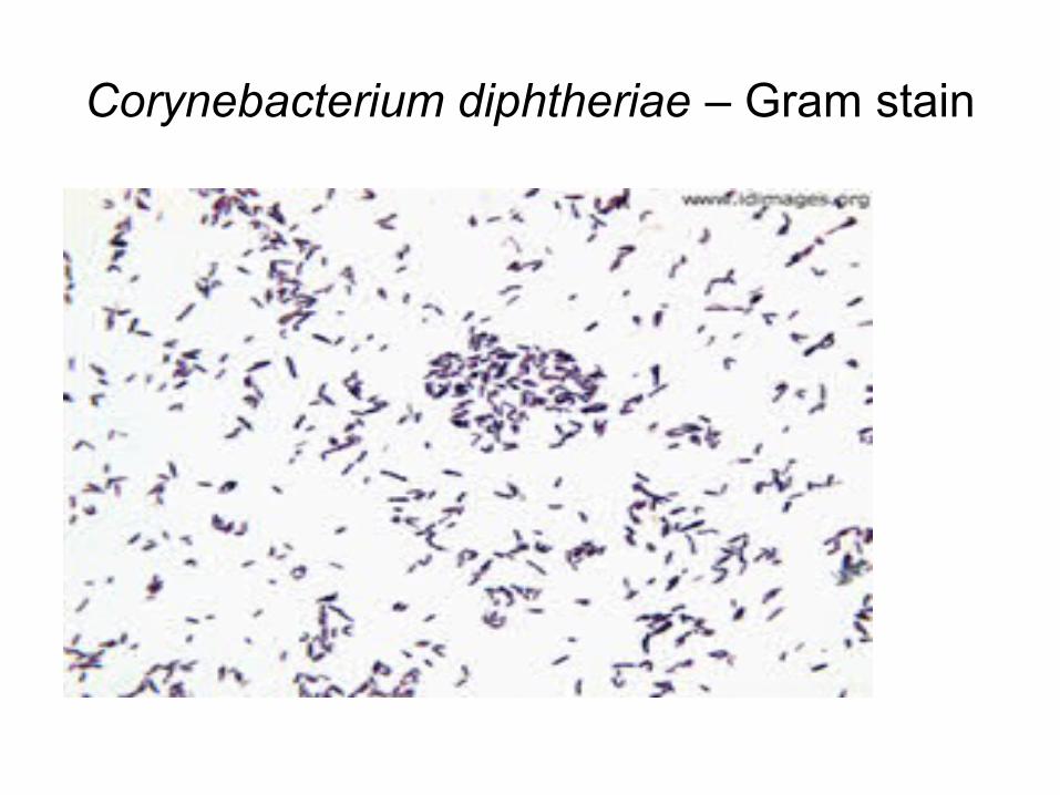
Corynebacterium diphtheriae – Gram stain

”Daisy flower” – Corynebacterium
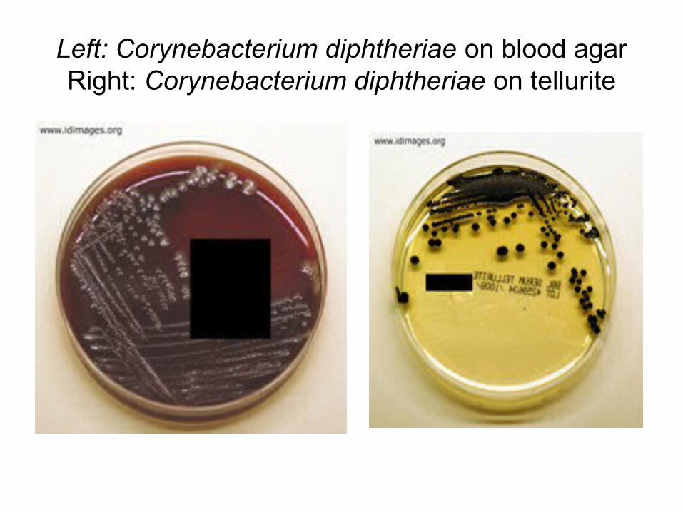
Left: Corynebacterium diphtheriae on blood agarRight: Corynebacterium diphtheriae on tellurite
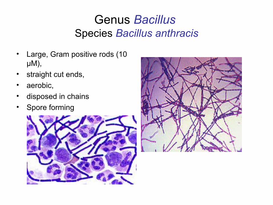
Genus Bacillus Species Bacillus anthracis
• Large, Gram positive rods (10 µM),
• straight cut ends, • aerobic, • disposed in chains• Spore forming
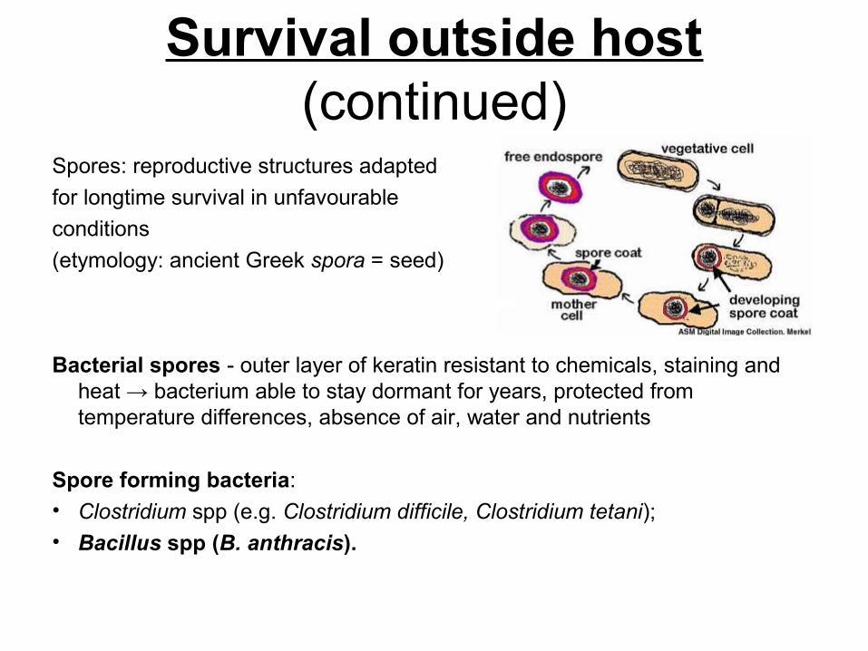
Survival outside host (continued)
Spores: reproductive structures adapted
for longtime survival in unfavourable
conditions
(etymology: ancient Greek spora = seed)
Bacterial spores - outer layer of keratin resistant to chemicals, staining and heat → bacterium able to stay dormant for years, protected from temperature differences, absence of air, water and nutrients
Spore forming bacteria: • Clostridium spp (e.g. Clostridium difficile, Clostridium tetani); • Bacillus spp (B. anthracis).

Bacillus anthracis (continued)
• High pathogenicity• Disease = zoonosis (infection of animals AND humans)• Clinical forms:
– Cutaneous anthrax – spores enter the body via skin lesions– Pulmonary anthrax – inhalatory infection– Digestive anthrax – ingestion of infected undercooked meat – + biological weapon (inhalatory infection) – agent of bioterrorism
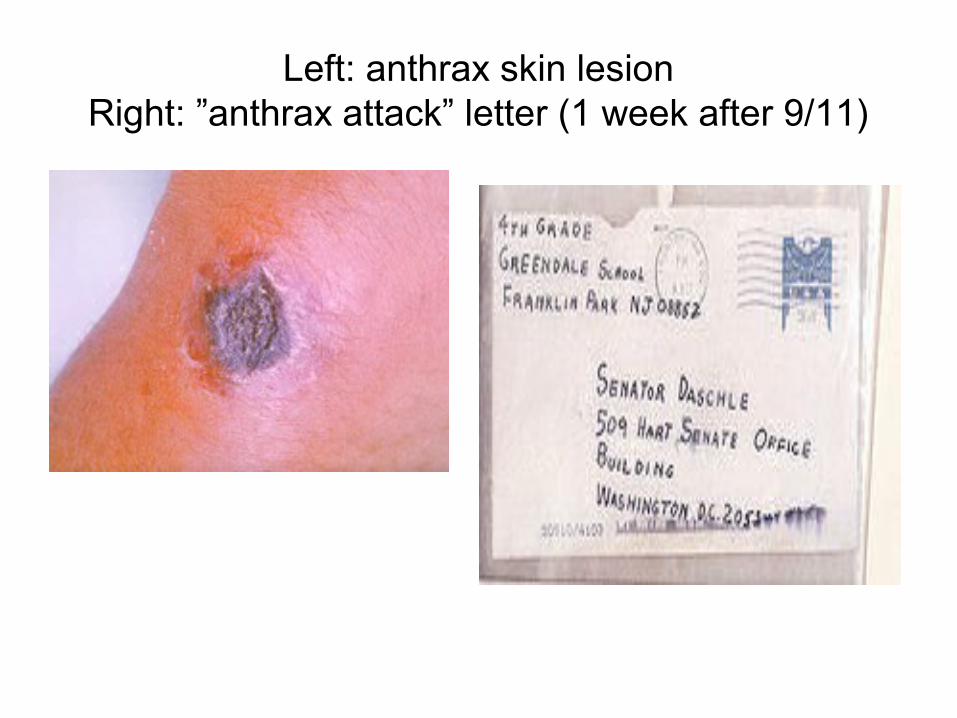
Left: anthrax skin lesionRight: ”anthrax attack” letter (1 week after 9/11)

Gram positive and Gram negative bacilli
Genera:• Corynebacterium• Bacillus• Listeria
Family EnterobacteriaceaeGenera:A. Highly pathogenic:
• Yersinia• Salmonella• Shigella
A. Facultatively pathogenic:• E.coli• Klebsiella• Proteus• Enterobacter• Serratia• Citrobacter

Family Enterobacteriaceae
• Gram negative rods, non-spore forming, non-fastidious• Wide spread in the environment (plants, soil, water,
human and animal intestines, on mucous membranes, etc)
• Clinical significance: – intestinal and extraintestinal infections
• Collection of specimens in intestinal infections: – stool (faeces)
• Collection of specimens in extraintestinal infections:– Urine, ear secretion, wound exudate, CSF, etc

Collection of stool (faeces)
• Disposable stool collection containers (simple / with transportation medium Carry Blair: non-nutritive medium which prevents overgrowth of Enterobacteriaceae but preserves viable enteric pathogens (Salmonella, Shigella, Vibrio, etc)
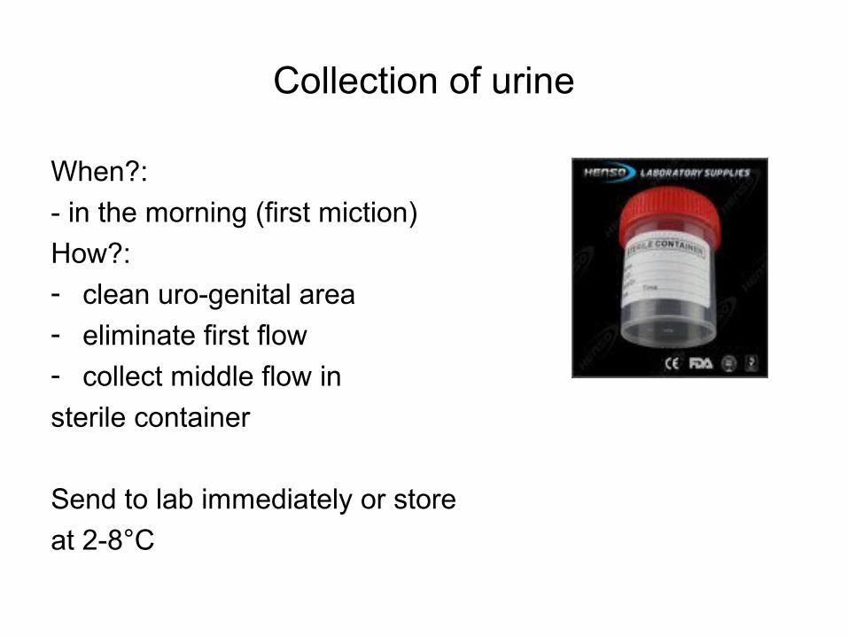
Collection of urine
When?:
- in the morning (first miction)
How?: - clean uro-genital area
- eliminate first flow
- collect middle flow in
sterile container
Send to lab immediately or store
at 2-8°C

Collection of cerebrospinal fluid (CSF)
Lumbar punction (spinal tap) • patient lies on the side, knees pulled up toward
chest, chin tucked downward • back cleaned and disinfected (iodine) + health
care provider injects local anesthetic into lower spine
• spinal needle inserted into lower back area• needle properly positioned, CSF pressure
measured and sample collected in sterile tube• needle removed, area cleaned, bandage placed
over puncture site

Genus Yersinia
Species:• Yersinia enterocolitica:
– colonizes the intestines; only some strains are pathogenic – may cause diarrhoea + apendicitis-like symptoms
• Yersinia pestis (causative agent of plague)– reservoir of germs: rodents (rats) – interpersonal transmission (human to human)– Routes of infection:
• Vectors: Flea bites →skin lesions (inflammation, necrosis, purulent secretion) + swollen lymph nodes (buboes) = bubonic plague →sepsis
• Airborne: Inhalation →pneumonia = pulmonary plague

Left: Oriental rat flea (vector of Y.pestis)Right, upper image: Y.pestis infected flea bite
Right, lower image: swollen lymph nodes (buboes)

Genus Salmonella
Clinical significance:
• 1. Food poisoning - infection limited to the intestine without going through the intestinal barrier
• 2. Enteric fevers – systemic infections (typhoid fever, paratyphoid fevers)

Genus Salmonella (continued)
Biological specimens:- Blood for hemoculture (enteric fevers)- Stool for coproculture (enteric fevers, food poisoning,
asymptomatic carriers)- Urine for uroculture (enteric fevers)
Culture media:- MacConkey: semitransparent colonies, colourless,
lactose-negative- Istrati-Meitert/ADCL: transparent, colonies with black
center = production of H2S
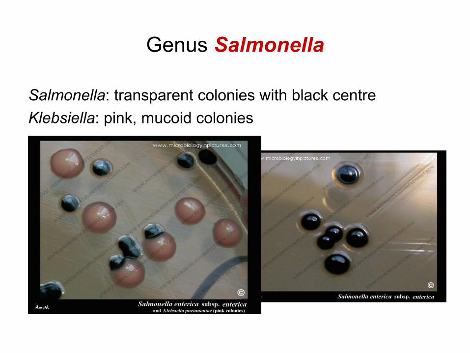
Genus Salmonella
Salmonella: transparent colonies with black centre
Klebsiella: pink, mucoid colonies

H2S production on Hektoen agar (Salmonella colonies)

Genus Shigella
• Clinical significance: – dysentery = multiple diarrhoeic, bloody / purulent stools, fever,
dehydration
• Culture media:– MacConkey – white/colourless colonies (Lactose-negative)– Istrati-Meitert/ADCL – no H2S

Shigella on Endo agar – no H2S production

Genus Escherichia; Species: E.coli
- Facultatively pathogenic - Enteric infections produced by 5 E.coli groups:
- EPEC = Entero-Pathogenic E.coli- ETEC = Entero-Toxigenic E.coli- EIEC = Entero-Invasive E.coli- EHEC = Entero-Hemorrhagic E.coli- EAEC = Entero-Adherent E.coli

MacConkey agarLeft: not inoculated; Right: E.coli (Lactose positive,
red colonies)
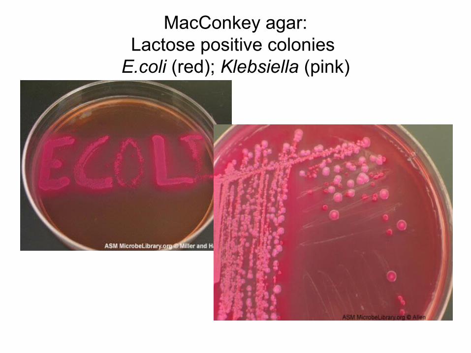
MacConkey agar:Lactose positive colonies
E.coli (red); Klebsiella (pink)

Genus Klebsiella
• Facultatively pathogenic: Colonizes the respiratory mucosa and the intestine
• In immunosuppresed patients (premature infants, elderly people) – potential for severe infections (pneumonia, sepsis, meningitis)
• Hospital acquired infections: surgical wound infections, urinary infections, sepsis
• Involvement in diarrhoeic diseasae - debated

Klebsiella – blood agar (non hemolytic mucoid colonies)

Klebsiella: Mucous colonies

Klebsiella – pink colonies (Lactose positive) on MacConkey agar

Genus Proteus
• Facultatively pathogenic• Colonizes the intestines• May cause infections in case of immunesuppression e.g.
urinary infections, synusitis, otitis, meningitis + hospital acquired infections
• Cultivation: characteristic ”invasion” of the culture medium – the swarming phenomenon – favours biofilm formation → catheter infections

Swarming of Proteus on culture plates (concentric growth rings)