Labeled Bone Images
65
© 2012 Pearson Education, Inc. Labeled Bone Images
-
Upload
ciaran-valentine -
Category
Documents
-
view
39 -
download
3
description
Labeled Bone Images. Figure 7-1 The Axial Skeleton. SKELETAL SYSTEM. 206. AXIAL SKELETON. 80. APPENDICULAR SKELETON. (see Figure 8 –1). 8. Cranium. Skull. 14. Face. Skull and associated bones. 29. Auditory ossicles. 6. Associated bones. 1. Hyoid. Sternum. 1. Thoracic - PowerPoint PPT Presentation
Transcript of Labeled Bone Images

© 2012 Pearson Education, Inc.
Labeled Bone Images

© 2012 Pearson Education, Inc.
Figure 7-1 The Axial SkeletonSKELETAL SYSTEM 206
APPENDICULAR SKELETON
(see Figure 8–1)
Cranium
Hyoid
Sternum
Ribs
Vertebrae
Sacrum
Coccyx
Associatedbones
Skull
FaceSkull and
associatedbones
Thoraciccage
Vertebralcolumn
AXIAL SKELETON
Auditoryossicles
8
14
6
1
1
24
24
1
1
29
25
26
80
An anterior view of the entireskeleton, with the axialcomponents highlighted. Thenumbers in the boxes indicatethe number of bones in the adultskeleton.
Sternum
Costalcartilages
Skull
Ribs
Lumbarvertebrae
Coccyx
Sacrum
Anterior (left) and posterior (right) views of the axial skeleton.The individual bones associated with the skull are not visible.
Cervicalvertebrae
Thoracicvertebrae

© 2012 Pearson Education, Inc.
Figure 7-1a The Axial SkeletonSKELETAL SYSTEM 206
APPENDICULAR SKELETON
(see Figure 8–1)
Cranium
Hyoid
Sternum
Ribs
Vertebrae
Sacrum
Coccyx
Associatedbones
Skull
FaceSkull and
associatedbones
Thoraciccage
Vertebralcolumn
AXIAL SKELETON
Auditoryossicles
8
14
6
1
1
24
24
1
1
29
25
26
80
An anterior view of the entire skeleton, with the axial components highlighted. The numbers in the boxes indicate the number of bones in the adult skeleton.

© 2012 Pearson Education, Inc.
Figure 7-3a The Adult Skull
PARIETALBONE(left)
PARIETALBONE(right)
OCCIPITALBONE
MANDIBLE
TEMPORAL BONE
Sagittal suture
Lambdoidsuture
Squamoussuture
Mastoid processStyloid process
Occipital condyle
External occipitalprotuberance
Posterior view

© 2012 Pearson Education, Inc.
Figure 7-3b The Adult Skull
PARIETALBONE(left)
PARIETALBONE(right)
Lambdoidsuture
Sagittalsuture
OCCIPITAL BONE
Coronalsuture
FRONTAL BONE
ZYGOMATICBONE
NASAL BONES
Superior view

© 2012 Pearson Education, Inc.
Figure 7-3c The Adult Skull
TEMPORALBONE
PARIETALBONE
OCCIPITALBONE
FRONTALBONE
Lateral view
Squamous suture
Lambdoid suture
Squamous part oftemporal bone
External acousticmeatus
Mastoid process
Zygomaticarch
Styloid processZygomatic process
of temporal boneTemporal processof zygomatic bone
Coronal suture
SPHENOID
NASAL BONE
LACRIMAL BONE
ETHMOID
MAXILLA
ZYGOMATIC BONE
MANDIBLE
Supra-orbital foramen
Infra-orbital foramen
Mental foramen
Mental protuberance

© 2012 Pearson Education, Inc.
Figure 7-3d The Adult Skull
Coronal suture
SPHENOID
NASAL BONE
LACRIMAL BONE
ETHMOID
MAXILLA
Supra-orbital foramen
Infra-orbital foramen
Mental foramen
Mental protuberance
ZYGOMATIC BONE
Optic canal
Inferior orbital fissure
Temporal process ofzygomatic bone
Mastoid process oftemporal bone
Middle nasal concha(part of ethmoid)Perpendicular plateof ethmoid Bony nasal
septumVOMER
Superior orbital fissure
PARIETAL BONE
TEMPORAL BONE
PALATINE BONE
INFERIORNASAL CONCHA
MANDIBLE
Anterior view
FRONTAL BONE

© 2012 Pearson Education, Inc.
Figure 7-3e The Adult Skull
ZYGOMATIC BONE MAXILLA
TEMPORAL BONE
PALATINE BONE
Zygomatic arch
Medial and lateralpterygoid processes
Foramen lacerum
Carotid canal
Mastoid process
Stylomastoid foramen
Occipital condyle
Foramen magnum
VOMER
SPHENOID
OCCIPITAL BONE
FRONTAL BONE
Jugular foramen
Foreman ovale
Styloid processMandibular fossa
Externalacoustic meatus
Lambdoid suture
External occipitalprotuberance
Inferior view

© 2012 Pearson Education, Inc.
Figure 7-4b The Sectional Anatomy of the Skull (Part 1 of 1)
Foramenmagnum
Carotid canal
Internal occipital crest
FRONTAL BONE
ETHMOID
SPHENOID
TEMPORAL BONE
PARIETAL BONE
OCCIPITAL BONE
Crista galli
Cribriform plate
Sella turcica
Foramen rotundum
Foramen lacerum
Foramen ovale
Foramen spinosum
Internalacoustic meatus
Jugular foramen
Hypoglossal canal
Superior view of a horizontal section through the skull, showing thefloor of the cranial cavity. Compare with part (a) and with Figure 7–3e.

© 2012 Pearson Education, Inc.
Figure 7-12c The Mandible and Hyoid Bone
Hyoid bone
© 2012 Pearson Education, Inc.
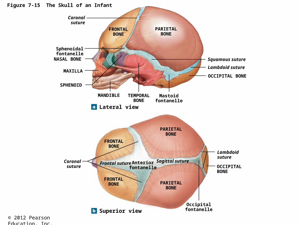
Figure 7-15 The Skull of an Infant
Sphenoidalfontanelle
NASAL BONE
MAXILLA
Coronalsuture
FRONTALBONE
PARIETALBONE
Squamous suture
Lambdoid suture
OCCIPITAL BONE
Mastoidfontanelle
TEMPORALBONE
SPHENOID
MANDIBLE
Lateral view
PARIETALBONE
FRONTALBONE
Sagittal sutureAnteriorfontanelle
Frontal sutureCoronalsuture
FRONTALBONE PARIETAL
BONE
Lambdoidsuture
OCCIPITALBONE
OccipitalfontanelleSuperior view

© 2012 Pearson Education, Inc.
Figure 7-16 The Vertebral Column
Spinal Curves
Primary curves developbefore birth, and secondarycurves after birth.
The cervical curve, asecondary curve, develops asthe infant learns to balance theweight of the head on thevertebrae of the neck.
The thoracic curve, aprimary curve,accommodates thethoracic organs.
The lumbar curve, asecondary curve, balancesthe weight of the trunk overthe lower limbs; it developswith the ability to stand.
The sacral curve, aprimary curve,accommodates theabdominopelvic organs.
Vertebral Regions
Regions are definedby anatomicalcharacteristics ofindividual vertebrae.
Cervical(7 vertebrae)
Thoracic(12 vertebrae)
Lumbar(5 vertebrae)
Sacral
Coccygeal
© 2012 Pearson Education, Inc.
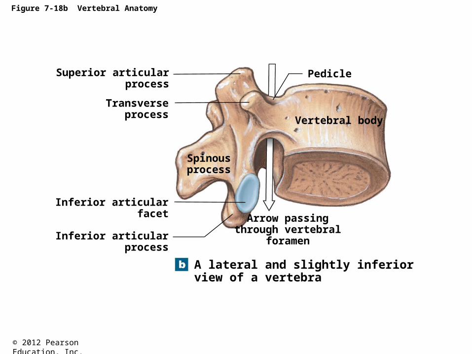
Figure 7-18b Vertebral Anatomy
A lateral and slightly inferiorview of a vertebra
Arrow passingthrough vertebral
foramen
Vertebral body
Pedicle
Spinousprocess
Inferior articularprocess
Inferior articularfacet
Transverseprocess
Superior articularprocess

© 2012 Pearson Education, Inc.
Figure 7-18c Vertebral Anatomy
Spinousprocess
Inferiorarticularprocess
Superiorarticularprocess
Transverseprocess
Inferiorarticular
facet
Vertebralforamen
Pedicle
Vertebralbody
An inferior view of avertebra

© 2012 Pearson Education, Inc.
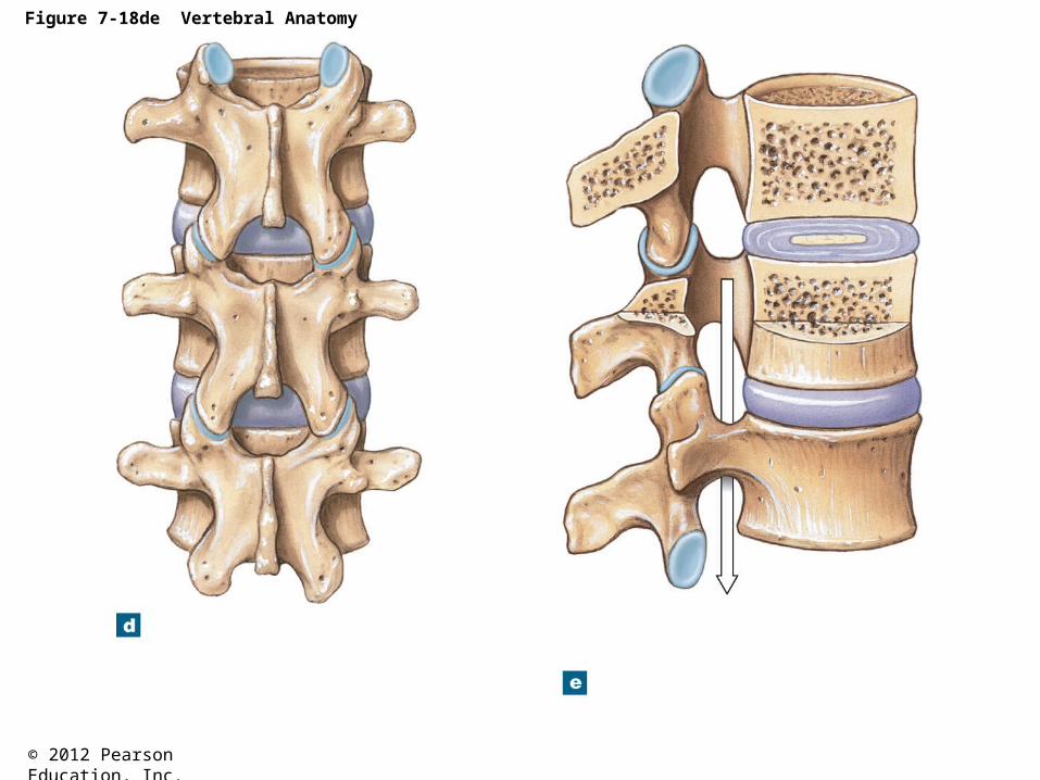
Figure 7-18de Vertebral Anatomy
A lateral and sectional viewof three articulatedvertebrae
Arrow passingthrough vertebral
canal
Vertebral body
Inferiorarticular
facetAn posterior view ofthree articulatedvertebrae
Inferiorarticularprocess
Vertebral body
Transverse process
Intervertebral disc
Spinous process
Intervertebraldisc
Intervertebralforamen
Lamina ofvertebral arch
Superior articular process
Superior articular facets

© 2012 Pearson Education, Inc.
Figure 7-19d The Cervical Vertebrae
Axis (C2)
Posteriorarch
Atlas (C1)
Dens ofaxis
Anteriorarch
Transverseligament
The atlas (C1) and axis (C2).

© 2012 Pearson Education, Inc.
Table 7-2 Regional Differences in Vertebral Structure and Function (Part 2 of 2)

© 2012 Pearson Education, Inc.
Figure 7-22a The Sacrum and Coccyx
Sacral cornu
Articularprocess
Entrance tosacral canal
Sacraltuberosity
Lateralsacral crest
Mediansacral crest
Coccygeal cornu
Sacralhiatus
A posterior view

© 2012 Pearson Education, Inc.
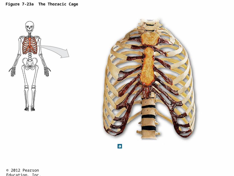
Figure 7-23 The Thoracic CageJugular notch
Clavicular articulation
Manubrium
Sternum Body
Xiphoidprocess
Costalcartilages
An anterior view, showing thecostal cartilages and the sternum
False ribs(ribs 8–12)
A posterior view, showing thearticulations of the ribs and vertebrae
True ribs(ribs 1–7)
False ribs(ribs 8–12)
True ribs(ribs 1–7)
Floating ribs(ribs 11–12)
Vertebrochondralribs
(ribs 8–10)
1
2
3
4
5
6
7
8
9
10
11
12
12
3
4
5
6
7
8
9
10
11
12
C7
T1
T2
T3
T4
T5
T6
T7
T8
T9
T10
T11
T12
L1
1
2
3
4
5
6
7
8
9
10
11
12
T12
T11
T1

© 2012 Pearson Education, Inc.
Figure 8-1 The Appendicular Skeleton (Part 1 of 2)
ClaviclePectoral
girdle
Upperlimbs
Scapula
Humerus
Radius
Ulna
Carpalbones
Metacarpalbones
Phalanges
Pelvicgirdle
Hip bone
SKELETAL SYSTEM
AXIAL SKELETON APPENDICULAR SKELETON
(see Figure 7–1)
206
80 126
4
2
60
16
10
28
2
2
2
2
22
© 2012 Pearson Education, Inc.
Figure 8-1 The Appendicular Skeleton (Part 2 of 2)
Lowerlimbs
Femur
Patella
Tibia
Fibula
Tarsal bones
Metatarsalbones
Phalanges
2
2
2
2
60
14
10
28

© 2012 Pearson Education, Inc.
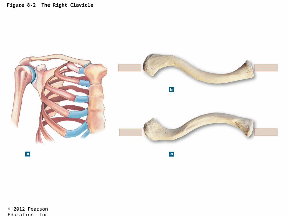
Figure 8-2 The Right Clavicle
Clavicle
The position of the clavicle withinthe pectoral girdle, anterior view.
MEDIALLATERAL
Scapula Jugular notch Acromial end Sternalend
Superior view of the right clavicle.
Acromial end
Conoidtubercle
Costaltuberosity
Sternalfacet
Sternal end
Inferior view of the right clavicle.Stabilizing ligaments attach to the conoidtubercle and the costal tuberosity.
LATERAL MEDIAL
Facet forarticulationwith acromion

© 2012 Pearson Education, Inc.
Figure 8-3 The Right Scapula
Acromion
Coracoidprocess
Superiorborder
Superiorangle
Lateralangle
Subscapularfossa
Lateral border
Body
Inferior angle
Anterior view
Acromion
Supraglenoidtubercle
Coracoidprocess
Spine
Medial border
Glenoidcavity
Lateral border
Inferior angle
Lateral view
AcromionSupraspinousfossa
Superiorborder
Coracoidprocess
Neck
Spine
InfraspinousfossaBody
Medialborder
Lateralborder
Posterior view

© 2012 Pearson Education, Inc.
Figure 8-4a The Right Humerus and Elbow Joint
Greater tubercle
Lesser tubercle
Intertuberculargroove
Head
Anatomicalneck
Surgicalneck
Deltoidtuberosity
Shaft
Anterior surface
Radial fossa
Lateralepicondyle
Condyle
Capitulum Trochlea
Coronoid fossa
Medialepicondyle
© 2012 Pearson Education, Inc.
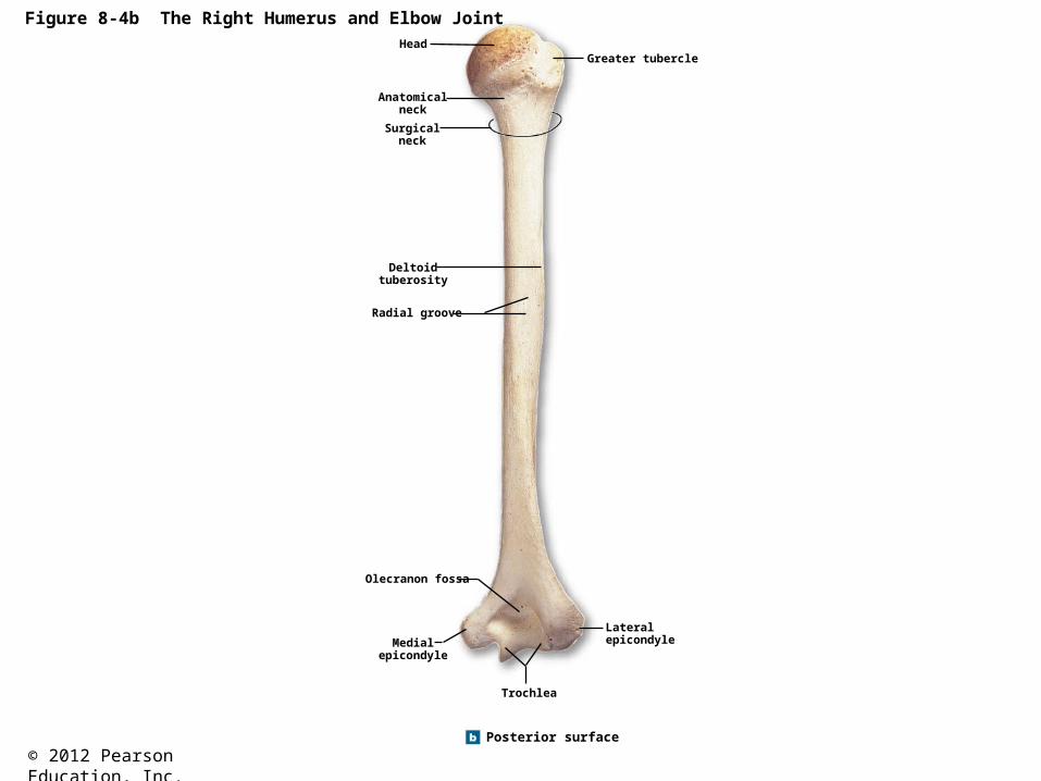
Figure 8-4b The Right Humerus and Elbow JointHead
Anatomicalneck
Surgicalneck
Deltoidtuberosity
Radial groove
Greater tubercle
Olecranon fossa
Medialepicondyle
Trochlea
Lateralepicondyle
Posterior surface

© 2012 Pearson Education, Inc.
Figure 8-5 The Right Radius and Ulna
Olecranon
Proximalradioulnar
joint
ULNA
Ulnar head
Styloid processof ulna
Posterior view
Radial head
Neck ofradius
Radial tuberosity
RADIUS
Interosseousmembrane
Ulnar notchof radius
Styloid processof radius
Olecranon
Trochlear notch
Coronoid process
Radial notch
Ulnar tuberosity
ULNA
ULNA
Lateral viewof ulna, showingtrochlear notch
Distal radio-ulnar joint
Ulnar head
Anterior view

© 2012 Pearson Education, Inc.
Figure 8-6b Bones of the Right Wrist and Hand
ULNA
Triquetrum
Pisiform
Hamate
Posterior view
VIV III II
I
RADIUS
Lunate
Scaphold
Trapezium
Trapezoid
Capitate
Metacarpalbones
Proximalphalanx
Distalphalanx
Middlephalanx

© 2012 Pearson Education, Inc.
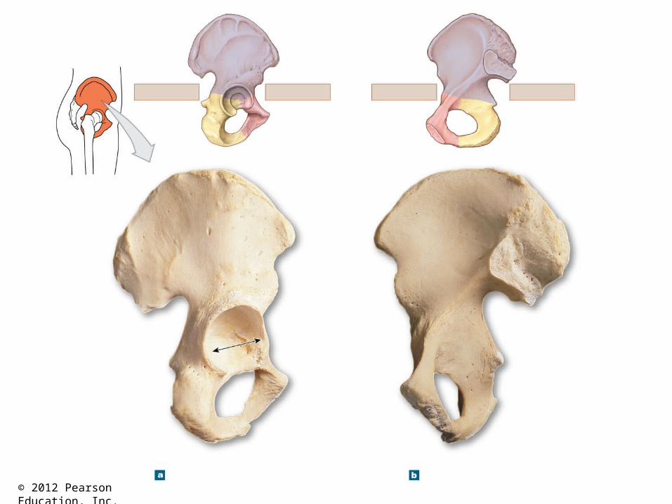
Figure 8-7 The Right Hip Bone
Ischium
Iliac crest
Anteriorsuperior
iliac spine
Inferiorgluteal line
Anterior inferioriliac spine
Acetabulum
Acetabular notch
Superior ramus of pubis
Pectineal line
Pubic tubercle
Inferior ramus of pubis
Obturator foramen
Ischial ramus
Ischial tuberosity
Lesser sciaticnotch
Ischial spine
Lunate surfaceof acetabulum
Greater sciatic notch
Posterior inferioriliac spine
Posteriorsuperior
iliac spine
Posteriorgluteal line
Anteriorgluteal line
Right hip bone, lateral view Right hip bone, medial view
ANTERIORPOSTERIOR
Ilium
PubisIschium
ANTERIOR POSTERIOR
Ilium
Pubis
Iliacfossa
Auricular surfacefor articulationwith sacrum
Iliactuberosity
Posteriorsuperioriliac spine
Posterior inferioriliac spine
Greater sciatic notch
Arcuate line
Ischial spine
Lesser sciatic notch
Ischial tuberosity
Ischial ramusLocationof pubic
symphysis

© 2012 Pearson Education, Inc.
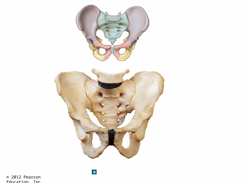
Figure 8-8a The Pelvis of an Adult MaleSACRUM
COCCYX
ILIUM
PUBIS
ISCHIUM
Hip bone(Figure 8–7)
Iliac crest
L5
SACRUMArcuate
line
Acetabulum
Pubictubercle
Obturatorforamen
Sacroiliacjoint
Iliacfossa
Pubicsymphysis
ILIUM
PUBIS
ISCHIUM
Anterior view

© 2012 Pearson Education, Inc.
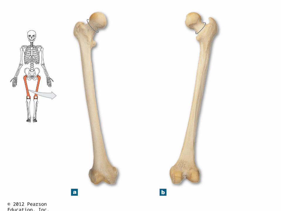
Figure 8-11 Bone Markings on the Right Femur
Neck
Greatertrochanter
Patellar surface
Lateral epicondyle
Lateral condyle
Anterior surface
Fovea capitis
Femoral head
Intertrochanteric line
Lesser trochanter
Shaft
Medial supracondylarridge
Adductor tubercle
Medial epicondyle
Medial condyle
Neck
Greater trochanter
Intertrochantericcrest
Gluteal tuberosity
Pectineal line
Linea aspera
Lateral supracondylarridge
Posterior surface
Popliteal surface
Intercondylar fossa
Lateral epicondyle
Lateral condyle

© 2012 Pearson Education, Inc.
Figure 8-12a The Right Patella (a, b) and Patella with Femur (c)
Patella

© 2012 Pearson Education, Inc.
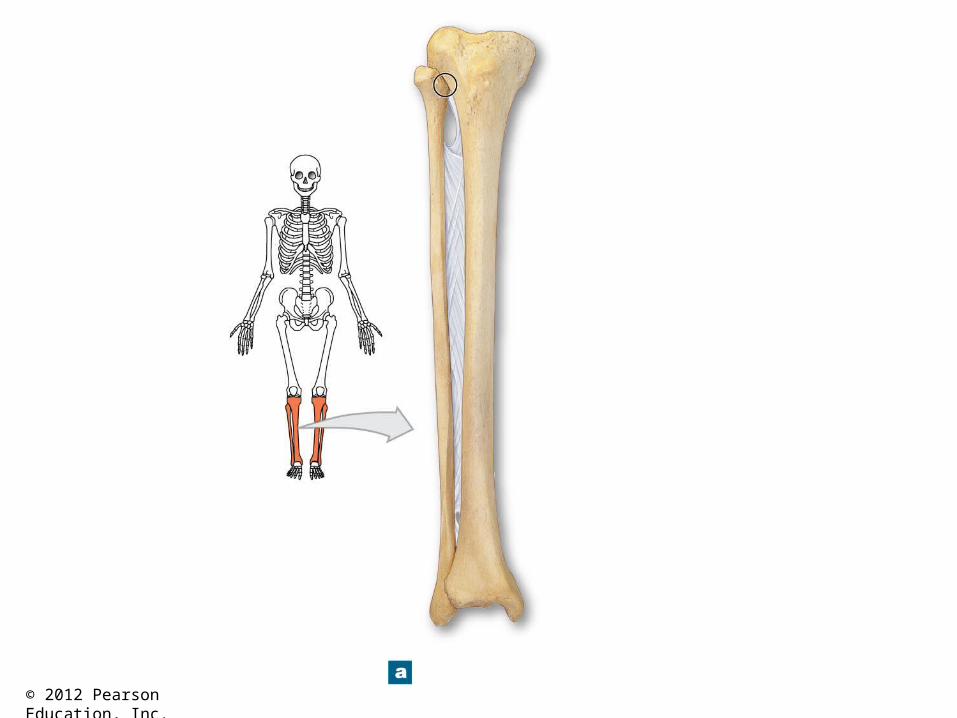
Figure 8-13 The Right Tibia and Fibula
Soleal line
Articular surface ofmedial tibial condyle
Medial tibialcondyle
Tibial tuberosity
Interosseousmembrane
Anterior margin
TIBIA
FIBULA
Medial malleolus(tibia)
Inferior articular surface
Anterior view
Lateral malleolus(fibula)
Superiortibiofibular
joint
Head of fibula
Lateral tibial condyle
FIBULATIBIA
Inferior tibiofibular joint
Lateral malleolus (fibula)
Posterior view
Intercondylar eminence
Articular surface oflateral tibial condyle
Lateral tibial condyle
Head of fibula
Anterior margin
Tibia
Interosseous membrane
Cross section oftibia and fibula
Fibula

© 2012 Pearson Education, Inc.
Figure 8-14 Bones of the Ankle and Foot
Calcaneus
Trochleaof talus
Hallux
Medial
Intermediate
Lateral
Cuneiform bones
Cuboid
Talus
Navicular
VIV III
II I
Metatarsal bones
Proximalphalanx
Distal phalanx
Superior view, right foot
Distal
Middle
Proximal
Phalanges
Medial view, right foot
Longitudinalarch
Transversearch
PhalangesMetatarsal
bones
Medialcuneiform
bone
Navicular Talus
Calcaneus

© 2012 Pearson Education, Inc.
Figure 7-3d The Adult Skull

© 2012 Pearson Education, Inc.
Figure 7-3c The Adult Skull

© 2012 Pearson Education, Inc.
Figure 7-3e The Adult Skull

© 2012 Pearson Education, Inc.
Figure 7-3a The Adult Skull

© 2012 Pearson Education, Inc.
Figure 7-3b The Adult Skull

© 2012 Pearson Education, Inc.

© 2012 Pearson Education, Inc.

© 2012 Pearson Education, Inc.

© 2012 Pearson Education, Inc.
Figure 7-12c The Mandible and Hyoid Bone

© 2012 Pearson Education, Inc.
Figure 7-16 The Vertebral Column

© 2012 Pearson Education, Inc.
Figure 7-18b Vertebral Anatomy

© 2012 Pearson Education, Inc.
Figure 7-18c Vertebral Anatomy
© 2012 Pearson Education, Inc.
Figure 7-18de Vertebral Anatomy

© 2012 Pearson Education, Inc.
Figure 7-19d The Cervical Vertebrae
© 2012 Pearson Education, Inc.
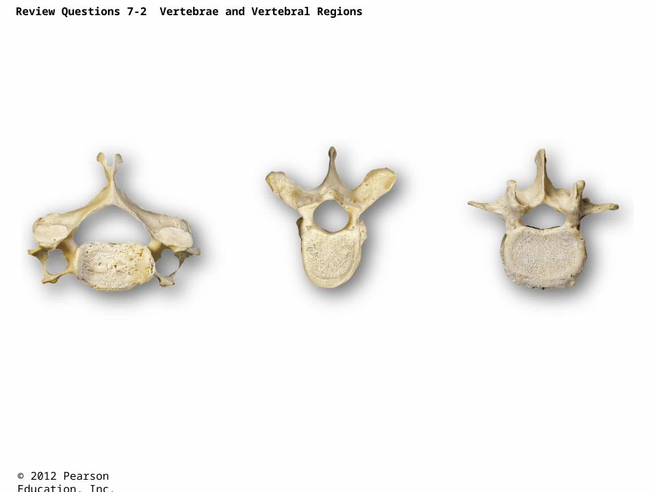
Review Questions 7-2 Vertebrae and Vertebral Regions

© 2012 Pearson Education, Inc.
Figure 7-22a The Sacrum and Coccyx

© 2012 Pearson Education, Inc.
Figure 7-23 The Thoracic Cage
© 2012 Pearson Education, Inc.
Figure 7-23a The Thoracic Cage

© 2012 Pearson Education, Inc.
Figure 7-23b The Thoracic Cage
© 2012 Pearson Education, Inc.
Figure 8-2 The Right Clavicle

© 2012 Pearson Education, Inc.
Figure 8-2a The Right Clavicle

© 2012 Pearson Education, Inc.
Figure 8-3 The Right Scapula

© 2012 Pearson Education, Inc.

© 2012 Pearson Education, Inc.
Figure 8-5 The Right Radius and Ulna

© 2012 Pearson Education, Inc.
© 2012 Pearson Education, Inc.
© 2012 Pearson Education, Inc.
© 2012 Pearson Education, Inc.

© 2012 Pearson Education, Inc.

© 2012 Pearson Education, Inc.
© 2012 Pearson Education, Inc.

© 2012 Pearson Education, Inc.