Lab protocol BLOOD - Dartmouth Collegeanatomy/Histo/protocols/Lab protocol 4.pdf · INTEGUMENT;...
9
Lab exercise 4 –BLOOD; BONE MARROW; RESPIRATORY SYSTEM; INTEGUMENT; MAMMARY GLANDS 1 Lab protocol In this lab we will first examine the components of blood before examining the bone marrow and respiratory system. BLOOD 1. Slide #101 [human blood smear, Wright’s stain]. Find and study under the oil-immersion lens the following formed elements. (Note. Do not use oil until you have first scanned the slide with low power objectives (10x -> 40x) to locate an optimal area for study . Identification of cells should be attempted only in areas where the red blood cells are a single layer deep and the white cells are undistorted and well-stained. WHITE BLOOD CELLS (WBC’s, LEUKOCYTES): neutrophils (polymorphonuclear leukocytes, PMNs or “polys”) eosinophils monocytes lymphocytes basophils (rare) As you study the various kinds of white blood cells, compare them in size to the surrounding red blood cells. Normal, undistorted, adult human red blood cells will be in the 7-8 μm diameter range. (In histological sections of paraffin embedded tissues the diameter of RBCs is in the range of 6-7 μm due to some shrinkage artifact.) Which type of white blood cell is the largest? Which is the smallest? The primary criteria for identifying the various kinds of white blood cells are: 1) the size of the cell, 2) the size and shape of the nucleus and the distribution of chromatin within it, and 3) the amount of cytoplasm and the number and staining reactions of the granules present within it. Be sure to identify neutrophils with their multi-lobed nuclei and perhaps also a “band” (“stab”) cell, which is an immature neutrophil with a horseshoe-shaped nucleus. Eosinophils are easily identifiable because of their obvious red cytoplasmic granules and bi-lobed nuclei. Basophils are so rare that you will probably not encounter one, but, if you do, it will have very dark cytoplasmic granules, obscuring the nucleus. Monocytes are the largest white cells often with bean-shaped nuclei and agranular cytoplasm. Finally, lymphocytes are the smallest WBC, slightly larger than the RBCs, and tend to be round with round nuclei that fill up almost the entire cell leaving scant cytoplasm. After you are satisfied with your ability to identify the various kinds of white blood cells, compare the percentages of the leukocytes in your smear to the expected differential values that you should have committed to memory. Abnormally high or low percentages of white blood cell types often accompany certain disorders and may be diagnostic. ERYTHROCYTES
-
Upload
vuongkhanh -
Category
Documents
-
view
233 -
download
0
Transcript of Lab protocol BLOOD - Dartmouth Collegeanatomy/Histo/protocols/Lab protocol 4.pdf · INTEGUMENT;...
Lab exercise 4 –BLOOD; BONE MARROW; RESPIRATORY SYSTEM; INTEGUMENT; MAMMARY GLANDS
1
Lab protocol
In this lab we will first examine the components of blood before examining the bone marrow and respiratory system.
BLOOD 1. Slide #101 [human blood smear, Wright’s stain]. Find and study under the oil-immersion
lens the following formed elements. (Note. Do not use oil until you have first scanned the slide with low power objectives (10x -> 40x) to locate an optimal area for study. Identification of cells should be attempted only in areas where the red blood cells are a single layer deep and the white cells are undistorted and well-stained. WHITE BLOOD CELLS (WBC’s, LEUKOCYTES): neutrophils (polymorphonuclear leukocytes, PMNs or “polys”) eosinophils monocytes lymphocytes basophils (rare) As you study the various kinds of white blood cells, compare them in size to the surrounding red blood cells. Normal, undistorted, adult human red blood cells will be in the 7-8 µm diameter range. (In histological sections of paraffin embedded tissues the diameter of RBCs is in the range of 6-7 µm due to some shrinkage artifact.) Which type of white blood cell is the largest? Which is the smallest? The primary criteria for identifying the various kinds of white blood cells are: 1) the size of the cell, 2) the size and shape of the nucleus and the distribution of chromatin within it, and 3) the amount of cytoplasm and the number and staining reactions of the granules present within it. Be sure to identify neutrophils with their multi-lobed nuclei and perhaps also a “band” (“stab”) cell, which is an immature neutrophil with a horseshoe-shaped nucleus. Eosinophils are easily identifiable because of their obvious red cytoplasmic granules and bi-lobed nuclei. Basophils are so rare that you will probably not encounter one, but, if you do, it will have very dark cytoplasmic granules, obscuring the nucleus. Monocytes are the largest white cells often with bean-shaped nuclei and agranular cytoplasm. Finally, lymphocytes are the smallest WBC, slightly larger than the RBCs, and tend to be round with round nuclei that fill up almost the entire cell leaving scant cytoplasm. After you are satisfied with your ability to identify the various kinds of white blood cells, compare the percentages of the leukocytes in your smear to the expected differential values that you should have committed to memory. Abnormally high or low percentages of white blood cell types often accompany certain disorders and may be diagnostic. ERYTHROCYTES
Lab exercise 4 –BLOOD; BONE MARROW; RESPIRATORY SYSTEM; INTEGUMENT; MAMMARY GLANDS
2
Erythrocytes take the shape of biconcave discs (why this shape?). They are essentially containers full of hemoglobin without any nucleus or evidence of internal organelles. One should note any abnormalities of the erythrocytes. Megaloblastic anemia will result in large RBCs, while iron deficiency will result in erythrocytes that are too small. A significant variation in shape of the erythrocytes should also be noted; this might include spherocytic changes, stippling or irregularly shaped cells (including sickle cells). When the area of central pallor occupies more than two-thirds of the cell, hypochromia is likely, and abnormalities in hemoglobin production should be suspected (such as iron deficiency). You will become acquainted with these and other less frequent abnormalities of erythrocytes when you study hematology. PLATELETS Finally, the number of platelets per oil immersion field should be noted. These are the smaller particles, also without a nucleus. These may be individual or clumped together. When successive high power fields contain fewer than four platelets, and they are not present elsewhere on the slide in clumps, platelets are said to be decreased. This will be confirmed with platelet counts.
2. Slide #100 [blood Smear, Human, Methylene blue]. Examine the slide first under low power and then under high power. Note: this slide was scanned at 100X (oil immersion). Immature red blood cells, called reticulocytes, can be seen in peripheral blood smears stained with a basic dye such as New Methylene Blue as seen here under oil immersion. These dyes bind to residual rRNA elements still present in these cells forming a reticular precipitate. Reticulocytes can be identified by the dark blue (basophilia) in the cytoplasm of some of the erythrocytes. About 1% of RBCs are reticulocytes. They usually remain as reticulocytes for one day, and nearly 1% or RBCs are turned over each day. Is this peripheral blood smear normal or abnormal?
BONE MARROW 3. Slide #103 [red bone marrow]. You will have either a section of a vertebral body showing red marrow in situ (odd boxes) or a section of a punch biopsy of red marrow (even boxes). Please examine both slides (they both appear on the virtual histology site). Do not use your oil immersion objective on this slide. This section is mainly to demonstrate general architecture of active marrow, not the detailed structure of hematopoietic cells.
Using the 10x objective identify: trabeculae and hematopoietic cells of the marrow. Assess the cellularity of the marrow, i.e., the proportion of hematopoietic cells compared to fatty tissue. Using the 40x objective identify in the marrow: Venous sinusoids are identified by the aggregations of mature RBCs in their lumens. You may also be able to see some arterioles.

Lab exercise 4 –BLOOD; BONE MARROW; RESPIRATORY SYSTEM; INTEGUMENT; MAMMARY GLANDS
3
Megakaryocytes. Giant cells with single large polyploid nuclei. What do these cells produce? Erythroblastic (erythroid) islands. With careful study of the stroma of bone marrow sections, an architectural organization to the seemingly random arrangement of myeloid tissue may be appreciated. Clusters of developing red blood cells, the erythroid islands, can be found in the stroma but in close proximity to the marrow sinusoids. A macrophage is typically found in the midst of the cluster but may be difficult to distinguish. In our slide collection erythroid islands are best observed in the punch biopsy specimen. It may be useful to first spend time familiarizing yourself with the cells of the erythroid lineage by examining the bone marrow smear, then return to this slide after you can more easily recognize their morphology. Do not spend a long time searching for erythroid islands. Simply understand their function as discussed in lecture. The remainder - cells of the granulocytic, monocytic, lymphocytic lines. You can easily see some juvenile and mature neutrophils and eosinophils.
4. Slide #75 [red bone marrow, rib]. This is a section of rib from a young rabbit. Compare the appearance of the red marrow elements to those seen in Slide #103. Blood sinusoids are easily seen. Note that even in the young animal there can be a considerable amount of fatty infiltration of the marrow. Interestingly, this animal must have received an IV injection of India ink before sacrifice. Note black ink particles phagocytosed by macrophages and, to a lesser extent, sinusoidal endothelium.
5. Slide #104 [red marrow, smear, Wright’s stain]. This is a smear of red marrow that has been aspirated along with some peripheral blood. You should first scan this slide using your 10x objective. In some parts the smear may be too thick, in other parts it may not be well

Lab exercise 4 –BLOOD; BONE MARROW; RESPIRATORY SYSTEM; INTEGUMENT; MAMMARY GLANDS
4
stained or it may have many black dots of precipitated stain. Select a GOOD region (very important!!) and examine it first with your 40x objective and then with your oil immersion objective. In order to positively identify these cells YOU MUST USE YOUR OIL IMMERSION OBJECTIVE. (You cannot return to 40X, but you can use your 10x by rotating nosepiece through the 4x objective).
Rather than trying to identify every cell that you see, work your way through the erythroid and granulocyte lineages searching for good examples of each developmental stage, remembering that many cells will be in between stages. Illustrations in your texts and atlases will remind you of the features characteristic of each stage. In the erythroid series, find proerythroblasts, basophilic erythroblasts, polychromatophilic erythroblasts, and orthochromatophilic erythroblasts. In the granulocyte line, find promyelocytes, myelocytes (neutrophilic/eosinophilic; basophils are too rare to spend time on), metamyelocytes (neutrophilic/eosinophilic), band cells and the mature, segmented forms. You are very unlikely to find myeloblasts and don’t waste time on this. Remember that the early stages are less abundant than the later stages, thus you may have to search awhile for proerythroblasts, promyelocytes, etc. Also, keep in mind that the earliest forms, the blasts, are difficult to distinguish from each other. An early proerythroblast, before it develops the royal blue cytoplasmic staining indicative of the red cell line, will look very much like a myeloblast, a lymphoblast or monoblast to the untrained eye (and probably to some trained eyes as well). Note that nucleoli can usually be seen in the early blast forms. These are lighter blue than the rest of the nucleus (this is different than H&E stain, where nucleoli are dark blue). We will not ask that you identify cells in the lymphocytic or monocytic lines as their features are difficult to discriminate from the early stages of the other lineages. KEEP THE FOLLOWING IN MIND WHILE EXAMINING A MARROW SMEAR: • The most abundant nucleated cells will be those of the granulocytic series. • The most abundant nucleated cells of both the granulocytic series and erythrocytic series
will be the more mature stages. • Although very early stages of blood cells (“stem cells”) may be rather small, only slightly
larger than a red cell, these cells do not show enough morphological characteristics to be classified. The earliest stages that are classifiable are all considerably larger than red cells.
• The size of nucleated cells of both erythrocytic and granulocytic stages decreases with progression down the developmental path. Compare the size of a nucleated marrow cell with that of a RBC.
• As cells mature the nucleus becomes smaller and more condensed. In trying to identify a marrow cell the first question is: ARE THERE CYTOPLASMIC GRANULES? If there are, it is one of the granulocytic series. If not, it is probably part of the red cell series. Granules may be difficult or impossible to see in late stages of neutrophil development, but nuclear characteristics of late stages should make for easy identification. Don’t mistake the blue and red mottling of a polychromatophilic erythroblast for granules.

Lab exercise 4 –BLOOD; BONE MARROW; RESPIRATORY SYSTEM; INTEGUMENT; MAMMARY GLANDS
5
HERE IS A TABLE THAT MAY HELP GRANULOCYTIC SERIES ERYTHROCYTIC SERIES GRANULES AZUROPHILIIC (large, darkly stained) NONE SPECIFIC (very small neutrophilic, larger eosinophilic) NUCLEUS OFTEN FLATTENED ROUND or HORSESHOE-SHAPED CYTOPLASM LIGHT BLUE DARK BLUE or ALMOST COLORLESS or MOTTLED PINK/BLUE
or RED FIND GOOD AREAS IN THE SMEAR- Under low, then high dry (40x), objectives find an area on the smear where the red cells are not piled on top of each other and where there are nucleated cells. SWITCH TO OIL IMMERSION OBJECTIVE- Since it can be difficult to identify some marrow cells even under oil, it is impossible to identify them under your 40x objective. KEEP IN MIND THE FOLLOWING DIFFERENCES BETWEEN A MARROW SMEAR AND A SECTION OF BONE MARROW:
• A marrow smear always contains variable number of mature cells of the peripheral blood.
• Megakaryocytes are fragile; you won’t find many in a marrow smear. Same goes for fat cells.
• Bone fragments would not be seen. • Finally- plasma cells are rarely found in either normal marrow or peripheral blood.
RESPIRATORY SYSTEM
6. Slide #167 [trachea, monkey, H &E]. This is a mixed set of slides. Some of you will have a complete cross section of the trachea (which may include the esophagus cut in cross section); others will have a piece of transversely sectioned trachea. In either slide identify the hyaline cartilage, which provides semirigid support for the wall of the trachea. The typical C-shape of the cartilages may not be apparent. The open part of the "C" is bridged by dense fibroelastic connective tissue (continuous with perichondrium) and contains the trachealis muscle (what muscle type?). What specific kind of epithelium lines the lumen of the trachea? The epithelium is supported by a thick basement membrane and a lamina propria containing an abundance of elastic fibers. The looser connective tissue external to the lamina propria is considered the submucosa. Mixed sero-mucous glands are usually found in the submucosa. You can check the same features in small piece of human trachea Slide #29 [H &E].

Lab exercise 4 –BLOOD; BONE MARROW; RESPIRATORY SYSTEM; INTEGUMENT; MAMMARY GLANDS
6
7. Slide #168 [lung, monkey, H&E]. This is a good general-purpose slide to begin study of the lung. Refer to the diagram (below) and the illustrations in your atlases and textbook. Find and study examples of segmental bronchi, 1o bronchioles, terminal and respiratory bronchioles. Give particular attention to the walls of these passages noting the changes in the components of the wall layers as one proceeds from larger to smaller airways. Where does the transition between conducting and respiratory divisions occur? Find pulmonary arteries that accompany the air duct system and pulmonary veins that run a more independent course through the lung. Look for bronchial arteries, lymphatic vessels, and nerves that accompany the largest bronchi. Look for accumulations of macrophages in the walls of some bronchioles. These are easily identified because of their accumulated dense foreign particles. Diffuse aggregations of T- and B-lymphocytes are also common in the walls of bronchi and bronchioles (“BALT”, bronchus-associated lymphoid tissue). Find the pleural surface of the lung and its covering of visceral pleura mesothelium (simple squamous epithelium). The epithelium rests on a layer of dense connective tissue with abundant elastic fibers. Note: this slide is not so useful for identifying the smaller air passages such as alveolar ducts, and alveolar sacs because these tend to be collapsed. Study these components in the subsequent slides.
8. Slide #169 [lung (inflated), human, Masson stain]. This is a grand old slide of inflated lung stained with a trichrome method (collagen stains blue). Inflation makes it much easier to
Lab exercise 4 –BLOOD; BONE MARROW; RESPIRATORY SYSTEM; INTEGUMENT; MAMMARY GLANDS
7
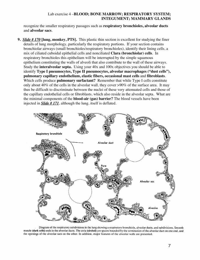
recognize the smaller respiratory passages such as respiratory bronchioles, alveolar ducts and alveolar sacs.
9. Slide # 170 [lung, monkey, PTS]. This plastic thin section is excellent for studying the finer
details of lung morphology, particularly the respiratory portions. If your section contains bronchiolar airways (small bronchioles/respiratory bronchioles), identify their lining cells, a mix of ciliated cuboidal epithelial cells and nonciliated Clara (bronchiolar) cells. In respiratory bronchioles this epithelium will be interrupted by the simple squamous epithelium constituting the walls of alveoli that also contribute to the wall of these airways. Study the interalveolar septa. Using your 40x and 100x objectives you should be able to identify Type I pneumocytes, Type II pneumocytes, alveolar macrophages (“dust cells”), pulmonary capillary endothelium, elastic fibers, occasional mast cells and fibroblasts. Which cells produce pulmonary surfactant? Remember that while Type I cells constitute only about 40% of the cells in the alveolar wall, they cover >90% of the surface area. It may thus be difficult to discriminate between the nuclei of these very attenuated cells and those of the capillary endothelial cells or fibroblasts, which also reside in the alveolar septa. What are the minimal components of the blood-air (gas) barrier? The blood vessels have been injected in Slide # 171, although the lung, itself is deflated.
Lab exercise 4 –BLOOD; BONE MARROW; RESPIRATORY SYSTEM; INTEGUMENT; MAMMARY GLANDS
8
INTEGUEMENT
10. Slide #33 [Thick skin, H &E]. Identify the following epidermal strata: basale (germinativum), spinosum, granulosum, lucidum (often difficult to discern), and corneum. In which of these layers would you expect to find mitotic figures? Study the spinosum at high magnification (40x objective). Note the so-called “intercellular bridges” (spines) between the keratinocytes from which this layer derives its name. Recall that this appearance reflects a shrinkage artifact as the cells pull away from each other during dehydration, with adjacent cells remaining connected only by their desmosomal attachments. Note the keratohyaline granules in the stratum granulosum. What is their composition/function? At what strata do the keratinocytes become so heavily keratinized that they die and become cornified plates (squames)? Identify the papillary and reticular layers of the dermis. What is the differnce in organization between these two layers? Find dermal papillae. Look for a Meissner corpuscle (touch receptor) in the dermal papilla. Now examine the hypodermis (superficial fascia). Note the abundance of blood vessels, nerves, and glands. What gland is found in this layer? Find their secretory units and ducts. Identify the secretory portion of the gland by locating dark cells (numerous), clear cells (rare) and myoepithelial cells (around the perimeter). What is the function of myoepithelial cells? You should also see the ducts of the eccrine glands that are lined with stratified cuboidal epithelium. Try to follow the ducts through the epidermis proper to their openings at the surface of the stratum corneum. Look at the skin of your fingertip using an inverted ocular as a hand lens. Where are the openings of the ducts located?
11. Slide #193 [Scalp, human, H&E]. This is good example of thin skin with abundant pilosebaceous units (hair follicles and associated sebaceous glands). Begin by comparing the thickness and appearance of the epidermal strata with that seen in the previous slide. Identify melanin granules within keratinocytes. In which layers are melanin granules found? This slide also demonstrates nicely the two layers of dermis: the papillary layer immediately beneath the epithelium and consisting of a network of more delicate collagen fibers compared to the subjacent reticular layer of dermis in which much larger, coarse bundles of collagen predominate. Next, study the hair follicles. Referring to the drawing (above) and the figures in your atlas or textbook identify the following: bulb (root), shaft, dermal papilla, hair matrix, root sheaths, sebaceous gland. It is unlikely that your section will have a perfect longitudinal section through a complete follicle, so you will have to piece the picture together from several different follicles. Look for arrector pili muscles (smooth muscle). Slide #58 (Box A) also is a section of scalp.
12. Slide # 194 [Skin (axillary), PTS]. This is another section of hairy thin skin, but with a new
feature, apocrine sweat glands that are found mainly in the axillary and perineal regions. Unlike eccrine (merocrine) sweat glands, these usually drain into hair follicles like sebaceous glands. The glands only begin to function after puberty. This plastic thick section affords excellent cytological detail. Note the considerably larger size of the secretory units of the apocrine sweat glands compared to those of eccrine sweat glands. Also identify the brightly eosinophilic myoepithelial cells subjacent to the lining epithelial cells of the secretory units.

Lab exercise 4 –BLOOD; BONE MARROW; RESPIRATORY SYSTEM; INTEGUMENT; MAMMARY GLANDS
9
13. Slide #197 [Lip, H&E]. This section is rather thick and over-stained with eosin but does show some interesting features. First find the surface epithelium. If you start at the part with hair follicles you will encounter a point of transition from the keratinized stratified squamous epithelium of the skin outside of the lip to the nonkeratinized stratified squamous epithelium of the lip (oral mucosa). This is an example of a mucocutaneous junction. Where else in the body do you find such junctions? The vermillion border extends from this line to where the very thin, keratinized skin of the lip meets the thicker skin over the face. This appears red because the richly vascularized dermal papilla can be seen through the thin overlying skin. This is a common site for carcinoma of the lip, particularly in heavy smokers.
14. The following slide will be ON DEMONSTRATION: Pacinian corpuscles. These structures, usually found in the deep dermis/hypodermis, are quite large and easily picked out even at 4x power. Where are these encapsulated touch-pressure receptors located in the body? What are their characteristic structural features?
MAMMARY GLAND
15. Slide #192 [Mammary gland, inactive /”resting”, monkey]. (odd boxes only, please share and/or look on the Virtual Histology site). This section from monkey includes skin (with keratinized stratified squamous epithelium) and mammary tissue. Note the lobar architecture of this mature, but inactive gland, and the many ducts lined with simple cuboidal epithelium. Note the almost complete lack of white adipose tissue in the monkey breast, which differentiates this from what would be seen in the human. In a woman who has never been pregnant (nulligravida), the resting breast contains ducts and rudimentary alveoli.
16. Slide #195 [Mammary gland, inactive/”resting”, human]. This section of human
mammary gland is most likely from a post-menopausal woman judging from the more abundant dense fibrous connective tissue and reduced white adipose tissue. Only isolated segments of ducts and an occasional hint of a lobe remain. This specimen would be better described as “atrophic” rather than “inactive”.
17. Slide #196 [Mammary gland, active (lactating), human H&E]. (MIXED SET; please
share). In response to elevated levels of estrogens and progesterone during pregnancy there is a rapid growth in length and branching of the duct system and proliferation of secretory alveoli. After parturition and the onset of lactation, the secretory alveoli and ducts become filled with milk. What specific kind epithelium lines the larger lactiferous ducts? In some places myoepithelial cells can be identified. These lie between the basement membrane and columnar epithelial cells. What is the function of these cells in the breast?