Lab notes guide to lab and diagnostic tests (2005)
223
-
Upload
aldawaa -
Category
Healthcare
-
view
229 -
download
2
Transcript of Lab notes guide to lab and diagnostic tests (2005)

Contacts • Phone/E-Mail
Name
Ph: e-mail:
Name
Ph: e-mail:
Name
Ph: e-mail:
Name
Ph: e-mail:
Name
Ph: e-mail:
Name
Ph: e-mail:
Name
Ph: e-mail:
Name
Ph: e-mail:
Name
Ph: e-mail:
Name
Ph: e-mail:
Name
Ph: e-mail:
Name
Ph: e-mail:
00Hop-FM 2/4/05 12:24 PM Page 2

Purchase additional copies of this bookat your health science bookstore ordirectly from F. A. Davis by shoppingonline at www.fadavis.com or by calling800-323-3555 (US) or 800-665-1148 (CAN)
A Davis’s Notes Book
Tracey Hopkins, BSN, RN
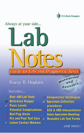
LABNotesGuide to LAB & DIAGNOSTIC TESTSGuide to LAB & DIAGNOSTIC TESTS
LABNotes
00Hop-FM 2/4/05 12:24 PM Page 3

F. A. Davis Company1915 Arch StreetPhiladelphia, PA 19103www.fadavis.com
Copyright © 2005 by F. A. Davis Company
All rights reserved. This book is protected by copyright. No part of it may bereproduced, stored in a retrieval system, or transmitted in any form or byany means, electronic, mechanical, photocopying, recording, or otherwise,without written permission from the publisher.
Printed in China by Imago
Last digit indicates print number: 10 9 8 7 6 5 4 3 2 1
Publisher, Nursing: Robert G. MartoneProject Editors: Danielle J. Barsky, Ilysa H. RichmanDesign Managers: Joan WendtCover Design: Paul FryConsultants: Beth Clark, RN, MSN, MED, PhD, Rachel Albert, Phd, RN
As new scientific information becomes available through basic and clinicalresearch, recommended treatments and drug therapies undergo changes.The author(s) and publisher have done everything possible to make thisbook accurate, up to date, and in accord with accepted standards at the timeof publication. The author(s), editors, and publisher are not responsible forerrors or omissions or for consequences from application of the book, andmake no warranty, expressed or implied, in regard to the contents of thebook. Any practice described in this book should be applied by the readerin accordance with professional standards of care used in regard to theunique circumstances that may apply in each situation. The reader isadvised always to check product information (package inserts) for changesand new information regarding dose and contraindications before adminis-tering any drug. Caution is especially urged when using new or infrequentlyordered drugs.
Authorization to photocopy items for internal or personal use, or the inter-nal or personal use of specific clients, is granted by F. A. Davis Company forusers registered with the Copyright Clearance Center (CCC) TransactionalReporting Service, provided that the fee of $.10 per copy is paid directly toCCC, 222 Rosewood Drive, Danvers, MA 01923. For those organizations thathave been granted a photocopy license by CCC, a separate system ofpayment has been arranged. The fee code for users of the TransactionalReporting Service is: 8036-1288-5/05 0 � $.10.
00Hop-FM 2/4/05 12:24 PM Page 4

Place 2 7/8�2 7/8 Sticky Notes here
for a convenient and refillable note pad
HIPAA Compliant
OSHA Compliant
Waterproof and Reusable
Wipe-Free Pages
Write directly onto any page of Lab Notes with
a ballpoint pen. Wipe old entries off with an
alcohol pad and reuse.
✓
✓
TOOLSOTHERTESTS
NUCLEARSCANS
CT/MRI/US
X-RAYS
LABSG-Z
LABSA-F
00Hop-FM 2/4/05 12:24 PM Page 5

Look for our other Davis’s Notes titles
Available Now!
RNotes®: Nurse’s Clinical Pocket GuideISBN: 0-8036-1060-2
LPN Notes: Nurse’s Clinical Pocket GuideISBN: 0-8036-1132-3
MedNotes: Nurse’s Pharmacology Pocket Guide ISBN: 0-8036-1109-9
MedSurg Notes: Nurse’s Clinical Pocket GuideISBN: 0-8036-1115-3
NutriNotes: Nutrition and Diet Therapy Pocket GuideISBN: 0-8036-1114-5
IV Therapy Notes: Nurse’s Clinical Pocket GuideISBN: 0-8036-1288-5
PsychNotes: Clinical Pocket GuideISBN: 0-8036-1286-9
ECGNotes: Nurse’s Clinical Pocket GuideISBN: 1-8036-1347-4
OrthoNotes: A Clinical Examination Pocket GuideISBN: 0-8036-1350-4
MA Notes: Medical Assistant’s Pocket GuideISBN: 0-8036-1281-8
00Hop-FM 2/4/05 12:24 PM Page 6

1A-type Natriuretic Peptide (ANP)22–77 pg/mL; SI units: 22–77 ng/L
. Lavender-top tube. Immediately send to lab on ice. Specimenmay need to be fasting; check with lab.■ Secreted by atrial myocardium; exerts an antihypertensive
effect by increasing the renal excretion of sodium and water.■ Secreted from the atria in response to atrial wall stretch.■ Less useful than B-type natriuretic.
Acid-fast Bacillus Stain (AFB)
Negative for acid-fast organisms
. Sputum specimen sent in a sterile collection cup. Gastricwashings/aspirates, urine, cerebral spinal fluid (CSF), other bodyfluids, or tissue samples may also be tested.■ Sputum samples should be induced in the early morning to
obtain the best specimen.■ Collect specimens for 3 days.■ Tests for Mycobacterium tuberculosis, atypical mycobacteria,
or other acid-fast bacteria. See Tuberculosis Culture.
Acid Phosphatase (ACP, Prostatic Acid Phosphatase, PAP)
�2.6 ng/mL or �0.5 IU/L; SI units: 2.2 – 10.5 U/L
. Red-top tube.■ Assess prostate cancer metastasis, treatment effectiveness.■ Infrequently done; usual test is PSA, prostate-specific antigen.
ACTH (Adrenocorticotropic Hormone)
AM: �80 pg/mL; SI units: �18 pmol/LPM: �50 pg/mL; SI units: �11 pmol/L
. Green-top tube.■ Assess signs and symptoms of adrenocortical dysfunction.■ Sample must be iced and sent to lab immediately.
ACTH Stimulation Test (Cosyntropin Stimulation Test)
�7 �g/dL over baseline in rapid screening test�40 �g/dL over baseline in 1- or 3-day test
. Green-top tube.■ Differentiate between pituitary-induced adrenal dysfunction
and adrenal insufficiency.■ Test involves obtaining a baseline ACTH level, administering
cosyntropin (a synthetic ACTH-like drug) over a prescribedperiod, and drawing repeat ACTH levels.
LABSA-F
01Hop-01 2/4/05 12:08 PM Page 1

2
Activated Partial Thromboplastin Time (aPTT)
See Partial Thromboplastin time (PTT).
Antithrombin III (AT-III)
21–30 mg/dL; SI units: 210–300 mg/L
. Blue- or red-top tube.■ Assess patients with thromboses.■ Patients with low levels are resistant to heparin therapy.
Alanine aminotransferase (ALT)
10–35 U/L; SI units: 0–0.58 mkat/L
. Red-top tube.■ Enzyme found in liver cells.■ Used in diagnosis of liver, biliary, and pancreatic disease.
Albumin
Adult: 3.5–5 g/dL; SI units: 35–50 g/LChild: 3.8–5.4 g/dL; SI units: 38–54 g/L
. Red-top tube.■ Main plasma protein; helps maintain osmotic pressure.
Decreased albumin causes fluid shifts and resultant edema.■ Levels decrease in renal or hepatic disease, acute infection,
malnutrition, malignancy, diabetes, and many other chronicand acute conditions.
Albumin Cobalt Binding Test (ACB Test) (Ischemia-modified
Albumin [IMA])
� 85 U/mL
. Red-top tube.■ A new cardiac marker test. The binding properties of albumin
change when it comes into contact with ischemic heart tissue(ischemia-modified albumin [IMA]) making it less able to bindwith cobalt. When a cobalt solution is added to the serum,cobalt binds to the normal albumin but not to IMA. More free,or unbound, cobalt indicates the presence of abnormalalbumin.
■ Level rises within a few minutes of the onset of cardiacischemia. Allows a greater window of time for therapeuticintervention as other markers only detect cardiac musclenecrosis, not ischemia.
■ Used in conjunction with ECG and troponin levels to evaluateetiology of chest pain.
LABSA-F
01Hop-01 2/4/05 12:08 PM Page 2

3Albumin/Globulin Ratio (A/G Ratio)�1
. Calculated from total protein and albumin levels.■ Total protein – albumin � globulins. Albumin ÷ globulins � A/G
ratio.■ Serum protein electrophoresis has replaced the A/G ratio.
Aldosterone, Serum7–30 ng/dL; SI units: 190–832 pmol/L (drawn in upright position)3–16 ng/dL; SI units: 80–440 pmol/L (drawn in supine position)
. Red-or green-top tube.■ Aldosterone is a potent mineralocorticoid that regulates sodium,
potassium, and water balance.■ Used with plasma rennin levels to distinguish between primary or
secondary (more common) hyperaldosteronism.
Aldosterone, Urine2–26 �g/24 hr; SI units: 6–72 nmol/24 hr
. 24-hr urine collection.■ Aldosterone is a potent mineralocorticoid that regulates sodium,
potassium, and water balance.■ Used to diagnose primary or secondary hyperaldosteronism.
Alkaline Phosphatase (ALP)Adult: 42–136 U/LChild: 50–230 U/L
. Red-top tube.■ Enzyme found predominately in the liver, biliary tract, and bone.■ Useful in assessing liver and bone disease.■ ALP isoenzymes distinguish between liver and bone disease. ALP1 is
hepatic; ALP2 is from bone.
Alpha-Fetoprotein (AFP, a-Fetoprotein)Men, Nonpregnant Females: �16 ng/mL; SI Units: �16 mLPregnant Females at 15–18 Weeks’ Gestation: 10–150 ng/mL; SI units:
10–150 mL
. Red-top tube.■ In men and nonpregnant females as a tumor marker to aid in
diagnosis of hepatocellular carcinoma, testicular tumor, ovariancancer. May be elevated in alcoholic cirrhosis.
■ In pregnancy to detect fetal neural tube defect, multiple pregnancy,fetal distress, fetal death.
LABSA-F
01Hop-01 2/4/05 12:08 PM Page 3

4
AmmoniaAdult: 15–45 �g/dL; SI units: 11–35 �mol/LChild: 29–70 �g/dL; SI units: 29–70 �mol/L
. Green-top tube.■ Ammonia forms when protein is broken down by bacteria in the
intestinal tract. It is then converted to urea by the liver and excretedby the kidneys.
■ Elevated in liver failure. Elevations manifest as encephalopathy(lethargy, confusion, tremors, coma).
Amylase, SerumAdult: 60–160 Somogyi U/dL; SI units: 30–70 U/L
. Red-top tube.■ Secreted by the pancreas and elevated in pancreatic disorders.■ Damaged or obstructed pancreatic cells cause amylase to spill into
lymph ducts and the peritoneum where excess amylase is picked upby by the blood.
Anion Gap (AG)8–16 mEq/L
. Calculated from electrolyte values.■ Anion gap equals the difference between the cations (sodium and
potassium) and the anions (chloride and bicarbonate).■ (Na� � K�) – (Cl- � HCO3
-) � AG■ Elevated AG (�17 mEq/L) is associated with metabolic acidosis.■ Decreased AG (�8 mEq/L) is associated with metabolic alkalosis
(see ABG section in this Tab).
Antibodies, Auto◆ Anticentomere antibody◆ Anti-DNA antibody◆ Antiglomerular basement membrane antibody◆ Antimicrosomal antibody◆ Antimitochondrial antibody◆ Antimyocardial antibody◆ Antineutrophil cytoplasmic antibody (ANCA)◆ Antinuclear antibody (ANA)◆ Antiparietal cell antibody◆ Antiscleroderma antibody (SCL 70)◆ Antismooth muscle antibody◆ Sjögren syndrome antibody (SS-A)
LABSA-F
01Hop-01 2/4/05 12:08 PM Page 4

5Negative. Reference ranges and measurement units forindividual tests vary as does the amount of detectable antibodythat constitutes a positive result. Results may be reported as theamount of antibody detected and if the values are considerednegative, positive, or equivocal.
. Red-top tube.■ Autoantibodies are proteins created by the immune system
that attack the body’s own tissues or organs.■ Autoantibodies represent a failure by the immune system
to distinguish between foreign proteins and the body’s owntissues.
■ Elevated autoantibody levels are found in people withautoimmune disorders such systemic lupus erythematosus(SLE).
■ Autoimmune disorders may be organ specific as in Graves’disease, or systemic, as in vasculitis.
Antidiuretic Hormone (ADH); ADH Suppression TestADH: 1–5 pg/mL; SI units: �1.5 ng/LADH suppression test: 80% of waterload excreted in 5 hr; Urineosmolality ≥100 mmol/kg; Urine to serum osmolality ratio �100;Urine specific gravity �1.003.
. Red-top tube, plastic.■ ADH controls the amount of water resorbed by the kidney.■ Inadequate ADH secretion results in diabetes insipidus (DI).■ Excess secretion of ADH related to various cancers (lung,
pancreas, urinary tract, blood) results in syndrome ofinappropriate ADH (SIADH).
Antistreptolysin O/Antistreptococcal O Titer (ASO)Adult and preschool age child: �100 IU/mLChild (school age) : �200 IU/mL. Red-top tube
■ Streptolysin is an enzyme secreted by beta-hemolytic strep-tococci. It causes an antibody response that begins to rise 1week after streptococcal infection and peaks in 2–3 weeks.
■ High serum ASO levels are seen with acute rheumatic fever,poststreptococcal glomerulonephritis, and collagendiseases.
LABSA-F
01Hop-01 2/4/05 12:08 PM Page 5

6
Apolipoproteins
Coventional SI Units
Apo A-I
◆ Male◆ Female
Apo B
◆ Male◆ Female
Lipoprotein (a)
◆ Caucasians
• Male
• Female◆ African-Americans
• Male
• Female
. Red-top tube.■ Proteins that transport cholesterol in the bloodstream.■ Used to evaluate the risk of corobary artery disease.■ Ratio of Apo-I to Apo B is calculated to further stratify risk.
LABSA-F
81–166 mg/dL80–214 mg/dL
46–174 mg/dL46–146 mg/dL
2.2–49.4 mg/dL2.1–57.3 mg/dL
4.6–71.8 mg/dL4.4–75 mg/dL
0.81–1.66 g/L0.8–2.14 g/L
0.46–1.74 g/L0.46–1.46 g/L
0.02–0.49 g/L0.02–0.57 g/L
0.05–0.72 g/L0.04–0.75 g/L
01Hop-01 2/4/05 12:08 PM Page 6

7
Arterial Blood Gases (ABGs)Normal ABG Results (US System of Measurements)
pH PaO2 PaCO2 O2 Saturation HCO3 Base Excess
7.35–7.45 80–100 mm Hg 35–45 mm Hg 95–100% 21–28 mEq/L -2 to �2 mEq/L
Normal ABG Results (SI Units)
pH PaO2 PaCO2 O2 Saturation HCO3 Base Excess
7.35–7.45 10.6–12.6 kPa 4.66–5.98 kPa 95–100% 21–28 mmol/L -2 to �2 mmol/L
Critical Levels: pH: �7.25 or �7.55; PaCO2: �20 or �60; PaO2: �45; HCO3: �15 or �40; Base Excess: � 3mEq/L
. Collect in an air-free heparinized syringe. Send in ice slurry to lab immediately.■ ABGs provide information about acid-base balance and the levels of O2 and CO2 in the blood.■ ABG results may indicate decreased O2 levels (hypoxia), decreased or increased CO2 levels (hypo- or
hypercapnia), acidosis (decreased pH), alkalosis (increased pH), and the degree of compensation.■ ABGs are drawn to establish the diagnosis and severity of respiratory failure and manage patients with
respiratory dysfunction, cardiac failure, renal or hepatic failure, trauma, multisystem failure, diabeticketoacidosis, sepsis, and other serious conditions.
pH■ An indicator of hydrogen ion concentration. Controlled primarily by the ratio of bicarbonate ions (HCO3
-)to carbonic acid (H2CO3). The body can tolerate only small changes in blood pH. Levels outside this rangelead to coma and death because vital proteins lose structural integrity and function.
■ Acidosis and alkalosis refer to processes that alter the pH of blood.■ Metabolic acidosis, metabolic alkalosis, respiratory acidosis, and respiratory alkalosis are the four ways in
which pH is altered. A patient often has two processes occurring simultaneously; for example, a LABS A-F
01Hop-01 2/4/05 12:08 PM Page 7

8
metabolic acidosis and a respiratory alkalosis. One process dominates and the other partiallycompensates.
■ In metabolic processes, the bicarbonate concentration in the blood changes. Bicarbonate is abase controlled by the kidneys. Decreases in bicarbonate result in metabolic acidosis andincreases result in metabolic alkalosis.
■ In respiratory processes, blood pH is affected by carbon dioxide (CO2) levels. Though CO2 istechnically a gas, it is regarded as a respiratory acid—the only acid that can be exhaled. It is thewaste product of cellular metabolism and is carried by the blood to the lungs for excretion. If thelungs are unable to excrete it, CO2 levels rise. Increased CO2 levels in the blood result inrespiratory acidosis. Decreased levels of carbon dioxide result in respiratory alkalosis.
PaO2
■ An indirect measure of oxygen content. Measures the tension (or partial pressure) of oxygen inthe blood.
PaCO2
■ Measures the partial pressure of carbon dioxide in the blood. CO2 content is controlled by thelungs, and PCO2 is therefore a measure of how adequately the lungs are ventilating.
O2 Saturation■ Indicates the oxygen content of the blood expressed as a percentage.
HCO3-
■ Indicates the bicarbonate ion concentration in the blood, which is regulated by the kidneys. It isdirectly related to blood pH.
Base Excess/Deficit■ A calculated result that indicates the number of buffering anions in the blood and reflects the
metabolic component of the patient’s acid-base balance.LABS A-F
01Hop-01 2/4/05 12:08 PM Page 8

9
LABSA-F
Aspartate Aminotransferase (AST)Adult, child: 0–35 U/L; SI units: 0–0.58 �kat/LNewborn: 15–60 U/L
. Red-top tube.■ Enzyme found in cardiac muscle, liver, and skeletal muscle.■ Used primarily to evaluate patients with symptoms of liver
disease (jaundice, liver enlargement, fatigue, weight loss,ascites, etc.).
B-type Natriuretic Peptide0–100 pg/mL SI units: 0–100 ng/L
. Lavender-top plastic tube. Put on ice immediately. Specimen mayneed to be fasting; check with lab.■ Secreted by ventricular myocardium; acts as a vasodilator and
increases renal excretion of sodium and water.■ Suppresses sympathetic tone and the renin-angiotensin
system.■ Aids in the diagnosis of CHF.
Bence Jones Protein (Immunofixation [IFE], Protein
Electrophoresis)Negative
. Random or 24-hr urine collection. Refrigerate during collection.■ Identifies types of proteins abnormally present in urine.■ Used in the diagnosis of monoclonal gammopathies,
lymphoproliferative diseases, multiple myeloma,macroglobulinemia of Waldenström, and amyloidosis.
Bilirubin,Total, Direct, IndirectAdult, child: Total: 0.3–1 mg/dL; SI units: 1.7–20.5 �mol/LDirect: 0.1–0.3 mg/dL; SI units: 1.7–5.1 �mol/LIndirect: 0.1–0.8 mg/dL; SI units: 1.7–12 �mol/L
Newborn: 1–12 mg/dL; SI units: 17.1–205 �mol/L Critical Level: Newborn: �15 mg/dL; SI units: �257 �mol/L
. Red-top tube.■ Bilirubin is a by-product of the the breakdown of hemoglobin.■ Most bilirubin is chemically attached (conjugated) to another
substance. This is called direct bilirubin. Unconjugatedbuilirubin is called indirect bilirubin. Conjugated bilirubin isexcreted in bile.
01Hop-01 2/4/05 12:08 PM Page 9

10
■ High bilirubin levels cause jaundice and are seen in liver diseaseand biliary obstruction.
■ In newborns, elevated bilirubin occurs with Rh or ABOincompatibility. Brain jaundice (kernicterus) develops at higherlevels and can result in mental retardation, cerebal palsy, orblindness.
Bilirubin, UrineNegative
. Random urine. Protect from light.■ Used to detect bilirubin in the urine, which is not normal and
indicates liver or biliary disease.
Bleeding Time1–9 min (Ivy)
. Assessed by making a 1mm deep incision and noting time it takesfor bleeding to stop.■ Prolonged bleeding time may indicate defective platelet function,
thrombocytopenia, von Willebrand’s disease.■ Affected by drugs including dextran, indomethacin, and NSAIDs.
Blood CulturesNegative
. Blood culture bottles—one aerobic and one anaerobic.■ Isolate and identify potentially pathogenic organisms causing
bacteremia; establish the diagnosis of endocarditis.■ Obtained when sepsis, meningitis, osteomyelitis, arthritis,
bacterial pneumonia, fever of unknown origin, or occult abscessis suspected.
■ Strict aseptic technique and skin preparation are essential tocollection.
■ Several sets of blood cultures are taken.
Blood Urea Nitrogen (BUN)Adult: 10–20 mg/dL; SI units: 3.6–7.1 mmol/LChild: 4–16 mg/dL; SI units: 1.4–5.7 mmol/LCritical Levels:
�40 mg/dL (not dehydrated/no history of renal disease)
�100 mg/dL (patient with history of renal disease)
�20 mg/dL increase in 24 hr (indicates acute renal failure)
. Red-top tube.
LABSA-F
01Hop-01 2/4/05 12:08 PM Page 10

11■ BUN, a by-product of protein metabolism, is excreted
primarily by the kidneys and therefore reflects kidneyfunction.
■ Elevated BUN (azotemia) occurs in most renal diseases;also rises with GI bleeding, dehydration, high protein diet,and CHF.
CD4/CD8 RatioSee Lymphocyte Immunophenotyping.
C. difficile toxin (Pseudomembranous Colitis Toxic Assay)Negative. Small amount of stool in a sterile container.
■ Assist in the diagnosis of antibiotic-related diarrhea.■ The C.difficile bacterium releases a toxin that causes the
epithelial lining of the colon to become necrotic.
C-reactive Protein (CRP); High-sensitivity CRP (hs CRP)�10 mg/L; SI units: �10 mg/L. Red-top tube. Some labs require a fasting sample.
■ Abnormal protein manufactured in the liver in response toinflammation and infection. Useful in evaluatingautoimmune and infectious diseases and in monitoringtreatment effectiveness.
■ CRP levels usually rise to 100 mg/L or more in the presenceof inflammation and infection. For this purpose, the plainCRP test is used because it measures CRP in the 10–1000mg/L range.
■ The high-sensitivity CRP test is used to assess risk ofatherosclerosis in otherwise healthy adults. It measuresCRP in the 0.5–10 mg/L range. Higher levels within thisrange are associated with an increased risk ofatherosclerosis.
CA 15–3�30 U/mL SI units: �30 kU/L. Red-top tube.
■ Tumor marker monitored to assess response to treatmentof invasive breast cancer.
■ Assess for recurrence of the disease.■ Relative fall of CA 15–3 value reflects treatment
effectiveness.
LABSA-F
01Hop-01 2/4/05 12:08 PM Page 11
12
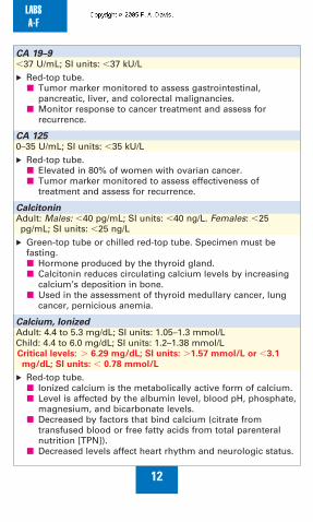
CA 19–9�37 U/mL; SI units: �37 kU/L. Red-top tube.
■ Tumor marker monitored to assess gastrointestinal,pancreatic, liver, and colorectal malignancies.
■ Monitor response to cancer treatment and assess forrecurrence.
CA 1250–35 U/mL; SI units: �35 kU/L. Red-top tube.
■ Elevated in 80% of women with ovarian cancer.■ Tumor marker monitored to assess effectiveness of
treatment and assess for recurrence.
CalcitoninAdult: Males: �40 pg/mL; SI units: �40 ng/L. Females: �25pg/mL; SI units: �25 ng/L
. Green-top tube or chilled red-top tube. Specimen must befasting.■ Hormone produced by the thyroid gland.■ Calcitonin reduces circulating calcium levels by increasing
calcium’s deposition in bone.■ Used in the assessment of thyroid medullary cancer, lung
cancer, pernicious anemia.
Calcium, IonizedAdult: 4.4 to 5.3 mg/dL; SI units: 1.05–1.3 mmol/LChild: 4.4 to 6.0 mg/dL; SI units: 1.2–1.38 mmol/LCritical levels: � 6.29 mg/dL; SI units: �1.57 mmol/L or �3.1
mg/dL; SI units: � 0.78 mmol/L
. Red-top tube.■ Ionized calcium is the metabolically active form of calcium.■ Level is affected by the albumin level, blood pH, phosphate,
magnesium, and bicarbonate levels.■ Decreased by factors that bind calcium (citrate from
transfused blood or free fatty acids from total parenteralnutrition [TPN]).
■ Decreased levels affect heart rhythm and neurologic status.
LABSA-F
01Hop-01 2/4/05 12:08 PM Page 12

13Calcium,TotalAdult: 8.2 to 10.5 mg/dL; SI units: 2.05–2.54 mmol/LChild: 8.6–11.2 mg/dL; SI units: 2.15–2.79 mmol/LCritical levels: �12 mg/dL; SI units: �2.99 mmol/L (coma, death).
�7mg/dL; SI units: �1.75 mmol/L (tetany, death)
. Red-top tube.■ 50% of calcium in blood is bound to albumin and is inactive;
the other 50%, called free or ionized calcium, is metabolicallyactive. Total calcium is a measurement of both bound and freecalcium.
■ Assess for diseases of the parathyroid gland or kidneys.
Carbon Dioxide Content (CO2)Adult: 22–30 mEq/L; SI units: 22–30 mmol/LChild: 20–28 mEq/L; SI units: 20–28 mmol/L. Red-top tube.
■ Used in the evaluation of pH and electrolytes.■ Blood CO2 measures the total amount of carbon dioxide in the
blood (bicarbonate [HCO3]], dissolved carbon dioxide gas[CO2], and carbonic acid [H2CO3]). It is essentially a measureof serum bicarbonate (HCO3) because 95% of CO2 is presentas HCO3.
■ Do not confuse with PaCO2, which measures the partialpressure of carbon dioxide.
■ Increased: compensation for respiratory acidosis andmetabolic alkalosis. Decreased: compensation for respiratoryalkalosis and metabolic acidosis.
Carbon Monoxide (Carboxyhemoglobin)Nonsmoker: �2%; Smoker: �9%; Toxic: �15%Critical Levels: �20%
. Lavender-top tube.■ Used to evaluate patients exposed to smoke, fumes, and fires.■ Levels �20% cause dizziness and headache; �30%,
tachycardia, hypotension, and confusion; >60%, coma anddeath.
Carcinoembryonic Antigen (CEA)Nonsmokers: �3 ng/mL; SI units �3 �g/LSmokers: �5 ng/mL; SI units �5 �g/L
. Red-top or lavender-top tube, depending on lab.
LABSA-F
01Hop-01 2/4/05 12:08 PM Page 13

14
■ Used to monitor treatment response and possible recurrenceof breast, colon, or pancreatic cancer.
■ Not a screening test since CEA can be elevated in benigndiseases and smokers.
■ Heparin use interferes with results. Hold for 2 days prior totest.
Cardiac BiomarkersSee individual tests for reference ranges.■ Enzymes, proteins, and hormones used in the diagnosis of acute
myocardial infarction.■ Biomarkers rise, peak, and return to normal in predictable time
frames allowing health care providers to monitor the progress ofthe infarction.
■ These laboratory tests include:◆ Albumin cobalt binding◆ Creatinine kinase (CK) (or creatine phosphokinase) and CK-MB
isoenzyme (less frequently used)◆ Troponin (most widely used to assess heart damage)◆ Myoglobin (less frequently used; may be ordered with
troponin)◆ B-type natriuretic peptide (used to assess heart function)
Catecholamines and Vanillylmandelic Acid (VMA)AdultsEpinephrine: 0–20 �g/24 hr; SI units: 0–109 nmol/dayNorepinephrine: 15–80 �g/24 hr; SI units: 89–473 nmol/dayDopamine: 65–400 �g/24 hr; SI units: 424–2612 nmol/dayVMA: �6.8 mg/24 hr; SI units: �35 �mol/24 hrChildren
Epinephrine: 4–10 yrs: �10 �g/24 hr; SI units: �54.6 nmol/dayNorepinephrine: 4–10 yr: 8–65 �g/24 hr; SI units: 47–384 nmol/dayDopamine: 1–4 yrs: 10–260 �g/24 hr; SI units: 65–1698 �mol/L/dayVMA: 2–18 yr: 1–5 mg/ 24 hr; SI units: �30 �mol/24 hr
. 24-hr urine collection.■ Used to screen for neuroendocrine tumors including
pheochromocytoma.■ Test is affected by multiple foods and drugs. Check with lab
about withholding medications or changing diet prior to andduring the test.
LABSA-F
01Hop-01 2/4/05 12:08 PM Page 14

15Cerebrospinal Fluid Analysis (CSF Analysis)Pressure: 50–180 mm H2OAppearance: Clear and colorlessTotal protein: 15–45 mg/dL; SI units: 150–450 mg/LProtein electrophoresis:
◆ Prealbumin: 2–7%◆ Albumin: 56–76%◆ Alpha1 globulin: 2–7%◆ Alpha2 globulin: 4–12%◆ Beta globulin: 8–18%◆ Gamma globulin: 3–12%; SI units: 0.3–0.12◆ Oligoclonal bands: none◆ IgG: �3.4 mg/dL; SI units: �34 mg/L
Glucose: 50–80 mg/dL; SI units: 2.8–4.4 mmol/LCell count: 0–5 WBCs; no RBCsChloride: 118–132 mEq/L; SI units: 118–132 mmol/LLactate dehydrogenase: 10% of serum levelLactic acid: 10–20 mg/dL; SI units: 1.1–2.2 mmol/LCytology: No malignant cellsCulture: No growthGram stain: NegativeIndia ink: NegativeVDRL: NonreactiveCritical Values: Positive Gram’s stain, India ink preparation, or
culture.
. Sterile test tubes, numbered in the order they were filled.Send to lab immediately. Do not refrigerate.■ Obtained by lumbar puncture, which requires careful
patient preparation, postprocedure care, and informedconsent.
■ Used to aid in the diagnosis of meningitis, intracranial orsubarachnoid bleeding, brain injury, neurosyphilis,degenerative brain diseases, CNS cancer or metastases,autoimmune disorders, multiple sclerosis, and otherconditions.
LABSA-F
01Hop-01 2/4/05 12:08 PM Page 15

16
Chlamydia Group AntibodyNegative: �0.91; Equivocal: 0.91–1.09; Positive: �1.10
. Red-top tube.■ Used in the diagnosis of chlamydial infection.■ Detects IgG antibodies to C. trachomatis, C. pneumoniae, and
C. psittaci.■ Other methods, including swabs from infected areas for C.
trachomatis, and urine samples are also used to detectChlamydia infections.
Chloride (Cl)Adult: 96–106 mEq/L; SI units: 96–106 mmol/LChild: 90–110 mEq/L; SI units: 90–110 mmol/LCritical levels: �80 mEq/L or �115 mEq/L
. Red-top or green-top tube.■ Aids in maintenance of electrical neutrality, fluid and acid-
base balance, and osmolality of body fluids (with sodium).Assessed with other electrolytes.
■ Decreased in vomiting, gastric suctioning, ketoacidosis, renaldisease with loss of sodium.
■ Increased with diarrhea, dehydration, total parenteralnutrition.
Cholesterol,Total; High-density Lipoprotein Cholesterol (HDL,
HDL-C); Low-density Lipoprotein Cholesterol (LDL, LDL-C); Very
Low-density Lipoprotein (VLDL)Total CholesterolAdult: �200 mg/dL; SI units: �5.2 mmol/LChild: 125–200 mg/dL; SI units: 3.27–5.2 mmol/LHDL Cholesterol
Adult: �50 mg/dL; SI units: �1.40 mmol/LLDL Cholesterol
Adult: �100 mg/dL; SI units: �2.56 mmol/LVLDL Cholesterol
25–50%
. Red-top tube. Fasting sample; no alcohol for 24 hr prior to test.■ Blood lipids synthesized in the liver and integral to the
formation of cell membranes, bile salts, and hormones.■ Implicated in atherosclerosis and MI.■ HDL levels �60 mg/dL are protective against heart disease.
LABSA-F
01Hop-01 2/4/05 12:08 PM Page 16
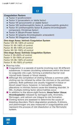
17Coagulation Factors
◆ Factor II (prothrombin)◆ Factor V (proaccelerin or labile factor)◆ Factor VII (proconvertin or stable factor)◆ Factor VIII (antihemophilic factor A, antihemophilic globulin)◆ Factor IX (antihemophilic factor B, plasma thromboplastin
component, Christmas factor)◆ Factor X (Stuart-Prower factor)◆ Factor XI (plasma thromboplastin antecedent)◆ Factor XII (Hageman factor)
One-stage Assay: Intrinsic Coagulation System
Factor VIII: 55–145% of controlFactor IX: 60–140% of controlFactor XI: 65–135% of controlFactor XII: 50–150% of controlOne-stage Assay: Extrinsic Coagulation System
Factor II: 50–200% of controlFactor V: 50–150% of controlFactor VII: 65–135% of controlFactor X: 45–155% of control
. Blue-top tube.■ Coagulation is a cascade of events involving over 30 different
substances. It causes circulating substances within the bloodto coagulate into a gel, forming a protective barrier overinjured body tissues or blood vessels.
■ Although the completion of clotting follows a common path,clotting can be initiated by either the intrinsic or the extrinsicpathway. Both pathways are usually triggered in tissue orblood vessel injury; however, in hemophilic diseases,alterations in intrinsic factors cause the bleeding disorder. InDIC, multiple clotting factor abnormalities occur.
■ In addition to the above eight factors, tissue factor (tissue
thromboplastin) released by damaged cells, thrombin,fibrinogen, and calcium are integral to clot formation.
■ Clotting factors are assessed to determine the cause ofbleeding disorders. Fibrin degradation products, D-dimers,and plasminogen are also measured in coagulopathies andrepresent the process of clot dissolution or the fibrinolyticsystem.
LABSA-F
01Hop-01 2/4/05 12:08 PM Page 17

18
Cold Agglutinin TiterNegative (�1:16). Red-top tube. Do not refrigerate.
■ Cold agglutinins are antibodies that cause red blood cells toagglutinate (clump together).
■ Used in the diagnosis of primary atypical pneumonia,Mycoplasma pneumoniae, hemolytic anemia, gangrene,Raynaud’s disease, and other diseases.
Complement,TotalAdult: 75–160 U/mL; SI units: 75–160 kU/L. Red-top tube.
■ Proteins involved in immunological and inflammatoryresponses.
■ Diagnose angioedema; assess treatment/status of variousdiseases including systemic lupus erythematosus nephritisand other types of nephritis.
■ Deficiency associated with increased susceptibility toinfection.
Complement C3 and C4C3: 55–120 mg/dL; SI units: 0.55–1.2 g/LC4: 20–50 mg/dL; SI units: 0.2–.05 g/L. Red-top tube.
■ Commonly assessed components of the complementsystem.
■ Increased levels associated with rheumatoid arthritis, somecancers, and acute viral hepatitis.
■ Decreased levels associated with SLE, glomerulonephritis,DIC, gram-negative sepsis.
LABSA-F
01Hop-01 2/4/05 12:08 PM Page 18

19Complete Blood Count with Differential (CBC with diff)
Test Conventional SI Units
Red Blood Cell(RBC)
Hemoglobin (Hgb)
Hematocrit (Hct)
MCV
MCH
MCHC
White Blood Cells(WBC)
WBC Differential
◆ Neutrophils,bands
◆ Neutrophils,segmented
◆ Lymphocytes
◆ Monocytes
◆ Eosinophils
◆ Basophils
Platelets
Critical levels:
Hgb: �5 g/dL or �20 g/dL
Hct: �15% or �60%
WBC: �500 mm3 or �50,000/ mm3
Platelets: �50,000 or �999,000/mm3
. Lavender-top tube.■ A CBC reveals
◆ Information about general health.◆ Number of red blood cells (RBC).
LABSA-F
Male: 4.6–6.2 � 106
cells /�L
Female: 4.2–5.9 � 106
cells / �L
Male: 13–18 g/dL
Female: 12–16 g/dL
Male: 45–52%
Female: 37–48%
80 to 100 �m3
27 to 31 pg/cell
32 to 36 g/dL
4,300–10,800cells/mm3
0–5%
54–65%
25–40%
2–8%
1–4%
0–1%
150,00–450,000/mm3
4.6–6.2 � 1012 cells /L
4.2–5.9 � 1012 cells /L
Male: 130–180 g/L
Female: 120–160 g/L
Male: 0.45–0.52
Female: 0.37–0.48
80 to 100 �m3
27 to 31 pg/cell
32 to 36 g/dL
4.3–10.8 � 109/L
0.03–0.08
0.54–0.65
0.25–0.40
0.02–0.08
0.010.04
0–0.01
150–450 x 109/L
01Hop-01 2/4/05 12:08 PM Page 19

20
◆ Number of white blood cells (WBC) and differential (seeWhite Blood Cells for more information).
◆ Total amount of hemoglobin in the blood (Hgb).◆ Fraction of blood composed of red blood cells (Hct).◆ Volume of Hgb in each RBC (MCV [mean corpuscular
volume]).◆ Weight of the Hgb in each RBC (MCH [mean corpuscular
hemoglobin]).◆ Proportion of Hgb contained in each RBC (MCHC [mean
corpuscular hemoglobin concentration]).◆ Number of platelets, which are critical to clot formation (see
Platelets for more information).■ MCV, MCH, and MCHC values are useful in the diagnosis of
various types of anemia. See below for description ofanemias.
Types of Anemia
Type of Anemia Possible Causes
Normocytic/normochromic
(normal cell size; normalamount of Hgb)
Microcytic/hypochromic
(small cell size; low amountof Hgb)
Microcytic/normochromic
(small cell size; normalamount of Hgb)
Macrocytic/normochromic
(large cell size; normalamount of Hgb)
Terms◆ Microcytic— MCV less than normal (�80 fL)◆ Normocytic— MCV within normal range (80–100 fL)◆ Macrocytic— MCV greater than normal (�100 fL)◆ Hypochromic—MCH less than normal (�27 pg)◆ Normochromic—MCH within normal range (27–31 pg)◆ Hyperchromic—MCH greater than normal (�31 pg)
LABSA-F
Acute blood loss, aplastic anemia,prosthetic heart valves, sepsis,tumor
Iron deficiency, lead poisoning,thalassemia
Erythropoietin deficiencysecondary to renal failure
Chemotherapy, folate deficiency,vitamin B12 deficiency
01Hop-01 2/4/05 12:08 PM Page 20

21Coombs’ Test, Direct (Direct Antiglobulin)Negative; no agglutination. Lavender-top tube.
■ Assess for immunoglobulins (antibodies) on surface of redblood cells
■ Positive results occur in hemolytic transfusion reactions,hemolytic anemias, erythroblastosis fetalis
Coombs’Test, Indirect (Indirect Antiglobulin, Autoantibody Test)Negative. Red-top tube.
■ Part of a cross-match for blood transfusion.■ Positive result indicates incompatibility.
Cortisol, FreeAdult: �50 �g/24; SI units: �138 nmol/dayChild: �38 �g/24 hr; SI units: �104 nmol/day. 24-hr urine collection.
■ Used to screen for adrenal hyperfunction.■ Result from urinary free cortisol test is a better indication of
cortisol secretion than a single plasma level.■ Best test for diagnosing Cushing’s syndrome.
Cortisol, PlasmaAdult: 8 AM–10 AM: 5–23 �g/dL; SI units: 138–635 mmol/L.
4 PM–6 PM: 3–13 �g/dL; SI units: 83–359 mmol/LChlid: 8 AM–10 AM: 15–25 �g/dL
4 PM–6 PM: 5–10 �g/dL. Green-top tube.
■ Powerful glucocorticoid secreted by the adrenal cortex inresponse to ACTH. Higher in the morning.
■ Affects gluconeogenesis, fat and protein metabolism; aidsin regulation of immune system; is increased duringphysical or emotional stress.
■ Assessed in the evaluation of Cushing’s syndrome andAddison’s disease.
LABSA-F
01Hop-01 2/4/05 12:08 PM Page 21

22
Creatinine Phosphokinase, (CPK, Creatine Kinase, CK);
CPK IsoenzymesCPKAdult: Male: 55–170 U/L; SI units: 55–170 U/L. Female: 30–135 U/L;SI units: 30–135 U/L
Newborn: 68–580 U/LIsoenzymes
CPK-MM: 100%; CPK-MB: 0%; CPK-BB: 0%
. Red-top tube.■ CPK is an enzyme critical to intracellular energy.■ The MB isoenzyme is a cardiac marker (but is not as specific
as troponin). Elevations occur in acute MI and are used in thediagnosis of MI.
■ CPK-MB ratio to total CPK is calculated to increase diagnosticaccuracy. A ratio of 2.5 correlates with cardiac damage.
■ MM elevations occur in crush injuries, seizures, malignanthypothermia.
Creatinine, SerumAdult: Male: 0.6–1.2 mg/dL; SI units: 53–106 �mol/L. Female: 0.5–1.1mg/dL; SI units: 44–97 �mol/L
Child: 0.3–0.7 mg/dL
. Red-top tube.■ Breakdown product of creatine phosphate in muscle.■ Produced at a constant rate by the body and excreted by the
kidney. Blood level rises in renal impairment.■ Creatinine level is a sensitive indicator of renal function but is
dependent on kidney function and muscle mass. Patients withdecreased muscle mass do not exhibit a rise in creatininelevels as readily as those with more muscle mass and shouldhave an estimated glomerular filtration rate (GFR) reported aswell.
Creatinine, Urine1–2 g/24 hr; SI units: 8.8–17.7 mmol/day
. 24-hr urine collection. Refrigerate.■ Creatinine is a by-product of muscle breakdown. It is filtered
(removed from plasma) by the kidneys and excreted in theurine.
■ Elevated levels of creatinine indicate impaired renal function.
LABSA-F
01Hop-01 2/4/05 12:08 PM Page 22

23Creatinine ClearanceMale: 107–139 mL/min; SI units: 1.78–2.32 mL/s. Female: 85–105mL/min; SI units: 1.45–1.78 mL/s.
. Timed urine sample (12 or 24 hr) with a blood sample takenthe morning of or sometime during the test.■ Creatinine clearance refers to the amount of blood that can
be cleared of creatinine in 1 min.■ It is calculated using the volume of urine, the amount of
creatinine excreted, and the amount of creatinine in theblood.
■ It is used to determine safe dosing of nephrotoxic drugs.Creatinine clearance of 10–20 mL/min is indicative of renalfailure and the need for dialysis.
Cryoglobulins� 0.4% or none detected■ Abnormal proteins present in various immune, hematologic,
collagen vascular, and oncologic disorders.■ Levels �5 mg/mL are associated with multiple myeloma and
leukemia.■ Levels between 1 and 5 mg/mL are associated with
rheumatoid arthritis.
Cystatin CAdult: 0.5–1 mg/LChild: 0–3 mo: 0.8–2.3 mg/L; 4–11 mo: 0.7–1.5 mg/L;1–17 yr: 0.5–1.3 mg/L. Red-top tube.
■ A cysteine protease inhibitor that is freely filtered(removed) by the glomeruli and thus can be used to assessfor changes in glomerular filtration rate.
■ High levels of cystatin C suggest impaired renal function.■ It is thought to be superior to creatinine as a marker of
glomerular filtration rate because it is not affected bymuscle mass, diet, or acute inflammatory processes.
Cystine10–100 mg/24 hr. 24-hr urine collection.
■ Used to detect cystinuria or identify cause of kidney stones.
LABSA-F
01Hop-01 2/4/05 12:08 PM Page 23

24
Cytomegalovirus (CMV) AntibodiesIgM � 1:8; IgG � 1:16. Red-top tube.
■ CMV is a virus in the herpes family.■ Active infection significant in pregnant women, potential
transplant patients and immunocompromised patients.■ If active infection is suspected, a second sample is assessed
in 10–14 days.
D-dimersNegative (�250 ng/mL; SI units: �250 �g/L). Blue-top tube.
■ A fibrin degradation product produced only after a clot hasformed and is in the process of being broken down.
■ Used in the diagnosis of deep vein thrombosis (DVT),pulmonary embolism (PE), or disseminated intravascularcoagulation (DIC).
Dexamethasone Suppression TestLow Dose
Overnight: 8 AM plasma cortisol: �5 �g/dLStandard: Urinary free cortisol on day 3: �10 �g/dayHigh Dose
Overnight: �50% reduction in plasma cortisol.Standard: �90% reduction in urinary free cortisol.. Red-top tube.
■ Administration of dexamethasone suppresses ACTH andshould suppress cortisol levels in healthy people.
■ Helpful in determining cause of increased cortisol levels(adrenal tumor, pituitary tumor, or ectopic ACTH-producingtumor).
LABSA-F
01Hop-01 2/4/05 12:08 PM Page 24

25Drug Levels,Therapeutic and ToxicConventional (US System of Measurements)
Drug Therapeutic Level Toxic Level
acetaminophen
alprazolam
amikacin
aminocaproic acid
aminophylline
amiodarone
amitriptyline
amoxapine
atenolol
bepridil HCl
carbamazepine
chloral hydrate
chloramphenicol
chlordiazepoxide
chlorpromazine
chlorpropramide
clonazepam
cyclosporine
desipramine
diazepam
digoxin
diltiazem
disopyramide
doxepin
ethosuximide
flecainide
gentamicin
LABSA-F
5–20 �g/mL10–50 ng/mLpeak: 20–30 �g/mLtrough: 4–8 �g/mL100–400 �g/mL10–20 �g/mL0.5–2.5 �g/mL120–150 ng/mL200–400 ng/mL200–500 ng/mL1–2 ng/mL5–12 �g/mL2–12 �g/mL10–20 �g/mL1–5 �g/mL50–300 ng/mL75–250 �g/mL15–60 ng/mL100–300 ng/mL150–300 ng/mL0.5–2 mg/L0.8–2 ng/mL50–200 ng/mL2–5 �g/mL150–300 ng/mL40–100 �g/mL0.2–1 �g/mLpeak: 6–10 �g/mLtrough: � 2 �g/mL
�40 �g/mL
�75 ng/mL
peak: �35 �g/mL
trough: �10 �g/mL
�400 �g/mL
�20 �g/ml
�2.5 �g/mL
�500 ng/ml
�500 ng/mL
�500 ng/mL
�2 ng/mL
�12 �g/mL
�20 �g/mL
�25 �g/mL
�5 �g/mL
�750 ng/mL
�250 �g/mL
�80 ng/mL
�85 or �500 ng/mL
�500 ng/mL
�3 mg/L
�2 ng/mL
�200 ng/mL
�7 �g/mL
�400 ng/mL
�150 �g/mL
�1 �g/mL
peak: �12 �g/mL
trough: �2 �g/mL
(Continued on following page)
01Hop-01 2/4/05 12:08 PM Page 25

26
Drug Therapeutic Level Toxic Level
haloperidol
hydromorphone
imipramine
kanamycin
lidocaine
lithium
meperidine
methotrexate
mexiletine
mezlocillin sodium
milrinone
morphine
nicardipine
nifedipine
nortriptyline
phenobarbital
phenytoin
primidone
procainamide
propafenone
propranolol
quinidine
salicylate
theophylline
tobramycin
tocainide HCl
trazadone
valproic acid
vancomycin
verapamil
LABSA-F
3–20 ng/mL1–30 ng/mL150–300 ng/mL20–25 �g/mL1.5–5 �g/mL0.5–1.5 mEq/L0.4–0.7 �g/mL� 0.01 �mol0.5–2 �g/mL35–45 �g/mL150–250 ng/mL10–80 ng/mL0.028–0.05 �g/mL0.025–0.1 �g/mL50–150 ng/mL15–30 �g/mL10–20 �g/mL5–12 �g/mL4–10 �g/mL0.5–3 �g/mL50–100 ng/m2–5 �g/mL10–30 mg/dL10–20 �g/mLpeak: 6–10 �g/mLtrough: � 2 �g/mL4–10 �g/mL500–2000 ng/mL50–100 �g/mLpeak: 20–40 �g/mLtrough: 5–10 �g/mL0.08–0.3 �g/mL
�42 ng/mL
�100 ng/mL
�500 ng/mL
�35 �g/mL
�5 �g/mL
�1.5 mEq/L
�1 �g/mL
�10 �mol in 24 hr
�2 �g/mL
�45 �g/mL
�250 ng/mL
�200 ng/mL
�0.05 �g/mL
�0.1 �g/mL
�500 ng/mL
�40 �g/mL
�20 �g/mL
�12 �g/mL
�10 �g/mL
�3 �g/mL
�150 ng/mL
�6 �g/mL
�35 mg/dL
�20 �g/mL
peak: �12 �g/mL
trough: �2�g/mL
�12 �g/mL
�4000 ng/mL
�100 �g/mL
peak: �80 �g/mL
trough: �10 �g/mL
�0.3 �g/mL
(Continued)
01Hop-01 2/4/05 12:08 PM Page 26

27SI Units (International System of Units)
Drug Therapeutic Range Toxic Level
amikacin
amitriptyline
carbamazepine
clonazepam
desipramine
diazepam
digoxin
disopyramide
ethosuximide
flecainide
gentamicin
imipramine
lignocaine
lithium
nortriptyline
phenobarbitone
phenytoin
primidone
procainamide
quinidine
salicylic acid
theophylline
tobramycin
valproic acid
vancomycin
LABSA-F
peak: 34–51 �mol/L
trough: 7–14 �mol/L
433–903 nmol/L
21–51 �mol/L
40–200 nmol/L
281–1125 nmol/L
0.35–3.5 nmol/L
1–2.6 nmol/L
9–18 �mol/L
280–708 �mol/L
0.5–2.4 �mol/L
peak:12–21 �mol/L
trough:�4 �mol/L
610–1670 nmol/L
6–21 �mol/L
0.5–1.5 mmol/L
190–570 nmol/L
86–172 �mol/L
40–80 �mol/L
23–55 �mol/L
17–42 �mol/L
6–15 �mol/L
1–2 mmol/L
28–111 �mol/L
peak: 13–21 �mol/L
trough: � 4 �mol/L
350–700 �mol/L
peak: 14–28 �mol/L
trough: 3–7 �mol/L
peak: �60 �mol/L
�805 nmol/L
�51 �mol/L
�260 nmol/L
�1500 nmol/L
�17.5 nmol/L
�2.6 nmol/L
� 21 �mol/L
�1062 �mol/L
�2.4 �mol/L
peak: �21 �mol/L
�1785 nmol/L
�39 �mol/L
�2 mmol/L
�1900 nmol/L
�172 �mol/L
�158 �mol/L
�55 �mol/L
�51 �mol/L
�29 �mol/L
�3.6 mmol/L
�111 �mol/L
peak: �21 �mol/L
�1386 �mol/L
peak: �28 �mol/L
01Hop-01 2/4/05 12:08 PM Page 27

28
LABSA-F
. Red-top tube.■ Drug levels are obtained both to enhance therapeutic
efficacy and to assess for toxicity/overdose/poisoning.■ Therapeutic drug monitoring (TDM) is the measurement of
serum drug levels so that dosages may be adjusted toachieve optimum clinical benefit.
■ Therapeutic drug monitoring is used with◆ cardiac glycosides (e.g., digoxin)◆ antiarrhythmics (e.g., lidocaine, procainamide)◆ anticonvulsants (e.g., phenytoin, carbamazepine)◆ lithium◆ theophylline◆ aminoglycoside antibiotics (gentamicin, tobramycin)◆ salicylates
■ For TDM, blood samples must be obtained at theappropriate time and after sufficient number of doses havebeen administered for valid interpretation of results.
■ Peak and trough levels are drawn with some drugs,especially antibiotics. Peak levels are drawn at the point ofmaximum drug absorption; trough levels are drawn justbefore the next dose.
Electrolytes, SerumSee individual tests for normal values.. Red-top tube.
■ Electrolytes are minerals present in body tissues and bloodas dissolved salts.
■ They are electrically charged particles that help maintainfluid and acid-base balance. They help move nutrients intocells and waste products out.
■ An electrolyte panel measures sodium (Na�), potassium(K�), chloride (Cl-), and bicarbonate (HCO3- ), which ismeasured indirectly as CO2.
■ See individual tests and Anion Gap for more information.
01Hop-01 2/4/05 12:08 PM Page 28

29Electrolytes, Urine
Conventional Units SI Units
SodiumChloridePotassiumCalcium
PhosphorusMagnesium
. 24-hour urine collection■ Provides information about hydration status, the kidneys’ ability
to conserve or excrete sodium.■ Calcium levels are increased in hyperthyroidism, immobilization,
multiple myeloma, Paget’s disease, and bone metastases.
Erythropoietin (EPO)5–35 IU/L
. Red-top tube.■ Hormone produced by the kidney to stimulate RBC production.■ Decreased in patients with renal disease, primary polycythemia.
Estradiol, Serum (Estrogen Fraction)Adult: Female: Follicular phase: 20–150 pg/mL; Midcycle phase:100–500 pg/mL; Luteal phase: 60–260 pg/mL; Pregnancy: 1st tri: 1–5ng/mL; 2nd tri: 5–15 ng/mL; 3rd tri: 10–40 ng/mL; Postmenopause:�30 pg/mL. Male: 15–50 pg/mL
. Red-top tube.■ An estrogen fraction useful in evaluating fetal well-being,
menstrual and fertility problems in women, precocious pubertyin girls, gynecomastia.
■ Used to assess amenorrhea to determine if cause is menopause,pregnancy, or a medical problem.
■ Serial measurements are used to monitor follicle development inthe ovary prior to in vitro fertilization.
■ Estrone, the major estrogen after menopause, and estriol, themajor estrogen in pregnant women, are the other majorestrogens.
LABSA-F
30–280 mEq/day110–250 mEq/day40–80 mEq/dayMale: �275 mg/day;
Female: �250 mg/day
0.9–1.3 g/day�150 mg/day
30–280 mmol/day110–250 mmol/day40–80 mmol/dayMale: �6.8 mmol/day;
Female: �6.2mmol/day
29–42 mmol/day3–4.3 mmol/day
01Hop-01 2/4/05 12:08 PM Page 29

30
Ethanol (blood alcohol)None. Red-top tube. Use povidone-iodine swab not alcohol to clean
venipuncture site.■ Levels �0.8% are considered to be proof of intoxication in
most states.
Febrile Agglutinin TiterNegative (�1:80). Red-top tube. Do not warm.
■ Febrile agglutinins are antibodies that cause RBCs toagglutinate (clump together) at high temperatures.
■ Febrile agglutinin titers are used in the diagnosis of somebacterial infections (salmonellosis, tularemia, RockyMountain spotted fever, typhus, brucellosis).
Fecal Fats (Fecal Lipids)2–6 g/24 hr; SI units: 7–21 mmol/day. 72-hr stool collection. Keep refrigerated during collection.
■ Assist in the diagnosis of malabsorption or pancreaticinsufficiency.
■ A high fat diet should be eaten for 3 days before andthroughout the collection time period and should refrainfrom laxative use.
Fecal Occult Blood (FOB, Stool for Occult Blood)Negative. Stool sample
■ Used to detect microscopic bleeding into the GI tract.■ Routine screening test for patients over 50 years old.■ Positive in ulcers, polyps, hemorrhoids, tumors,
inflammatory bowel disease, diverticulosis, and otherdisorders of the GI tract.
FerritinAdult: Female: 10–150 ng/mL; SI units: 10–150 �g/L. Male: 12–300ng/mL; SI units: 12–300 �g/L
Child �1 year: 7–140 ng/mL; SI units: 7–140 �g/L
LABSA-F
01Hop-01 2/4/05 12:08 PM Page 30

31. Red-top tube.
■ Indicates available iron stores in the body.■ Level below 10 ng/mL diagnostic of iron deficiency anemia.
Fibrin Split Products (Fibrin Degradation Products, FDP, FSP)�5 �g/mL; SI units: �5 mg/L. Blue-top tube (check with lab).
■ Blood clots in the vascular system stimulate the productionof plasmin, which breaks down clots into fibrin splitproducts.
■ Elevated amounts of fibrin split products in the bloodindirectly indicate the presence of blood clots or afibrinolytic disorder such as DIC.
Fibrinogen150–400 mg/dL; SI units: 1.5–4 g/LCritical Levels: �100 mg/dL
. Blue-top tube.■ Fibrinogen is essential to clot formation. Decreased
fibrinogen levels result in prolonged PT and PTT.■ Usedful in diagnosis of DIC.
Folic Acid (Folate)� 2 ng/mL (radioimmunassay); SI units: �5 mmol/L. Red-top tube.
■ Normal levels essential for properly functioning red andwhite blood cells.
■ Decreased in malabsorption, malnutrition, liver disease,cancer.
Follicle-stimulating Hormone (FSH)Adult: Males: 5–15 IU/L. Females: follicular or luteal phase: 5–20IU/L, midcycle: 30–50 IU/L, postmenopause: �50 IU/L.
Prepubertal children: �6 IU/L. Red-top tube.
■ Pituitary hormone involved in maturation of ovarian folliclein women and spermatogenesis in men.
LABSA-F
01Hop-01 2/4/05 12:08 PM Page 31

32
LABSG-Z
Gamma-Glutamyl Transpeptidase (GGT, SGGT)Adult: Male: 9–50 IU/L. Female: 8–40 IU/L. Red-top tube.
■ Liver enzyme sensitive to biliary and liver disorders,including alcoholic liver disease.
GastrinFasting: �100 pg/mL; SI units: 47.7 pmol/LPostprandial: 95–140 pg/mL; SI units: 45.3–66.7 pmol/L. Red-top tube.
■ Hormone that stimulates secretion of gastric acid.■ Elevated in pernicious anemia, Zollinger-Ellison syndrome,
stomach cancer, peptic ulcer, atrophic gastritis, renalinsufficiency, steroid administration, H2 blockers.
GlucagonAdult: 50–100 pg/mL; SI units: 50–100 ng/LChild: 0–148 pg/mL; SI units: 0–148 ng/L. Lavender-top tube, chilled. Fasting sample.
■ Hormone secreted by the alpha cells of the islets ofLangerhans.
■ Glucagon deficiency may occur with pancreatitis, pancreaticcancer, cystic fibrosis.
■ Elevated glucagon levels occur with glucogonoma (cancerof the alpha islet cells), diabetes, acute pancreatitis,cirrhosis, stress, renal failure, rejection of transplantedkidney.
Glucose, FastingAdult: 70–105 mg/dL; SI units: 3.9–5.8 mmol/LChild � 2 years old: 60–100 mg/dLCritical Levels: �50 or �400 mg/dL
. Red-top tube. Fasting sample.■ Assessed to diagnose or monitor Type 1 and 2 diabetes.■ An elevated fasting blood glucose level above 126 mg/dL
on at least two occasions typically indicates diabetes.
02Hop-02 2/4/05 12:10 PM Page 32

LABSG-Z
33Glucose-6-Phosphate Dehydrogenase (G-6-PD) Screening Test
and G-6-PD AssayScreening Test
NegativeAssay
7.0–20.5 U/g of hemoglobin; SI units: 0.45–1.29 mU/mol (referenceranges vary with testing methodology)
. Lavender- or green-top tube.■ G-6-PD is an enzyme found in RBCs; its function is to protect
hemoglobin from oxidation.■ People with a G-6-PD deficiency are susceptible to hemolysis
when exposed to an oxidative stressor such as systemicinfections, septicemia, metabolic acidosis, and exposure tooxidant drugs (aspirin, sulfa drugs, antimalarials, thiazidediuretics, quinidine, antipyretics, sulfanomides,chloramphenical, phenacetin, probenicid, and tolbutamide).
Glucose Tolerance Test, Standard OralFasting: �126 mg/dL; SI units: �7 mmol/L2-hr: �200 mg/dL; SI units �1.1 mmol/L
. Red-top tube. Fasting sample.■ Blood glucose levels are assessed twice. The first is a fasting
sample, the second sample is taken 2 hr after ingestion of75 g of glucose. Samples may be assessed at other times aswell.
■ Useful for screening for gestational diabetes but usuallyunnecessary for diagnosing diabetes as fasting blood glucose�126 mg/dL or a random blood glucose level �200 mg/dL isususally considered diagnostic.
Glycosylated Hemoglobin (A1C, GHb, Glycohemoglobin, HBA1)Nondiabetic: � 5%Diabetes well controlled: 2.5–6%Diabetes not well controlled: � 8%
. Lavender-top tube.■ Prolonged blood glucose elevation causes a greater
percentage of RBCs to become saturated with glucose(glycohemoglobin).
■ Used for monitoring average diabetic control for preceding 3months as blood cells typically live for 2–3 months.
02Hop-02 2/4/05 12:10 PM Page 33
34
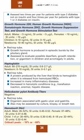
■ Asessed two times per year for patients with type 2 diabetesnot on insulin and four times per year for patients with type1 or 2 diabetes on insulin.
Growth Hormone (GH, Human Growth Hormone [HGH],
Somatotropin Hormone [SH]), Growth Hormone Suppression
Test, and Growth Hormone Stimulation Test
Adult: Males: �5 ng/mL; SI units: �5 �g/L. Females: �10 ng/mL;SI units: �10 �g/L
Chlidren: 0–10 ng/mL; SI units: 0–10 �g/LNewborns: 10–40 ng/mL; SI units: 10–40 �g/L
. Red-top tube.■ Growth hormone is produced in episodic bursts by the
pituitary gland. ■ Assessed to evaluate possible dwarfism, growth retarda-
tion, or gigantism in children and acromegaly in adults.
HaptoglobinAdult: 60–270 mg/dL; SI units: 0.6–2.7 g/LNewborn: 0–10 mg/dL; SI units: 0–0.1 g/L. Red-top tube.
■ A protein produced by the liver that binds to hemoglobinwhen it is released from hemolyzed RBCs.
■ Increased in many inflammatory diseases.■ Decreased in hemolytic conditions (e.g., transfusion
reaction, anemia), hepatic disease.
Helicobacter pylori Antibody TitersNegative. Red-top tube.
■ Organism associated with gastic ulcer and gastritis.■ Also may be assessed by culture, biopsy, or breath test.
Hematocrit (Hct)Adult: Male: 45–52%; SI units: 0.45–0.52. Female: 37–48%;SI units: 0.37–0.48.
Child: 1–6 yr: 30–40%; SI units: 0.30–0.40; 6–18 yrs: 32–44%;SI units: 0.32–0.44
Critical Levels: �15% or �60%
LABSG-Z
02Hop-02 2/4/05 12:10 PM Page 34

LABSG-Z
35. Lavender-top tube.
■ Hematocrit is the percentage of whole blood that is made upof red blood cells. It is expressed as a percentage or a decimalfraction. (A hematocrit value of 35% means that there is 35 mLof red blood cells in 100 mL of blood.)
■ Increased in dehydration and increased production of RBCs.■ Decreased in anemia, when RBC production is impaired or
there is increased destruction of RBCs, in chronic disease,blood loss, and fluid volume excess.
■ See Complete Blood Count.
Hemoglobin (Hgb)Adult: Male: 14–18 g/dL; SI units: 8.7–11.2 mmol/L. Female: 12–16g/dL; SI units: 7.4–9.9 mmol/L.
Child: 1–14 ys: 11.3–14.4 g/dL; SI units: 113–144 mmol/L.Critical Levels: �5 or �20 g/dL
. Lavender-top tube.■ Hemoglobin is the main protein in erythrocytes. It carries
oxygen to and removes carbon dioxide from red blood cells.■ Increased in dehydration, COPD, high altitudes, polycythemia
vera.■ Decreased in fluid volume excess, hematologic cancers,
hemolytic disorders, blood loss, anemia.■ See Complete Blood Count.
Hemoglobin Electrophoresis (Hemoglobinopathy Profile)Hgb A1: 95–98%Hgb A2: 2–3%Hgb F: 0.8–2%; Newborn: 50–80%; � 6 mo old: 1–2%Hgb C, S, or E: 0%
. Lavender-top tube.■ Used to screen for abnormal hemoglobins.■ Hemoglobin A, A2 and F are normal hemoglobins. Hgb F is the
predominant fetal hemoglobin.■ Hgb S is the predominant hemoglobin found in people with
sickle cell disease.■ Hgb C and Hgb E produce mild hemolytic anemia and
splenomegaly. They are considered relatively benign. Hgb E isextremely common in Southeast Asia.
02Hop-02 2/4/05 12:10 PM Page 35
36
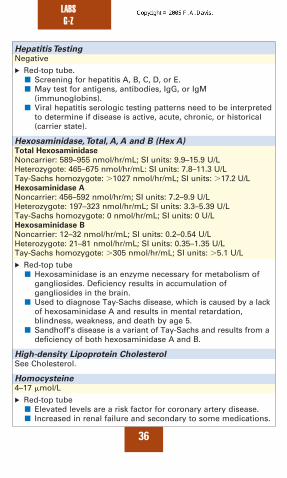
Hepatitis TestingNegative
. Red-top tube.■ Screening for hepatitis A, B, C, D, or E.■ May test for antigens, antibodies, IgG, or IgM
(immunoglobins).■ Viral hepatitis serologic testing patterns need to be interpreted
to determine if disease is active, acute, chronic, or historical(carrier state).
Hexosaminidase,Total, A, A and B (Hex A)Total Hexosaminidase
Noncarrier: 589–955 nmol/hr/mL; SI units: 9.9–15.9 U/LHeterozygote: 465–675 nmol/hr/mL: SI units: 7.8–11.3 U/LTay-Sachs homozygote: �1027 nmol/hr/mL; SI units: �17.2 U/LHexosaminidase A
Noncarrier: 456–592 nmol/hr/m; SI units: 7.2–9.9 U/LHeterozygote: 197–323 nmol/hr/mL; SI units: 3.3–5.39 U/LTay-Sachs homozygote: 0 nmol/hr/mL; SI units: 0 U/LHexosaminidase B
Noncarrier: 12–32 nmol/hr/mL; SI units: 0.2–0.54 U/LHeterozygote: 21–81 nmol/hr/mL; SI units: 0.35–1.35 U/LTay-Sachs homozygote: �305 nmol/hr/mL; SI units: �5.1 U/L
. Red-top tube■ Hexosaminidase is an enzyme necessary for metabolism of
gangliosides. Deficiency results in accumulation ofgangliosides in the brain.
■ Used to diagnose Tay-Sachs disease, which is caused by a lackof hexosaminidase A and results in mental retardation,blindness, weakness, and death by age 5.
■ Sandhoff’s disease is a variant of Tay-Sachs and results from adeficiency of both hexosaminidase A and B.
High-density Lipoprotein CholesterolSee Cholesterol.
Homocysteine4–17 �mol/L
. Red-top tube■ Elevated levels are a risk factor for coronary artery disease.■ Increased in renal failure and secondary to some medications.
LABSG-Z
02Hop-02 2/4/05 12:10 PM Page 36

LABSG-Z
37Human Chorionic Gonadotropin, Serum (HCG)Nonpregnant state: � 0.01 IU/mL4 weeks pregnant: 0.10–1.0 IU/mL16 weeks pregnant: 10–50 IU/mL. Red-top tube.
■ Hormone produced by the placenta.■ Levels peak at about 16 weeks and then decline.
Human Immunodeficiency Virus (HIV) TestingHIV Antibody, ELISA: negativeHIV Western Blot: negativeHIV Antigen (P-24 Antigen): negativeHIV Viral load: �50 copies/mL. Red-top tube.
■ Used to diagnose HIV infection.■ Viral load tests are used to inform treatment strategies and
monitor disease progression.
Human Leukocyte Antigens (HLA)Match or nonmatch. Green-top tube.
■ Used to assess tissue compatibility.■ Useful in assessing compatibility for organ transplants.
5-Hydroxyindoleacetic Acid2–8 mg/24 hr; SI units: 10.4–41.6 mmol/24 hr. 24-hr urine collection.
■ Used in the diagnosis of carcinoid tumors (a tumor found inthe appendix or intestinal wall that secretes high levels ofserotonin leading to symptoms including hypertension,facial flushing, abdominal cramps, and heart valvedamage).
■ Certain foods and medications must be avoided beforeurine collection. These include: bananas, pineapple, redplums, avocado, walnuts, kiwi, tomatoes, cough medicines,muscle relaxants, acetaminophen, caffeine, fluorouacil,iodine solutions, phenacetin, MAO inhibitors, isoniazid, andphenothiazine drugs such as Compazine and Thorazine.
02Hop-02 2/4/05 12:10 PM Page 37

38
ImmunoglobulinsIgG (�10 years old): 650–1700 mg/dL; SI units: 6.5–17 g/LIgA (�10 years old): 40–390 mg/dL; SI units: 0.40–3.90 g/LIgM (�2 years old): 25–210 mg/dL; SI units: 0.25–2.1 g/LIgD: (Adults): 0.5–3 mg/dL; SI units: 0.005–0.03 g/LIgE: (Adults): 0.01–0.04 mg/dL; SI units: 0–430 mg/L. Red-top tube.
■ Used to evaluate immunity.■ Monitor other diseases such as multiple myeloma,
lymphoma, bacterial infection, malnutrition, sarcoidosis.
International Normalized Ratio (INR)See Prothrombin Time.
Iron Tests (Serum Iron,Total Iron Binding Capacity, Serum
Transferrin,Transferrin Saturation)Serum Iron
Adult: 35–165 �g/dL; SI units: 10–27 �mol/LChild 6 mo–2 yr: 40–100 �g/dL; SI units: 7.16–17.9 �mol/LTotal Iron Binding Capacity (TIBC)
250–460 �g/dL; SI units: 45–82 �mol/LSerum Transferrin
200–430 mg/dL; SI units: 2–3.8 g/LTransferrin Saturation
Male: 30–50%Female: 20–35%. Red-top tube.
■ Iron is critical to proliferation and maturation of red bloodcells.
■ 65% of iron is found in hemoglobin. Most of the rest isstored as ferritin in in the liver, bone marrow, and spleen.
■ Transferrin is the major transporting protein of iron.■ Increased in excessive iron intake and decreased
production of erthrocytes. Decreased in iron deficiencyanemia, normochromic anemia associated with chronicdiseases.
LABSG-Z
02Hop-02 2/4/05 12:10 PM Page 38

LABSG-Z
3917-Ketosteroids (17-Ketogenic Steroids, 17-KGS,
Corticosteroids)Adult: Male: 8–25 mg/24 hr; SI units: 27–85 �mol/24 hr. Female:5–15 mg/24 hr; SI units: 17–52 �mol/24 hr.
Child: �1–3 mg/24 hr (age dependent; the younger the child, thelower the normal range); SI units: 3–10 �mol/24 hr.
. 24-hr urine collection.■ Aids in the diagnosis of adrenal cortex dysfunction.■ Increased in Cushing’s syndrome, stress, adrenocortical
cancer, testicular and ovarian cancers, infection, pituitaryhyperfunction.
■ Decreased in Addison’s disease, nephrosis, pituitaryhypofunction.
Lactate Dehydrogenase (LD, LDH), LDH IsoenzymesLDH
Adult: 100–190 U/L but may differ significantly from lab to labLDH Isoenzymes
LDH-1: 17–27%LDH-2: 27–37%LDH-3: 18–25%LDH-4: 3–8%LDH-5: 0–5%
. Red-top tube.■ Enzyme present in many body tissues.■ Elevated levels occur in many disease states including MI,
cancer, liver disease, muscle disease, and trauma.
Lactic Acid (Lactate)Venous: 0.5–1.5 mEq/L or 8.1–15.3 mg/dL; SI units: 0.5–1.5 mmol/LArterial: 0.5–2 mEq/L or 11.3 mg/dL; SI units: 0.5–2 mmol/LCritical Levels: (venous or arterial) �5 or �45 mg/dL; SI units:
�5 mmol/L
. Green or gray-top tube. Send to lab on ice.■ Sensitive indicator of tissue hypoxia.■ Accumulation of excess lactic acid due to hypoxia coupled
with decreased hepatic clearance leads to lactic acidosis.■ Lactic acid levels are increased in hemorrhage, shock, sepsis,
DKA, strenuous exercise, cirrhosis.
02Hop-02 2/4/05 12:10 PM Page 39

40
LeadAdult: 0–25 �g/dL; SI units: 0–1.2 mmol/LChild: 10–20 �g/dL; SI units: 0.48–0.966 mmol/LCritical Levels: Adult: �40 �g/dL; Child: �30 �g/dL
. Lavender-, navy-, or green-top tube (check with lab).■ Excessive lead accumulation results in neurologic impairment;
children are especially sensitive to lead poisoning.■ Lethargy and coma are seen in adults with levels �60 mg/dL.
Legionnaire’s Antibody TestNegative
. Red-top tube.■ Requires two specimens; first is taken at acute onset and second
is taken at least 3 weeks later.■ Considered diagnostic of Legionnaire’s disease (acute respiratory
infection with pneumonia) if the titer quadruples or if a singletiter is �1:256.
Lipase0–160 U/L; SI units: 0–160 U/L
. Red-top tube. Fasting sample.■ Pancreatic enzyme elevated in acute pancreatitis.
LipoproteinsSee individual lipoproteins: Cholesterol, Triglycerides, andPhospholipids.
Liver Function Tests (LFTs)See individual tests for reference ranges.
. Red-top tube.■ A panel of tests used to evaluate liver function. Includes:
◆ Alanine aminotransferase (ALT) ◆ Alkaline phosphatase (ALP)◆ Aspartate aminotransferase (AST)◆ Bilirubin◆ Albumin◆ Total protein
■ Used in the evaluation of symptoms associated with liver disease(jaundice, nausea, vomiting and/or diarrhea; loss of appetite;ascites, hematemesis, melena; fatigue or loss of stamina; historyof alcohol or drug abuse).
LABSG-Z
02Hop-02 2/4/05 12:10 PM Page 40

LABSG-Z
41Low-density Lipoprotein (LDL)See Cholesterol.
Luteinizing Hormone (LH)Adult: Male: 7–24 IU/L; Females: 5–20 IU/L, with the midcyclepeak approximately three times the baseline level. (Referenceranges vary with lab methodology.)
. Red- or lavender-top tube.■ Ordered to evaluate fertility problems in men and women.
Lyme Disease AntibodyNegative/low titer (titer of 1:128 is borderline). Red-top tube.
■ A positive serology by ELISA is not definitive.■ A Western blot can confirm the diagnosis of Lyme disease.
Lymphocyte Assay (CD4 marker, CD4/CD8 Ratio)T cells: 60–80% or 600–2400 cells/�LB cells: 4–16% or 50–250 cells/�LCD4: 493–1191 �LCD8: 182–785 �L; CD4/CD8 Ratio: �1. Lavender-top tube.
■ Assessed in the diagnosis of AIDS, leukemias, lymphomas.■ Used to guide drug therapy decisions in HIV infection and
AIDS.■ See White Blood Cells and Complete Blood Count.
Magnesium, Serum1.6–2.2 mg/dL; SI units: 0.66–0.91 mmol/LCritical Levels: �1 or �5 mg/dL
. Red-top tube.■ Electrolyte critical to many metabolic processes including
nerve impulse transmission, muscle relaxation,carbohydrate metabolism, and electrolyte balance.
■ Low levels may cause cardiac irritability, weakness,arrhythmias, seizures, and delirium.
■ Renal patients cannot excrete magnesium efficiently andthus are at risk for hypermagnesemia.
02Hop-02 2/4/05 12:10 PM Page 41

42
Methemoglobin�1.5% of total hemoglobin. Levels �15% result in systemicsymptoms; levels �70% are fatal.
. Lavender- or green-top tube. Deliver to lab on ice.■ Methemoglobinemia occurs when iron in hemoglobin is
oxidized to its ferric form. Methemoglobin binds so firmly withoxygen that less of it is available to tissues. Excess levelscause hypoxia.
■ Methemoglobinemia can be hereditary, but usually is acquiredfrom drugs and chemicals, such as nitrites and anilinederivatives, which includes virtually all local anesthetics.
■ Excessive use of local anesthetics has caused fatalmethemoglobinemia.
Microalbumin (MA, Urine Albumin, Albumin
to Creatinine Ratio)Microalbumin: 0–30 mg/dayAlbumin to creatinine ratio: 0–-30 �g albumin/mg creatinine; SIunits: �2.5 mg albumin/mmol creatinine
. 24-hr or timed urine specimen.■ This test measures minute amounts of albumin in the urine
and is an early indicator of kidney damage, detecting kidneydamage up to 5 years earlier than routine urine protein tests.
■ Is used to identify diabetics at risk for nephropathy so thatappropriate intervention (ACE inhibitors to controlhypertension and tight glycemic control).
■ Levels �300 mg/day (SI units: �300 mg/L) indicate irreversiblenephropathy.
■ The timed test (4-hr or overnight urine sample) is lessaccurate than the 24-hr urine study. It uses a microalbumin tocreatinine ratio to correct for variations in urine dilution.
Myoglobin, Serum�90 �g/L; SI units: �90 �g/L
. Red-top tube.■ Protein found in cardiac and skeletal muscle. Binds to oxygen
and provides a reserve of oxygen during exercise.■ Rises in 3 hrs after cardic muscle damage and is therefore one
of the first markers to rise after an MI.■ Ordered in conjunction with troponin.
LABSG-Z
02Hop-02 2/4/05 12:10 PM Page 42

LABSG-Z
43Myoglobin, UrineNegativeRandom urine sample.
■ Myoglobin is released into the circulation, filtered by theglomeruli, and excreted by the kidneys following muscle injury.
■ Elevations occur in skeletal muscle ischemia and trauma, MI,muscular dystrophy, rhabdomyolysis, and malignanthyperthermia.
5-Nucleotidase�17U/L
. Red-top tube.■ Indicator of liver damage secondary to biliary obstruction.
Osmolality, Serum278–298 mOsm/kg H2O; SI units: 279–298 mmol/kgCritical Levels: �265 or �320 mOsm/kg H2O
. Lavender- or green-top tube. Send to lab on ice.■ Measures the concentration of particles in solution and therefore
indicates hydration status.■ Osmolality increases with dehydration and decreases with fluid
overload.
Osmolality, Urine24-hr urine: 300–900 mOsm/kgRandom sample: 50–1200 mOsmol/kg water
. Random, timed or 24-hr urine collection. Refrigerate specimenduring collection.■ Osmolality is a measure of the number of particles in solution.■ Used to assess electrolyte and fluid balance, the kidneys’ ability
to concentrate urine, renal disease, diabetes insipidus (DI), andsyndrome of inappropriate antidiuretic hormone secretion(SIADH).
■ Determination of both urine and serum osmolality aids ininterpretation of results.
Parathyroid Hormone (Parathormone, PTH)10–55 pg/mL: SI units: 10–65 ng/L
. Red-top tube. Fasting specimen. Calcium level should be drawn atthe same time.■ Secreted by the parathyroid gland; regulates calcium and
phosphorus. Useful for diagnosing parathyroid problems.
02Hop-02 2/4/05 12:10 PM Page 43

44
Partial Thromboplastin Time (PTT)20–35 secCritical Levels: �100 sec
. Blue-top tube.■ Used to monitor therapeutic heparin, hirudin, or argatroban
anticoagulation.■ Therapeutic levels of anticoagulant are indicated by PTT of
1.5– 2.5 times the control.
Parvovirus B19 AntibodyNegative
. Red-top tube.■ Parvovirus B19 is responsible for fifth disease in children.
Infection is self-limited and does not require treatment.■ Parvovirus infection can cause fetal harm, infectious arthritis
in adults, and severe anemia in immunocompromisedpatients.
Phenylketonuria Test (PKU Test, Guthrie Test)Guthrie blood test: ≤2 mg/dL; SI units: ≤121 �mol/LPKU urine test: No green color.
. Heel stick to obtain a few drops of blood for filter paper or wetdiaper for test stick or ferric oxide test.■ PKU is a heritable disease characterized by an inability of the
body to convert phenylalanine, present in protein foods, intotyrosine. Excess phenylalanine results in mental retardation.
■ All states mandate testing of newborns.■ Children with PKU must be maintained on a low-
phenylalanine diet for 6–to 8 years.
Phosphorus, Serum (Phosphate, PO4)Adult: 2.5–4.5 mEq/dL; SI units: 0.78–1.52 mmol/LChild: 4.5–6.5 mg/dL; SI units: 1.45–2.1 mmol/LCritical Levels: �1 mg/dL
. Red-top tube.■ Phophorus is critical to cellular metabolism, cell membrane
integrity, and bone and teeth formation.■ Increased in renal failure, hyperparathyroidism, diuretic abuse.■ Decreased in hypoparathyroidism.
LABSG-Z
02Hop-02 2/4/05 12:10 PM Page 44

LABSG-Z
45Plasminogen80–120% of normal for plasma. Blue-top tube.
■ Plasminogen is the inactive precursor of plasmin. Plasminparticipates in fibrinolysis.
■ Used to evaluate hypercoaguable states such as thrombosisand DIC.
Platelet Antibodies (Antiplatelet Antibodies)Negative
. One red-top tube and one lavender-top tube.■ Antibodies to platelets can result from an autoimmune
response or a reaction to transfused blood products.■ The antibodies cause destruction of donor and native
platelets.■ Positive platelet antibodies are found in AIDS, acute myeloid
leukemia, immune complex diseases, drug-inducedthrombocytopenia, posttransfusion purpura, and idiopathicthrombocytopenia purpura.
Platelets150,000–450,000/mm3; SI units: 150–450 � 109/LCritical Levels: �50,000 or �999,000/mm3
. Lavender-top tube.■ Platelets are critical to hemostasis and blood clot formation.■ The number of platelets may be normal but their function
impaired. Impaired platelet function is assessed by obtainingbleeding times.
■ Increased platelets occur in many inflammatory disorders andmyeloproliferative states as well as in acute or chronic bloodloss, hemolytic anemias, after splenectomy, sudden exercise,cirrhosis, and iron deficiency.
■ Thrombocytopenia (decreased platelets) occurs in aplasticanemia, megaloblastic and severe iron deficiency anemias,uremia, autoimmune thrombocytopenias, DIC, thromboticthrombocytopenic purpura, following massive hemorrhage, insevere infection, and as a side effect of many different drugsincluding: abciximab, alcohol, allopurinol, carbamazepine,cimetidine, heparin, histamine blockers, mostchemotherapeutic agents, nonsteroidal anti-inflammatories,procainamide, quinidine, quinine, ranitidine, rifampin.
02Hop-02 2/4/05 12:10 PM Page 45

46
Porphyrins, Urine
Total porphrins Male: 8–149 �g/24 hr; Female: 3–78 �g/24 hrUroporphyrin Male: 4–46 �g/24 hr; Female: 3–22 �g/24 hrCoproporphyrin Male: �96 �g/24 hr; Female: �60 �g/24 hrPorphobilinogen 0–2 mg/24 hr; SI units: 0.8–8.0 �mol/day
. 24-hr or random urine. Keep refrigerated. Protect from light.■ Porphyrins are important in the synthesis of heme.■ Porphyrias are genetic enzyme deficiencies.■ Lead poisoning is also associated with excess urine
porphyrins.
Potassium, Serum (K�)Adult: 3.5–5.0 mEq/L: SI units: 3.5–5.0 mmol/LChild: 3.4–4.7 mEq/L; SI units: 3.4–4.7 mmol/LCritical Levels: �2.5 or �6.5 mEq/L
. Red-top tube.■ Very narrow normal range; small changes in potassium
level can have profound effects on body functions.■ Effects of potassium include transmission of nerve
impulses; contraction of skeletal, smooth, and cardiacmuscle; and maintenance of acid-base balance andosmolarity.
■ Potassium levels may be decreased secondary to vomiting,diarrhea, diuretic use, insulin administration, burns, ascites,and other clinical conditions.
■ Increased levels occur with excessive IV administration,acute or chronic renal failure, potassium-sparing diuretics,infection, dehydration, transfusion of hemolyzed blood.
Prealbumin20–40 mg/dL; SI units: 150–360 mg/L. Red-top tube. Fasting sample.
■ Used to assess for malnutrition and to evaluate nutritionalstatus of hospitalized patients, patients scheduled forsurgery, and patients who are chronically ill.
■ Also used to monitor nutrition in patients receivingparenteral nutrition or who are on hemodialysis.
LABSG-Z
02Hop-02 2/4/05 12:10 PM Page 46

LABSG-Z
47ProgesteroneAdult: Male: � 0.3–1.2 ng/mL. Female: Follicular phase: 0.2–1.4ng/mL; Luteal phase: 3.3–25.6 ng/mL; Midluteal phase: 4.4–28ng/mL; 1st trimester: 11.2–90.0 ng/mL; 2nd trimester: 25.5–89.4ng/mL; 3rd trimester: 48.4–422.5 ng/mL; Postmenopausal:0–0.7 ng/mL
. Red-top tube.■ Progesterone prepares the uterus for implantation of a
fertilized egg and may be used to confirm ovulation.■ Increased in molar pregnancy, adrenal hyperplasia.■ Decreased in placental deterioration, toxemia of pregnancy,
fetal death, ovarian cancer, threatened spontaneous abortion.
Prostate-Specific Antigen (PSA)0–4 ng/mL; SI units: �4 �g/L
. Red-top tube.■ PSA is a glycoprotein made by cells in the prostatic ducts.■ Test is used in the diagnosis and monitoring of prostate
cancer.■ Levels �10 ng/mL are associated with prostate cancer.
Protein Electrophoresis
Protein Type % of Total As g/dL
Albumin 58–74% 3.5–5.5 g/dL
Alpha-1 globulin 2.0–3.5% 0.2–0.4 g/dL
Alpha-2 globulin 5.4–10.6% 0.5–0.9 g/dL
Beta globulin 7–14% 0.6–1.1 g/dL
Gamma globulin 8–18% 0.7–1.7 g/dL
. Red-top tube. May require random or 24-hr urine as well.■ Protein electrophoresis is a method for separating the proteins
found in serum or urine. The proteins form a specific patternin the electrical field. The pattern is then interpreted.
■ Used to identify abnormal proteins and to detect monoclonalproteins (the abnormal production of one immunoglobulin).
■ Ordered when multiple myeloma is suspected, when totalprotein or albumin levels are abnormal, or when urine proteinlevels are elevated.
02Hop-02 2/4/05 12:10 PM Page 47

48
Protein, Urine30–140 mg/24 hr; SI units: 0.01–0.14 g/24 hr
. Random or 24-hr urine collection. Refrigerate during collection.■ Urine normally contains only scant quantities of urine.■ Used to assess renal function, preeclampsia, multiple
myeloma.■ See Bence Jones Protein.
Prothrombin Time (PT); International Normalized Ratio (INR)Prothrombin time10–14 secCritical Levels: �30 secINR
2–3 for patients with PE, DVT, valvular heart disease; 2.5–3.5 forpatients with prosthetic heart valve or recurrent systemicembolism.
. Blue-top tube.■ The PT is the amount of time it takes blood to clot when mixed
with a thromboplastin reagent. Normal values vary widely withdifferent thromboplastin reagents. The INR uses a conversionfactor that accounts for the differences in reagents. This allowsthe health care provider to evaluate and compare test resultsregardless of the reagents used by different laboratories.
■ Used primarily to evaluate warfarin therapy.■ Therapeutic levels of warfarin are indicated by a PT one and a
half to two times times the control.
Renin (Plasma Renin Activity)Adult: Normal sodium diet, upright: 1–6 ng/mL/hr; SI units: 0.77–4.6mmol/L/hr. Sodium-restricted diet, upright: 0.1–3.0 ng/mL/hr
. Lavender top-tube. Note position of patient (supine or upright)and normal or sodium-restricted diet.■ Used in the diagnosis and treatment of hypertension.
Reticulocyte CountAdult: 0.5–1.5% of all RBCsChild:0.5–2.0% of all RBCs
. Lavender top tube.■ Reticulocytes are juvenile RBCs.■ Used in to evaluate the etiology of anemia, erythropoietin
therapy, and recovery from myelosuppression.
LABSG-Z
02Hop-02 2/4/05 12:10 PM Page 48

LABSG-Z
49Rheumatoid Factor (RF)�1:20 titer (agglutination method)
. Red-top tube.■ Used in the diagnosis of rheumatoid arthritis.■ RF is an antibody that attaches to immunoglobulin G (IgG).■ Also elevated in collagen vascular diseases, infections, cancer,
and MI.
Rotavirus AntigenNegative
. Small amount of stool in a sterile container. Refrigerateimmediately after collection.■ Common cause of diarrhea in infants and young children.■ Highly contagious; transmitted by the fecal-oral route.
SARS CoV, SerumNegative coronavirus antibodies
. Gold-top tube.■ Specimens should be collected when the diagnosis is first
suspected and at later times if indicated.■ An antibody response may be detected during the first week
of illness, but a response is more likely by the end of thesecond week up to 28 or more days after onset of symptoms.
Sedimentation Rate (Erythrocyte Sedimentation Rate, ESR)Adults (Westergren method): Males under 50 yr: �15 mm/hr. Males:over 50 yr: �20 mm/hr. Females under 50 yr: �20 mm/hr; Femalesover 50 yr: �30 mm/hr.
Children (Westergren method): Newborn: 0 to 2 mm/hr; 6 mo–to 12yr: 3–13 mm/hr.
. Lavender-top tube.■ Measures the rate at which red blood cells settle in saline or
plasma at the bottom of a test tube. In certain disease states(inflammation, infection, cancer), erythrocytes stack together,increasing their weight and the rate at which they settle. Thus,increased sedimentation rate is an indicator of thesediseases/conditions.
■ Used to monitor inflammatory or malignant disease,rheumatic fever, and heart attack.
■ Useful in monitoring tuberculosis, tissue necrosis,rheumatologic disorders, and other diseases.
02Hop-02 2/4/05 12:10 PM Page 49

50
Sickle Cell Screening TestSee Hemoglobin Electrophoresis.
Sodium, Serum136–145 mEq/L; SI units: 136–145 mmol/LCritical Levels: �120 or �160 mEq/L
. Red- or green-top tube.■ Sodium is critical to body water distribution, maintenance
of osmotic pressure, neuromuscular function, acid-basebalance, and electrolyte balance.
■ Decreased in many clinical conditions including diarrhea,vomiting, nasogastric suction, SIADH, diuretics, andcongestive heart failure.
■ Increased in excessive dietary or IV intake, Cushing’ssyndrome, diabetes insipidus, and excessive sweating.
Sodium, UrineSee Electrolytes, urine.
Sputum Culture and Sensitivity (Sputum C&S and Gram stain)Normal flora. Sputum specimen in sterile container.
■ To identify pathogenic organisms in the respiratory tract.■ Discard specimens that are mostly saliva; obtain specimen
from from the bronchi after instructing the patient to cougheffectively.
■ Some patients may require suctioning to obtain anappropriate sputum sample.
Stool Culture (Stool for C&S, Stool for Ova
and Parasites [O&P])Normal intestinal flora. Small amount of stool specimen in a sterile container with a
screw-top lid.■ Evaluate cause of diarrhea.
LABSG-Z
02Hop-02 2/4/05 12:10 PM Page 50

LABSG-Z
51Sweat ElectrolytesSodium
Normal: �70 mEq/L; Abnormal: �90 mEq/L; Equivocal: 70–90 mEq/LChloride
Normal: �50 mEq/L; Abnormal: �60 mEq/L; Equivocal: 50–60 mEq/L
. Collection of sweat on filter paper after iontophoresis (stimulationof sweating with pilocarpine).■ Screening or diagnostic test for cystic fibrosis (CF).■ Children with CF have increased sodium and chloride in their
sweat.
T3 Radioimmunoassay (Triiodothyronine)75 to 220 ng/dL; SI units: 1.2–3.4 nmol/L
. Red-top tube.■ Major hormone produced by the thyroid gland.■ Used primarily to aid in the diagnosis of hyperthyroidism.
T3 Resin Uptake (T3RU, RT3U, Resin T3 Uptake)24–37%
. Red-top tube.■ Part of the thyroid function tests.■ Measures the percentage of thyroid hormone bound to protein
and therefore indicates the percentage of binding sitesavailable.
■ T3 uptake �37% indicates number of available binding sites islow, which occurs in hyperthyroidism.
■ T3 uptake �37% indicates number of available binding sites ishigh, which occurs in hypothyroidism.
T4,Total (Thyroxine Screen)Adults: 4.5 to 11.2 �g/dL; SI units: 58–154 nmol/LInfant (1 wk–4 mo): 9–16 �g/dLCritical Levels: Adults: �18 �g/dL; Infant: �3.5 or �18.0 �g/dL
. Red-top tube.■ Major hormone produced by the thyroid gland. Controls basal
metabolic rate.■ Used to screen for/diagnose hypothyroidism or
hyperthyroidism.■ Total T4 measure all the T4 both protein bound (96–99%) and
free (1–4%).
02Hop-02 2/4/05 12:10 PM Page 51

52
T4, Free (Free Thyroxine)Adult: 0.8–2.8 ng/dL; SI units: 10–36 pmol/LNeonate (to 4 days old): 2–6 ng/dL; SI units: 26–77 pmol/LChild 2 wk–20 yr: 0.8–2.0 ng/dL
. Red-top tube.■ Total T4 levels can be affected by protein levels in the blood
and therefore give false results.■ Free or unbound T4 is measured in patients with protein
abnormalities to get a more accurate indicator of thyroidhormone.
TestosteroneAdult: Male: 300–1100 ng/dL; SI units: 9.71–38.14 nmol/L. Female:15–70 ng/dL; SI units: 0.52–2.43 nmol/L
Child (10–11 yr old): Male: 5–50 ng/dL. SI units: 0.17–1.73 nmol/L.Female: 5–25 ng/dL; SI units: 0.17–0.87 nmol/L
. Red- or green-top tube.■ Used to assess early or late puberty in boys and for evaluation
of impotence and infertility in men.■ Used in the evaluation of excess hair growth, virilization (male
body characteristics) and irregular menses in women.
Throat and Nares CultureNormal flora
. Collect specimen in commercial culture swab.■ Identify pathogenic organisms from oropharynx and evaluate
nares for staph colonization.■ To obtain throat culture, swab vigorously over both tonsils
and the posterior pharynx avoiding the tongue and uvula.■ For nares, insert swab into nostril until it touches the posterior
nares. Leave swab in place for 15 sec, rotate, and remove.■ Neisseria gonorrhoeae and C. diptheriae require special media.
Thyroid Function TestsSee individual tests for reference ranges.
. Red-top tube.■ The thyroid gland produces three hormones: thyroxine (T4),
triiodothyronine (T3) and calcitonin. T3 and T4 are collectivelyreferred to as thyroid hormone.
■ Thyoid hormone controls cellular metabolic activity and iscritical to brain development and growth.
LABSG-Z
02Hop-02 2/4/05 12:10 PM Page 52

LABSG-Z
53■ Thyroid hormone secretion is controlled by TSH (thyroid-
stimulating hormone) from the anterior pituitary gland.■ Laboratory thyroid function tests include TSH, T3, T4, Free
Thyroxine, T3 Resin Uptake, and Thyroid Antibodies.
Thyroid Antibodies (Antithyroglobulin Antibody and
Antimicrosomal Antibody)Negative. Red-top tube.
■ Confirm the cause of thyroid dysfunction.■ Microsomal antibodies are produced in response to
microsomes released from damaged thyroid cells.
Thyroid-stimulating Hormone (TSH)0.4–4.2 �U/mL: SI units: 0.4–4.2 mU/LNeonates: �20 �U/mL: SI units: �20 mU/L. Red-top tube.
■ Best screening test for thyroid function.■ Increased levels indicate hypothyroidism; decreased levels
indicate hyperthyroidism.■ Also used to monitor thyroid hormone replacement therapy.
TORCH Screen (Toxoplasmosis, Rubella, Cytomegalovirus,
Herpes Viruses)Negative. Red-top tube.
■ Detect maternal and infant infection.■ High levels of immunoglobulin IgM against any of the
microorganisms indicates congenital infection.
TransferrinAdult: 220–430 mg/dL: SI units: 2.2–4.3 g/LNewborn: 125–175 mg/dL: SI units: 1.25–2.75 g/L. Red-top tube. Fasting sample.
■ Transferrin transports circulating iron to bone marrow forhemoglobin synthesis or to the liver, spleen, and bonemarrow for storage.
■ Increased in iron deficiency anemia.■ Decreased in liver damage, malnutrition, renal disease, and
infection.
02Hop-02 2/4/05 12:10 PM Page 53

54
TriglyceridesAdult: �150 mg/dL; SI units: �1.7 mmol/LChild (over 10 yr): Male: 32–148 mg/dL: SI units: 0.36–1.67mmol/L. Female: 37–124 mg/dL: SI units: 0.42–1.4 mmol/L
. Red-top tube. Fasting sample.■ Triglycerides are fats and are assessed as part of a lipid
profile.■ Levels �1000 mg/dL are associated with pancreatitis.
Troponins (TnI,TnT)Cardiac troponin T: �0.2 ng/mLCardiac troponin I: �0.03 ng/mL. Yellow-top tube.
■ Proteins that help regulate cardiac contractility.■ Sensitive biomarker of cardiac muscle injury.■ Toponins become elevated earlier and remain elevated
longer than CPK-MB, which allows for earlier diagnosis andinitiation of thrombolytic therapy.
Tuberculosis Culture (TB Culture, AFB Smear)No acid-fast bacilli observed on smear or isolated in culture.. Sealed or screw-topped container for all specimen types
(sputum, urine, other body tissue or fluid). Urine and sputumcultures are collected serially (usually for 3 days). Sputumspecimen should be obtained in the early morning; inducedsputum may be required if specimen is inadequate. Send tolab immediately.■ Tuberculosis is diagnosed by culturing Mycobacterium
tuberculosis from a specimen.■ AFB smear (smear for acid-fast bacilli) is performed in
addition to the culture. A second specimen may be requiredfor the AFB smear if the volume of specimen is in adequate.AFB is used to monitor therapy effectiveness.
■ Antibiotic sensitivity may be performed as well.
LABSG-Z
02Hop-02 2/4/05 12:10 PM Page 54

LABSG-Z
55Tumor Necrosis Factor-a (TNF-a, Cachectin)�8.1 pg/mL. Red-top tube
■ TNF-a is cytokine (an immune system protein) involved insystemic inflammatory processes.
■ Inhibition of TNF-a is part of the treatment strategy forautoimmune diseases such as rheumatoid arthritis.
■ Elevated in sepsis, autoimmune diseases, infectiousdiseases, and transplant rejection.
Uric Acid, SerumAdult: Male: 4.0–8.5 mg/dL; SI units: 0.24–0.51 mmol/L. Female:2.8–7.3 mg/dL; SI units: 0.16–0.43 mmol/L
Child: 2.5–5.5 mg/dL; SI units: 0.12–0.32 mmol/LCritical Levels: �12 mg/dL
. Red-top tube.■ Uric acid is the end product of purine metabolism.■ Useful to assess for gout and to monitor patients with renal
failure or to monitor if uric acid levels are too high afterchemotherapy or radiation.
Uric Acid, UrineLow purine diet: 250–500 mg/day; Unrestricted diet: 250–750mg/day
. 24-hr urine. Refrigerate during collection.■ Assess for elevated levels of urine uric acid in patients with
renal calculus formation or identify patients at risk for stoneformation.
■ Uric acid kidney stones occur in gout or secondary tomalignant diseases, ulcerative colitis, Crohn’s disease, andsurgical jejunoileal bypass.
02Hop-02 2/4/05 12:10 PM Page 55

56
Urinalysis (UA)
Characteristic Normal Finding
Appearance and color Clear; yellow, strawpH 4.6–8.0Protein 2–8 mg/dLSpecific gravity 1.005–1.030Leukocyte esterase NegativeNitrite NegativeGlucose NegativeKetones NegativeWhite blood cells 3–4White blood cell casts NegativeRed blood cells 1–2Red blood cell casts NegativeCrystals Few/Negative
. Random urine sample.■ Urinalysis provides information about the renal/urinary
system.■ Protein content in urine is indicative of decreased renal
function.■ Specific gravity measures the concentration of particles in the
urine and is an indicator of the kidney’s ability to concentrateurine. It also reflects overall hydration status. Low specificgravity indicates that the urine is dilute; high specific gravitymeans that the urine is concentrated (volume depletion).
■ Leukocyte esterase, nitrite, and white blood cells in the urineare an indication of urinary tract infection.
■ Red blood cells indicate of damage to the renal tubules.■ Crystals indicate the presence of renal stones.■ Casts are clumps of cells formed in the tubules. Hyaline casts
indicate protein in the urine. WBC and RBC casts are generallyindicative of upper urinary tract infection. RBC casts are alsopresent in other serious kidney disorders. Renal tubular
epithelial cell casts reflect damage to the tubules and arefound in renal tubular necrosis, viral disease, and transplantrejection.
LABSG-Z
02Hop-02 2/4/05 12:10 PM Page 56

LABSG-Z
57Urine Culture and Sensitivity (Urine C&S)Negative or No Growth
. Clean catch mid-stream urine sample, or aseptic aspiration ofurine from a closed urinary drainage system, in a sterilecontainer.■ Test for pathogenic bacteria in patients with suspected UTI or
abnormal urinalysis results (positive leukocyte esterase,increased number of WBC in urine, bacteria in urine).
■ Test includes determining to which antibiotics the bacteria aresensitive.
■ Bacterial counts �100,000/mL of a single organism indicate aUTI.
Urobilinogen0–4 mg/24hr; SI units: 0.09–4.23 �mol/24 hr
. 24-hour urine collection.■ An increase in urobilinogen indicates hepatocellular dysfunc-
tion or an increased bilirubin production due to hemolysis.■ Absence of urobilinogen indicates complete biliary obstruction.
VDRL (Venereal Diagnosis Research Laboratory, Rapid Plasma
Reagin, RPR)Negative
. Red-top tube.■ Used to diagnose syphilis infection.■ Blood test detects antibodies to Treponema pallidum;
therefore indicates current or past infection.■ Lumbar puncture and CSF analysis are necessary to detect
neurosyphilis.
Viral CultureNegative
. Lavender-top tube for blood; other specimens (cerebrospinalfluid; dermal, ocular, genital, mucosal, oral, or rectal lesions;respiratory washings; stool, tissue, urine, or biopsy specimens)sent in sterile container, viral culturette swab, or lab-providedmedium). Transport specimens immediately or refrigerate.■ Assess body tissue for adenovirus, cytomegalovirus,
enteroviruses, herpes simplex virus, influenza, mumps,parainfluenza, RSV, etc.
02Hop-02 2/4/05 12:10 PM Page 57

58
Viral Load (Plasma Viral Load, PVL)Results range from No Detectable Virus and Low Levels (200–500copies/mL) to High Levels (�10,000 copies/mL )
. Lavender-top tube.■ Indicates the amount of HIV-RNA present in the blood.■ Used to monitor the status of HIV disease and inform
therapy changes.■ Tests are performed serially with treatment changes to
monitor treatment response.
Vitamin B12
160–900 pg/mL; SI units: 118–690 pmol/L. Red-top tube.
■ B12 is essential to the proper formation and function ofRBCs.
■ B12 deficiency can result from insufficient intake of foodscontaining B12; malabsorption from celiac disease, reducedstomach acid, lack of intrinsic factor (pernicious anemia) orsurgical resection of the stomach or small intestine; liverand kidney diseases, alcoholism, and certain medications.
■ Ordered to establish cause of macrocytic anemia.
West Nile VirusNegative. Red-top tube.
■ Involves testing for IgM and IgG West Nile virus (WNV)antibodies or testing for WNV genetic material.
■ Indicates current or recent infection.■ May be assessed repeatedly to determine if antibody levels
are stable, rising, or falling.
LABSG-Z
02Hop-02 2/4/05 12:10 PM Page 58

LABSG-Z
59White Blood Cell Count and Differential
Conventional
Absolute (Percentage) SI Units
Total WBC
Total Neutrophils
◆ Neutrophils,bands
◆ Neutrophils,segmented
Lymphocytes
◆ T cells
◆ B cells
Monocytes
Eosinophils
Basophils
. Lavender-top tube.■ White blood cells are crucial to defending the body from
foreign organisms, tissues, and other substances.■ An elevated white blood cell count (leukocytosis) usually
represents an increase in one of the types of WBCs rather thanan increase in all the types of cells.
■ An increased lymphocyte count is seen in infectiousmononucleosis, viral hepatitis, cytomegalovirus infection,other viral infections, pertussis, toxoplasmosis, brucellosis, TB,syphilis, lymphocytic leukemias, chronic bacterial infection,and multiple myeloma.◆ An increased neutrophil count may indicate acute infection,
eclampsia, gout, myelocytic leukemia, rheumatoid arthritis,rheumatic fever, acute stress, thyroiditis, trauma.• “Left shift” occurs when there is more than 10–12%
bands or when the sum of bands plus segmentedneutrophils is �80%.
• The left shift represents an increase in the percentage ofimmature band neutrophils to mature segmentedneutrophils and occurs in bacterial infection and toxemiabut can also occur in acute stress situations.
4,300–10,800/mm3
2500–8000 (55–70%)
0–700 (0–5%)
1800–7000 (54–65%)
1000–4000 (25–40%)
600–2400 cells/�L
50–250 cell/�L
100–700 (2–8%)
50–500 (1–4%)
25–100 (0–1%)
4.3–10.8 � 109/L
2.5–80 � 109/L
0.03–0.08 � 109/L
0.54–0.65 � 109/L
0.25–0.40 � 109/L
600–2400 cells/�L
50–250 cell/�L
0.02–0.08 � 109/L
0.01–0.04 � 109/L
0–0.01 � 109/L
02Hop-02 2/4/05 12:10 PM Page 59

60
• T cells, specifically CD-4 T cells are monitored inpatients who are HIV positive.
◆ An increased eosinophil count occurs in allergicdisorders, parasitic infection,and Hodgkin’s disease.
◆ An increased monocyte count may indicate chronicinflammatory disease, parasitic infection, tuberculosis,and viral infection.
■ Decreased white blood cell count is called leukopenia.◆ Decreased lymphocytes is the hallmark of AIDS. It also
occurs in acute infections, Hodgkin’s disease, leukemia,sepsis, systemic lupus, renal failure, and radiationsickness.
◆ Decreased neutrophils may occur in aplastic anemia,influenza, chemotherapy, overwhelming bacterialinfection, and secondary to medications including:• Analgesics and anti-inflammatories• Antibiotics• Anticonvulsants• Antimetabolites• Antineoplastics• Antithyroid drugs • Arsenicals• Barbiturates• Cardiovascular drugs• Diuretics
Wound CultureNo growth, routine or normal skin flora, routine or normal florafor body area cultured.
. Collect exudate or tissue sample in a sterile specimen cup(with screw-top lid) or swab in culture tube. Aspirated exudatemay be sent to lab in syringe with needle removed. Source ofspecimen must be identified.■ To identify pathogenic organisms in wounds.■ Common organisms cultured from wounds include
Escherichia coli, Proteus, Klebsiella, Pseudomonas,Enterobacter, enterococci, other streptococci, Bacteroides,Prevotella, Clostridium, Staphylococcus aureus, andcoagulase-negative Staphylococcus.
LABSG-Z
02Hop-02 2/4/05 12:10 PM Page 60

LABSG-Z
61Xylose Tolerance test
2 hr (plasma) 5 hr (urine)
Adult �32–58 mg/dL �4 g/5 hr (16–32%)
. Gray-top tube; fasting sample and sample 2 hr after ingestionof xylose. Plastic urine container for urine sample.■ After ingestion, xylose is absorbed by the small bowel and
30– 40% excreted in the urine. Intestinal malabsorption isindicated by low blood levels and decreased urineexcretion.
■ Decreased levels seen in celiac diseases, tropical sprue,Crohn’s disease, immunoglobulin deficiency, pellagra,radiation enteritis, surgical bowel resection, vomiting,delayed gastric emptying, thyroid disease, ascites, andincreased intestinal motility.
Zinc60–130 mg/dL; SI units: 9–20 mmol/L. Royal blue–top tube.
■ Zinc contributes to RNA and DNA synthesis and is essentialto tissue repair.
■ Used to evaluate suspected nutritional deficiency, especiallyfor burn patients and patients receiving enteral or parentalnutrition.
■ Useful in evaluating patients with delayed wound healingor growth retardation.
02Hop-02 2/4/05 12:10 PM Page 61

62
X-RAYS
General Description and Process
■ X-rays are used to evaluate the structure of bones and softtissues.
■ X-rays are similar to visible light, but have a shorterwavelength.
■ The patient is placed between x-ray machine and speciallytreated film.
■ Gamma rays created in the x-ray machine pass through thepatient’s body.
■ Different internal structures absorb the x-rays in varyingamounts, which results in shadows of varying shades of graybeing cast on the film.
■ The amount of x-rays absorbed and therefore the image onthe film varies according to the composition and density oftissues. The greater the density, the more radiopaque theimage; the lesser the density, the more radiolucent the image.◆ Dense structures such as bone absorb most of the x-rays
and appear white on film.◆ Air-filled structures do not absorb x-rays and appear black.◆ Fat, muscle, and fluids appear as shades of gray.
■ Images may be enhanced with contrast medium.
Radiodensity
Air appears black and is the most radiolucent
Fat appears very dark gray
Water appears medium gray
Bone appears light medium gray
Contrast appears very light gray
Heavy metals (foreign objects, joint prostheses)appear white and are the most radiopaque
03Hop-03 2/4/05 12:11 PM Page 62

63Terms
Attenuation: process by which radiation loses power as it travelsthrough body tissues and interacts with them. Attenuationcreates the contrast observed in x-ray images.
Contrast: the difference in brightness between the light and darkareas of a picture or image.
Contrast-mediated x-rays: radiologic studies of soft tissues inwhich contrast media have been used to enhance the structuraldetails visible on film.
Contrast medium: substance that alters the contrast byincreasing or decreasing attenuation. Barium sulfate and iodineare the most commonly used contrast media.
Fluoroscopy: x-rays are passed through the patient to a viewingscreen so that movement of structures can be observed.
Plain x-rays: radiologic studies performed without contrast mediaor other supplemental technique. Used for bones or air-filledstructures (e.g., chest, abdomen).
Tomography: radiographic imaging technique in which the x-raysource and the film are moved in opposite directions. Thisresults in a two- dimensional image of a specific plane. (See CTscans.)
Xeroradiography: technique that uses a photoelectrical processsimilar to photocopying. Useful for soft tissue imaging such asmammography.
Basic Positions for X-rays
AP (anterior-posterior): X-ray passes through patient from frontto back.
PA (posterior-anterior): X-ray passes through patient from back tofront.
Lateral: Patient is positioned on either side and so that the x-raypasses from one side of body through the other.
Oblique: X-ray is angled between PA and lateral positions.(see Figures)
X-RAYS
03Hop-03 2/4/05 12:11 PM Page 63

64
X-RAYS
Anterior-Posterior Position (AP)
Anterior-posterior position.
Posterior-Anterior Position
Posterior-anterior position.
03Hop-03 2/4/05 12:11 PM Page 64

X-RAYS
65Lateral Positions
Right lateral position.
Left lateral position.
03Hop-03 2/4/05 12:11 PM Page 65

66
X-RAYS
Posterior Oblique Positions
Right posterior oblique position.
Left posterior oblique position.
03Hop-03 2/4/05 12:11 PM Page 66

X-RAYS
67Anterior Oblique Positions
Right anterior oblique position.
Left anterior oblique position.
03Hop-03 2/4/05 12:11 PM Page 67

PLAIN X-RAYS
General Patient Preparation for Plain X-rays
■ Determine if the patient is pregnant or may be pregnant. X-ray iscontraindicated unless benefit to mother outweighs risk to fetus.
■ The reproductive organs are shielded during x-ray unlessabdominal films are needed on a female.
■ If scheduled for abdominal films, determine if the patient hashad a barium contrast study or taken medications containingbismuth in the previous 4 days. (Films will need to be postponeduntil all contrast is excreted.)
■ Tell the patient that he or she will have to stay very still whilefilms are being taken (approximately 1 sec for each view).
■ Remove metal objects and jewelry. If jewelry has psychologicalor spiritual significance for the patient, it can usually be held bythe patient or moved to a part the body not under examination.
Abdominal X-ray
Plain film of the abdomen; also called abdominal flat plate or KUBfor kidneys, ureter, and bladder.
PURPOSE■ Assess cause of abdominal pain.■ Evaluate liver or kidney size, shape, and position.
PROCEDUREPatient lies supine on the x-ray table. Males will have the testiclesshielded. One AP (anterior-posterior) image is taken unlessabdominal x-ray is part of an Obstruction Series.
PRETEST PREPARATION■ See General Pretest Patient Education in Tools Tab.■ Tell patient that he or she will have to take a deep breath and
hold it or exhale and not breathe for 2 or 3 sec while pictures aretaken.
■ Tell patient not to expect discomfort during test.
POSTTEST CARE■ No special posttest care required.
68
X-RAYS
03Hop-03 2/4/05 12:11 PM Page 68

X-RAYS
69Bone Densitometry (Bone Mineral Density,
BMD, DEXA Scan)
Uses an enhanced type of x-ray technology called dual-energy x-rayabsorptiometry to measure bone density. A portable DEXA devicecan be used to measure bone density in the wrist, heel, or fingers.The larger device, available in hospitals, usually assesses densityof the lower spine or hip. Although DEXA is the most widely usedscan, other methods for assessing bone density, includingultrasound and computed tomography, are also available.
PURPOSE■ Identify risk for osteoporosis.■ Monitor rate of bone loss in patients with osteoporosis.
PROCEDUREThe patient lies on a padded table. The x-ray generator is below thetable and the detector is above. A narrow beam of low-dose x-raysfrom below the padded table passes through the bone while anarm above the table scans the body, detecting the x-ray beam.Software interprets the data, providing both an image of the boneand an analysis the density of the bone matrix.
Test results are reported in terms of standard deviations from thenorm. A score of greater than -1 is considered normal. A scorebetween -1 and -2.5 is classified as osteopenia, the first stage ofbone loss. A score of less than -2.5 indicates osteoporosis.
PRETEST PREPARATION■ See General Pretest Patient Education in Tools Tab.■ The patient will have to remove jewelry if it is in the x-ray field. If
jewelry has psychologic or spiritual significance, the patient mayhold the jewelry during the procedure.
■ Although x-rays are not painful, some patients may experiencediscomfort related to repositioning.
■ The patient will have to stay still during the test.
POSTTEST CARE■ No special posttest care required.
03Hop-03 2/4/05 12:11 PM Page 69

70
Bone X-rays (Clavicle, Scapula, Foot, Hand,Toe,
Finger, Mandible)
Plain films of bones or joints including the clavicle, scapula, foot, hand,finger, toe, and mandible. See Long Bone X-rays.
PURPOSE■ Assess for fracture, tumor, infection, structural abnormalities,
degenerative diseases.■ Evaluate pain, loss of function, deformity.
PROCEDUREThe patient may stand or sit, or lie on the x-ray table depending upon the
anatomic part being studied.
PRETEST PREPARATION■ See General Pretest Patient Education in Tools Tab.■ The patient will have to remove jewelry if it is in the x-ray field. If
jewelry has psychologic or spiritual significance, the patient may holdthe jewelry during the procedure.
■ Although x-rays are not painful, some patients may experiencediscomfort related to repositioning.
POSTTEST CARE■ No special posttest care required.
Plain film of hand showing metacarpal fracture
X-RAYS
03Hop-03 2/4/05 12:11 PM Page 70

X-RAYS
71Chest X-ray
Plain film of the chest. Different possible views include:◆ PA (posterior-anterior): diminishes image of heart making
other structures clearer.◆ AP (anterior-posterior): enhances cardiac image.◆ Lateral: heart is less magnified.◆ Oblique: aids in evaluation of lung masses and infiltrates.◆ Lateral decubitus (cross-table lateral): used when fluid in chest
cavity is suspected.
PURPOSE■ Assess lung fields, cardiac border, large arteries, clavicle, ribs,
diaphragm, and mediastinum.■ Diagnose pulmonary or cardiac disorders including heart
failure, COPD, pneumonia, TB, and neoplastic disease.■ Evaluate placement of feeding tubes, chest tubes, central
venous catheters, pacemaker wires, endotracheal tubes, etc.
PROCEDUREPatient stands or is placed in appropriate position. Reproductiveorgans and thyroid gland may be shielded. Images are taken,frequently PA and lateral.
PRETEST PREPARATION■ See General Pretest Patient Education in Tools Tab.■ Tell patient that he or she will have to take a deep breath and
hold it for 2 or 3 sec while pictures are taken.■ Tell patient that metal necklaces will need to be removed. If
jewelry has psychologic or spiritual significance, the patientmay hold the jewelry during the procedure.
■ Tell patient not to expect discomfort during test.
POSTTEST CARENo special posttest care required.
03Hop-03 2/4/05 12:11 PM Page 71

72
Chest X-ray with Anatomic Landmarks
Chest x-ray showing anatomic sructures.
Abnormal Chest X-ray
Abnormal densities of lung cancer.
X-RAYS
Trachea
AorticarchPulmonaryarteryLeftventricleGastricair bubble
Superiorvena cava
Rightventricle
Diaphragm
03Hop-03 2/4/05 12:11 PM Page 72

X-RAYS
73Joint X-rays
Plain films of a joint or joints (hip, knee, shoulder, elbow, ankle, wrist,joints in the feet and hands, etc).
PURPOSE■ Assess for fracture, infection, cysts, tumor, degenerative diseases.
PROCEDUREThe patient lies on the x-ray table while various views of the joint are
taken.
PRETEST PREPARATION■ See General Pretest Patient Education in Tools Tab.■ The patient will have to remove jewelry if in the x-ray field.■ Although the x-ray is not painful, some patients may experience dis-
comfort when painful joints are manipulated and repositioned for x-ray.
POSTTEST CARE■ No special posttest care required.
Long Bone X-rays
Plain film of a long bone such as the femur, tibia/fibula, humerus, orradius/ulna.
PURPOSE■ Evaluate bone pain or assess for fracture, tumor, infection, joint or
bone deformity.
PROCEDUREPatient may lie on the x-ray table or sit depending on extremity to be
filmed. Several views may be taken.
PRETEST PREPARATION■ See General Pretest Patient Education in Tools Tab.■ Although the x-ray is not painful, some patients may experience
discomfort from having an injured or painful extremity positioned forx-ray.
POSTTEST CARE■ No special posttest care required.
03Hop-03 2/4/05 12:11 PM Page 73

74
Mammography
X-ray (xeroradiography) of the breasts.
PURPOSE■ Detect tumors, cysts, and other breast disorders.■ Differentiate between malignant and benign lesions.
PROCEDUREThe patient either sits or stands in front of the x-ray machine.One breast at a time is rested on a flat surface that contains the x-ray
plate. A compressor firmly presses against the breast to flatten the tissue.X-ray films are taken from several angles while the patient holds her
breath.
PRETEST PREPATATION■ Remove metal objects and jewelry from around the neck or chest. If
jewelry has psychologic or spiritual significance for the patient, it canbe held by the patient or moved to a part the body not underexamination.
■ Tell the patient not to wear talcum powder or deodorant the day of test.■ Explain that some patients experience brief discomfort from breast
compression and that the test takes 15 min.
POSTTEST CARE■ No special posttest care is required.
Mammogram showing breast cancer
X-RAYS
03Hop-03 2/4/05 12:11 PM Page 74
X-RAYS
75Obstruction Series
Plain film of the abdomen plus abdominal films in other positions.
PURPOSE■ Assess for bowel obstruction, paralytic ileus, bowel perforation.
PROCEDUREPatient stands erect and lies supine while abdominal films are taken.
Other views may include cross-lateral (lateral decubitus) and lowerchest.
PRETEST PREPARATION■ See General Pretest Patient Education in Tools Tab.■ Tell patient that he or she will have to take a deep breath and hold it or
exhale and not breathe for 2 or 3 sec while pictures are taken.■ Tell patient not to expect discomfort during test.
POSTTEST CARE■ No special posttest care required.
Orbital X-rays
Plain film of the orbits of the eyes.
PURPOSE■ Assess for fracture, foreign body, tumor, congenital abnormality.
PROCEDUREPatient may lie on the table or sit in a chair. Several views may be taken
with the head immobilized by sand bags or foam pads.
PRETEST PREPARATION■ See General Pretest Patient Education in Tools Tab.■ The patient will have to remove jewelry from the head and neck and
dentures.■ Although the x-ray is not painful, some patients may experience
discomfort if recent history of head or neck trauma.
POSTTEST CARE■ No special posttest care required.
03Hop-03 2/4/05 12:11 PM Page 75

76
Paranasal Sinuses X-rays
Plain films of facial sinuses.
PURPOSE■ Assess for fracture, infection, cysts, tumor, foreign body.
PROCEDUREPatient sits in an x-ray chair. The patient’s head is placed in a paddedbrace for proper immobilization. Several views may be taken.
PRETEST PREPARATION■ See General Pretest Patient Education in Tools Tab.■ The patient will have to remove jewelry from the head and neck
and dentures.■ Although the x-ray is not painful, some patients may experience
discomfort if recent history of head or neck trauma.
POSTTEST CARENo special posttest care required.
Skull X-rays
Plain film of cranial vault, facial bones.
PURPOSE■ Assess for fracture, infection, tumor.
PROCEDUREPatient may lie on the x-ray table or sit in a chair. Several views aretaken, which require the patient to assume different positions andmay require varying degrees of neck flexion. The head will beimmobilized with sand bags or foam pads.
PRETEST PREPARATION■ See General Pretest Patient Education in Tools Tab.■ The patient will have to remove jewelry from the head and neck,
and dentures.■ Although the x-ray is not painful, some patients may experience
discomfort if recent history of head or neck trauma.
POSTTEST CARE■ No special posttest care required.
X-RAYS
03Hop-03 2/4/05 12:11 PM Page 76

X-RAYS
77Spinal X-rays
Plain films of the cervical, thoracic, lumbar, sacral or cocygeal spine.
PURPOSE■ Assess back and neck pain, assess for fracture, tumor, spinal alignment
abnormalities, degenerative diseases.
PROCEDUREThe patient lies on the x-ray table while various views of the spine are
taken.
PRETEST PREPARATION■ See General Pretest Patient Education in Tools Tab.■ The patient will have to remove jewelry if it is in the x-ray field. If
jewelry has psychological or spiritual significance, the patient may holdthe jewelry during the procedure.
■ Although the x-ray is not painful, some patients may experiencediscomfort related to repositioning for x-ray.
POSTTEST CARE■ No special posttest care required.
Degenerative disc disease of the cervical spine. Decreased disc space at C5–6 withosteophyte formation anteriorly (white arrow) and posteriorly (black arrow).
03Hop-03 2/4/05 12:11 PM Page 77

78
CONTRAST-MEDIATED X-RAYS
Contrast Agents
Contrast agents enhance visualization of structures that do notshow up well on x-ray alone. They do this by blocking the passageof x-rays, which causes the structures to appear white on the film.The most commonly used contrast media contain barium (forgastrointestinal studies) or iodine (for other studies). Contrastagents containing iodine are referred to as iodinated contrastmedia (ICM) and are capable of causing severe adverse reactions.
Iodinated contrast media are categorized as ionic or nonionic andhigh osmolality or low osmolality. Ionic contrast media dissolveinto charged particles in the bloodsteam. They are associated witha higher incidence of adverse reations because the chargedparticles can disrupt cardiac and neural electrical activity. Nonionic
contrast agents do not dissolve into charged particles and areconsidered safer. Osmolality is the concentration of particles ormolecules of solids dissolved or suspended in solvent or liquid. Acontrast with a high osmolality has a higher number of particles insolution and is associated with adverse reactions (see section onOsmolality below).
Mild adverse reactions (see Table) are not uncommon; severereactions are rare but potentially life-threatening. Reactions aremore common when contrast is administered IV.Because of the risk of adverse reaction, informed consent is
required when an iodinated contrast medium is used.
Osmolality and Adverse Reactions
Although adverse reactions often look like an allergic response, theeffects are related to the osmolality of the injected substancerather than an allergy to iodine.
◆ Introduction of highly osmolar fluids into the bloodstreamcauses fluid shifts resulting in fluid overload and compensatoryblood vessel dilation, which causes cardiovascular effects (seeTable).
X-RAYS
03Hop-03 2/4/05 12:11 PM Page 78

X-RAYS
79◆ Vasonconstricting substances are released into the system as a
result of the arterial dilation. This can result in diminished bloodsupply to the kidneys and kidney damage.
◆ Homeostasis will be disrupted less if the contrast agent has anosmolality similar to that of blood plasma, which is 300 osmol/kg.
◆ Low-osmolarity contrast has an average osmolality of 750 osmol/kg or less, whereas high-osmolarity contrast has an osmolality of1000–2400 osmol/kg.
◆ Anaphylactoid reactions also occur, although the mechanism is notwell understood; true iodine allergies are very rare.
Contrast Agents
High-Osmolality Agents Low-Osmolality Agents
(Higher risk of reaction) (Lower risk of reaction)
◆ Diatrizxoate sodium (Hypaque)
◆ Iodamide meglumine(Renovue)
◆ Iothalamate meglumine(Conray)
◆ Oral cholecystographicagents (Telepaque)
Risk Factors for Adverse Reactions to Contrast
The following conditions are associated with an increased risk ofadverse reaction to ICM:
■ Previous reaction■ Asthma (severe reactions are five to nine times more common)■ Allergy■ Diabetes (for nephropathic reactions)■ Renal failure (for nephropathic reactions)■ Severe cardiac disease■ Dehydration (for nephropathic reactions)■ Extremes of age (newborn or �65 years old)■ Seizure disorder■ Liver disease■ Multiple myeloma (if renal involvement)
◆ ioxaglate meglumine (Hexabrix)
◆ gadodiamide (Omniscan)◆ gadoteoridol (ProHance)
◆ iodixanol (Visipaque)◆ iopamidol (Isovue)
◆ ipromide (Ultravist)◆ ioversol (Optiray)
03Hop-03 2/4/05 12:11 PM Page 79
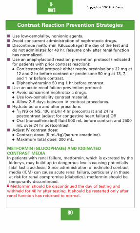
80
Contrast Reaction Prevention Strategies
■ Use low-osmolality, nonionic agents.■ Avoid concurrent administration of nephrotoxic drugs.■ Discontinue metformin (Glucophage) the day of the test and
do not administer for 48 hr. Resume only after renal functionhas normalized.
■ Use an anaphylactoid reaction prevention protocol (indicatedfor patients with prior contrast reaction):◆ Corticosteroid protocol: either methylprednisolone 32 mg at
12 and 2 hr before contrast or prednisone 50 mg at 13, 7,and 1 hr before contrast.
◆ Diphenhydramine 50 mg 1 hr before contrast.■ Use an acute renal failure prevention protocol:
◆ Avoid concurrent nephrotoxic drugs.◆ Use low-osmolality contrast material.◆ Allow 2–5 days between IV contrast procedures.
■ Hydrate before and after procedure:◆
1/2 NS or NS, 100 mL/hr 4 hr precontrast and 24 hrpostcontrast (adjust for congestive heart failure) OR
◆ Oral (noncaffeinated) fluid 500 mL before contrast and 2500mL over 24 hr postcontrast.
■ Adjust IV contrast dose:◆ Contrast dose: (5 mL/kg)/(serum creatinine).◆ Maximum total dose: 300 mL.
METFORMIN (GLUCOPHAGE) AND IODINATED
CONTRAST MEDIAIn patients with renal failure, metformin, which is excreted by thekidneys, may build up to dangerous levels causing potentiallyfatal lactic acidosis. Since administration of iodinated contrastmedia (ICM) can cause acute renal failure, particularly in thoseat risk for renal compromise (diabetics), metformin should betemporarily discontinued.Metformin should be discontinued the day of testing and
withheld for 48 hr after testing. It should be restarted only afterrenal function has returned to normal.
X-RAYS
03Hop-03 2/4/05 12:11 PM Page 80

X- RAYS
81
Reactions to Iodinated Contrast Media (ICM)
Signs and Symptoms Treatment
Anaphylactoid
ReactionMild symptoms: scattered urticaria,
pruritus; rhinorrhea; nausea,vomiting; diaphoresis; coughing; anddizziness.
Moderate symptoms: persistentvomiting; diffuse urticaria; headache;facial and or laryngeal edema; mildbronchospasm; dyspnea;palpitations, tachycar-dia, bradycardia; hypertension;abdominal cramps.
Severe symptoms: life-threateningarrhythmias (V-tach); hypotension;overt bronchospasm; laryngealedema; pulmonary edema; seizures;syncope; cardiopulmonary arrest
✓ Observe for progression to more severereaction, which requires immediateintervention.
✓ Administer antihistamines for itching orprochlorperazine for nausea and vomitingif necessary.
✓ Provide O2 at 10–12 L/min via face mask.✓ Monitor vital signs every 5 min.✓ Administer antihistamines, initiate IV
access with NS or D5W.✓ Possible administration of epinephrine,
steroids, and bronchodilators.
✓ Administer epinephrine 0.3–0.5 mg subQevery 10–20 min and methylprednisolone50 mg IV.
✓ Give beta agonist inhaler (e.g., albuterol)for bronchospasm.
✓ Call a code.✓ Manage ABCs: Monitor breathing and
vital signs continuously.✓ Maintain or initiate IV access.Prepare for intubation and resuscitation.
(Continued on following page)
03Hop-03 2/4/05 12:11 PM Page 81

82
Signs and Symptoms Treatment
Cardiovascular
Reaction
Bradycardia,hypotension,vasovagalreactions
Nephropathic
Reaction
Delayed
Reaction
Occurs within7 days of theinjection ofICM.rography
X- RAYS
Decreased heart rate (�60 bpm) andhypotension possibly accompaniedby nausea, vomiting, diaphoresis,and mental status changes. Canlead to cardiovascular collapse anddeath if untreated.
Decreased urine output. Diagnosis ofcontrast-associated renal failure:serum creatinine elevation of 25%within 3 days of contrast.
Flu-like symptoms: fatigue, upperrespiratory tract congestion, fevers,chills, nausea, vomiting, diarrhea,abdominal pain; pain in the injectedextremity; rash; dizziness andheadache; pruritus; parotitis; jointpain; constipation; and depression.
✓ Provide O2 at 10–12 L/min via facemask.
✓ Monitor vital signs every 5 min.✓ Administer IV fluids.✓ Atropine for vasovagal responses.
✓ Discontinue other nephrotoxic drugs ifpossible.
✓ Space studies up to 5 days apart toallow kidneys to recover.
✓ Hydrate patients 12 hr before and 2 hrafter study.
✓ Monitor lab values, intake, and output.
✓ Usually resolves spontaneously withlittle or no treatment.
(Continued)
03Hop-03 2/4/05 12:11 PM Page 82

X-RAYS
83Arthrography
X-ray with contrast to examine a joint and its surrounding softtissues (cartilage, ligaments, bursa, joint capsule). Commonlydone of the knee and shoulder but hip, wris,t and ankle are oftendone as well. Informed consent required. Potential Complications:
Infection, reaction to contrast (unlikely since contrast is not
administered IV).
PURPOSE■ Assess persistent, unexplained joint pain.■ Evaluate injuries to joint structures.■ Assess for tumor, tears, ruptures, cysts, and other abnormalities.
PROCEDUREThe patient lies supine on the x-ray table with the joint exposed.Lidocaine is injected to anesthetize the skin. Next, a needle isinserted into the joint space to aspirate synovial fluid for analysisand to minimize dilution of the contrast. The syringe is replacedwith a syringe containing contrast, which is injected into the joint.
To distribute the contrast throughout the joint, the patient eitherwalks a bit or has the joint manipulated through its range ofmotion.
Films are taken with the joint in various positions.
PRETEST PREPARATION■ See General Pretest Patient Education in Tools Tab.■ Ask patient about previous reactions to contrast or other risk
factors for adverse reaction to contrast (see page 79).■ Inform the patient that test will take about 30 min.■ Prepare the patient to expect some discomfort when the contrast
is injected.
POSTTEST CARE■ Assess vital signs and pain level.■ Assess patient for reaction to contrast (see Table, pages 81-82).■ If ordered, apply ice to joint and administer an analgesic.■ If knee has been evaluated, the patient will likely have an elastic
wrap applied. Inform patient to leave wrap on per physician’sorder.
03Hop-03 2/4/05 12:11 PM Page 83

84
■ Advise patient to minimize joint rotation and/or weightbearing for 1–2 days.
■ Teach patient to monitor for and report signs and symptomsof infection (fever, pain, swelling warmth over joint, rednessor drainage at puncure site).
Negative contrast arthrogram of the knee
X-RAYS
03Hop-03 2/4/05 12:11 PM Page 84

X-RAYS
85Arteriogram (Angiogram, Arteriography)
Rapid-sequence x-rays following administration of IV contrast toevaluate vascular conditions such as aneurysm, stenosis,blockage, or neoplasm. Digital subtraction angiography (DSA)removes the bony structures from the picture, therebyenhancing visualization. Performed in interventional radiologysuite or other special procedures area. See also CardiacCatheterization in Other Studies Tab. Common studies include:
◆ Renal arteriogram
◆ Carotid arteriogram
◆ Cerebral arteriogram
◆ Lower extremity arteriogram
◆ Pulmonary arteriogram
Informed consent required. Potential complications include
reaction to contrast, hemorrhage from arterial puncture, arterial
embolism, renal failure secondary to IV contrast, stroke,
thrombosis and arterial occlusion distal to puncture site,
hematoma, pseudoaneurysm, neurovascular damage.
PURPOSE■ Evaluate patency, size, and shape of blood vessels.■ Assess for plaque, emboli, or other explanation for impeded
blood flow.
PROCEDUREThe patient is placed supine on the x-ray table. An intravenousline, if not already present, is inserted and the patient isconnected to an ECG monitor.
After prepping, a small incision is made in the groin to accessthe femoral artery or into the arm to access the brachial artery.
Under fluoroscopy, a wire is threaded into the artery and alongthe arterial system until it is near or in the artery to beexamined. A catheter is then threaded over the wire and thewire is removed. The contrast material is injected either byhand or with a preset automated injector while a rapid series ofx-rays are taken.
The patient may receive sedation or possibly pain medication atthe initiation of or during the procedure.
03Hop-03 2/4/05 12:11 PM Page 85

86
Throughout the procedure, the patient’s vital signs are assessedand the peripheral pulses distal to the catheter insertion site aremonitored.
After the study is complete, the catheter is removed andpressure is applied over the puncture site for 15 min. Apressure dressing is then applied.
PRETEST PREPATATION■ See General Pretest Patient Education in Tools Tab.■ Ask patient about iodine or shellfish allergies.■ Check that the patient has been NPO for the prescribed
amount of time (2–8 hr).■ Locate and mark peripheral pulses distal to the proposed
puncture site to facilitate pulse assessment during and afterangiogram. (If pulses are absent before the procedure,document this finding. Assess both by palpation and withDoppler.)
■ Compare warmth, color, and strength of pulse in extremity tobe used for vascular access to the nonprocedure extremity.Document preexisting differences so that postprocedureassessments can be properly interpreted.
■ Check that preprocedure coagulation studies are available. Ifresults are out of range, document and notify physician andpersonnel in the unit where the procedure will be performed.
■ Ask if patient is taking metformin (Glucophage). Informphysician if yes and check for orders to hold the medicationfor 48 hr postprocedure. Ascertain that alternative tometformin has been ordered to cover blood glucoseelevations.
■ Assess vital signs.■ If a cerebral arteriogram is scheduled, assess the patient’s
neurologic status carefully to establish the baseline againstwhich postprocedure assessments can be compared.
■ Tell the patient:◆ the test takes 1–2 hr◆ there is minor discomfort associated with the insertion of
the catheter but that sedative and analgesic medication willbe available
◆ he or she will feel a sensation of warmth when the contrastis injected but that it will only last a few seconds
X-RAYS
03Hop-03 2/4/05 12:11 PM Page 86

X-RAYS
87◆ to report feelings of dizziness, itching, nausea, or other
unusual feeling to radiology staff◆ he or she may feel bladder fullness during the procedure
secondary to the contrast’s diuretic effect■ Explain postprocedure care and the importance of maintaining
bedrest for several hours after the procedure.
POSTTEST CARE■ Asssess the patient’s distal pulses and vital signs every 15 min
until stable and per facility protocol.■ Assess puncture site every 15 min for swelling, hematoma, or
bleeding. Assess if pressure dressing is intact. Apply sandbagto site. Report bleeding or disruption of pressure dressingimmediately. If site is bleeding, call physician immediately
and apply strong pressure with both hands until help arrives.
■ Assess neurovascular status of extremity: In addition topalpating peripheral pulses, note skin temperature, color, andcapillary refill time; ask about pain, numbness, or tingling;have the patient demonstrate mobility of hand or foot(depending on puncture site) without flexing or extending theextremity.
■ Continue to monitor the patient for several hours. Assistpatient to stand and walk when ambulation can be resumed.
■ For the patient who will be discharged to home following theprocedure, explain that he or she must monitor the puncturesite for bleeding, pain, swelling, and abnormal skin color ortemperature change in the arm or leg.
■ Advise the patient not to engage in strenuous activity for12–24 hr and not to take a hot bath or shower.
03Hop-03 2/4/05 12:11 PM Page 87

88
Barium Enema (BE)
X-ray and fluoroscopy of the colon after instillation of bariumsulfate to enhance images. Fluoroscopy is used to visualize thebarium as it moves through the large intestine. Air may beinsufflated into the colon to provide additional contrast and makethe mucosa more visible. Potential Complication: retained barium
in the colon causing obstruction or impaction.
PURPOSE■ Assess for tumor, polyps, inflammatory disorder, cause of rectal
bleeding, change in bowel habits, stricture, tumor, diverticula,and megacolon.
■ BE is contraindicated in patients wih suspected bowel perfora-tion. In this case, evaluation of the large intestine is accompli-shed using water-soluble ICM such as Gastrografin and no bowelprep.
PROCEDUREThe patient lies on the x-ray table for a plain film of the abdomen.Next, barium is instilled into the colon using a rectal tube or, if thebarium is administered through an ostomy, a Foley catheter.
Transit of the barium through the large intestine into the terminalileum is observed under flouroscopy. Spot films from variousviews and with the patient in different positions are taken ofsignificant findings. After all films are taken, the barium isaspirated from the colon and the rectal tube is removed. Thepatient will expel residual barium. An additional film is taken afterthe evacuation of barium.
PRETEST PREPARATION■ See General Pretest Patient Education in Tools Tab.■ The patient will have a clear liquid diet the evening before
the test and nothing by mouth, including medication, aftermidnight.
■ The patient should drink plenty of fluids the day before the test.■ The day before the test, the patient will begin the bowel prep,
which involves drinking magnesium citrate or other product inthe afternoon and taking biscodyl tablets (usually three 5-mgtabs) in the early evening.
X-RAYS
03Hop-03 2/4/05 12:11 PM Page 88

X-RAYS
89■ The morning of the test, the patient will use a biscodyl
suppository and/or a cleansing enema until the returnedmaterial is clear of fecal matter.
■ The patient should be told that the instillation of the barium(and air, if used) can cause cramping and bloating.
■ Reassure patient that radiology staff know that most peoplefind the test embarassing and will help the patient be ascomfortable as possible.
■ Inform the patient that testing will take 45–90 min.
POSTTEST CARE■ Assess for dehydration or electrolyte imbalance, especially in
older adults.■ Retained barium can cause an impaction or obstruction. The
patient should take a mild laxative or an enema to ensure thatno barium is retained. Stool may appear light colored for afew days.
03Hop-03 2/4/05 12:11 PM Page 89

90
Barium Swallow
X-ray with contrast medium (barium solution) to enhance imagesof the esophagus (also called esophagography). A water-solubleiodinated contrast medium (ICM) such as Gastrografin may beused if perforation or obstruction is suspected since barium, ifdeposited in the soft tissues, can cause a serious inflammatoryresponse.
PURPOSE■ Assess for anatomic deformity, stricture, tumor, foreign body,
reflux disease, swallowing disorder, esophageal spasm.■ Evaluate epigastric pain or regurgitation.
PROCEDUREA plain film is taken with the patient standing or lying supine onthe x-ray table. The patient then stands in front of a fluoroscopyscreen and swallows the contrast. The structure and function ofthe esophagus are evaluated and spot films may be taken. Thepatient is then strapped to the x-ray table and the table is tilted invarious positions as additional films are taken.
PRETEST PREPARATION■ See General Pretest Patient Education in Tools Tab.■ Ask patient about iodine or shellfish allergies if ICM will be used.■ The patient will be NPO for 8 hr prior to the procedure.■ The patient will have to remove jewelry from the head and neck.■ Although the procedure is not painful, some patients may not
like drinking the contrast media.
POSTTEST CARE■ If the patient has had barium: inform the patient that a mild
laxative and extra fluids will help with the elimination of thebarium. Inform the patient that stools will appear white or lightcolored. Instruct patient to notify health care provider ifconstipated.
■ If the patient has had an ICM (iodinated contrast medium):Monitor for anaphylactic, cardiovascular, nephropathic, ordelayed reactions to contrast. See preceding Table, Reactions toIodinated Contrast Media (ICM). Inform patient that somepatients experience diarrhea from water-soluble ICM.
X-RAYS
03Hop-03 2/4/05 12:11 PM Page 90

X-RAYS
91Cystography (Cystourethrogram, Voiding
Cystourethrogram, Cystogram)
X-ray and fluoroscopy with contrast medium to visualize bladderand urethra structure and function. (Also see NuclearCystogram in Nuclear Studies Tab.) Informed Consent required.
Potential Complications: UTI, reaction to contrast (unlikely
since contrast is not administered IV).
PURPOSE■ Evaluate structure and excretory function of the bladder and
urethra■ Assess for vesicoureteral reflux.■ Detect obstruction to urine flow (stones, stricture, stenosis,
prostatic enlargement).■ Evaluate cause of recurrent UTI.
PROCEDUREThe patient is placed supine or in the lithotomy position on thex-ray table. A catheter is placed and approximately 300 mL(adult dose) of contrast material is instilled into the bladder. X-rays are taken of the bladder while it is filled with contrast.
If a voiding cystourethragram is ordered, the catheter is thenremoved and the patient voids while more films are taken.
PRETEST PREPARATION■ See General Pretest Patient Education in Tools Tab.■ Ask patient about previous reactions to contrast or other risk
factors for adverse reaction to contrast (see page 79).■ Inform the patient that test will take about 30 min.■ Explain that patient will feel a brief sensation of pushing when
catheter is inserted.
POSTTEST CARE■ Assess patient for reaction to contrast.■ Monitor urine output and inform patient to monitor at home.■ Tell patient to drink plenty of fluids unless contraindicated.■ Assess for signs/symptoms of UTI and inform patient to report
fever, malaise, frequency, urgency, bladder or flank pain.
03Hop-03 2/4/05 12:11 PM Page 91
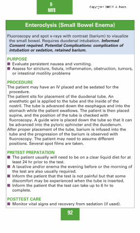
92
Enteroclysis (Small Bowel Enema)
Fluoroscopy and spot x-rays with contrast (barium) to visualizethe small bowel. Requires duodenal intubation. Informed
Consent required. Potential Complications: complication of
intubation or sedation, retained barium.
PURPOSE■ Evaluate persistent nausea and vomiting.■ Assess for stricture, fistula, inflammation, obstruction, tumors,
or intestinal motility problems
PROCEDUREThe patient may have an IV placed and be sedated for theprocedure.
The patient sits for placement of the duodenal tube. Ananesthetic gel is applied to the tube and the inside of thenostril. The tube is advanced down the esophagus and into thestomach while the patient swallows. The patient is then placedsupine, and the position of the tube is checked withfluoroscopy. A guide wire is placed down the tube so that it canbe advanced into the pyloric sphincter and the duodenum.
After proper placement of the tube, barium is infused into thetube and the progression of the barium is observed withfluoroscopy. The patient may need to assume differentpositions. Several spot films are taken.
PRETEST PREPATATION■ The patient usually will need to be on a clear liquid diet for at
least 24 hr prior to the test.■ A laxative and/or enema the evening before or the morning of
the test are also usually required.■ Inform the patient that the test is not painful but that some
discomfort may be experienced when the tube is inserted.■ Inform the patient that the test can take up to 6 hr to
complete.
POSTTEST CARE■ Monitor vital signs and recovery from sedation (if used).
X-RAYS
03Hop-03 2/4/05 12:11 PM Page 92

X-RAYS
93■ Retained barium can cause an impaction or obstruction. The
patient should take a mild laxative or an enema to ensure thatno barium is retained.
■ Inform the patient that stool may appear light colored for afew days.
■ Patients with chronic constipation should take a mild laxativefor several days until stool color returns to normal.
Hysterosalpingogram
X-ray and fluoroscopy with contrast to visualize uterine cavityand fallopian tubes. Potential Complications: Infection,
perforation of the uterus, reaction to contrast (unlikely since
contrast is not administered IV).
PURPOSE■ Assess patency of fallopian tupes and structural integrity of
uterus.■ Detect displaced IUD, tubal preganancy.■ Assess cause of repeated miscarriage.
PROCEDUREThe patient lies on the table in the lithotomy position. Aspeculum is placed in the vagina to visualize the cervix. Aflexible catheter is placed through the cervical opening into theuterine cavity.
Contrast is slowly injected through the catheter into the uterus.An x-ray is taken as the uterine cavity is filling. Additionalcontrast is injected so that the fallopian tubes fill and more x-rays are taken including an oblique view.
Once the instruments are removed, the patient remains on thetable for several minutes until cramping subsides.
PRETEST PREPATATION■ Ascertain that the patient is not pregnant and that last
menstrual period occurred 7–10 days prior to test date.■ Inform the patient that testing takes approximately 30 min.■ Prepare the patient to expect mild to moderate cramping,
which can be relieved with medications.
03Hop-03 2/4/05 12:11 PM Page 93

94
■ Some radiologists request a bowel prep (laxative the eveningbefore, suppository or enema in the morning) and/or acleansing douche
■ A pretest sedative such as oral diazepam may be given.
POSTTEST CARE■ Assess vital signs, pain level, amount and characteristics of
vaginal discharge.■ Some women experience dizziness; assess patient for
dizziness.■ Most women experience cramping for several minutes after
the procedure, but some may have cramps for several hours.A mild analgesic such as ibuprophen can be given.
■ Teach patient to assess for and report signs or symptoms ofinfection such as fever, pelvic pain and tenderness, orpurulent discharge.
■ Teach patient to assess for and report signs or symptoms ofuterine perforation: abdominal pain and distension, vaginalbleeding, signs and symptoms of peritonitis.
■ Tell the patient to expect to have vaginal discharge for a fewdays after the test. This discharge is the contrast agent and isharmless. It may be blood tinged but should not be franklybloody.
X-RAYS
03Hop-03 2/4/05 12:11 PM Page 94

X-RAYS
95Myelogram
X-rays and fluoroscopy with contrast to provide visualization of thespinal subarachnoid space and the spinal canal. Myelography isoften combined with computed tomography (CT scan), which isthen called called CT myelography. Magnetic resonance imaging(MRI) has replaced myelography for many indications. Informed
consent required. Potential Complications: Severe headache; CSF
leak; brain herniation; seizures; meningitis; puncture of the spinal
cord, aorta, or vena cava; severe reaction to contrast.
PURPOSE■ Assess signs and symptoms consistent with narrowing of the
spinal canal.■ Assess patients with peripheral neurologic deficits.■ Detect spinal stenosis, herniated disk, tumor, or infection.■ Assess changes in bone structure caused by arthritis or
ankylosing spondylosis
PROCEDUREThe patient is placed prone or in a side-lying position for lumbarpuncture. Under fluoroscopy to verify proper placement, a needleis inserted and 15 mL of cerebrospinal fluid (CSF) is removed. Thepatient is positioned prone and secured to the x-ray table withstraps. Then 15 mL of water-soluble contrast agent is then injectedinto the spinal canal and the x-ray table is tilted to distribute thecontrast. Progress of the contrast is fluoroscopically observed.Lesions or obstructions to flow are apparent and spot films aretaken. After all views are obtained, the needle is removed and thesite is dressed.
PRETEST PREPARATION■ See General Pretest Patient Education in Tools Tab.■ Ask patient about previous reactions to contrast or other risk
factors for adverse reaction to contrast (see page 79).■ In addition to other medical history questions, ask the patient
about history of seizures or use of medications that can lowerseizure threshold. Ascertain that patient has discontinued seizurethreshhold–lowering drugs for 48 hr before the test (see Tablebelow).
03Hop-03 2/4/05 12:11 PM Page 95

96
■ In addition to drugs that lower the seizure threshold,anticoagulants should also be withheld for 48 hr beforemyelography.
■ Foods and fluids restrictions vary with the type of contrastused. Some patients will be NPO for 8 hr prior to theprocedure and will require a laxative or enema. Some patientsmay have clear liquids before the test and no bowel prep.Check with the radiology department for details.
■ Let the patient know that some patients experience discomfortduring the study and that sedative or analgesic medicationwill be provided if necessary.
■ Inform the patient that an IV may be placed and fluids ormedications (sedatives or anticonvulsants) may be given.
■ Have the patient void before the test.■ Tell the patient that the test takes 45 min (longer if combined
with CT scan) and that he or she will be monitored in theradiology department or the hospital for several hours afterthe procedure.
POSTTEST CARE■ Proper positioning of the head after myelogram is very
important in preventing a headache. If an oil-based contrastmaterial was used (rare), the patient must remain flat in bedfor 24 hr.
■ If a water-based contrast material was used, the patient mustremain in bed for 24 hr, but the head of the bed can beelevated 15–30 degrees.
■ Assess vital signs, neurologic status, and pain level every 30min for 4 hr, then every 4 hr for 24 hr.
■ Assess for numbness, tingling, or diminished mobility oflower extremities.
■ Assess patient’s ability to void.■ Encourage fluid intake to help excrete contrast.■ Assess puncture site for bleeding or drainage.■ Assess patient for reaction to contrast (see Table, pages
81-82).■ Document and report any postprocedure problems promptly.
X-RAYS
03Hop-03 2/4/05 12:11 PM Page 96

X-RAYS
97Medications That Lower Seizure Threshold and May Be
Withheld for 48 Hr Prior to a Myelogram
amitriptylineamitriptyline/chloridiazepoxideamitriptyline/perphenazineamoxapineamphetamineamphetamine mixturebupropionchlorpromazineclomipramineclozapinecycloserinecyclosporinedesipraminedextroamphetaminedoxapramdoxepinephedrinefentanylfluoxetinefluphenzainefluvoxaminehaloperidolimipenem-cilastatinimipraminelithiumloxapinemaprotiline
meperidinemesoridazinemethamphetaminemethylpheridaremirtazapinemolindonenefazodonenortriptylineolanzapineparoxetineperphenazinepimozideprochlorperazinepromazineprotriptylinequetiapineriperidonesertralinetheophyllinethioridazinethiothixenetrazodonetrifluoperazinetriflupromazinetramadoltrimipraminevenlafaxine
03Hop-03 2/4/05 12:11 PM Page 97

98
Operative or T-Tube Cholangiogram
X-ray and fluoroscopy of the common bile duct using aniodinated contrast medium (Hypaque) to enhance visualization.Done during gallbladder surgery or 7–10 days aftercholecystectomy. Informed Consent required. Potential
Complications: severe adverse reaction to contrast.
PURPOSE■ Evaluate biliary duct patency before removal of T-tube or, if
performed during surgery, to visualize biliary tree and thusminimize risk of operative injury to the common bile duct.
■ Assess for strictures, gallstones, fistula, or tumor.
PROCEDURET-Tube Cholangiogram:The T-tube is clamped, usually the daybefore the procedure. In the radiology department, the patientis placed on the x-ray table. The T-tube is unclamped andcontrast medium is injected into it. The contrast is visualizedwith fluoroscopy as it moves through the duct system. The tubeis clamped while various pictures (prone, side-lying, erect) ofthe right upper quadrant (RUQ) are taken.
Intraoperative cholangiogram: During surgery a catheter isinserted into the common bile duct. Contrast is injected into thecatheter and x-rays are taken.
PRETEST PREPARATION■ See General Pretest Patient Education in Tools Tab.■ Ask patient about iodine or shellfish allergies if ICM.■ T-tube cholangiogram: NPO for 1 meal prior to test.
Administer an enema 1 hr before test.■ Tell the patient that he or she may experience pressure as the
contrast is injected. Some patients may experience nausea,vomiting, cramps, or diarrhea from the contrast.
■ Inform the patient that testing usually takes about 30 min.
POSTTEST CARE■ Monitor for anaphylactic, cardiovascular, nephropathic, or
delayed reactions to contrast. See preceding Table, Reactionsto Iodinated Contrast Media (ICM).
X-RAYS
03Hop-03 2/4/05 12:11 PM Page 98

X-RAYS
99■ Monitor for dehydration or electrolyte imbalance if patient
experiences vomiting and/or diarhhea.■ Check if T-tube has been removed (if postop findings are
normal). Apply a dry sterile dressing to the site.■ Check orders to see if T-tube is to be left clamped or
unclamped.■ Assess T-tube insertion site for pain or swelling.
Oral Cholecystogram (OCG)
X-ray of the gallbladder with iodinated contrast medium(Telepaque or Oragrafin) to enhance visualization. Infrequentlyused since ultrasound, CT scan, and nuclear scans give betterresults (refer to appropriate Tabs). Informed Consent required.
Potential Complication: severe adverse reaction to contrast.
PURPOSE■ Assess RUQ/epigastric pain and other symptoms associated
with gallbladder disorders.
PROCEDUREAfter proper preparation the evening before the test (see below),the patient lies on the x-ray table while pictures are taken fromvarying views. The patient may be given a high-fat meal tostimulate gallbladder contractions, which are observed withfluoroscopy. Additional films are taken as the contrast movesthrough the common bile duct.
PRETEST PREPARATION■ See General Pretest Patient Education in Tools Tab.■ Ask patient about iodine or shellfish allergies.■ The patient will eat a low-fat meal the evening before the test
and will be NPO after midnight.■ The patient will take six Telepaque tablets 5 min apart 2–3 hr
after the evening meal.■ An enema may be prescribed the morning of the test.■ Although the procedure is not painful, some patients may
experience nausea, vomiting, cramps, or diarrhea from thecontrast.
■ Inform the patient that testing may take up to 2 hr.
03Hop-03 2/4/05 12:11 PM Page 99

100
POSTTEST CARE■ Monitor for anaphylactic, cardiovascular, nephropathic, or
delayed reactions to contrast. See preceding Table, Reactionsto Iodinated Contrast Media (ICM).
■ Monitor for dehydration or electrolyte imbalance if patientexperiences vomiting and/or diarhhea.
Percutaneous Transhepatic Cholangiogram (PTC)
Fluoroscopic visualization of the bile ducts with spot films ofsignificant findings. Performed in interventional radiology.Contrast medium is administered through a fine needleinserted through the skin into the liver and into a bile duct.Informed consent required. Potential complications: sepsis,
bleeding, peritonitis, severe adverse reaction to contrast.
PURPOSE■ Distinguish between obstructive and nonobstructive jaundice
and pancreatitis.■ Assess patients in whom ERCP (see Scopes Tab) has been
unsuccessful.■ Assess cause of obstructive jaundice.■ Evaluate persistent upper abdominal pain after
cholecystectomy.■ Establish external drainage of the biliary system as a palliative
measure for patients with nonresectable malignant disease.
PROCEDUREThe patient lies on the x-ray table. A peripheral IV line is insertedand a sedative and/or pain medication are administered(sometimes a sedative is administered before the test). A localanesthetic is administered into the right upper abdominal wall.
The patient holds his or her breath and a fine needle is insertedbetween the 10th and 11th intercostal space, midclavicluar line.The needle is advanced until it enters a bile duct, after whichthe introducer is removed and contrast medium is injected intothe biliary system.
The movement of the contrast through the bile ducts is observedon the fluoroscope, which shows whether the bile moves freely
X-RAYS
03Hop-03 2/4/05 12:11 PM Page 100

X-RAYS
101or is obstructed. The patient is helped to assume a variety ofpositions while spot films are taken.
After all films have been taken, the contrast is aspirated from thebiliary ducts, the needle is removed, and a dry sterile dressing isapplied.
A catheter may be left in place if external biliary drainage isrequired.
PRETEST PREPARATION■ See General Pretest Patient Education in Tools Tab.■ Check that patient has been NPO for 8 hr and clotting times, PT,
and platelet count results are normal and available on the chart.Coagulation studies are frequently abnormal in patients with
liver disease.
■ Make sure that pretest antiobiotcs have been administered.■ Ask patient about previous reactions to contrast or other risk
factors for adverse reaction to contrast (see page 79).■ Ascertain that within the preceding week the patient has not
taken NSAIDs, aspirin, or medications that affect bleeding times.■ Administer pretest sedative, if ordered.■ Explain that the patient may feel discomfort when the needle
penetrates the liver, pressure, or brief discomfort in the rightsubscapular area. Reassure the patient that medication will begiven to enhance comfort.
■ The patient will need to remain still and hold his or her breathfor short periods during the test.
■ Tell the patient testing usually takes between 30 and 60 min.
POSTTEST CARE■ Monitor for anaphylactic, cardiovascular, nephropathic or
delayed reactions to contrast. See preceding Table, Reactions toIodinated Contrast Media (ICM).
■ Assess vital signs frequently according to policy.■ The patient should maintain bedrest for 6 hr postprocedure.■ Monitor for bleeding from puncture site, swelling around
puncture site. Notify physician or NP immediately if thepatient has right abdominal or shoulder pain (indicatesbleeding), fever, dizziness, drop in blood pressure, tachycardia,or heme � stool.
■ Continue antibiotics on schedule.
03Hop-03 2/4/05 12:11 PM Page 101

102
PTC Approach
Percutaneous transhepatic cholangiogram.
Needle insertion site for percutaneous transhepatic cholangiogram.
X-RAYS
Dilatedbile ducts
Commonbile duct
Liver
Gallbladder
03Hop-03 2/4/05 12:11 PM Page 102

X- RAYS
103
Pyelogram, Antegrade and Retrograde
Retrograde or antegrade pyelography is performed when IVP results are inadequate or IVP iscontraindicated. Informed Consent required. Potential Complications: severe adverse reaction
to contrast; adverse reaction to anesthesia, other risks of surgery (retrograde pyelogram).
Antegrade Pyelogram Procedure and Care
Procedure Pre- and Posttest Care
Antegrade
Pyelogram
Visualizes stones,tumors,obstructions in theurinary system.
Performed ininterventionalradiology.
Kidneys are located by US orCT; overlying skin is cleanedwith antiseptic and localanesthetic is injected.
Under fluoroscopy, a needlewith a manometer attachedis inserted into the kidneyand pressure readings aretaken.
Contrast is injected into thekidney.
X-rays are taken of kidney’supper- collecting system.
A nephrostomy tube may beleft in the kidney fordrainage.
✓ Follow preoperative orders regardingbowel prep and NPO status.
✓ Patient will require standard post-procedure assessment and VSmonitoring.
✓ Monitor needle insertion site forbleeding or hematoma. Providenephrostomy tube care, if indicated.
✓ Monitor fluid intake and urinaryoutput for at least 24 hr.
✓ Assess urine for blood.✓ Call physician or NP if patient has not
voided within 8 hr of the procedure, ifbladder is distended or painful, or ifvoiding small amounts.
✓ Assess for signs of infection, such aschills, fever, and hypotension.
03Hop-03 2/4/05 12:11 PM Page 103
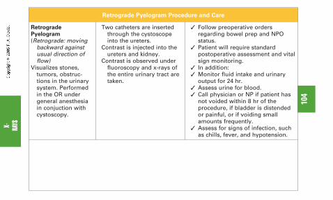
104
Retrograde Pyelogram Procedure and Care
Retrograde
Pyelogram
(Retrograde: movingbackward againstusual direction offlow)
Visualizes stones,tumors, obstruc-tions in the urinarysystem. Performedin the OR undergeneral anesthesiain conjuction withcystoscopy.
X- RAYS
Two catheters are insertedthrough the cystoscopeinto the ureters.
Contrast is injected into theureters and kidney.
Contrast is observed underfluoroscopy and x-rays ofthe entire urinary tract aretaken.
✓ Follow preoperative ordersregarding bowel prep and NPOstatus.
✓ Patient will require standardpostoperative assessment and vitalsign monitoring.
✓ In addition:✓ Monitor fluid intake and urinary
output for 24 hr.✓ Assess urine for blood.✓ Call physician or NP if patient has
not voided within 8 hr of theprocedure, if bladder is distendedor painful, or if voiding smallamounts frequently.
✓ Assess for signs of infection, suchas chills, fever, and hypotension.
03Hop-03 2/4/05 12:11 PM Page 104

X-RAYS
105Pyelogram, Intravenous (IVP, Excretory
Urogram, EU, ExU)
X-ray with contrast medium to visualize the kidneys, renal pelvis,ureters, and bladder. Also called excretory urography (ExU) orIV urogram (IVU). Being replaced by CT urogram (see CT Tab)Informed Consent required. Potential Complications: severe
adverse reaction to contrast.
PURPOSE■ Evaluate structure and excretory function of the renal system.■ Assess patients with flank/kidney pain.■ Asses for kidney stones.■ Investigate cause of hematuria.■ Evaluate suspected obstruction, structural abnormalitiy, or
trauma to the urinary system.
PROCEDUREThe patient is placed supine on the x-ray table and an abdominalflat plate is taken to determine if any barium, stool, stones, orgas will obscure visualization. Next, an IV line is inserted andthe contrast is injected. The contrast reaches the kidneys via thebloodstream, where it is filtered out of the blood and excretedthrough the ureters into the bladder. Films are takenapproximately every 5 min until the contrast reaches thebladder. These x-rays reveal tumors, cysts, stones, or otherstructural and functional abnormalities. Once the contrastreaches the bladder, the patient is asked to void and anotherfilm is taken. This will reveal if the bladder empties completely.
PRETEST PREPARATION■ See General Pretest Patient Education in Tools Tab.■ Ask patient about previous reactions to contrast or other risk
factors for adverse reaction to contrast (see page 79).■ The patient should be well-hydrated and should increase fluid
intake the day before the test. Check BUN; if elevated, notifyradiologist.
■ The patient will not be allowed foods in the 8 hr preceding thetest and may be NPO. In some cases, the patient may have aclear liquid breakfast.
03Hop-03 2/4/05 12:11 PM Page 105

106
■ A bowel prep may be ordered the evening before or the morningof the test.
■ Ascertain that patients on metformin (Glucophage) havewithheld the medication the day of the test.
■ The patient should void immediately before the test.■ Inform the patient that the test will take 30 to 60 min.
POSTTEST CARE■ Monitor for anaphylactic, cardiovascular, nephropathic, or
delayed reactions to contrast. See preceding Table, Reactions toIodinated Contrast Media (ICM).
■ Ensure adequate oral or IV hydration.■ Monitor urine output and inform patient to monitor at home.■ Ensure that renal function is evaluated before metformin is
resumed 48 hr after the test.■ Patient may resume pretest activity level and diet.
Upper GI Series (UGI)
X-ray and fluoroscopy with contrast medium (usually bariumsulfate) to enhance images of the lower esophagus stomach,duodenum, and upper jejunum. Allows visualization of thedigestive system at work as barium moves through it. A water-soluble iodinated contrast medium (ICM) such as Gastrografinmay be used if perforation is suspected since barium, if depositedin the soft tissues, can cause a serious inflammatory response.When combined with evaluation of the entire small bowel is calledUGI with small bowel follow-through.
PURPOSE■ Assess for ulcers, stricture, tumor, reflux disease, hernia, and
inflammation.
PROCEDUREThe patient sits or stands behind the fluoroscopy machine and isasked to swallow the barium (8–16 oz). Passage of the bariumthrough the upper GI tract is filmed as the patient drinks.
After ingestion of the barium, x-ray images are obtained with thepatient in different positions (supine, side-lying, prone) and fromdifferent views (PA, AP, lateral, and oblique).
X-RAYS
03Hop-03 2/4/05 12:11 PM Page 106

X-RAYS
107If needed, enhancement of the structures can be obtained byhaving the patient swallow baking soda crystals or drink bariumthrough a perforated straw (called double-contrast). The airsmoothes out the gastric rugae (wrinkles and folds), whichmakes the anatomy clearer.
PRETEST PREPARATION■ See General Pretest Patient Education in Tools Tab.■ Ask patient about iodine or shellfish allergies if ICM will be
used.■ The patient will be NPO for 8 hr prior to the procedure.■ The patient will have to remove jewelry on the trunk, head, or
neck. If jewelry has psychological or spiritual significance, thepatient may hold the jewelry during the procedure.
■ Although the procedure is not painful, some patients may notlike drinking the barium.
■ Inform the patient that testing may take up to 5 hr, althoughthe study usually is completed within 2 hr.
POSTTEST CARE■ If the patient has had barium: inform the patient that a mild
laxative and extra fluids will help with the elimination of thebarium. Inform the patient that stools will appear white orlight colored. Instruct patient to notify health care provider ifconstipated.
■ If the patient has had an ICM (iodinated contrast medium):Monitor for anaphylactic, cardiovascular, nephropathic, ordelayed reactions to contrast. See preceding Table, Reactionsto Iodinated Contrast Media (ICM). Inform patient that somepatients experience diarrhea from water-soluble ICM.
■ Instruct the patient to notify health care provider ifconstipated.
03Hop-03 2/4/05 12:11 PM Page 107

108
Venogram, Lower Extremity
X-ray with contrast to enhance visualization of venous system.Requires informed consent. Potential complications: severe
adverse reaction to contrast, renal failure, dislodgement of
venous thrombi.
PURPOSE■ Detect deep vein thrombosis (DVT)
PROCEDUREThe patient is placed on the x-ray table, which is tilted at anangle between 30 and 45 degrees.
A tourniquet is placed above the ankle and sometimes on thethigh. (This is to diminish blood flow in the peripheral veinsthereby allowing contrast to fill the deep veins.) An IV is startedin the foot and the contrast medium is injected over 2–4 min. Aseries of x-rays are taken as the contrast moves through theveins. Alternatively, the progression of the contrast can beviewed using fluoroscopy with spot films taken as needed.
PRETEST PREPARATION■ See General Pretest Patient Education in Tools Tab.■ Ask patient about iodine or shellfish allergies if ICM will be
used.■ The patient may be asked to take only clear liquids for 2 hr
prior to the procedure.■ Although the procedure is not painful, some patients may
have mild discomfort as the catheter is inserted.■ Inform the patient that testing takes 30–90 min.
POSTTEST CARE■ Assess patient’s vital signs after the procedure. Monitor for
reaction to contrast.■ Encourage fluids.■ Observe puncture site for bleeding, hematoma, signs or
symptoms of infection.
X-RAYS
03Hop-03 2/4/05 12:11 PM Page 108

CT/MRI/US
109
COMPUTED TOMOGRAPHY (CT SCANS)
Overview of CT Scanning
■ A CT scan is a specialized x-ray that takes cross-sectionalpictures of all types of tissue.
■ CT scans are sometimes called CAT scans. The “A” refers tothe word axial, which is a particular orientation of the image.Other orientations are available now, so the A has beendropped.
■ It is used extensively in diagnosing disease and injury of the:◆ Brain, cerebral blood vessels , eyes, inner ear, and sinuses.◆ Neck, shoulders, cervical spine, and blood vessels.◆ Chest, heart, aorta, lungs, and mediastinum.◆ Thoracic and lumbar spine.◆ Upper abdomen, liver, kidney, spleen, and pancreas.◆ Skeletal system including bones of the hands, feet, ankles,
legs and arms, and jaws.◆ Pelvis and hips, reproductive system, bladder, and GI tract.
■ It is also used to diagnose cancers, including lung, liver, andpancreatic cancer and measure tumor size and assessinvolvement in other nearby tissues.
■ Fluoroscopic CT imaging is used to guide minimally invasiveprocedures such as:◆ Drainage of fluid collections (cysts, abscesses, lymphoceles,
hematomas).◆ Biopsy.◆ Injection of pain medications into a spinal disc space.◆ Dynamic study of knee or elbow motion, swallowing, or
study of the larynx.◆ Embolization to stop bleeding, for example, after liver or
spleen trauma.■ Computed tomography angiography (CTA) is a less invasive
method for examining blood vessels.■ Patients over 300 lb may be unable to undergo CT scanning.■ It is important that patients lie very still during imaging. Any
motion will blur the images necessitating repeat scans.■ The CT scan itself usually takes only a few minutes to
complete but the patient should allow up to 2 hr for the test.
04Hop-04 2/4/05 12:12 PM Page 109
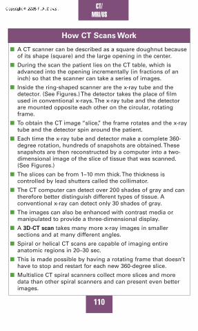
110
How CT Scans Work
■ A CT scanner can be described as a square doughnut becauseof its shape (square) and the large opening in the center.
■ During the scan the patient lies on the CT table, which isadvanced into the opening incrementally (in fractions of aninch) so that the scanner can take a series of images.
■ Inside the ring-shaped scanner are the x-ray tube and thedetector. (See Figures.) The detector takes the place of filmused in conventional x-rays. The x-ray tube and the detectorare mounted opposite each other on the circular, rotatingframe.
■ To obtain the CT image “slice,” the frame rotates and the x-raytube and the detector spin around the patient.
■ Each time the x-ray tube and detector make a complete 360-degree rotation, hundreds of snapshots are obtained. Thesesnapshots are then reconstructed by a computer into a two-dimensional image of the slice of tissue that was scanned.(See Figures.)
■ The slices can be from 1–10 mm thick. The thickness iscontrolled by lead shutters called the collimator.
■ The CT computer can detect over 200 shades of gray and cantherefore better distinguish different types of tissue. Aconventional x-ray can detect only 30 shades of gray.
■ The images can also be enhanced with contrast media ormanipulated to provide a three-dimensional display.
■ A 3D-CT scan takes many more x-ray images in smallersections and at many different angles.
■ Spiral or helical CT scans are capable of imaging entireanatomic regions in 20–30 sec.
■ This is made possible by having a rotating frame that doesn’thave to stop and restart for each new 360-degree slice.
■ Multislice CT spiral scanners collect more slices and moredata than other spiral scanners and can present even betterimages.
CT/MRI/US
04Hop-04 2/4/05 12:12 PM Page 110

CT/MRI/US
111
X-Ray tubes
Detectors
Exterior of a CT Scanner
Interior of a CT Scanner
04Hop-04 2/4/05 12:12 PM Page 111

112
How the CT Image Is ObtainedCT
/MR
I/US
Monitor
Detectors
X-Ray tube
Colimater
04Hop-04 2/4/05 12:12 PM Page 112

CT/
MRI/U
S
113
Views
Sagittal, coronal, and axial views.
Axial view Sagittal view Coronal view
04Hop-04 2/4/05 12:12 PM Page 113

114
CT/MRI/US
*Source: RSNA News - August 2004 Patients and Physicians UninformedAbout CT Risks.http://www.rsna.org/publications/rsnanews/aug04/ct_dose-1.html.Accessed 8/28/04**Source: Studies Find Radiation Doses From CT Scans Often Too HighFor Children. http://imaginis.com/ct-scan/news/news1.26.01.asp. Accessed8/28/04
Radiation Exposure During CT Scans
■ CT studies expose the patient to significantly more radiationthan conventional x-rays. For example, a chest CT can exposethe patient to 100 times more radiation than a plain x-ray.*
■ Pregnant women should not undergo CT scanning because ofpotential damage to the fetus.
■ Lead shields are used to protect reproductive organs fromradiation exposure.
■ Pediatric patients are often exposed to much higher doses than
necessary because default settings are intended for adults.**
◆ Excessive exposure to radiation during childhood can lead tocancer later in life.
◆ Scanning protocols should be adjusted for each child’s weightand body type.
◆ Health care providers should weigh the risks and benefits ofusing CT scans in children, especially when multiple CT scansare anticipated.
CT Scans with Contrast Media
■ CT scan studies may be enhanced with contrast agents.■ CT contrast agents are used to make organs, blood vessels,
and/or tissue types appear whiter to better show neoplasm,disease, or injury. See next page for overview of contrast andrefer to pages 78-82 in Tab 3 for a complete discussion ofcontrast media, its uses, and risks.
■ Contrast can be administered intravenously, orally, rectally, or byinhalation, which is called Xenon CT (for the name of the gasinhaled and is used for lung and brain imaging).
■ In studies of pelvic organs, patients may receive IV, oral, andrectal contrast.
04Hop-04 2/4/05 12:12 PM Page 114

CT/MRI/US
115IV CONTRAST■ Associated with greater risk of reaction than other routes.■ Given to visualize blood vessels and tissues such as brain,
spine, liver, and kidneys.■ Administered by loading into a power-assisted injector, which
automatically administers the contrast.■ Total volume of contrast injected ranges from 75–150 mL.■ Patients may experience a warm feeling and a metallic taste
when contrast is injected, which is harmless.■ Patients may also experience mild itching, hives, nausea,
vomiting, or dizziness, which can be treated withantihistamines.
■ Other reactions include cardiovascular, nephropathic, ordelayed responses or severe anaphylactoid responses.
■ Studies using IV contrast can only be performed afterobtaining informed consent.
■ Assess renal function studies. Report elevations inBUN/creatinine to physician and radiology staff.
■ Determine if patient takes metformin (Glucophage), whichmust be withheld for 48 hr after administration of IV contrast.
ORAL CONTRAST■ Barium and Gastrografin are the contrast agents used to
enhance images of the abdomen and pelvis.■ Barium is the most commonly used agent. It has a milk
shake– like consistency and is flavored to enhance palatability.■ Gastrografin has the consistency of water. It is an iodinated
contrast medium (i.e., it contains iodine).■ Patient preparation varies but usually involves restriction of
food and fluids for several hours prior to testing.■ Patients usually drink 1000–1500 mL of contrast.
RECTAL CONTRAST■ Barium or Gastrografin is also administered rectally when CT
scanning the large intestines or pelvic organs.■ It is administered in the radiology department.■ After the study is complete, the contrast is drained out of the
large intestine.■ Preparation involves restricting foods and fluids and taking
laxatives and/or cleansing enemas before testing.
04Hop-04 2/4/05 12:12 PM Page 115

116
Patient Preparation for CT Scan
■ Tell the patient that he or she should:◆ Wear comfortable, loose-fitting clothing if an outpatient.◆ Remove clothing, jewelry, or anything else that might
interfere with imaging (belts, earrings, bra, glasses,dentures, hairpins, clothing with zippers and snaps). Ifjewelry has psychological or spiritual significance for thepatient, it usually can be held by the patient or moved to apart the body not under examination.
◆ Inform the physician of any medications that are beingtaken, both prescription and nonprescription.
◆ Inform the physician if pregnant, diabetic, or allergic to anyfoods or drugs.
◆ Inform the physician about previous reactions to contrast.■ Explain to the patient that he or she will:
◆ Be positioned on a narrow table that slides into the scanner.◆ Be monitored by a technologist and a radiologist who will
be directing the study from an adjacent room (where themonitor and computer are).
◆ Be able to communicate with the technologist at all timesvia an intercom.
◆ Experience no pain from the study, although positioningmight feel a little awkward.
◆ Have an IV started if contrast will be given.◆ Be asked to hold his or her breath and lie very still for up to
30 sec.■ On rare occasions, the contrast may cause a life-threatening
anaphylactoid reaction manifested by laryngeal edema,shortness of breath, and hypotension. Tell the patient to
alert the technologist via the intercom if he or she has trouble
breathing.Tell the patient if this occurs, the exam will be
stopped and medication given.
■ If scheduled for abdominal films, determine if the patient hashad a barium contrast study or taken medications containingbismuth in the previous 4 days. (Films will need to bepostponed until all contrast is excreted.)
■ After the CT images are reviewed, the patient will be releasedfrom the imaging department or center.
CT/MRI/US
04Hop-04 2/4/05 12:12 PM Page 116

CT/MRI/US
117Abdominal/Pelvic CT
Computed x-ray of the abdomen and or pelvic organs, usually withoral contrast and IV contrast and sometimes with rectal contrastas well. Used for looking at solid organs, such as the liver,pancreas, spleen, kidneys, adrenal glands, and pelvic organs andfor evaluating the abdominal aorta and vena cava. Also used toassess lymph nodes in the abdomen. Less useful for evaluatingthe stomach and intestines, although the CT scan can providesome information about these structures as well. Informed
consent usually required. Potential complications: severe adverse
reaction to contrast.
PURPOSE■ Assess abdominal/pelvic pain or suspected liver, kidney, or
gallbladder disease.■ Evaluate abdominal trauma.■ Assess signs of inflammation or infection inside the abdomen.■ Assess for cancer; stage or monitor tumors during therapy.
PROCEDUREBefore the procedure begins, the patient will drink the oral contrastand, if IV contrast is to be given, will have an IV placed. Thepatient then lies on the CT table. The technologist will helpposition the patient’s body, if necessary, with pillows. The tablewill be advanced slowly into the gantry (the ring-shaped CTscanner) in millimeters as the images are taken. The technologistwill tell the patient when to hold his or her breath, and will alsocontrol the position and advancement of the table. The IV contrastmay be administered before or during the test.
PRETEST PREPARATION■ See General Pretest Patient Education in Tools Tab.■ Ask patient about previous reactions to contrast or other risk
factors for adverse reaction to contrast (see page 79).■ Unless no IV contrast will be given, the patient should be well
hydrated and should increase fluid intake the day before the test.Check BUN; if elevated, notify radiologist.
■ Ascertain that patients on metformin (Glucophage) havewithheld the medication the day of the test.
■ Tell the patient the test is painless.
04Hop-04 2/4/05 12:12 PM Page 117

118
■ Oral contrast preparation may vary, but generally the patient will beasked to drink three cups of oral contrast over a 30-min period 90min before the CT appointment. The patient then will be given morecontrast to drink in the radiology department.
■ Check with the radiology department about restriction of food andfluids. Some departments ask that the patient remain NPO exceptfor the contrast and medications while others may allow the patientto eat lightly (juice and crackers) after the initial few cups of oralcontrast have been consumed at home.
■ The patient should void immediately before the test.■ Inform the patient that the test will take 5–30 min.
POSTTEST CARE■ When the exam is over, the technologist will check the images while
the patient waits. If no more images are needed, the patient will bedischarged from the radiology department.
■ If IV contrast is given, provide the patient with and encourageintake of oral fluids to help excrete the contrast.
■ If oral or rectal barium was given, tell the patient that stools mayappear light-colored and to inform a health care provider ifconstipated.
■ Monitor for reaction to IV contrast.
CT scan of abdomen. Arrows point to adrenal gland. a �aorta, v � venacava, p � pancreas, k � kidney, L � liver, s � spleen.
CT/MRI/US
04Hop-04 2/4/05 12:12 PM Page 118

CT/MRI/US
119Angiogram, CT (CTA)
X-ray with contrast to visualize arterial blood flow of the brain,lungs, kidneys, or extremities. CT angiography is much lessinvasive than traditional arteriography, which involves arterialpuncture and threading a catheter through the artery. In CTA,iodinated contrast is quickly injected into a peripheral veinwhile numerous and very thin “slices” are imaged. Informed
consent may be required. Potential complications: severe
adverse reaction to contrast.
PURPOSE■ Detect atherosclerotic disease.■ Evaluate for pulmonary embolism.■ Assess blood flow in the renal, pelvic, or carotid arteries.■ Identify aneurysms.■ Identify aortic dissection.■ Identify cerebral arteriovenous malformation.■ Evaluate stent functionality.■ Evaluate trauma patients for arterial damage.
PROCEDUREThe patient is positioned on the CT table with the body part tobe examined placed inside the opening. First, a test image andsmall dose of contrast will be taken to determine optimalpositioning and timing. Next, the contrast is loaded into anautomatic injector and the contrast is administered quicklywhile the area is scanned.
PRETEST PREPARATION■ See General Pretest Patient Education in Tools Tab.■ Ask patient about previous reactions to contrast or other risk
factors for adverse reaction to contrast (see page 79).■ The patient should be well-hydrated and should increase fluid
intake the day before the test.■ Check BUN; if elevated, notify radiologist.■ Ascertain that patients on metformin (Glucophage) have
withheld the medication the day of the test.■ Tell the patient the test is painless and will take 10–20 min.
04Hop-04 2/4/05 12:12 PM Page 119

120
POSTTEST CARE■ When the exam is over, the technologist will check the images
while the patient waits. If no more images are needed, thepatient will be discharged from the radiology department.
■ Provide the patient with and encourage intake of oral fluids tohelp excrete the contrast.
■ Monitor for reaction to IV contrast.
Brain CT
Computed x-ray of the brain, usually with contrast. Images showslices of brain as though viewed from the top of the head. Axenon CT or a perfusion CT scan can reveal cerebral blood flow.Helical or spiral CT scans result in three-dimensional imagesthat aid greatly in localizing brain tumors. Informed consent
may be required. Potential complications: severe adverse
reaction to contrast.
PURPOSE■ Assess patients with head trauma for bleeding, brain damage,
or skull fractures.■ Assess patients with stroke symptoms.■ Evaluate sinuses.■ Evaluate facial injuries and as an adjunct to planning for
surgical reconstruction.■ Assess patients with neurologic symptoms including severe
headache or change in mental status.■ Detect aneuysms, infection, tumor, or structural defects.■ Plan radiation therapy for brain cancer.■ Guide needle biopsies of the brain.
PROCEDUREThe patient lies supine on the CT table and the head is immo-bilized in a brace. An IV will be started if contrast will be used.
The table is advanced into the scanner and a series of picturesare taken at different intervals and at varying levels over thehead. The images are displayed on a viewing monitor and thenrecorded on film. IV contrast is then injected and a secondseries of pictures is taken.
CT/MRI/US
04Hop-04 2/4/05 12:12 PM Page 120

CT/MRI/US
121PRETEST PREPARATION■ See General Pretest Patient Education in Tools Tab.■ Ask patient about previous reactions to contrast or other risk factors
for adverse reaction to contrast (see page 79).■ Unless IV contrast will not be given, the patient should be well
hydrated and should increase fluid intake the day before the test.■ Check BUN; if elevated, notify radiologist.■ Ascertain that patients on metformin (Glucophage) have withheld the
medication the day of the test.■ Tell the patient the test is painless and will take 10–20 min without
contrast or up to 40 min with contrast because scans are taken beforeand after contrast.
POSTTEST CARE■ When the exam is over, the technologist will check the images while
the patient waits. If no more images are needed, the patient will bedischarged from the radiology department.
■ If IV contrast was given, provide the patient with and encourage intakeof oral fluids to help excrete the contrast.
■ Monitor for reaction to IV contrast.
CT showing intracerebral hemorrhage within the left parietal lobe.
04Hop-04 2/4/05 12:12 PM Page 121

122
New Technology: CT Cerebral Perfusion Imaging
■ Perfusion CT scanning is a relatively new technique that canbe used to provide qualitative and quantitative informationabout the blood flow in the brain.
■ How it works: Using traditional CT scanning methods andspecial software, the patient’s brain is scanned every fewseconds before, during, and after the administration of IVcontrast. The data obtained are then analyzed to determinecerebral blood volume (how much blood is in the brain), themean transit time of the blood through the cerebralcapillaries, and the cerebral blood flow. Areas of interest canbe isolated; for example, if the physician wants to knowadequacy of blood flow from a specific artery.
◆ Can be performed using standard CT scanners.◆ More widely available and less expensive than MRI.◆ Less invasive than CT angiography.◆ Uses a relatively small amount of contrast agent.◆ Other brain-scanning techniques such as PET, SPECT, or
xenon CT require special equipment not found in manyhospitals, cost more, or are more difficult for patients.
◆ Assess blood flow prior to carotid surgery.◆ Assess stroke risk.◆ Assess ischemic damage and potential benefit of tPA
therapy.◆ Assess cerebrovascular reserve (the degree to which the
brain blood flow system is stressed) by administeringacetazolamide to dilate blood vessels and maximizeflow. Some patients’ vessels don’t dilate in response toacetazolamide, indicating they already are maximallydilated and letting as much blood through as possible.
◆ Potentially can be used to map brain tumor growth andchange.
Source: Hoeffner EG et al. Cerebral perfusion CT: technique and clinicalapplications. Radiology.2004; 231: 632–644.
CT/MRI/US
Ap
plicati
on
sA
dvan
tag
es
04Hop-04 2/4/05 12:12 PM Page 122

CT/MRI/US
123Chest CT
Computed x-ray of the chest, usually with contrast. A spiral CTgives better quality images in less time (20 sec), which is animportant consideration for children, elderly, or critically illpatients who cannot hold their breath for a long time. Informed
consent may be required. Potential complications: severe adverse
reaction to contrast.
PURPOSE■ Follow-up to abnormal or inconclusive conventional chest x-rays.■ Evaluate signs and symptoms of disease of the chest.■ Detect and evaluate primary tumors in the lung and
mediastinum or metastatic tumors from other parts of the body.■ Assess tumor response to treatment.■ Guide needle biopsy to the lung.■ Assess for pneumonia, tuberculosis, emphysema, bronchiectasis,
and diffuse interstitial lung disease.■ Assess for inflammation or other diseases of the pleura.■ Evaluate patients with chest injury to assess injury to organs,
bones (including the spinal column), and major blood vessels.■ Evaluate suspected aneurysm.■ Detect pulmonary emboli (see CT Angiogram [CTA]).
PROCEDUREThe patient is positioned on the table and the table is advanced intothe scanner while a series of pictures are taken. The images aredisplayed on a viewing monitor and then recorded on film. If IVcontrast is to be used, the technologist injects the contrast and asecond set of pictures is taken.
PRETEST PREPARATION■ See General Pretest Patient Education in Tools Tab.■ Ask patient about previous reactions to contrast or other risk
factors for adverse reaction to contrast (see page 79).■ If IV contrast will be given, the patient should be well hydrated
and should increase fluid intake the day before the test.■ Check BUN; if elevated, notify radiologist.■ If IV contrast will be used, ascertain that patients on metformin
(Glucophage) have withheld the medication the day of the test.■ Tell the patient the test is painless and will take 15–30 min.
04Hop-04 2/4/05 12:12 PM Page 123

124
POSTTEST CARE■ When the exam is over, the technologist will check the images
while the patient waits. If no more images are needed, thepatient will be discharged from the radiology department.
■ If IV contrast was given, provide the patient with and encourageintake of oral fluids to help excrete the contrast.
■ Monitor for reaction to IV contrast.
Skeletal CT (Bones or Joints)
Computed x-ray of the bones or joints provides more detailedinformation than standard x-rays. Contrast material may be givenwith the CT scan to help define blood vessels or soft tissue areas.Informed consent may be required if IV contrast is given. Potential
complications: severe reaction to contrast.
PURPOSE■ Assess bones, cartilage, muscles, tendons, and joints for
damage, lesions, fractures, or other abnormalities.■ Detect primary or metastatic tumors.■ Detect hidden fractures or bone infection (osteomyelitis).■ Assess difficult to evaluate joints such as the emporomandibular
joint, sternoclavicular joint, or sacroiliac joint.
PROCEDUREThe patient is positioned on the CT table. An IV is started if contrastis to be given. The table is then advanced into the scanner and thetomograms are taken.
PRETEST PREPARATION■ See General Pretest Patient Education in Tools Tab.■ Ask patient about previous reactions to contrast or other risk
factors for adverse reaction to contrast (see page 79).■ If IV contrast will be given, the patient should be well hydrated
and should increase fluid intake the day before the test.■ Check BUN; if elevated, notify radiologist.■ If IV contrast will be used, ascertain that patients on metformin
(Glucophage) have withheld the medication the day of the test.■ Tell the patient the test is painless and will take 15–30 min.
CT/MRI/US
04Hop-04 2/4/05 12:12 PM Page 124

CT/MRI/US
125POSTTEST CARE■ When the exam is over, the technologist will check the images
while the patient waits. If no more images are needed, thepatient will be discharged from the radiology department.
■ If IV contrast was given, provide the patient with andencourage intake of oral fluids to help excrete the contrast.
■ Monitor for reaction to IV contrast.
Spinal CT
Computed x-ray of the spine, usually with contrast IV orintrathecal contrast. A spinal CT gives better quality images inless time (20 sec), which is an important consideration forchildren, elderly, or critically ill patients who cannot hold theirbreath for a long time. Spinal CT does not always show enoughdetail to properly assess the spinal cord. For this reason, MRIimaging is preferred. Informed consent may be required.
Potential complications: severe adverse reaction to contrast.
PURPOSE■ Detect spinal damage in trauma patients.■ Detect primary or metastatic tumors in the vertebral column.■ Identify spinal stenosis (narrowing of the spinal canal),
vertebral fracture, infection, or degenerative disease such asarthritis.
■ Guiding diagnostic procedures such as the biopsy oraspiration of fluid from a localized infection (abscess).
PROCEDUREThe patient lies on his or her back on the CT table. An IV isstarted. Although pictures are taken initially without contrast,intrathecal or IV contrast is administered after the first set ofpictures is taken. For intrathecal injection, the patient lies on hisor her stomach or side. The injection is usually given in thelumbar spine but may occasionally be injected into the cervicalspine. After skin preparation, the needle is inserted. Underfluoroscopic CT guidance, the physician injects the contrast andthe needle is removed.
04Hop-04 2/4/05 12:12 PM Page 125

126
The patient is asked to move into different positions, or the tablemay be tilted at different angles to move the contrast materialinto the areas of interest. The table is then advanced into thescanner and the tomograms are taken.
PRETEST PREPARATION■ See General Pretest Patient Education in Tools Tab.■ Ask patient about previous reactions to contrast or other risk
factors for adverse reaction to contrast (see page 79).■ Unless IV contrast will not be given, the patient should be well
hydrated and should increase fluid intake the day before thetest.
■ Check BUN; if elevated, notify radiologist.■ If IV contrast will be used, ascertain that patients on
metformin (Glucophage) have withheld the medication theday of the test.
■ Tell the patient the test is painless and will take 15–30 min.
POSTTEST CARE■ When the exam is over, the technologist will check the images
while the patient waits. If no more images are needed, thepatient will be discharged from the radiology department.
■ If IV contrast was given, provide the patient with andencourage intake of oral fluids to help excrete the contrast.
■ For patients who have had intrathecal puncture, assess VS,sensation, and mobility in lower extremities and generalneurologic status including headache, irritability, or seizureactivity.
■ Monitor for reaction to IV contrast.
CT/MRI/US
04Hop-04 2/4/05 12:12 PM Page 126

CT/MRI/US
127Body CT Scan, Whole
Computed x-ray of the entire body without contrast. Whole bodycomputed tomography (WBCT) is sometimes advocated as ascreening tool, although professional organizations and the FDAgenerally state that no evidence exists to support the use of CTscanning on asymptomatic individuals (i.e., as a screening tool). Insome imaging centers, patients can obtain a whole body scanwithout a physician’s order. Informed consent is not usually
required.
PURPOSE■ Screen for disease.
PROCEDUREThe procedure is similar to all other CT scans, except that contrastis not given.
PRETEST PREPARATION■ Pretest preparation is primarily educational; for example,
explaining what the machine looks like, the way to communicatewith the technologist, positioning, etc.
■ The patient undergoing elective WBCT should be told thelimitations of the study, the risks of radiation exposure, and thepossibility that benign conditions, formerly unsuspected, willrequire follow-up interventions to definitively rule out pathologicprocesses. These interventions could subject the patient to riskshe or she otherwise would not have encountered.
■ Follow-up may be limited to noninvasive testing or may involveinvasive procedures and the risks of anesthesia. Even otherradiologic exams carry risks of radiation exposure and potentialallergic reaction to injected contrast material.
POSTTEST CARE■ Posttest care is also primarily educational involving explaining
when test results will be available.■ The patient who has undergone a WBCT for asymptomatic
screening should be reminded that normal findings carry thepossibility of inaccuracy and false reassurance and thatsuspicious findings will require follow-up.
04Hop-04 2/4/05 12:12 PM Page 127

128
MAGNETIC RESONANCE IMAGING (MRI)
Overview of MRI
■ MRI is a noninvasive imaging technology that gives detailedpictures of internal structures.
■ MRI is used to evaluate:◆ Head trauma (assess for bleeding or swelling).◆ Neurologic symptoms suggestive of cerebral aneurysm,
stroke, tumor, and suspected spinal cord lesion or injury.◆ Cardiac or major blood vessel disease (vena cava
obstruction, renal vein thrombosis, renal arterialobstruction, portal vein obstruction, aneurysms).
◆ Renal disease (hydronephrosis, glomerulonephritis, acutetubular necrosis).
◆ Cancer of the pancreas, adrenal glands, and gallbladder.◆ Biliary disease, lymphadenopathy, and staging of prostate,
uterine, or bladder cancer.◆ Musculoskeletal disorders including problems with joints,
soft tissues, or bones.■ MRI generally is not helpful in evaluating small and large
bowel function.■ MRI scans produce very detailed images of body structures.
The images look similar to CT scan images, but MRI scans aredone without using ionizing radiation. Ionizing radiation,
which is used in conventional x-rays and CT scans, is radiationwith enough energy to completely remove an electron from itsorbit. Ionization can be destructive to living tissue andincreases the risk of developing cancer.
■ In addition to producing images of structures, MRI is also usedto “see” the physiology of the body through techniques suchas functional MRI (fMRI). For example, fMRI can show whichparts of the brain are functioning during various activities. Thisis accomplished by asking the patient to perform certain tasks(tapping fingers, answering simple questions) during imaging.
■ MR angiography (MRA) produces 3-D reconstructions of bloodvessels, is noninvasive, and doesn’t require contrast.
■ MR spectroscopy (MRS) reveals the biochemistry of specificorgans or tissues.
CT/MRI/US
04Hop-04 2/4/05 12:12 PM Page 128

CT/MRI/US
129How MRI Works
■ The body contains millions of hydrogen atoms in all types oftissue. MRI is based on the fact that hydrogen atoms foundthroughout the body in water can be affected by a magneticfield. The net result is that an MRI image shows differences inthe water content and distribution in various body tissues
■ The MRI machine looks like a large cylinder with a table thatcan advance into the bore, or opening, of the cylinder. Thecylinder contains the powerful magnet.
■ After proper preparation and positioning, the patient is placedin the MRI machine and into the strong electromagnetic fieldgenerated by the magnet.
■ In response to this magnetic pull, the hydrogen atoms in thebody align with the magnetic field, either in the samedirection or opposite to the direction of the field.
■ A powerful radio signal is then sent through the patient’sbody at the desired level (or slice). This results in thehydrogen atoms being raised to a higher state of energy.
■ When the radio signal is turned off, the hydrogen atomsreturn to their original energy state and the extra energy, orresonance, is released in the form of radio waves.
■ This resonance is picked up by radio receivers in the MRImachine and transmitted to a computer. These energydifferences end up appearing as different shades of gray onthe MRI image.
■ Through a series of complex calculations, the computerconstructs an image derived from the magnetic resonanceand displays it on a screen. It is then photographed andrecorded on x-ray film.
■ Different tissues resonate at different frequencies, which is thebasis for how the detailed images are produced.
■ Current MRI machines generate these images as 3-Dprojections. A 3-D MRI image can be “sliced” and examined indetail, which has been described as “virtual” exploratorysurgery.
04Hop-04 2/4/05 12:12 PM Page 129

130
Safety Issues Associated with MRI
Several injuries and a few deaths have occurred during MRIbecause of the powerful magnets used. Not only are themagnets capable of attracting metal objects quite a distancefrom the magnet, but the magnetic field also interferes with theoperation of certain devices. Overall, five hazards associatedwith MRI have been identified:
■ Flying projectiles—metallic objects are forcefully pulled by themagnet, becoming airborne and hurtling toward the openingof the MRI machine. Several patients have been killed orinjured by oxygen tanks, scissors, IV poles, traction weights,stethoscopes, and other magnetic objects, which can reachspeeds of up to 40 miles per hour.
■ Twisting or movement of metallic implants such as aneurysmclips in response to the strong magnetic fields of the MRequipment.
■ Burns—anything with metal in it can become hot and causeburns. Burns from various sources including contact with anelectrically conductive cable, tattoos heating up due to theiron oxide content, ECG or pulse oximeter leads conductingcurrents, and patient contact with the magnet bore have beenrecorded.
■ Malfunction of devices, including PCA pumps, ventilators, andpacemakers, leading to serious patient harm or death.
■ Undetected artifact from various sources leading to change inMRI image and possible misdiagnosis.
MRI is contraindicated in the presence of the following patient
conditions or treatment modalities:
◆ Cardiac pacemaker.◆ Implanted cardiac defibrillator.◆ Aneurysm clips.◆ Carotid artery vascular clamp.◆ Neurostimulator.◆ Insulin or infusion pump.◆ Implanted drug infusion device.◆ Bone growth/fusion stimulator.◆ Cochlear or ear implant.◆ IUD or diaphragm.
CT/MRI/US
04Hop-04 2/4/05 12:12 PM Page 130

CT/MRI/US
131MRI Contrast
■ Contrast is used in MR studies to help differentiate diseasedtissue from healthy tissue.
■ In conventional x-rays and CT scanning, contrast mediaweaken x-rays as they pass through the body. In MRI, contrastworks by altering the time it takes for hydrogen atoms toreturn to their original energy state. Because the time factor ischanged, the contrast-enhanced tissues send out differentsignal intensities, which cause them to appear much brighteron the final image.
■ Contrast also enables detection of smaller-sized tissuechanges. For example, tumors �1–2 cm in size will show upfairly clearly without contrast, whereas contrast-enhancedstudies may reveal even smaller tumors.
■ Most MRI contrast media contain gadolinium, a heavy metal(chemical symbol Gd). Other agents, those with organspecificity, are also available.
■ MRI contrast agents are administered intravenously by theMRI technician during the test. Unlike iodinated contrastagents, patients do not experience any flushing, itching, ormetallic taste when MRI contrast is administered.
■ Although reactions to MRI contrast are much rarer thanreactions to iodinated contrast agents, the possibility of areaction, including serious, anaphylactoid or cardiovascularreactions, should always be considered, especially in thosepatients with a known clinical hypersensitivity, a history ofasthma, or other allergic respiratory disorders.
■ Since contrast is largely excreted by the kidneys, renalfunction should be monitored when patients with impairedrenal function receive contrast.
■ Patient with sickle cell anemia and other hemoglobindisorders may have an increased risk of vaso-occlusivecomplications.
04Hop-04 2/4/05 12:12 PM Page 131

132
Pretest Patient Preparation for MRI
■ Tell the patient that he or she will change into a hospital gown.■ There are no food or fluid restrictions prior to MRI. Occasionally, the
patient may be asked to restrict food or fluids.■ Explain that the patient will have to remove jewelry, watch, hairpins
or hair clips, keys, coins, wallet, credit cards, eyeglasses, hearingaid, removable dental work, belts and buckles, or other objectssuspected of containing metal.
■ The patient must be thoroughly screened for internal metallicobjects. Ask the patient about prosthetic valves or joints, implantedinfusion devices, shrapnel, bone screws, metal plates, etc. Allimaging centers will have a screening form for this purpose thatmust be completed.
■ Explain that the technologist will start an IV if contrast is used.
Procedure for MRI
■ After positioning the patient on the table, the technologist places anapparatus called a surface coil (a special radio antenna) around thearea of interest.
■ The table is advanced into MR gantry (opening).■ The technologist will leave the room, but the patient can
communicate with the technologist via the intercom.■ A mild sedative can be given prior to the MRI scan to help control
anxiety and claustrophobia.■ The patient will hear knocking noises as the MR sequences are run.
Most scans require two to six different sequences, each lasting 2–15min.
■ The patient must lie completely still during scanning.■ The MRI scanning time ranges from 30 to 90 min.
Posttest Care After MRI
■ After the study is complete, the patient will be asked to wait untilthe images are reviewed. Once it is determined that the images areadequate, the patient can leave.
■ Tell the patient when the results will be available and whom he orshe should contact with questions.
■ No physical posttest care is required.
CT/MRI/US
04Hop-04 2/4/05 12:12 PM Page 132

CT/MRI/US
133
◆ Distinguish an adrenal adenoma from ametastatic lesion.
◆ Pheochromocytomas and otherparagangliomas.
◆ Identify biliary system in patients whocannot tolerate endoscopic retrogradecholangiopancreatography (ERCP).
◆ Retained gallstones, recurrentcholedocholithiasis, strictures, biliary andpancreatic neoplasms, and chronicpancreatitis all may be assessed, as canfluid collections or aneurysms.
◆ Tumor, infection, inflammation, trauma,stroke, multiple sclerosis, optic or auditorynerve dysfunction, change in mentalstatus, headaches, and symptomsassociated with vascular occlusion.
◆ In functional MRI (fMRI), assess the part ofthe brain controlling thought, speech,movement, or sensation. This is helpful inplanning surgical or radiation treatmentfor brain lesions.
◆ Lung masses.◆ Follow-up to other diagnostic study.◆ Assess for abnormal lymph nodes.◆ Stage tumors.
(Continued on following page)
Adrenal, Biliary, Brain, Chest, Kidney, Liver,
Musculoskeletal, Spine, and Vascular System
MRI Scans
For all the following MRI scans, refer to Pretest Preparation,Procedure, and Posttest Care on the preceding page andGeneral Pretest Patient Education in Tools section.
MRI Scans
MRI Ordered Indications/Comments
Adrenal glands
Biliary system
MRCP—Magnetic
Resonance
Cholangio-
pancreatography
Brain
Chest
04Hop-04 2/4/05 12:12 PM Page 133

134
MRI Ordered Indications/Comments
Kidney
Liver
Musculoskeletal
structures
Spine
Vascular system
MRA–Magnetic
Resonance
Angiography
CT/MRI/US
◆ Characterize mass found on other study,renal imaging for patients who can’t takex-ray contrast, and assess patency of renalveins or inferior vena cava.
◆ Assess for tumor thrombus in renal cellcarcinoma.
◆ Ultrasound and CT are the primaryimaging studies for the liver but MRI,using liver-specific contrast, can beused to diagnose atypical liverlesions, cirrhosis, tumors, andhemangiomas and to assess for portal veinpatency.
◆ Internal derangement of the knee(meniscal injuries, ligament tears).
◆ Shoulder injuries (rotator cuff tears,tendonitis).
◆ Hip pain (osteoporosis, osteoarthritis,avascular necrosis).
◆ Assess pain, swelling, and deformity inany joint including temporomandibularjoint.
◆ Disc disease, spinal stenosis, tumors,infection, inflammation, and multiplesclerosis.
◆ Assess all areas of the spine—cervical,thoracic, lumbar.
◆ Noninvasive assessment of many vascularabnormalities.
◆ Circle of Willis, carotid arteries are themost common sites.
◆ Assess thoracic and abdominal aorticaneurysms, renal arteries, and lowerextremity arteries and coarctation.
◆ Presence and extent of renal arterystenosis.
(Continued)
04Hop-04 2/4/05 12:12 PM Page 134

CT/MRI/US
135Example of an MRI Image
MRI of the cervical spine, sagittal view. Note cerebellum (A), spinal cord(B), marrow of C2 vertebral body (C), and dense intervertebral disc ofC4–5(D).
04Hop-04 2/4/05 12:12 PM Page 135

136
New Technology: Cardiac Magnetic Resonance Imaging
MRI has been used successfully to image all areas of the body,including the heart and the vascular system. However, due tothe complexities involved in imaging the beating heart,difficulties that are magnified when trying to image smallcoronary arteries, MRI as an aid to diagnosing coronary arterydisease or heart attack is still an unrealized goal.
Nonetheless, research into enhancing the use of MRI in thediagnosis of cardiac disease, especially to diagnose it beforesymptoms develop, continues in many university settings.
Potential Uses of Cardiac MRI■ Diagnose heart attack in patients with chest pain.■ Diagnose coronary artery disease using a technique called
“black-blood” MRI (in the resulting image, the blood appearsblack and the artery wall appears white).
■ Identify plaques that are vulnerable to rupture.■ Assess angioplasty sites for restenosis.■ Assess the microvascular circulation of the heart.■ Replace fluoroscopy to guide procedures such as angioplasty.
Potential Advantages of Cardiac MRI■ Replace multiple other tests including cardiac catheterization,
echocardiogram, MUGA scan, and thallium scan.■ Does not involve exposure to ionizing radiation.■ Does not expose the patient to iodinated contrast agents.■ Does not expose the patient to the risks associated with
arterial puncture and cannulation.
Disadvantages of Cardiac MRI■ Because metal objects cannot be in the MRI scanning room,
patients who require cardiac monitoring or who have stents,staples, or a pacemeaker will be ineligible for the test.
■ Other known disadvantages such as expense and somepatients’ inability to tolerate the enclosed space of the MRI.
CT/MRI/US
Source: Cardiac MRI - Ready for prime time? Heart Disease/Cardiology, Richard N. Fogoros, M.D. http://heartdisease.about.com/library/weekly/aa031201a.htm accessed 8/1/04
04Hop-04 2/4/05 12:12 PM Page 136

CT/MRI/US
137
ULTRASOUND (US)
Overview of Ultrasound Imaging
■ An ultrasound study, also called a sonogram, is a noninvasivediagnostic procedure that uses sound waves to create gray-scale images of internal structures. The high-pitched soundwaves cannot be heard by the human ear.
■ How it works:
◆ Ultrasound uses high frequency sound waves to obtainimages of internal structures.
◆ The ultrasound is produced by the transducer, which isattached to the ultrasound equipment.
◆ The transducer is pressed against the skin overlying thestructure to be studied. For some studies, the transducer isplaced internally.
◆ The transducer sends a pulse of ultrasound waves into thetissues. When the sound waves hit a boundary betweentissues, the ultrasound waves bounce back as a series ofechoes and are picked up by the transducer, amplified, anddisplayed on the monitor.
◆ Using the time it takes for sound to travel through tissueand the time it takes for the echoes to return, the machinecalculates the distance from the probe to the tissue ororgan.
◆ These distances and intensities of the echoes are displayedon the screen.
■ It is used to evaluate the shape and position of organs andtissues and to detect masses, edema, stones, anddisplacement of tissue. In obstetrics, ultrasound is used toevaluate fetal development and well-being.
■ Ultrasound is also used to guide biopsies.■ Tissues that strongly reflect ultrasound are called hyperechoic.■ Tissues that reflect ultrasound poorly are called hypoechoic.■ Fluid, which does not reflect sound, is anechoic.■ Echocardiography is ultrasound of the heart. It is used to eva-
luate cardiac chamber size, wall thickness, wall motion, valveconfiguration and motion, and the proximal great vessels.
■ Vascular ultrasonography is ultrasound of blood vessels.
04Hop-04 2/4/05 12:12 PM Page 137

138
CT/MRI/US
To recorder
Transducer
Emitter crystal
Blood vessel
Blood flow RBCs
Skin and muscle tissue
Receiver crystal
■ Conventional ultrasonography is ultrasound of organs otherthan the heart such as:◆ Liver, gallbladder, and spleen.◆ Kidneys and bladder.◆ Prostate, uterus, ovaries, and breasts.◆ Musculoskeletal structures.
■ 3-D ultrasound uses special software and the 2-Dl imagesobtained from ultrasound to construct 3-D images of internalstructures.
■ Doppler ultrasound is a technique used to determine speed ofblood flow through arteries and veins by converting thesound waves into audible sounds or line graphs. Dopplercolorflow imaging, harmonic Doppler, and spectral Dopplerare different modalities that enhance images and providemore information.
Doppler ultrasound device to detect bllod flow velocity.
04Hop-04 2/4/05 12:12 PM Page 138

CT/MRI/US
139Pretest Preparation for Ultrasound Tests
■ Many ultrasound studies require no food or fluid restrictionsbut some preparation may be indicated.
■ Pelvic ultrasounds usually require a full bladder, so the patientmay be asked to drink several glasses of water and not voidbefore the test.
■ Some abdominal ultrasounds require that the patient eat alow- fat meal or restrict intake for a few hours before the test.
Procedure for Ultrasound Tests
■ The patient changes into a hospital gown and then ispositioned on the exam table.
■ The appropriate body area is exposed for examination.■ Acoustic conducting gel is applied on the skin overlying the
structure to be examined.■ The sonographer then applies the transducer gently to the
area, moving it as needed to obtain the images.■ After the exam is completed, the gel is removed.■ Most tests take approximately 30 min.
Posttest Care After Ultrasound Tests
■ Most studies do not require specific posttest care.■ Patients having a transesophageal echocardiogram or a
dobutamine stress echocardiogram will require monitoring fora short time after the study.
■ All patients require information after a study is completed,including when results will be available and whom to call forinformation or to report side effects or complications.
04Hop-04 2/4/05 12:12 PM Page 139

140
Echocardiograms
Overview
■ An echocardiogram (“echo”) is an ultrasound of the heart. Inechocardiography, the high-frequency sound waves bounce offcardiac structures to provide information about the size, shape,and position of the heart, the thickness of the walls of the heart,and the function of heart valves, atria, and ventricles.
■ Doppler echocardiography is performed with an echo todetermine the direction and velocity of blood flow in the heart.
■ There are several types of echocardiograms, which will bedescribed in the following pages:
1. Transthoracic Echocardiogram2. Transesophageal Echocardiogram (TEE)3. Dobutamine Stress Echocardiogram
New Technology: Echocardiogram Contrast
■ The use of contrast in echocardiography is not new; however,new advancements in ultrasound techniques and contrast agentshave improved the quality of contrast ultrasound studies.
■ Obtaining good echocardiographic images is often difficult in thepatient who is obese, has a barrel chest from COPD or otherchest deformity, is in ICU, or is on a respirator. These technicallydifficult studies can be enhanced with contrast.
■ Ultrasound contrast is made of gas-filled microbubbles smallerthan a red blood cell. The microbubbles are encapsulated in ashell of albumin, phospholipid, or other substance, which helpsprevent them from being destroyed in the high–blood flowcirculation. The bubbles improve the quality of the images.
■ Ultrasound contrast agents (USCA) travel into the right heartafter IV injection, into the pulmonary circulation, and then intothe left heart.
■ Echocardiography with contrast is most commonly used for theevaluation of left ventricle function.
■ The new contrast agents are generally well tolerated, but somepatients may experience allergic reactions to contrast withalbumin.
CT/MRI/US
04Hop-04 2/4/05 12:12 PM Page 140

CT/MRI/US
141Transthoracic Echocardiogram
Ultrasound with or without contrast. Informed consent generally
not required. Potential complication: reaction to ultrasound
contrast.
PURPOSE■ Evaluate cardiac structures, function, and blood flow.■ Evaluate ejection fraction (EF), a measure of the heart’s
pumping ability (reference ranges: males: 63–77% females:55–75%).
■ Assess for thinning or thickening of heart walls and dilatedatria or ventricles.
■ Assess for abnormal wall motion or scar tissue.■ Assess for stenosed or leaking valves and valvular
vegetations.■ Assess for atrial-septal defect, aortic dilation or dissection.■ Assess for pericardial effusion, thrombi, tumors, masses.
PROCEDUREThe patient wears a hospital gown and lies on the exam table.ECG leads are attached and the sonographer applies acousticgel to the transducer and the patient’s skin. The heart isassessed from several different views with the patient lying onhis or her back or left side.
PRETEST PREPARATION■ See General Pretest Preparation in Tools Tab.■ There is no need to restrict food or fluids or withhold
medication.■ Tell the patient the test is painless, without risk, and takes
approximately 1 hour or up to 90 min if a Doppler Echo isperformed.
POSTTEST CARE■ Echocardiography does not require any specific follow-up
care.■ Tell the patient the test results will be available in 24–48 hr.
04Hop-04 2/4/05 12:12 PM Page 141

142
Transesophageal Echocardiogram (TEE)
Ultrasound using an internal transducer. Since the esophagus iscloser to (lies behind) the heart, the TEE can provide better images.Informed consent required. Potential complications: esophageal
perforation or bleeding, complications from IV sedation.
PURPOSE■ Evaluate cardiac structures, function, and blood flow. May be
done during surgery.■ Assess for thinning or thickening of heart walls, dilated atria or
ventricles, abnormal wall motion, scar tissue, stenosed orleaking valves, valvular vegetations, atrial-septal defect, aorticdilation or dissection, pericardial effusion, thrombi, tumors, andmasses.
PROCEDUREThe patient lies on his or her left side on the exam table. An IV islbe started and monitoring apparatus including ECG leads, bloodpressure cuff, and pulse oximetry are placed on the patient. Thepatient receives IV sedation and the pharynx is sprayed with alocal anesthetic.
The flexible transducer probe is advanced into the pharynx and intothe esophagus. The patient assists placement by swallowing. Thetransducer surface is manipulated into position by the externalcontrols and the heart is studied from various angles. Ultrasoundcontrast (USCA) may be administered to improve image quality.
The transducer is removed once all images have been obtained.
PRETEST PREPARATION■ The patient should not eat or drink anything after midnight the
night before the test.■ Medications may be taken with a small sip of water.■ Because the patient will be NPO, oral hypoglycemic agents and
insulins should be held until after the test. Consult with primarycare physician.
■ Tell the patient that his or her dentures will have to be removed.■ Tell the patient the test lasts approximately 45 min, but to be
prepared to spend approximately 2 hr in the exam area.
CT/MRI/US
04Hop-04 2/4/05 12:12 PM Page 142

CT/MRI/US
143POSTTEST CARE■ After the procedure, the patient will remain in the procedure
room until he or she is fully awake and alert.■ The patient’s blood pressure, respirations, and pulse will be
monitored until stable.■ The patient cannot eat or drink for 2 hr after the test, and the
gag reflex should be assessed before taking foods or fluids.■ Once the gag reflex has returned, the patient may eat soft
foods but should avoid hot liquids for 24 hr.■ Patients discharged to home after the test must have another
person available to drive them.
Transesophageal echocardiogram compared to transthoracicechocardiogram.
04Hop-04 2/4/05 12:12 PM Page 143

144
Dobutamine Stress Echocardiogram (DSE)
See Transthoracic Echocardiogram for description of the pro-cedure. Dobutamine stress echocardiogram evaluates cardiacfunction before and after IV administration of dobutamine.Informed consent required. Potential complications:
dobutamine-induced side effects including hypertension,
dysrhythmias, lightheadedness, chest pain, nausea, dyspnea.
■ The test is ordered to evaluate coronary artery disease inpatients unable to exercise on a treadmill. Dobutamine“stresses” the heart, increasing oxygen demand and thereforeheart rate and blood pressure.
■ The patient cannot smoke for at least 4 hr prior to test.■ The patient should not eat or drink anything except water 4 h
prior to test. Coffee, tea, or carbonated or caffeinatedbeverages are not allowed.
■ For the diabetic patient, the primary care physician and thecardiologist performing the study should be consultedregarding insulin/oral hypoglycemic medication adjustments;however, usually the insulin dose will be halved and thepatient may have orange juice in the morning.
■ The primary care physician or cardiologist performing the testshould be consulted about withholding or adjusting beta-blockers before the test. Beta-blockers include atenolol(Tenormin), metoprolol (Lopressor, Toprol), nadolol (Corgard),and propranolol (Inderal). Check the patient’s medications
including all natural and over-the-counter products and notify
the echocardiographer for orders about withholding cardiac
medications.
■ The patient will receive an IV and dobutamine will beadministered during the echocardiogram.
■ Patients may feel tachycardic, short of breath, or have aheadache. The patient should report all physical sensations tothe echocardiographer.
■ After the study, the patient will remain in the testing area untilany side effects subside.
■ If the patient returns to a patient care floor, his or her vitalsigns should be monitored.
CT/MRI/US
04Hop-04 2/4/05 12:12 PM Page 144

CT/MRI/US
145Vascular Ultrasound Studies (Doppler
Ultrasound, Duplex Scan)
Overview
■ Vascular ultrasound studies provide information about thestructure and patency of blood vessels by combining standardultrasound, which produces an image of the blood vessel, andDoppler ultrasound, which produces a graph or audible soundthat reflects the velocity of blood flow though the bloodvessel.
■ The velocity of the blood can be used to determine if a bloodvessel is occluded and the degree to which it is occluded.
■ These studies are referred to as Doppler Ultrasound or Duplex
Scans.■ Continuous-wave (CW) Doppler (bedside Doppler) refers to a
hand-held device used to hear the blood flow in theextremities when pulses cannot be palpated. It is frequentlyperformed at the bedside. Acoustic gel is applied over thepresumed arterial site and the transducer is lightly applied tothe area until blood flow is detected. Notify health care
provider if pulses cannot be detected by Doppler.
■ Color Doppler is the application of color (red or blue) tostandard ultrasound images of blood vessels to show thespeed and direction of blood flow.
■ Power Doppler is a technique that is more sensitive atdetecting low blood flow than color Doppler and is used toevaluate blood flow through organs.
■ The procedure for Duplex scans is similar to that of anyultrasound.
■ The most common duplex scans involve the carotid arteries,the arteries of the legs, or the venous system of the legs.
■ Transcranial Doppler and a duplex scan of the abdominalvessels are also done.
04Hop-04 2/4/05 12:12 PM Page 145

146
Carotid, Arterial, or Venous Duplex Scan
For all the following Doppler studies, refer to Pretest Preparation,Procedure, and Posttest Care for Ultrasound on page 139 andGeneral Pretest Patient Education in Tools section.
Vascular Duplex Scans
Test Ordered Indications/Comments
Carotid Duplex
Scan
Arterial Duplex
Scan of Lower
Extremities
Venous Duplex
Scan of Lower
Extremities
CT/MRI/US
◆ Plaque or stenosis, occlusion, aneurysm, andthrombus.
◆ May include assessment of major vertebralarteries.
◆ Provide quantitative measurements of theseverity of stenosis.
◆ Assessment of the femoral artery and itsbranches down into the calf.
◆ Plaque or stenosis, occlusion, aneurysm, arterialinsufficiency, presence of collateral circulation,patency of graft site, and embolism.
◆ May include measurement of the ABI (ankle-brachial index). ABI is the ratio of the highestsystolic pressure in the ankle vessel to thehighest brachial systolic pressure. Used topredict the severity of peripheral arterialdisease. A normal resting ABI is 1 or higher.
◆ May also be done on upper extremities
◆ DVT, thrombophlebitis, and venousinsufficiency,
◆ Changes in venous blood flow characteristicsbefore and after respiration are assessed toprovide information about venous occlusion.Compression using gentle pressure is alsoused.
◆ Limitations of the study include:• Less accurate for detecting thrombi in the calf.• May not be able to distinguish between old
and new clots.• Not helpful in diagnosing postoperative
asymptomatic DVT.
04Hop-04 2/4/05 12:12 PM Page 146

CT/MRI/US
147Carotid Duplex Scan
US of carotid artery. Yellow area indicates blockage.
04Hop-04 2/4/05 12:12 PM Page 147

148
CT/MRI/US
◆ Detect tumor; evaluate symptoms of UTI;assess bladder for residual urine.
◆ Must have a full bladder for study.◆ Evaluate abnormality detected by
mammography; drainage of breast cystsand biopsy of breast masses.
◆ Not approved for breast cancerscreening.
◆ Detect masses; differentiate betweencyst and solid tumor; and aid in thediagnosis of kidney disease includinginfection, hydronephrosis, and renalcalculi.
◆ Evaluate abdominal pain, suspectedgallstones, abdominal mass, enlargedspleen, abnormal liver function tests, orjaundice; assess for ascites, abdominalinfection, and tumors.
◆ Patient may need to restrict food andfluid before test.
◆ Evaluate rotator cuff tear, shoulder pain,muscle tear, mass in soft tissues,tendonitis, or acute injury.
Other Ultrasound Studies
Bladder, Breast, Kidney, Liver/Biliary,
Musculoskeletal, Obstetric, Pelvic, Prostate,
Scrotal, and Thyroid Ultrasound Tests
For all the following ultrasound studies, refer to PretestPreparation, Procedure, and Posttest Care for Ultrasound onpage 139 and General Pretest Patient Education in Toolssection.
Common Ultrasound Tests
Test Ordered Indication/Comments
Bladder
Breast
Kidney
Liver/BiliarySystem(Abdominal US)
Musculoskeletal
04Hop-04 2/4/05 12:12 PM Page 148

CT/MRI/US
149Test Ordered Indication/Comments
Obstetric
Scrotal orTesticular
Pelvic
Prostate
Thyroid
◆ Estimate fetal age; number of fetuses;evaluate abnormal bleeding, abnormalfetal heart beat, diabetes, follow-up forpossible fetal abnormality.
◆ Evaluate testicular pain, mass, swelling,or trauma; infertility assessment.
◆ Pelvic pain or mass; ectopic pregnancy,abnormal bleeding, screening for ovariancancer; infertility work-up.
◆ Uses an intravaginal transducer.◆ Follow-up to elevated PSA; assess
prostate after abnormal rectal exam.◆ Uses an transrectal (internal) transducer.◆ Assess mass or nodule; follow-up to
abnormal thyroid blood tests, history ofneck irradiation, or abnormality seen onother tests.
04Hop-04 2/4/05 12:12 PM Page 149

NUCLEARSCANS
150
Overview of Nuclear Medicine Scans
■ In nuclear medicine scanning, a radioactive isotope (called aradionuclide, a radiopharmaceutical, or a tracer) is introducedinto the body orally, by inhalation, or intravenously.
■ The isotope travels through the bloodstream and accumulates(called “uptake”) to varying degrees in different tissues. Theradionuclide then begins to decay, emitting radiation that canbe detected with a gamma camera.
■ The gamma camera contains a detector called a scintillationcrystal. It scans the area of interest detecting the signalsemitted by the radiation.
■ The radiation signals are converted into light, the light isconverted into an electric signal, and the electric signal isdigitized. The computer can then provide an image of bodystructure and function that can be viewed on a monitor andmanipulated.
How Nuclear Scans Show Disease
■ A nuclear scan reveals disease because the degree to whichtissues take up radiation reflects the metabolic activity of thattissue. Hypofunctioning tissue takes up less radionuclide thannormal tissue and hyperfunctioning tissue, like cancers, takesup more.
■ In normal tissues, the radionuclide is evenly distributed andshows up as uniformly gray. In abnormal tissues, theradionuclide shows up as either bright white— a “hot spot”indicating hyperfunction, or dark—a “cold spot” indicatinghypofunction.
■ The nuclear medicine image can be colorized to clearly showfunctional activity.
■ Nuclear medicine body structure images are not as detailedas those obtained with magnetic resonance (MR) or computedtomography (CT), but the functional information obtainedthrough nuclear scanning is very helpful in detecting disease.
05Hop-05 2/4/05 12:13 PM Page 150

NUCLEARSCANS
151Safety Issues with Nuclear Scans
■ Nuclear medicine studies involve exposure to considerably lowerdoses of radiation than conventional x-rays or CT scans.
■ Patients should be told to drink several glasses of water after nuclearscans to help excrete the radioactive isotope. All of the isotope will beexcreted within a few days.
■ Tell the patient that the isotope is only weakly radioactive and that theirexposure does not represent a risk to themselves or family members.
■ Inform the patient that the radiation dose for a nuclear scan is muchless than the dose received when undergoing radiation treatment.
■ Pregnant or nursing women should not have nuclear scans.■ Allergic reaction to the radioisotope is rare.
Whole skeleton bone scan. Diagnosis for this patient was degenerative jointdisease in multiple sites (see arrows indicating increased uptake in the cervicalspine, wrist, ankle, and toes).The arrowhead indicates a total joint prosthesis atthe knee.
05Hop-05 2/4/05 12:13 PM Page 151

NUCLEARSCANS
Nuclear Medicine Imaging Techniques
■ SPECT (single photon emission computed tomography) acquiresmultiple views using a rotating gamma camera. It provides 3-Dcomputer-reconstructed images of function.
■ PET (positron emission tomography) is the most sensitivetechnique for imaging metabolic and biochemical pathways.◆ Biochemical activity in diseased tissues and cells changes
before structure changes; therefore PET scanning can revealdisease in its very early stages.
◆ Using different types of radionuclides and two opposingdetectors, PET provides 3-D computer-reconstructed images ofphysiology and function. Radio tracers used for clinical PETimaging include:
◆ O-15–labeled water, used to measure blood flow in theperiphery.
◆ N-13-labeled ammonia, used to measure blood flow in theheart.
◆ FDG, a radioisotope-labeled glucose molecule, used tomeasure metabolic processes throughout the body. Forexample, the brain uses glucose for energy. By tagging aglucose molecule with a radioactive tracer and then scanningthe patient’s brain, PET can identify abnormal areas of thebrain by revealing areas that are underutilizing glucose.
◆ Many other tracers are under development.■ FDG-PET is gaining wide use in the diagnosis, staging, and
treatment planning of many cancers. The higher rate of glucosemetabolism in cancer cells makes cancer cells appear as hotspots in PET images. Because extremely minute amounts of FDGcan be detected, FDG-PET scans are capable of detecting cancerslong before anatomic changes are apparent.
■ FDG-PET has applications in cardiology, neuropsychiatry, andother disease processes as well.
■ Dual-Modality PET/CT imaging combines both PET and CTscanning in one device, merging the incomparable molecularimaging of PET with the detailed anatomic images of CT. PET/CTwill dramatically change diagnosing, staging, treatment, andrestaging of cancer patients. In addition to the improveddiagnostic capability, PET/CT has the advantage of decreasingoverall scanning times for cancer patients who typically undergomultiple scans throughout the course of their disease.
152
05Hop-05 2/4/05 12:13 PM Page 152

NUCLEARSCANS
153Bone Scan (Whole Body Bone Scan)
Nuclear scan to detect abnormal bone. Can show bone changesearlier than conventional x-ray.
PURPOSE■ Assess patients with unexplained bone pain.■ Assess for infection, tumor, bone metastasis, and fracture.■ Assess for conditions such as Paget’s disease, tuberculosis,
renal osteodystrophy, bone infarction, etc.
PROCEDUREThe patient receives an IV injection of the radioactive isotope(tracer). It takes 2–4 hr for the tracer to circulate and beabsorbed by the bones, so the patient must wait until thecorrect time to scan. The patient is asked to drink water duringthis time to help excrete any unabsorbed isotope.
For scanning, the patient lies on the imaging table and thegamma camera passes over the body taking images. The entirebody or a specific area can be scanned.
In a 3-phase bone scan, a series of images is taken at differenttimes—first immediately after the tracer is injected to assessblood supply, 10 min after the tracer is injected, and again in2–4 hr to assess osteoblastic activity.
PRETEST PREPARATION■ See General Pretest Patient Education in Tools Tab.■ Inform the patient about the time it takes for scanning.
POSTTEST CARE■ There is no specific posttest care.
05Hop-05 2/4/05 12:13 PM Page 153

NUCLEARSCANS
154
Brain PET (Positron Emission Tomography
of the Brain, FDG Brain Scan)
PET (positron emission tomography) is a nuclear scan that showsbiochemical function and activity. In brain PET, fluorine-18– labeledfluorodeoxyglucose (FDG), a radioactive version of glucose, isadministered and disperses in the brain. The brain uses glucose asits main energy source; however, normal and abnormal cellsabsorb FDG differently.
PURPOSE■ Assess brain tumors and masses; assess effectiveness of
chemotherapy; and distinguish benign from malignant tumors.Assess for tumor recurrence versus scar tissue.
■ Assess brain injury.■ Localize the site of seizure activity.■ Diagnose Alzheimer’s disease and other dementias.■ Evaluate psychiatric disorders such as schizophrenia or autism.■ Evaluate movement disorders such as Parkinson’s disease.■ Map the areas of the brain to help guide surgery.
PROCEDUREThe patient has an IV started and the radioactive tracer (FDG) isadministered. Uptake time for the PET brain scan is about 30 min;during which time the patient lies quietly in a dimly lit room. Oncescanning begins, the table moves the patient to different positionsin the scanner so that distribution of the tracer can be accuratelydetermined. Scanning time is approximately 20 min.
PRETEST PREPARATION■ See General Pretest Patient Education in Tools Tab.■ The patient should be NPO for 4 hr before testing.■ Medications can be taken with a small amount of water.■ Diabetic patients should have a blood glucose level between 100
and 200 mg/dL as elevated glucose levels can interfere with thetest.
POSTTEST CARE■ There is no specific posttest care.
05Hop-05 2/4/05 12:13 PM Page 154

NUCLEARSCANS
155Brain SPECT Scan (Brain Perfusion Study)
Reveals cerebral blood flow and provides a 3-D image of the brain.Does not show the anatomy of the blood vessels like angiographybut shows the pattern of perfusion throughout the brain.Damaged brain tissue shuts down its own blood supply; therefore,areas of the brain with vascular defects (decreased perfusion)indicate brain damage. Certain patterns of decreased perfusion areconsidered classic findings associated with specific diseases; forexample, Alzheimer’s disease, in which brain SPECT typically findsbilateral hypoperfusion of the temporal and parietal lobes with orwithout involvement of frontal lobes. Informed consent required.
PURPOSE■ Diagnose Alzheimer’s disease and other forms of dementia.■ Locate seizure foci.■ Evaluate cerebral ischemia and stroke patients.■ Evaluate brain injury.■ Evaluate patients with psychiatric and mood disorders.■ Detect recurrent brain tumor.■ Diagnose brain death.
PROCEDUREThe patient lies on the scanning table and the gamma camera ispositioned around the patient’s head. The technologist thenadministers the radioactive tracer intravenously. The cameraimmediately begins imaging the brain as the radioisotope (thetracer) travels into the cerebral circulation. After a waiting periodof 30 min, the patient is repositioned under the camera andpictures are taken again.
To locate seizure foci: Requires two SPECT scans, one to showblood flow during a seizure and one to show blood flow withoutseizure activity. These will be done on two different days.
To evaluate cerebrovascular disease: Requires two SPECT scans,one to show the patient’s baseline cerebral blood flow and one toshow blood flow after administration of acetazolamide (Diamox).Acteazolamide dilates blood vessels and markedly increasescerebral blood flow; however regions supplied by significantlystenosed blood vessels will not show an increase in perfusion.
05Hop-05 2/4/05 12:13 PM Page 155

156
NUCLEARSCANS
PRETEST PREPARATION■ See General Pretest Patient Education in Tools Tab.■ Tell the patient nuclear scanning is not painful and that the
test will take about 1 hr.
POSTTEST CARE■ No special posttest care is required.
Gallium Scan
Total body nuclear scan used to detect inflammation, which is anindicator of infection or tumor. Radioactive gallium accumulatesin areas of inflammation, infection, and benign or malignanttumors. Informed consent required.
PURPOSE■ Assess patients with fever of unknown origin and scan for
occult abscess or infection.■ Evaluate bronchogenic cancer, lymphoma, liver tumors, and
malignant melanoma.
PROCEDUREThe patient receives an IV injection of the radioactive isotope.Scanning may begin as early as 4–6 hr later; however, since ittakes longer for gallium to be taken up by the tissues, scanningis usually performed at 24-, 48-, and 72-hr intervals. Forscanning, the patient lies on the imaging table while thegamma camera moves over and around the body.
Scanning takes 1 hr to 90 min.
PRETEST PREPARATION■ See General Pretest Patient Education in Tools Tab.■ Gallium accumulates in the large intestine before excretion;
therefore a laxative the evening before and an enema 1 hrbefore scanning may be required to avoid interference withthe results.
POSTTEST CARE■ There is no specific posttest care.
05Hop-05 2/4/05 12:13 PM Page 156

NUCLEARSCANS
157Gastric Emptying Scan
Nuclear scan to assess symptoms of delayed gastric emptying.
PURPOSE■ Assess for diabetic gastroparesis, pyloric stenosis, or ileus.
PROCEDUREThe patient ingests an egg sandwich and juice that have been“labeled” with a radionuclide. The emptying of the food from thestomach is then followed over 1–2 hr.
PRETEST PREPARATION■ See General Pretest Patient Education in Tools Tab.■ The patient should be NPO for 4 hr before the test.■ Tell the patient nuclear scanning is not painful and that the test
will take about 1 hr.
POSTTEST CARE■ No special posttest care is required.
Gastrointestinal Bleeding Scan (Abdominal or
GI Scintigram,Tagged Red Blood Cell Scan)
Nuclear scan that uses the patient’s red blood cells and aradioisotope to identify the source of GI bleeding. Because theblood is tagged with a radioactive isotope, the gamma camera candetect the blood when it pools in the GI tract.
PURPOSE■ Localize the site of active gastrointestinal bleeding.■ Aids in endoscopic or surgical repair of bleeding since visually
locating bleeding source can be quite difficult for the surgeon.■ May be done prior to angiography.
PROCEDUREThe patient has an IV started in the nuclear medicine department.The patient’s blood is “tagged” with a radioactive isotope, usuallyby mixing the isotope (technetium-99m) with a sample of thepatient’s RBCs and then reinjecting it into the patient. Theradionuclide remains in the circulation and will extravasate intothe lumen of the bowel at the site of the GI bleeding.
05Hop-05 2/4/05 12:13 PM Page 157

158
Scanning is performed immediately, after a delay, or severaltimes at intervals over the day.
PRETEST PREPARATION■ See General Pretest Patient Education in Tools Tab.■ Tell the patient nuclear scanning is not painful and that the
test will take about 1 hr.■ Patients with a suspected GI bleed may be quite sick and
unstable. Assess vital signs, obtain latest hemoglobin andhematocrit results, and observe for signs and symptoms ofshock.
■ Arrange for someone to accompany the patient to nuclearscanning if the patient is unstable.
POSTTEST CARE■ No special posttest care required.
Liver/Spleen Scan
Nuclear scan of the liver and spleen.
PURPOSE■ Detect abscess, cyst, benign tumor, cirrhosis, or hepatitis.■ Assess liver and spleen following abdominal trauma.■ Detect primary or metastatic tumor.
PROCEDUREThe patient has an IV started in the nuclear medicinedepartment. The radioactive isotope is administered IV and istaken up by the liver and spleen within a few minutes. After 15min, the images will be taken. SPECT scanning provides thebest images.
PRETEST PREPARATION■ See General Pretest Patient Education in Tools Tab.■ Tell the patient nuclear scanning is not painful and that the
test will take about 1 hr.
POSTTEST CARE■ No special posttest care is required.
NUCLEARSCANS
05Hop-05 2/4/05 12:13 PM Page 158

NUCLEARSCANS
159Lung Scan (V/Q Scan, Ventilation
and Perfusion Lung Scans)
Nuclear scans that assess pulmonary perfusion and ventilation.Each is a separate test but typically both are performed at thesame time to evaluate the possibility of pulmonary embolism (PE).The results of the perfusion scan are compared to the results ofthe ventilation scan to estimate the probability of PE. Informed
consent required.
PURPOSE■ Evaluate patients with signs and symptoms of PE.■ Assess pulmonary perfusion.■ Assess lung function; evaluate asthma, pneumonia, obstructive
lung disease, cystic fibrosis, and other lung diseases.
PROCEDUREVentilation scan:The patient inhales the radioactive tracer througha face mask. The patient will have to hold his or her breath andassume different positions while the gamma camera images thelungs. In certain pathologic lung conditions, pneumonia andemphysema, for example, the radioactive gas will not be evenlydistributed as diseased tissue or exudates obstruct the airwaysand impede ventilation. In PE, the airways are not obstructed andthe ventilation scan is normal.
Perfusion scan:The patient lies on the imaging table and receivesan IV injection of a radioactive tracer. The gamma camera scansthe lungs to image the distribution of the tracer throughout thepulmonary blood supply. If the perfusion scan is abnormal, i.e.,there are areas of the lungs not being perfused normally, itindicates obstruction of blood flow. If the areas of obstructedblood flow correlate with the areas of poor ventilation, it suggestslung disorders such as pneumonia or emphysema. If theventilation scan is normal but the perfusion scan shows areas ofobstructed blood flow, it suggests pulmonary embolism.
PRETEST PREPARATION■ See General Pretest Patient Education in Tools Tab.■ Explain the procedure to the patient; there are no special pretest
preparations.
05Hop-05 2/4/05 12:13 PM Page 159

160
POSTTEST CARE■ There is no specific posttest care.■ Patients who require a lung scan are potentially unstable.
Monitor the patient closely before and after the test. Implementinterventions to improve respiratory status without delay.
Ventilation scan showing no filling defects
Perfusion scan of same patient showing defect in the RUL consistent withpulmonary embolism.
NUCLEARSCANS
05Hop-05 2/4/05 12:13 PM Page 160

NUCLEARSCANS
161Multigated Blood Pool Imaging (MUGA Scan,
Cardiac Blood Pool Scan, Ventriculogram,
Radionuclide Angiogram [RNA]).
Nuclear scan that images the blood within the cardiac chambersand provides information about contractile (pumping) functionof the myocardium. The study can be performed at rest and/orwith exercise for comparison.
PURPOSE■ Assess effects of chemotherapy on cardiac muscle.■ Assess for weakened cardiac muscle, areas of previous
myocardial infarction, and ventricular aneurysm.■ Determine ejection fraction (EF).
PROCEDUREThe patient lies on the table, has an IV started, and has ECGelectrodes placed on the chest. Blood will be withdrawn so thatit can be labeled with a radioisotope. After the blood isreinjected, the gamma camera will move over the patient’schest and begin imaging the heart.
The gamma camera is synchronized with the ECG to triggerimaging of the heart and blood flow at specific times during thecardiac cycle. Images of the heart in motion can then be viewedto determine whether the ventricle is contracting properly. Thecomputer will analyze the amount of blood ejected from theventricles to provide the ejection fraction (EF).
PRETEST PREPARATION■ See General Pretest Patient Education in Tools Tab.■ The patient should be NPO for 4 hr before the test.■ Tell the patient nuclear scanning is not painful and that the
test will take 1–2 hr.
POSTTEST CARE■ No special posttest care is required.
05Hop-05 2/4/05 12:13 PM Page 161

162
Myocardial Perfusion Scan (Stress Thallium
Scan, Sestamibi Scan,Treadmill-Mibi)
Nuclear SPECT scan that provides information about coronaryblood flow, cardiac function, and ventricle size. The radionuclideused may be thallium, tetrofosmin, or cardiolite. Myocardialperfusion scans include:
◆ Resting scan: Performed at rest to detect areas of ischemicmyocardium.
◆ Exercise scan: Performed with the patient exercising to assessfor areas of ischemia.
◆ Persantine, adenosine, or dobutamine scan: Administered topatients who are unable to exercise. These drugs arevasodilators and will increase coronary artery blood flowwithout exercising.
Informed consent is required. Potential complications include
dysrhythmias and myocardial ischemia. Contraindicated in acutemyocardial infarction, unstable angina, uncontrolled cardiacarrhythmia, severe aortic stenosis, symptomatic heart failure,acute pulmonary embolus, myocarditis or pericarditis, or aorticdissection.
PURPOSE■ Evaluate chest pain, assess for coronary artery disease, evaluate
collateral circulation, and assess graft patency.■ Evaluate myocardial infarction.
PROCEDUREThe patient has an IV started, electrodes placed on the chest, and ablood pressure monitor placed around the left arm.
For an exercise test: The patient begins exercising on a treadmill orexercise bike. The rate and incline of the treadmill or the resistanceto pedaling are increased incrementally. The patient’s heart rate,blood pressure, and perceived degree of exercise are monitoredthroughout. At the point of maximum exertion, the radionuclide isinjected and the patient continues to exercise for another minuteor two. The intensity of the exercise is decreased until the patientstops exercising. The patient is then escorted to the scanning roomand placed supine on the table. The gamma camera rotates aroundthe patient, taking multiple images. If an area of the heart is
NUCLEARSCANS
05Hop-05 2/4/05 12:13 PM Page 162

NUCLEARSCANS
163ischemic, it will take up less of the radionuclide and will appearas a dark area, which is called a defect. After a waiting period,the patient is scanned again.
For a resting test: the patient does not exercise but lies on thescanning table. The radionuclide will be administered and,depending on which radionuclide is used, the gamma camerawill begin scanning within 10–60 min.
For a persantine, adenosine, or dobutamine scan: A restingperfusion scan is usually performed first. Next, instead ofexercising, the patient is given persantine, adenosine, ordobutamine IV over several minutes. The patient mayexperience flushing, chest pressure, shortness of breath,dizziness, nausea, or headache during administration of thevasodilators. Adenosine and dipyridamole (Persantine) cancause hyperventilation and bronchospasm and should not begiven to patients with asthma or bronchospasm. Dobutamine iscontraindicated in patients with uncontrolled hypertension,unstable carotid artery disease, significant ventricular ectopy,and glaucoma.
After the infusion is complete, the radionuclide is administeredand the patient is scanned.
PRETEST PREPARATION■ See General Pretest Patient Education in Tools Tab.■ Tell the patient not to eat or drink for 4 hr prior to the test.■ Beta blockers, ACE inhibitors, and calcium channel blockers
may diminish test efficacy. Check with physician aboutwithholding these medications.
■ The patient should not drink caffeinated beverages for 24 hrbefore the test or take medications that contain theophyllinefor 48 hr before the test. Caffeine and theophylline interferewith and weaken the effects of adenosine and dipyridamole.
■ Vital signs and ECG should be obtained before the test.
POSTTEST CARE■ No special posttest care is required. However, patients with
known cardiac disease should be assessed for up to 24 hrafter testing for signs and symptoms of cardiac ischemia.
05Hop-05 2/4/05 12:13 PM Page 163

164
Renogram and Renal Perfusion Scan
(Furosemide Renal Scan, Captopril Renal Scan,
Renal Scintigraphy)
Renal nuclear scans assess perfusion, structure, and function of thekidneys. They are the preferred diagnostic studies in children andpatients with preexisting renal disease because exposure toiodinated contrast is avoided.
A Captopril renal scan is used to diagnose renal hypertension. Inpatients with renal artery stenosis, glomerular filtration ismaintained by angiotensin. By administering captopril, an ACEinhibitor, this compensatory mechanism is temporarily removedand a transient decrease in the GFR will be seen.
A furosemide renal scan is used to increase output and observe foroutflow obstruction.
PURPOSE■ Assess patients with acute or chronic renal failure.■ Aid in the diagnosis of renal artery stenosis, renal vein
thrombosis, and renal artery embolism or infarction.■ Evaluate kidneys after trauma.■ Assess for infection such as renal abscess or nephritis.■ Evaluate chronic UTI.■ Assess transplanted kidney for acute or chronic rejection.
PROCEDUREThe patient has an IV started and the radioisotope is administered.The patient either sits or lies prone on the scanning table.Scanning occurs at various times, depending on which test isbeing performed. If the patient is having a furosemide test, themedication is administered IV. Captopril is given by mouth andscanning is done 1 hr later.
PRETEST PREPARATION■ See General Pretest Patient Education in Tools Tab.■ To enhance the accuracy of the results, the patient should be
well hydrated before the study.■ The patient should void before the study.
POSTTEST CARE■ No special posttest care is required.
NUCLEARSCANS
05Hop-05 2/4/05 12:13 PM Page 164

NUCLEARSCANS
165Other Nuclear Scans
The nuclear scans cited below are conducted similarly to othernuclear scans: The patient receives a radioactive isotope and isscanned by a gamma camera while lying on the scanningtable. Generally, there is no pretest preparation; call thenuclear medicine department if in doubt. There is no specificposttest care, although some facilities suggest drinking plentyof fluids to help flush out the isotope.
Cardiac Scans
Test Name Indications
FDG-PositronEmissionTomography (PET)Scan/ MyocardialViability
First-pass Study(Cardiac Flow,Shunt Imaging)
Infarct Avid PYPScan (MyocardialInfarction Scan)
◆ PET scans give information aboutcoronary artery blood flow. Also usedto assess myocardial viability prior torevascularization procedures.
◆ Viable areas of the heart will havegreater uptake of the radioisotope andappear as hot spots. Cold spots (darkerareas with less uptake) indicate areasthat are not viable.
◆ Assess heart chamber disorders,especially septal defects.
◆ Radiopharmaceutical is injected andmultiple images are taken as the tracermakes its “first pass” through the greatvessels and chambers.
◆ Used to evaluate chest pain, equivocalEKG changes, or increased CPK; assessfor extension of MI, and rule out ordiagnose acute myocardial infarction.Tc-99m PyP, the radioisotope, is takenup by necrotic myocardial cells.
◆ Timing of scan is very important;should be done within 48–72 hr afteronset of symptoms.
(Continued on following page)
05Hop-05 2/4/05 12:13 PM Page 165

166
Endocrine Scans
Test Name Indications
FDG-PositronParathyroid
Thyroid Scan
GI and Hepatobiliary Scans
Test Name Indications
Hepatobiliary Scan(HIDA Scan,Cholescintigraphy)
Hepatic ArterialPerfusion Scan
GastroesophagealReflux Scan
Meckels Scan(Gastric MucosalImaging)
NUCLEARSCANS
◆ Detect and localize hyperfunctioningparathyroid adenomas.
◆ Determine thyroid size, function, andposition and evaluate functional statusof thyroid nodules.
◆ Evaluate of thyroid and neck masses.◆ Evaluate of patients with history of
head and neck irradiation.◆ Quantitative thyroid uptake (I-131
uptake).
◆ Assess for acute or chroniccholecystitis, sphincter of Oddi spasm,biliary dyskinesia, and bile leak orobstruction.
◆ HIDA is the radioactive tracer that isinjected IV, travels to the liver, and isexcreted through the biliary system.
◆ Assess for suspected liver metastasis.◆ Frequently done simultaneously with
CT portography.◆ Detect and quantitate reflux.◆ Evaluate suspected aspiration
pneumonia.◆ Diagnose Meckle’s diverticulum in the
small bowel.◆ Very sensitive and specific test.
(Continued)
(Continued on following page)
05Hop-05 2/4/05 12:13 PM Page 166

NUCLEARSCANS
167Test Name Indications
Peritoneal ShuntEvaluation
Schilling’s Test
Tagged RBCLiver/Spleen Scan
Urea Breath Test(UBT, H. pyloriBreath Test)
Tumor/Infection
Octreotide Scan
Oncoscint Scan
RadionuclideLymphangiogram
White Blood CellScan
◆ Assess patency of peritoneal shunts.◆ Requires injection of radionuclide into
ascites fluid.◆ Evaluate cause of vitamin B12
deficiency. Requires IM administrationof vitamin B12 and ingestion ofradioactive tracer bound to vitamin B12
followed by 24-hr urine collection.◆ The radioactive B12 should be excreted
in the urine.◆ Assess for hepatic hemangioma.◆ Uses tagged red blood cells and SPECT◆ Assess for H. pylori bacteria in the
stomach.◆ The patient provides a breath sample
(breathes into a tube) after ingesting acapsule with trace amounts of aradioactive substance.
◆ Image, detect, follow-up, andmonitoring of neuroendocrine tumors.
◆ Detect recurrent colorectal and ovariancancer.
◆ Evaluate lymph channels in patientswith malignant melanoma or breastcancer.
◆ Evaluate patients with inflammatorybowel disease.
◆ Detect abscess or acute (�4–6 weeks)infection.
◆ Evaluate fever of unknown origin andassess for acute osteomyelitis orprosthesis or graft infection.
05Hop-05 2/4/05 12:13 PM Page 167

168
OTHERTESTS
ARTHROSCOPY
■ Visualization of a joint space, usually the knee or shoulder,with an arthroscope. The arthroscope is connected to a videocamera and the joint is viewed on a monitor. If a procedure isplanned, instruments are inserted through the arthroscope orthrough separate incisions. Removal of cartilage, repair oftorn ligaments, and removal of inflamed or damaged lining(synovectomy) are a few of the procedures performed duringarthroscopy.
■ Arthroscopy is performed using general, spinal, or regionalanesthesia. Preoperative and postoperative care are required.Potential complications include infection and the risks of
anesthesia.
BIOPSIES
■ Biopsy is the removal of tissue for microscopic examination.■ Types of biopsies include:
◆ Needle biopsy: Includes fine needle and core needlebiopsies. Fine needle aspiration (FNA) uses a thin needleand a syringe to withdraw a small amount of fluid andsmall pieces of tissue from the tumor. In a core biopsy, theneedle is larger in diameter and removes a small cylinder oftissue. It is done using local anesthesia. If the tumor cannotbe palpated, FNA or core biopsy can be done underfluoroscopic, ultrasonic, or CT guidance.
◆ Excisional or incisional biopsy: Excisional biopsy involvesremoval of the entire tumor. Incisional biopsy involvesremoving a small piece of the tumor. Depending on thetumor location, excisional or incisional biopsy is doneunder local, regional, or general anesthesia.
◆ Endoscopic biopsy: Involves removal of tissue during anendoscopic procedure of the gastrointestinal, genitourinary,or the respiratory tract.
◆ Laparoscopic and thoracoscopic biopsy: Removal of tissueduring laparoscopy, which is performed to view and obtainsamples from the abdomen, or thorascopy, which isperformed to view and remove samples from the chest.Both require a small incision.
06Hop-06 2/4/05 12:13 PM Page 168

OTHERTESTS
169BRONCHOSCOPY
■ Direct visualization of the larynx, trachea, and bronchi with abronchoscope. Used diagnostically for biopsy, aspiration ofsputum for C&S and cytology, and direct examination oftissues and structures. Used therapeutically for removal ofobstructing tissue, secretions, or foreign object.
■ Bronchospy is performed using sedation and local anesthesia.Requires pre- and postprocedure care. Potential complications
include hypoxemia, aspiration, pneumothorax, and complica-
tions associated with anesthesia. Assess gag reflex afterbronchoscopy and withhold all foods or fluids until patient isawake and alert and gag reflex has returned.
CARDIAC CATHETERIZATION
■ Catheterization of the right or left heart to visualize thecoronary arteries, the great vessels, and the chambers of theheart.
■ The catheter is inserted into the groin or arm and guidedunder fluoroscopy into the heart. The procedure involvestaking blood pressure measurements within the heart’s majorarteries, sampling blood for oxygen content, coronaryangiography, which requires use of an iodinated contrast, anda left ventriculogram, which also requires contrast andprovides information about coronary blood flow.
■ Pre- and postprocedure care are required. Complications
include stroke, reaction to contrast, perforation of blood
vessels, heart attack, air embolism, dysrhythmias,
thrombosis, and death.
COLONOSCOPY
■ Direct visualization of the colon from the anus to the cecumusing a colonoscope. Used to identify causes of lower GIbleeding, screen for colon cancer, assess patients withulcerative colitis, remove polyps, and biopsy suspicioustissue.
■ Patients receive conscious sedation for the procedure.Requires a thorough bowel prep. Requires pre- andpostprocedure care. Potential complications include
perforated colon and the risks of conscious sedation.
06Hop-06 2/4/05 12:13 PM Page 169

170
COLPOSCOPY
■ Direct visualization of the cervix and vagina using acolposcope, which includes a magnifying lens to betterexamine suspicious tissue. Is performed after an abnormalPap smear and to evaluate daughters of women who receiveddiethylstilbestrol while pregnant.
■ Does not require specific pre- or postprocedure care. If abiopsy is performed (conization), the patient may experiencebleeding for several hours. The patient should refrain fromintercourse for 1 wk.
CYSTOMETROGRAM (CMG)/ URODYNAMIC STUDIES
■ Assessment of detrusor muscle (the bladder muscle) functionto evaluate cause of incontinence or other bladderdysfunction.
■ The patient is catheterized and sterile saline is instilled intothe bladder. The patient is asked to report sensations offullness and urge to void.
■ The test evaluates sensation, bladder function, and pressuresduring filling. It also measures total bladder capacity, detrusorcompliance, and the ability to empty the bladder normally.Uninhibited bladder contractions, abnormal bladder wallcompliance, and stress incontinence are measured.
■ Also includes urine flowmetry, in which the patient voids intoa special commode. Urine flow rate is then measured, which,when low, suggests diminished bladder contractility or outletobstruction. High flow rates suggest poor urethral function,high voiding pressures, or a combination of both.
ELECTROENCEPHALOGRAM (EEG)
■ Study that measures the brain’s electric activity. Electrodesplaced on the scalp transmit the signals of electric activity tothe EEG machine, which records the amplitudes on graphpaper.
■ Used to evaluate seizure disorders, identify brain abcessesand tumors, evaluate head injuries, monitor cerebral bloodflow during surgeries that affect cerebral blood flow (carotidendarterectomy), and establish brain death.
OTHERTESTS
06Hop-06 2/4/05 12:13 PM Page 170

OTHERTESTS
171ELECTROMYOGRAM (EMG)
■ Study that measures the electric activity of skeletal muscles atrest, during voluntary contraction, and with electric stimulation.
ELECTROPHYSIOLOGY STUDY (EPS)
■ Electric “mapping” of defects in the heart’s conduction system.A catheter with multiple electrodes in its tip is threaded underfluoroscopy into the right heart via a peripheral vein. The loca-tion of sites responsible for dysrhythmias can be identified bypacing the heart and then inducing dyrhythmias. Radiofrequncywaves can be used to obliterate the sites.
■ Requires pre- and postprocedure care. Potential complications
include stroke, hemorrhage, ventricular tachycardia or fibrilla-
tion, and myocardial perforation.
ENDOSCOPIC RETROGRADE
CHOLANGIOPANCREATOGRAPHY (ERCP)
■ ERCP combines x-rays and endoscopy to visualize the biliarypancreatic ducts. Used primarily to diagnose and treatgallstones, strictures, tumors, or cysts.
■ A flexible fiberoptic endoscope is inserted into the esophagus,through the stomach, and into the descending duodenum.
■ A catheter is inserted through the endoscope into the commonbile duct.
■ After the catheter is in place, contrast is injected into the biliaryducts and pancreas.
■ Release of strictures, removal of obstruction, and tissuesamples (biopsy) may be taken during the procedure.
■ Pre- and postprocedure care are required. Potential complica-
tions include pancreatitis, infection, bleeding, and duodenal
perforation.
ESOPHAGEAL FUNCTION STUDIES
■ Manometric tests that measure the pressure of the loweresophageal sphincter and esophageal motility. Performed bypassing a pressure-sensitive catheter into the esophagus andslowly withdrawing it.
■ Esophageal function studies include pH monitoring, esophagealacidity assessment, and an acid-perfusion test, which helps todistinguish esophageal pain from cardiac pain.
06Hop-06 2/4/05 12:13 PM Page 171
172
ESOPHAGOGASTRODUODENOSCOPY (EGD)
■ Endoscopic examination of the esophagus, stomach, andduodenum. EGD is both diagnostic and therapeutic.Interventions possible during EGD include coagulation ofbleeding vessels, biopsy, dilatation of stictures, stentplacement (in the biliary system), and gastrostomy.
■ Pre- and postprocedure care are required. Potentialcomplications include perforation, aspiration, orcomplications associated with conscious sedation.
EVOKED POTENTIAL STUDIES (EP STUDIES)
■ Studies that measure electric signals along nerve pathways.Used in the diagnosis of demyelinating diseases, stroke,spinal cord injury, hearing loss, Parkinson’s disease,peripheral nerve damage, and other conditions in which nervedamage is a feature.
■ Neural activity is evoked by visual stimulation, auditory stim-ulation, or somatosensory stimulation (mild electric shock).
FETAL MONITORING
■ Cardiac monitoring of the fetus for indicators of stress. Typesof monitoring include:◆ External Fetal Monitoring: Electrodes that sense fetal heart
rate and contractions are placed on the abdomen. Assesshow well the fetus is tolerating contractions.
◆ Nonstress Test (NST): Measures the FHR accelerations withnormal (fetal) movement. FHR should accelerate by15 bpm during a 20-min interval. If the baby does not moveduring the initial testing time, massaging the abdomen orusing a loud noise to stimulate fetal movement may bedone.
◆ Contraction Stress Test (CST): Contractions are inducedeither by nipple stimulation (nipple stimulation test) oroxytocin (oxytocin challenge test [OCT]). The FHR shouldnot show late deceleration during contractions, which mayindicate hypoxia.
◆ Internal Fetal Monitoring: Involves placing an electrodedirectly on the fetal scalp through the cervix. Performed ifexternal monitoring is not working reliably. The electrode isplaced transvaginally. The wire is strapped to the mother’sthigh and attached to a monitor.
OTHERTESTS
06Hop-06 2/4/05 12:13 PM Page 172

OTHERTESTS
173HOLTER MONITOR
■ Ambulatory ECG monitoring in which the patient hascontinuous ECG monitoring for 24 hr. The patient has ECGleads placed on his or her chest. The leads are attached to theHolter monitor, which is a small baterry-operated recorder.The patient keeps a diary for 24 hr noting symptoms andengaging the event marker on the monitor when he or sheexperiences cardiac symptoms. The recordings are laterreviewed by a cardiologist.
■ Holter monitoring is used to identify transient dysrhythmias.
OXIMETRY
■ Noninvasive method of monitoring the percentage ofhemoglobin (Hb) that is saturated with oxygen. The pulseoximeter is a probe linked to a computer. The probe is placedon the patient’s finger or ear lobe. The oximetry unit displaysthe percentage of Hb saturated with oxygen.
■ Values under 90% indicate severe hypoxemia.
PULMONARY FUNCTION TESTS
■ Assessment of the functional status of the lungs using aspirometer connected to a recorder. Used in the work-up ofpatients with respiratory symptoms, to quantitate degree ofrespiratory impairment, to assess pulmonary status beforesurgery, and in the ongoing assessment of patients withknown pulmonary disease.
■ Basic functional status is determined by measuring:◆ The amount of air that can be moved in and out of the
lungs.◆ How quickly the air is moved in and out.
■ Spirometry with flow volume loops measures expiratoryvolumes and flow rates. In a sitting position with nose clipson, the patient breathes into a mouthpiece making a maximalinspiratory and expiratory effort. Flow volume loop is amethod that graphically illustrates the patient’s inspirationand expiration. While the patient uses the spirometer, the flowrate is digitized and plotted against the volumes. The result isa graph showing a continuous loop from inspiration toexpiration. The shape of the flow volume loop is important ininterpreting results.
06Hop-06 2/4/05 12:13 PM Page 173

174
■ PFTs are taken again after administration of a bronchodilatorto determine if there is improvement in the expired volumesand flow rates. They are also repeated after inhalation ofmethacholine to induce bronchial hyperreactivity. Asthmaticpatients will demonstrate hyperreactivity at much lowerdoses.
■ Spirometry results are reported in absolute values and as apredicted percentage of normal. However, normal values varydepending on gender, race, age, and height. Also, referenceranges vary with the laboratory. Reference formulas must beapplicable to the patient population being tested.
■ Many specialized PFTs acquire other types of information;however, basic spirometric tests still provide considerabledetail and include:
Basic Pulmonary Function Tests
Test Description
FVC—Forced VitalCapacity
FEV1—ForcedExpiratory Volumein 1 Sec
FEV1/FVC—the ratioof FEV1 to FVC(also called %FEV1)
FRC—FunctionalResidual Capacity
RV– Residual Volume
TLC—Total LungCapacity
PEFR—PeakExpiratory Flow Rate
OTHERTESTS
After taking the deepest breathpossible, the FVC is the volume of airthat can be forcibly and maximallyexhaled until no more can beexpired.
The volume of air that can be forciblyexhaled from the lungs in the firstsecond of a forced expiration.
Indicates the percentage of the totalFVC exhaled during the first secondof forced exhalation.
The amount of air left in the lungs afternormal expiration.
The amount of air left in the lungs aftermaximal expiration.
The total amount of air in the lungs atmaximal inspiration
The maximum exhaled flow rate.
06Hop-06 2/4/05 12:13 PM Page 174

OTHERTESTS
175TILT TABLE TEST
■ Performed to evaluate vasovagal or neurocardiogenicsyncope (fainting), which occurs when heart rate slows andblood pressure decreases secondary to an abnormal reflex ofthe nervous system that causes blood vessels to dilate.
■ The patient has an IV started, has continuous ECG and bloodpressure monitoring, and is secured to the table. The table istilted to 60–80 degrees for 45 min. If the patient does not faint,isoproterenol is administered; the patient is then tilted againfor another 45 min. If the patient does not faint, the test isconsidered negative. If the patient faints, the tilt table will belaid flat and the patient will be monitored. Usually recovery isimmediate.
TRIPLE SCREEN TEST
■ Blood test for pregnant women that checks the levels of hCG(human chorionic gonadotropin) and estriol, which areproduced by the placenta, and the level of alpha-fetoprotein(AFP), which is produced by the fetus.
■ The purpose is to identify babies at increased risk forchromosomal defects such as Down’s syndrome or birthdefects such as neural tube defects.
06Hop-06 2/4/05 12:13 PM Page 175

176
TOOLS
General Pretest Preparation
All patients undergoing diagnostic testing have similar needs forinformation, explanation, and instruction. Instructions shouldbe provided orally and in writing and in language the patientcan understand (no medical jargon). At the time the test isordered, tell the patient:
■ The name of the test, why it’s done, and how it will help withthe patient’s health problem.
■ How the test is done and what the patient will feel during thetest.
■ How to prepare for the test.■ Risks associated with the test.■ Factors that will affect the test, such as not following pretest
preparations, moving during the test, interfering medications,timing of the test, etc.
■ When test results will be available and who will inform thepatient of the results.
■ How to schedule the appointment for the testing and where togo for the testing.
In addition to providing factual information, always bear in mindthat patients about to undergo diagnostic testing may beanxious about the test and anxious about the potential results.Ask and discuss with the patient:
■ What fears or concerns he or she has regarding the testincluding:◆ Fear of pain.◆ Fear of test results that indicate illness.◆ Implications of the results;i.e., the next step—possible
treatment plans, need for further testing, medications, etc.■ Do not minimize the patient’s concerns. Testing and the
possibility of illness are extremely stressful.At the time of the test, the technician or radiology staff personwill perform a focused assessment, which varies according tothe test. Patients undergoing invasive testing or testing withiodinated contrast need to have a focused assessment beforethe test including assessment of vital signs, pertinent bloodtests, medical history, and history of allergic or other reaction tocontrast.
07Hop-07 2/4/05 12:14 PM Page 176

TOOLS
177Venipuncture Procedure
IDENTIFY THE PATIENT■ Check the patient’s wrist band against the order sheet or lab
requisition; make sure the patient’s name and hospitalnumber are legible.
■ Check if the patient is on anticoagulants or has a history
of bleeding disorders. Special care must be taken to achieve
hemostasis.
OBTAIN AND PREPARE EQUIPMENT■ Gloves■ Labels■ Tourniquet■ 2 � 2 gauze pad■ Alcohol sponge■ Adhesive strip■ Sharps container■ 21- or 22-gauge safety-engineered needle, safety needle
holder■ Appropriate color evacuated collection tubes. (See Tube Top
Colors, pp. 183-184.)
SELECT VENIPUNCTURE METHOD■ Evacuated tube system: Evacuated tubes have color-coded
tops and fill by vacuum. The tube is used with a collectiontube holder and a safety needle to collect blood directly intothe tube. The collection tube holder helps in handling theneedle and collection tube and allows for changing tubes toobtain multiple samples with one “stick.” When the collectiontube is disengaged from the needle, a small rubber sleevecovers the needle and prevents loss of blood while the tube ischanged.
■ Syringe method: Uses a 10-mL syringe and a needle topuncture the vein and aspirate blood. The needle is theninserted into the colored tube top for transportation to the lab.
■ Butterfly method: Uses a small buttlerfly needle and a syringeto obtain the venous sample.
07Hop-07 2/4/05 12:14 PM Page 177

178
SELECT THE SITE■ Place the patient in a supine or semi-Fowler’s position with
ventral surfaces of both arms up.■ Examine both arms for suitability. Never draw blood from
the arm if it is on the side of a mastectomy, is edematous, has
an IV below the venipuncture site, has a vascular access or
fistula, or has a hematoma.
■ Tie a tourniquet approximately 4 inches above the intendedvenipuncture site. Be sure that it is tight but not uncom-fortable.
■ Ask the patient to make a fist.■ Assess the antecubital area of the arm. The basilic, cephalic,
and median cubital veins are all usually near the skin surface.■ Palpate the vein. It should rebound (bounce).■ If the antecubital veins of both arms are not suitable, assess
the forearms first and then the hands and wrist.If a suitable vein cannot be found, DO NOT ATTEMPT BLOOD
DRAW. Remove the tourniquet and inform the nurse in charge or
call the physician.
PREPARE THE SITE■ Observe Standard Precautions.■ Don gloves.■ Open an alcohol pad and rub the site in concentric circles,
working outward from the center. To avoid patient discomfort,allow the area to air dry for 30–60 sec (or wipe with a sterile,dry gauze pad).
■ Povidone-iodine preps may be used. Check if patient is allergic.
PERFORM THE VENIPUNCTURE■ With your nondominant hand, stabilize the vein by holding
the vein between the index finger and thumb.■ With your dominant hand grasp the collection tube holder and
turn so that the bevel is UP. Holding the needle at a 15-degreeangle, enter the skin directly above the vein and in the samedirection as the blood flow. Advance needle into vein. Entryshould be smooth and quick.
■ See Other Venipuncture Methods on page 181 for proceduresfor obtaining blood with a syringe using a butterfly or aneedle.
TOOLS
07Hop-07 2/4/05 12:14 PM Page 178

TOOLS
179
Venipuncture technique.The thumb of the left hand stabilizes the vein.
07Hop-07 2/4/05 12:14 PM Page 179

180
ENGAGE EVACUATED COLLECTION TUBE■ Ease the collection tube further into the safety needle holder.■ Remove the tube when it is filled.■ If the tube contains an additive, gently invert the tube 8–10
times to mix the additive with the blood.■ Engage subsequent collection tubes as needed.■ Obtain specimens in the following order:
◆ sterile blood culture tubes◆ nonadditive clotting tubes (red)◆ coagulation tubes and tubes containing citrate (blue)◆ serum-separator tubes (speckled)◆ tubes containing heparin (green)◆ tubes containing EDTA (lavender, royal blue)◆ tubes containing acid citrate dextrose (yellow)◆ tubes containing sodium fluoride and potassium oxalate
(gray)
REMOVE NEEDLE■ Release tourniquet as last tube is filling and disengage the
collection tube from the safety needle.■ Apply a 2 � 2 gauze pad over the site and withdraw needle.■ Apply direct pressure with 2 � 2 dressing.
CONTROL BLEEDING■ Raise the arm above the heart level for approximately 3–5 min
to control bleeding, if necessary. Hold gauze in place untilbleeding has completely stopped.
■ Fold a single 2 � 2 inch gauze pad into quarters and tape ittightly to the site.
DISCARD USED NEEDLES SAFELY■ DO NOT RECAP, cut, or bend needles. If recapping is done,
use approved “One-Handed Method” or recapping block.NEVER RECAP WITH TWO HANDS.
■ Do not separate needle from the blood tube holder. Disposeof the entire unit in the proper puncture-resistant SHARPScontainer.
LABEL SPECIMENS AT THE BEDSIDE■ Send to the lab with a properly filled out requisition.
TOOLS
07Hop-07 2/4/05 12:14 PM Page 180

TOOLS
181Other Venipuncture Methods
Venipuncture Using a Syringe
■ Use a syringe when the patient’s veins are small or fragilebecause suction from an evacuated tube can cause the vein tocollapse. Using a syringe allows control of the amount of suction.
■ Steps for entry into the the vein are the same as with theevacuated tube system.
■ Once in the vein, pull back on plunger to obtain desired amountof blood. Release tourniquet and withdraw needle while applyinga folded gauze 2 � 2 and pressure to venipuncture site.
■ Transfer blood to appropriate tubes by first replacing needle withlarge-bore (18 gauge) needle (prevents hemolysis).
■ Insert needle into the stopper and allow tube to fill by vacuum.■ Follow the correct order of tube top color.
Venipuncture Using an IV Infusion Set (Butterfly)
■ Used for venipuncture from a very small or fragile vein.■ For small children use a 23-gauge butterfly IV infusion set
attached to a 1- or 3-mL syringe (prevents excessive suction).■ Steps for entry into the the vein are the same as with the
evacuated tube system.■ Grasp the wings between thumb and index finger, hold skin and
vein taut with other hand, and penetrate the skin with the needle.■ As soon blood is visible in the tube, pull back on the plunger or
engage the vacuum tube. Release the tourniquet, remove theneedle, and complete the procedure.
Skin Puncture
■ Blood for laboratory analysis is sometimes obtained by skinpuncture rather than venipuncture.
■ Sites for skin puncture include the finger, heel (in infants), or earlobe. To perform skin puncture:◆ Select the site.◆ Apply a warm compress to the site to increase blood flow.◆ Clean the area with 70% alcohol.◆ Thoroughly dry the site with a gauze sponge.
07Hop-07 2/4/05 12:14 PM Page 181

◆ Firmly prick the site with a lancet.◆ To avoid diluting the blood sample with tissue fluid, wipe
away this first drop of blood and do not squeeze or milk thesurrounding tissue.
◆ Collect the blood in a capillary tube, on a slide, onto a teststrip, or into a small container.
◆ If using a capillary tube, hold it horizontally and touch theend of the tube to the drop of blood without touching theskin. The tube will fill by capillary action.
Hemolysis
■ Hemolysis is the destruction of the encasing membrane of thered blood cell and the subsequent release of hemoglobin intothe plasma.
■ Hemolyzed blood specimens can give false values andtherefore are not acceptable for testing.
■ Hemolyzed specimens are often the result of incorrectvenipuncture technique or mishandling of the specimen.
■ Tips to avoid rejected specimens include:◆ Use a 20- to 22-gauge needle.◆ If air leaks from around the needle or loss of vacuum
occurs, replace the tube and collect a new specimen.◆ Do not remove the needle from the vein with the evacuated
tube still engaged in the safety needle.This will cause air toenter the tube, which can damage red blood cells.
◆ When using a syringe to withdraw blood, pull back on theplunger gently and evenly to avoid damaging cells.
◆ Allow venipuncture site to dry after cleaning. Alcohol usedcan contribute to hemolysis.
◆ Do not collect a specimen from an area of the arm that hasa hematoma.
◆ Consider discarding specimens if the blood enters the tubetoo slowly or if accessing the vein was difficult. Both thesescenarios often result in damage to the red blood cells andhemolysis. If good blood flow is established, collect anothersample from the same site and discard the first sample. Ifgood blood flow cannot be established, select a newvenipuncture site.
182
TOOLS
07Hop-07 2/4/05 12:14 PM Page 182

TOOLS
183Tube Top Colors: Additives and Uses
Common Blood Collection
Red
(None)
Red Marble Top or Gold(Clot activator and gel
for serum separation)
Light Blue(Sodium Citrale
Green(Sodium Heparin or
Lithium Heparin
Lavender (EDTAethylenediamineteraacetic acid)
Gray (PatassiumOxalater sodiumFluoride or sodiumFluoride
Yellow(SPS-Sodium
Polyanetholesulfonate)
Yellow Marble Topor Orange
Thrombin
Light GreenLithium heparin and
gel for plasmaseparation
Pink (EDTA)
Tan(Sodium Heparin
(glass tubes)EDTA (plastic))
Royal Blue(Sodium Heparin
EDTA None)
Color (Additive Color (Additive
07Hop-07 2/4/05 12:14 PM Page 183

184
TOOLS
Tube Top Colors: Uses
Examples of Common Labs
Red:Blood bank, type and cross, or used as a discard tube
Red Marble-top or Gold:Chemistry, Ca, BUN, creatinine
Light Blue:Coagulation studies, PT, PTT, INR, fibrinogen
Lavender:Hematology, CBC, H&H platelet counts
Gray:Chemistry, blood glucose
Green:Chemistry ionized Ca
07Hop-07 2/4/05 12:14 PM Page 184

TOOLS
185Urine Sample Collection
Random Urine Collection
■ If time permits, collect the first voided specimen of the morning.■ Collect the specimen in a nonsterile container with a screw top
lid.■ If the specimen cannot be sent to the lab immediately, refrigerate
until taken to the lab.■ Specimens can be refrigerated for 24 hr. They should be received
in the laboratory within 1 hr of removal from refrigeration.
Clean Catch Midstream Urine Collection
Female PatientsTell the patient to:
■ Wash her hands well, remove the lid from the collection cup, andopen the towelette package.
■ Separate the labia and keep the labia separated until the urine iscollected.
■ Cleanse the meatus with one towelette using a front to backwipe.
■ Repeat with next towelette.■ Keep the labia separated and begin to urinate into the toilet.■ Move the collection cup into the urinary stream making sure the
cup does not come in contact with clothing, legs, or genitals.■ Fill the container halfway, remove the cup from the urinary
stream, and finish voiding into the toilet.■ Wash her hands, replace the lid on the container, and dry the
outside of the container.
Male PatientsTell the patient to:
■ Wash his hands well, remove the lid from the collection cup, andopen the towelette package.
■ Pull back the foreskin, if present, to expose the urinary meatus.■ Wash the meatal opening with the towelette using a circular
motion.■ Repeat with the remaining towelettes.■ Begin voiding into the toilet.
07Hop-07 2/4/05 12:14 PM Page 185

186
■ Move the collection cup into the urinary stream making sure thecup does not come in contact with clothing, legs, or genitals.
■ Fill the container halfway, remove the cup from the urinarystream, and finish voiding into the toilet.
■ Wash his hands, replace the lid on the container, and dry theoutside of the container.
Urine Collection from a Closed Urinary Drainage System
■ Wash hands.■ Clamp the drainage tubing below the port.■ Obtain gloves, syringe, needle, alcohol wipe, and collection cup.■ Label collection container.■ Don gloves and clean the port with an alcohol swab.■ Insert the needle into port and aspirate at least 10 mL of urine.■ Transfer the urine from the syringe into the specimen cup.■ Unclamp the catheter.■ Discard needle in sharps container. Wash hands.■ Send the specimen to the lab
Timed Urine Specimen Collection (2-, 6-, 12-, or 24-Hr Urine)
■ Urine samples collected over a specific period of time are calledtimed specimens.
■ Times may be as short as 2 hr or as long as several days.■ At the start of the collection, have the patient void and discard
this urine sample. This is the start time and should be noted onthe time on the collection container. For 24-hr urines, this ideallywould be the first void of the morning.
■ Collect all urine from that point onward for the specified periodof time. Unless instructed otherwise, keep the urine refrigeratedor on ice in the bathroom.
■ At the end of the time period, void and add this specimen to thecontainer. Note the time of the lasted voided urine. If collecting a24-hr urine sample, the last urine collected should be voided atthe exact same time as the first urine was discarded.
■ Once the collection time period is complete, send the urine tothe lab immediately.
■ Note that usual fluid intake is allowed during the urine collectionperiod; however dietary restrictions may apply. (Check with lab.)
TOOLS
07Hop-07 2/4/05 12:14 PM Page 186
TOOLS
187Respiratory Secretions Collection
Nasopharyngeal or oropharyngeal swabs or sputum specimensmay be collected for viral and/or bacterial diagnostic testing.
Nasopharyngeal or Oropharyngeal Swabs
■ Nasopharyngeal swabs: Insert a swab into the nostril parallel tothe palate. Leave the swab in place for a few seconds to absorbsecretions. Swab both nostrils.
■ Oropharyngeal swabs: Swab the posterior pharynx and tonsillarareas, avoiding the tongue.
■ If using a culturette swab, insert into plastic container and breakthe seal that releases the viral transport media. If culturetteswabs are not available, place the swabs immediately into sterilevials containing 2 mL of viral transport media. Break theapplicator sticks off near the tip to permit tightening of the cap.
■ Label each specimen container and send to the lab.
Sputum Sample
■ Explain to the patient the difference between sputum and oralsecretions. Only a small amount of sputum is required, but itmust be sputum and not saliva.
■ Have the patient rinse his or her mouth with water (to helpreduce contamination by normal oral flora).
■ Tell the patient to take three or four deep breaths and coughlightly to help raise secretions from the bronchial tree.
■ Next have the patient cough forcefully and directly into a sterilescrew-cap sputum collection cup or sterile dry container.
■ Label the specimen and send to the lab.■ If the patient is unable to raise a sputum sample, the sample
may be obtained by the following means:◆ Induced sputum: must be ordered by physician, usually done
by a respiratory therapist. Deep coughing is induced byirritation.
◆ Bronchial washings: done by the physician during abronchoscopy.
07Hop-07 2/4/05 12:14 PM Page 187

188
Normal Flora
Microbiology reports identify the disease-causing organismsfrom cultures. Many organisms are found in different bodysites, but do not produce disease (unless there is an abnormallyhigh amount). The microbiology report may say “normal flora”to mean that no disease-producing organism was found, or itmay list all organisms found and not distinguish which arenormal flora. The following list identifies organisms thatnormally live in and on the body.
SKIN FLORA■ Alpha hemolytic streptococci■ Coagulase-negative staphylococci■ Bacillus species■ Corynebacteria species
RESPIRATORY FLORA■ Alpha hemolytic streptococci (not Enterococcus)■ Nonhemolytic streptococci■ Corynebacteria species■ Neisseria species■ The following pathogens may be part of the routine flora if
they do not predominate:◆ Coagulase-negative staphylococci◆ Haemophilus influenzae◆ Haemophilus parainfluenzae◆ Moraxella catarrhalis◆ Neisseria meningitidis◆ Streptococcus pneumoniae
GENITOURINARY TRACT FLORA■ Alpha hemolytic streptococci (not Enterococcus)■ Nonhemolytic streptococci■ Coagulase-negative staphylococci (if not predominating)■ Corynebacteria species■ Lactobacilli
TOOLS
07Hop-07 2/4/05 12:14 PM Page 188

TOOLS
189Safety Issues When Collecting Specimens
The OSHA (Occupational Safety and Health Act) BloodbornePathogens Standard mandates that all laboratory specimens betreated as potentially infectious. Follow these guidelines tominimize exposure and spread of organisms:
■ Wear gloves whenever there is potential for exposure to
blood, body fluids, or tissue specimens.
■ Use goggles, gown, and mask in areas where contact with
aerosols or large amounts of body fluids may occur (for
example, in the OR, procedure room, or emergency
department).
■ Do not recap or cut needles prior to disposal. Always discard
needles in the proper needle-disposal containers. Do not
remove safety needles from collection tube holders. Dispose
of holder and needle.
■ Wash hands frequently!
■ Tightly cap all specimen containers to prevent leakage orspills.
■ Make sure specimen container is not leaking or cracked. If itis, place in another container before sending to lab.
■ Before sending specimens to the lab, place collectioncontainer in a sealed bag labeled with a biohazard sticker.
■ Make sure laboratory requisition forms are not contaminated.If they are, prepare new ones.
■ Consider a hepatitis B virus vaccination. It is recommendedfor health care personnel in contact with patients and bodyfluids.
07Hop-07 2/4/05 12:14 PM Page 189

190
Physiologic Changes in Older Adults
Changes in Lab Values in Older Adults
Laboratory Values Age-associated Change
Alkaline phosphatase
Serum albumin
Serum magnesium
Uric acid
Total cholesterol
HDL cholesterol
Triglycerides
Fasting blood glucose
2-hr postprandial bloodglucose
PaO2
Creatinine clearance
Leukocyte count
ESR(erythrocyte sedimenta-
tion rate)
Source: Brigden ML, Heathcote JC. (2000). Problems in interpretinglaboratory tests. What do unexpected results mean? PostgraduateMedicine 107(7). http://www.postgradmed.com/issues/2000/06_00/brigden.htm. Accessed 9/22/04
TOOLS
↑ up to 20% in men and up to 37%in women
Slight ↓
↓ 15%
Slight ↑
↑ 30–40 mg/dL by age 55 in womenand age 60 in men
↑ 30% in men and ↑ 30% in womenbetween ages 30 and 80
↑ 30% in men and 50% in womenbetween ages 30 and 80
↑ 2 mg/dL per decade after age 30
↑ up to 100 mg/dL plus age in yearsafter age 40
↓ 25% between ages 30 and 80
↓ 10 mL/min/1.73 m3 per decade
Slight ↓
↑ (upper limit 40 mm/hr in men and45 mm/hr in women)
07Hop-07 2/4/05 12:14 PM Page 190

TOOLS
191Abbreviations Used in Lab Results
� less than� greater thanc centi (10�2 or 0.01)CrCl creatinine clearanced deci (10�1 or 0.1)dL deciliterEq equivalentsf femto (10�15 or
0.000000000000001)g or gm gramgm/dL grams per deciliterIU International Unitk kilo (103 or 1000)
L literm milli (10�3 or 0.001)mEq milliequivalentmg milligrammc or � micro (10�6 or
0.000001)mmol millimolemol molemOsm milliosmolesn nano (10�9 or
0.000000001)Osm Osmolesp pico (10�12 or
0.000000000001)
Reference Ranges Explained
■ A reference range for any given test is the average of theresults from a large population of healthy individuals plus orminus 2 standard deviations.
■ Reference range rather than normal range is the preferredterm.
■ Many factors, including the reagents and equipment used bydifferent labs, influence reference ranges and each lab mustestablish its own reference ranges.
■ Only a few analytes (i.e., substances that are analyzed)have nationally standardized testing methods and reportformats.
■ Glucose, cholesterol, and prostate-specific antigen are threecommonly performed tests that have been standardized.
■ In most cases, the patient’s result must be compared to thereference range supplied by the laboratory that ran the test foraccurate interpretation.
■ Labs also use different units of measure to report many testresults.
07Hop-07 2/4/05 12:14 PM Page 191

192
Reusable Lab Test Results Forms
Blood Chemistry Screen/Metabolic Panel
Patient’s Name:
Date/Time Date/Time
Albumin
ALP (alkaline phosphatase)
ALT (alanineaminotransferase)
AST (also called SGOT)
Bilirubin, total
BUN (blood urea nitrogen)
Calcium
Chloride
Creatinine
Glucose
Potassium
Protein, total
Sodium
TOOLS
07Hop-07 2/4/05 12:14 PM Page 192

TOOLS
193Hematology Panel
Patient’s Name:
Date/Time Date/Time
RBC
Hgb
Hct
MCV
MCH
MCHC
RDW
Platelets
WBC
Neutrophils, bands
Neutrophils, segmented
Lymphocytes
Monocytes
Eosinophils
Basophils
07Hop-07 2/4/05 12:14 PM Page 193

194
Cardiac Biomarkers
Patient’s Name:
Date/Time Date/Time
CK (creatinephosphokinase)
CK-MB
Troponins (TnI/TnT)
Myoglobin, serum
ACB (albumin cobaltbinding)*
BNP (B-type natriureticprotein)
*May be called IMA for ischemia-modified albumin.
Coagulation Profile
Patient’s Name:
Date/Time Date/Time
PT/INR
PTT
Platelets
D-dimer
Fibrin split products
Fibrinogen
TOOLS
07Hop-07 2/4/05 12:14 PM Page 194

TOOLS
195ABG Results
Patient’s Name:
Date/Time Date/Time
pH(7.35–7.45)
PaO2
(80–100 mm Hg)
PaCO2
(35–45 mm Hg)
O2 Saturation(95–100%)
HCO3
(21–28 mEq/L)
Base Excess(�2 to �2 mEq/L)
Quick ABG Interpretation
pH HCO3 PaCO2
Respiratory acidosis ↓ ↑ or normal ↑Compensated normal ↑ ↑
or slightly ↓Respiratory alkalosis ↑ ↓ or normal ↓Compensated normal ↑ ↑
or slightly ↓Metabolic acidosis ↓ ↓ ↓ or normalCompensated normal ↓ ↓
or slightly ↓Metabolic alkalosis ↑ ↑ ↑ or normalCompensated normal ↑ ↑
or slightly ↓
07Hop-07 2/4/05 12:14 PM Page 195

196
TOOLS
Normal Rate 60–100 bpm 0.04 sec
Normal P-R 0.12–0.20 sec 0.20 sec
Normal QRS 0.08–0.12 sec
P wave atrial depolarization; QRS ventricular
depolarization; T wave ventricular repolarization
P QRS T P
P-R interval
R
S Q
Rate..................................................................... Between 60–100
Rhythm.............................................................................. Regular P waves............................................................................. Present P-R..................................................................................... Normal
QRS ..........................................................Normal (0.08–0.12 sec)
P waves before every QRS, P-R <0.20
Quick ECG Interpretation
Normal Sinus Rhythms
07Hop-07 2/4/05 12:14 PM Page 196

TOOLS
197Basic ECG Assessment
Follow these steps for basic electrocardiogram interpretation.1. Determine ventricular rate.2. Determine QRS duration and shape.3. Identify P waves and determine if a P wave precedes every
QRS complex.4. If more than 1 P wave precedes a QRS complex, determine
ratio of P waves to QRS complex (ex., 4:1, 3:1, 2:1).5. Is P wave shape consistent?6. Determine atrial rate and rhythm.7. Determine P-R intervals and if they are consistent.
Analyzing the P-R Interval (PRI)
• PRI is consistent and between 0.12 and 0.20 sec (3–5 smallboxes): This is considered a normal PRI.
• PRI is �0.12 sec (3 small boxes): consider junctional rhythm.• PRI is longer than 0.20 sec (5 small boxes), it remains
consistent in length from PRI to PRI: Consider 1� AV block.• PRI undergoes progressive lengthening until a QRS is dropped:
Consider 2� AV block, type I.• PRI is consistent; however, there are additional P waves that
do not preceed a QRS complex: Consider 2� AV block, type II.• PRI is not consistent, nor is there any correlation between the
P wave and the QRS: Consider 3� AV block (CHB).
Analyzing the QRS Complex
• QRS between 0.08 and 0.12 (2–3 small boxes): Considernormal.
• QRS �0.12 sec, “wide and bizarre”: Consider ventricularectopy.
• QRS �0.12 sec (3 small boxes), with notched or “rabbit ears”appearance: Consider BBB.
• QRS preceded by 1–2 very narrow “spikes”: Think pacemaker.
07Hop-07 2/4/05 12:14 PM Page 197

198
TOOLS
Selected References
Brigden ML, Heathcote JC. (2000). Problems in interpretinglaboratory tests. What do unexpected results mean?Postgraduate Medicine Online Vol 107, No 7.http://www.postgradmed.com/issues/2000/06_00/brigden.htm
Cavanaugh B (2003). Nurse’s Manual of Laboratory andDiagnostic Tests. Philadelphia: FA Davis Company.
CDC. Severe Acute Respiratory Syndrome (SARS) Public HealthGuidance for Community-Level Preparedness and Response toSevere Acute Respiratory Syndrome (SARS). (2004, June 8).Supplement F: Laboratory Guidance. Version 2. AppendixF4— Guidelines for Collecting Specimens from Potential SARSPatients.
http://www.cdc.gov/ncidod/sars/guidance/f/app4.htm
FDA. T03-14 (2003). Talk Paper Clears New Lab Test to Help RuleOut Heart Attack. http://www.fda.gov/bbs/topics/ANSWERS/2003/ANS01200.html
Fogoros RN (2004). Cardiac MRI — Ready for prime time? HeartDisease / Cardiology. http://heartdisease.about.com/library/weekly/aa031201a.htm
Gosbee J, DeRosier J (2004). MR Hazard Summary. The NationalCenter for Patient Safety (NCPS). http://www.patientsafety/mrihazardsummary.html
Hash R. (1999). Intravascular radiographic contrast media: issuesfor family physicians. J Am Board Fam Pract 12(1):32–42.
Hoeffner EG et al. (2004). Cerebral perfusion CT: technique andclinical applications. Radiology 231:632–644.
08Hop-Ref 2/4/05 12:32 PM Page 198

TOOLS
199Maddox TG. (2002). Adverse reactions to contrast material:
recognition, prevention, and treatment. Am Fam Physician66(7):1229–1234.
Mylonakis E. (2001). Plasma Viral Load Testing in theManagement of HIV Infection
Am Fam Physician 3:483–490. http://www.aafp.org/afp/20010201/483.html
National Cancer Institute (2002). Radiation Risks and PediatricComputed Tomography (CT): A Guide for Health CareProviders.http://www.nci.nih.gov/cancertopics/causes/radiation-risks-pediatric-CT
OSHA. Safety and Health Bulletin 10-15-03. (2003). Disposalof Contaminated Needles and Blood Tube Holders Usedfor Phlebotomy. http://www.osha.gov/dts/shib/shib101503.html
RSNA News (2004, August). Patients and Physicians UninformedAbout CT Risks. http://www.rsna.org/publications/rsnanews/aug04/ct_dose-1.html
Schnell, ZB et al. (2003). Davis’s Comprehensive Handbook ofLaboratory and Diagnostic Tests. Philadelphia: FA DavisCompany.
Schnitker, JB, Light, DW (2001).Nonneurologic indications forMRI: Technological advances have broadened applications.Postgraduate Medicine Online Vol 109, No 6. http://www.postgradmed.com/issues/2001/06_01/schnitker.htm
Smeltzer S, Bare B (2004). Brunner and Suddarth’s Textbook ofMedical Surgical Nursing. 10th ed. Philadelphia: LippincottWilliams & Wilkins.
Taylor RL et al (2002). Validation of a high-throughput liquidchromatography–tandem mass spectrometry method forurinary cortisol and cortisone. Clin Chem 48:1511–1519.
08Hop-Ref 2/4/05 12:32 PM Page 199

200
TOOLS
Illustration Credits
Pages 64, 65, 66, 67, 70, 72, 77, 135, 151 from: McKinnis, L,Fundamentals of Orthopedic Radiology, FA Davis, Philadelphia,PA, 1997
Page 74 from: Taber’s Cyclopedic Medical Dictionary, FA Davis,Philadelphia, PA, 1997
Page 138 from: Cavanaugh B (2003). Nurse’s Manual ofLaboratory and Diagnostic Tests. Philadelphia: FA DavisCompany
Pages 183, 184 courtesy of Ehren Myers, R.N.
Page 121 from: Atlas of Clinical Neurology, Edition 2.Philadelphia: Current Medicine
Page 147 from: Atlas of Heart Disease, Volume 1, Edition 4.Philadelphia: Current Medicine
Page 118 from: Atlas of Cardiac Imaging, Volume 1, Edition 3.Philadelphia: Current Medicine
Page 160 from: Atlas of Anesthesia, volume 4, Edition 1.Philadelphia: Current Medicine
Page 84 from: Atlas of Rheumatology, Edition 3. Philadelphia:Current Medicine
09Hop-credit 2/4/05 12:33 PM Page 200
Index
TOOLS
201
A
A/G Ratio, 3A1C, 33Abbreviations
laboratory results, 191Abdomen
obstruction series, 75plain film, 68
Abdominal flat plate, 68Abdominal/pelvic computed
tomography, 117–118ABGs, 7ABI, 146ACB Test, 2Acid-fast bacillus stain, 1Acid phosphatase, 1ACP, 1ACTH, 1ACTH stimulation test, 1Activated partial thromboplas-
tin time, 2Additives
tube top colors, 183–184Adenosine
myocardial perfusion scan,162, 163
ADH suppression test, 5Adrenal glands
magnetic resonance imag-ing, 133
Adrenocorticotropic hormone,1
AFB, 1AFP, 3AG, 4
Agglutinin titerfebrile, 30
Alanine aminotransferase, 2Albumin, 2
urine, 42Albumin cobalt binding test, 2Albumin/globulin ratio, 3Albumin to creatinine ratio,
42Aldosterone
serum, 3urine, 3
Alkaline phosphatase, 3ALP, 3Alpha-fetoprotein, 3ALT, 2Ammonia, 4Amylase
serum, 4Anaphylactoid reaction
iodinated contrast media,81
Anechoic tissues, 137Anemia types, 20Angiogram, 85–87, 119–120
radionuclide, 161Angiography
computed tomography, 109,119–120
magnetic resonance, 128,134
Anion gap, 4ANP, 1Antibodies
auto, 4–5
10Hop-Index 2/4/05 12:16 PM Page 201

202
Antidiuretic hormone, 5Antiglobulin
direct, 21indirect, 21
Antimicrosomal antibody, 53Antiplatelet antibodies, 45Antistreptolysin O/antistrepto-
coccal O titer, 5Antithrombin III, 2Antithyroglobulin antibody, 53Apolipoproteins
Apo A-1, 6Apo B, 6lipoprotein (a), 6
APTT, 2Arterial blood gases
base excess/deficit, 7–8HCO3, 7–8O2 saturation, 7–8PaCO2, 7–8PaO2, 7–8pH, 7–8quick interpretation
reusable results form, 195reusable results form, 195
Arterial duplex scan, 146Arteriogram, 85–87Arteriography, 85–87Arthrography, 83–84Arthroscopy, 168ASO, 5Aspartate aminotransferase, 9AT-111, 2A-type natriuretic peptide, 1Autoantibody test, 21
B
Barium enema, 88–89Barium swallow, 90
Bence Jones protein, 9Biliary system
magnetic resonance imag-ing, 133
Bilirubintotal, direct, indirect, 9–10urine, 10
Biopsies, 168Bladder
ultrasound, 148Bleeding time, 10Blood
fecal occult, 30Blood alcohol, 30Blood chemistry screen/meta-
bolic panelreusable results form, 192
Blood countcomplete
with differential, 19–20Blood cultures, 10Blood gases
arterial. see Arterial bloodgases
Blood urea nitrogen, 10–11BMD, 69Body
wholebone scan, 151, 153
Body computed tomography,127
Bonelong
x-rays, 73x-rays, 69–70
Bone densitometry, 69Bone mineral density, 69Bone scan
whole body, 151, 153
TOOLS
10Hop-Index 2/4/05 12:16 PM Page 202

TOOLS
203Bones
computed tomography,124–125
Braincomputed tomography,
120–122FDG scan, 154magnetic resonance imag-
ing, 133positron emission tomogra-
phy, 154SPECT scan, 155–156
Brain perfusion study,155–156
Breastsultrasound, 148x-rays, 74
Bronchial washings, 187Bronchoscopy, 169B-type naturiuretic peptide,
9BUN, 10
C
C. difficile toxin, 11CA 125, 12CA 15–3, 11CA 19–9, 12Cachectin, 55Calcitonin, 12Calcium
ionized, 12total, 13
Captopril renal scan, 164Carbon dioxide content, 13Carbon monoxide, 13Carboxyhemoglobin, 13Carcinoembryonic antigen,
13–14
Cardiac biomarkers, 14reusable results form, 194
Cardiac blood pool scan, 161Cardiac catheterization, 169Cardiac magnetic resonance
imaging, 136Cardiac scans, 165Cardiolite
myocardial perfusion scan,162
Cardiovascular reactioniodinated contrast media, 82
Carotid arteriogram, 84Carotid duplex scan, 146, 147Catecholamines, 14CBC with diff, 19CD4/CD8 ratio, 11, 41CD4 marker, 41CEA, BCerebral arteriogram, 85Cerebrospinal fluid analysis,
15Chest
computed tomography, 123magnetic resonance imag-
ing, 133Chest x-ray, 71
abnormal, 72with anatomic landmarks, 72
Chlamydia group antibody, 16Chloride, 16Cholangiogram
operative or T-tube, 98–99percutaneous transhepatic,
100–101approach, 102
Cholangiopancreatographyendoscopic retrograde, 171magnetic resonance, 133
10Hop-Index 2/4/05 12:16 PM Page 203

204
Cholecystogramoral, 99–100
Cholescintigraphy, 166Cholesterol
total, 16Clavicle
x-rays, 70Coagulation factors, 17Coagulation profile
reusable results form, 194Coagulation system
extrinsic, one-stage assay,17
intrinsic, one-stage assay, 17Cold agglutinin titer, 18Colonoscopy, 169Colposcopy, 170Complement
C3 and C4, 18total, 18
Complete blood countwith differential, 19–20
Computed tomography,109–126abdominal/pelvic, 117–118brain, 120–122cerebral perfusion imaging,
122chest, 123–124contrast agents, 114–115CT scanner
description, 110exterior, 111interior, 111
3D-CT scan, 110fluoroscopic, 109image, 112IV contrast, 115oral contrast, 115
overview, 109patient preparation, 116positron emission, 152radiation exposure, 114rectal contrast, 115single photon emission, 152skeletal, 124–125spinal, 125–126views, 113whole body, 127xenon, 114–115
Computed tomographyangiography, 109, 119–120
Computed tomography myel-ography, 95–96
Computed tomography scan-ner, 110, 111
Contraction stress test, 172Contrast agents, 78, 79
computed tomography,114–115
echocardiograms, 140iodinated
adverse reactions, 80–82magnetic resonance imag-
ing, 131Contrast-mediated x-rays,
78–108adverse reactions, 78–79
prevention strategies,80–82
risk factors, 79contrast agents, 78osmolality, 78–79
Coombs’ testdirect, 21indirect, 21
Core biopsy, 168Corticosteroids, 39
TOOLS
10Hop-Index 2/4/05 12:16 PM Page 204

TOOLS
205Cortisol
free, 21plasma, 21
Cosyntropin stimulation test,1
CPK isoenzymes, 22C-reactive protein, 11Creatine kinase, 22Creatinine
serum, 22urine, 22
Creatinine clearance, 23Creatinine phosphokinase, 22CRP, 11Cryoglobulins, 23CSF Analysis, 15Cystatin C, 23Cystine, 23Cystogram, 91Cystography, 91Cystometrogram, 170Cystourethrogram, 91Cytomegalovirus antibodies,
24
D
D-dimers, 24Delayed reaction
iodinated contrast media, 82DEXA scan, 69Dexamethasone suppression
test, 24Diagnostic testing
general preparation, 176Digital subtraction angiogra-
phy, 85Dipyridamole
myocardial perfusion scan,162, 163
Dobutaminemyocardial perfusion scan,
162, 163Dobutamine stress echocar-
diogram, 144Doppler ultrasound, 138, 145
bedside, 145color, 145continuous-wave, 145power, 138transcranial, 138
Drug levelstherapeutic and toxic
conventional measure-ments, 25–26
SI units measurements,27
Drug monitoringtherapeutic, 28
Dual-modality PET/CT, 152Duplex scans, 138, 145. see
also Doppler ultrasoundcarotid, 146, 147vascular, 146
E
Echocardiograms, 196, 197dobutamine stress, 144echocardiogram contrast,
140overview of, 140transesophageal, 142–143transthoracic, 141, 143
Echocardiography, 137Electroencephalogram, 170Electrolytes
serum, 28sweat, 51urine, 29
10Hop-Index 2/4/05 12:16 PM Page 205

206
Electromyogram, 171Electrophysiology study, 171Endocrine scans, 166Endoscopic biopsy, 168Endoscopic retrograde cholan-
giopancreatography, 171Enteroclysis, 92–93Eosinophils
increased, 60EPO, 29Erythrocyte sedimentation
rate, 49Erythropoietin, 29Esophageal function studies,
171Esophagogastroduodeno-
scopy, 172Esophagography, 90ESR, 49Estradiol
serum, 29Estrogen fraction, 29Ethanol, 30EU, 105Evoked potential studies,
172Excisional biopsy, 168Excretory urogram, 105–106ExU, 105
F
Fatsfecal, 30
FDG-PET, 152FDG-positron
parathyroid, 166FDG-positron emission
tomography scan, 165FDP, 31
Febrile agglutinin titer, 30Fecal fats, 30Fecal lipids, 30Fecal occult blood, 30Ferritin, 30–31Fetal monitoring, 172
external, 172internal, 172
a-Fetoprotein, 3Fibrin degradation products,
31Fibrin split products, 31Fibrinogen, 31Fine needle aspiration, 168Finger
x-rays, 70First-pass study, 165Flora
normal, 188Flowmetry
urine, 170
Fluorine-18 -labeled fluo-rodeoxyglucosebrain scan, 154
Fluoroscopic computedtomography, 109
Folate, 31Folic acid, 31Follicle-stimulating hormone,
31Foot
x-rays, 70Forms
reusable lab test results,192–195
Free thyroxine, 52FSH, 31Furosemide renal scan, 164
TOOLS
10Hop-Index 2/4/05 12:16 PM Page 206
TOOLS
207G
G-6-PD assay, 33Gallium scan, 156Gamma-glutamyl transpepti-
dase, 32Gastric emptying scan, 157Gastric mucosal imaging,
166Gastrin, 32Gastroesophageal reflux scan,
166Gastrointestinal bleeding
scan, 157–158Gastrointestinal scans,
166–167Genitourinary tract
flora, 188GGT, 32GHb, 33Glucagon, 32Glucophage
x-ray reactions, 80–82Glucose
fasting, 32
Glucose-6-phosphate dehy-drogenase screening test,33
Glucose tolerance teststandard oral, 33
Glycohemoglobin, 33–34Glycosylated hemoglobin,
33–34Growth hormone, 34Growth hormone stimulation
test, 34Growth hormone suppression
test, 34Guthrie test, 44
H
Handx-rays, 70
Haptoglobin, 34HBA1, 33Hct, 34HDL Cholesterol, 16Helicobacter pylori antibody
titers, 34Helicobacter pylori breath
test, 167Hematocrit, 34–35Hematology panel
reusable results form, 193Hemoglobin, 35
glycosylated, 33–34Hemoglobin electrophoresis,
35Hemoglobinopathy profile, 35Hemolysis, 182Hepatic arterial perfusion
scan, 166Hepatitis testing, 36Hepatobiliary scans, 166–167Hexosaminidase
total, A, A and B, 36Hgb, 35Hgc, 37High-density lipoprotein
cholesterol. see CholesterolHigh-sensitivity CRP, 11HLA, 37Holter monitor, 173Homocysteine, 36hs CRP, 11Human chorionic
gonadotropinserum, 37
Human growth hormone, 34
10Hop-Index 2/4/05 12:16 PM Page 207

208
Human immunodeficiencyvirus testing, 37
Human leukocyte antigens, 375-Hydroxyindoleacetic, 37Hyperechoic tissues, 137Hypoechoic tissues, 137Hysterosalpingogram, 93–94
I
IMA, 2Immunofixation, 9Immunoglobulins, 38Incisional biopsy, 168Infarct avid PYP scan, 165International normalized ratio
(INR), 38, 48. seeProthrombin time
Ionizing radiation, 128Iron tests, 38Ischemia-modified albumin,
2IV infusion set
butterflyvenipuncture, 181
IV urogram, 105–106IVP, 105
J
Jointscomputed tomography,
124–125x-rays, 73
K
K�, 4617-Ketogenic steroids, 3917-Ketosteroids, 39Kidney
magnetic resonance imag-ing, 134
ultrasound, 148
L
Laboratory resultsabbreviations, 191
Laboratory testsreference ranges, 191reusable results forms,
192–195Laboratory values
older adults, 190Lactate, 39Lactate dehydrogenase, 39Lactic acid, 39Laparoscopic biopsy, 168LDH isoenzymes, 39LDL, 41LDL Cholesterol, 16Lead, 40Legionnaire’s antibody test, 40LFTs, 40Lipase, 40Lipids
fecal, 30Lipoproteins. see individual
proteinsLiver
magnetic resonance imag-ing, 134
nuclear medicine scans,158
Liver/biliary systemultrasound, 148
Liver function tests, 40Low-density lipoprotein. see
CholesterolLower extremity arteriogram,
85Lung scan, 159–160Luteinizing hormone, 41Lyme disease antibody, 41
TOOLS
10Hop-Index 2/4/05 12:16 PM Page 208

TOOLS
209Lymphangiogram
radionuclide, 167Lymphocyte assay, 41Lymphocyte immunopheno-
typing, 11Lymphocytes
decreased, 60increased, 59
M
MA, 42Magnesium
serum, 41Magnetic resonance angiogra-
phy, 128, 134Magnetic resonance cholan-
giopancreatography,133
Magnetic resonance imageexample, 135
Magnetic resonance imaging,128–136adrenal glands, 133biliary system, 133brain, 133cardiac, 136chest, 133contrast agents, 131functional, 128functioning, 129kidney, 134liver, 134musculoskeletal, 134overview, 128patient preparation, 132posttest care, 132procedure, 132safety issues, 130spine, 134
uses, 128vascular system, 134
Magnetic resonance spec-troscopy, 128
Mammography, 74Mandible
x-rays, 70Meckels scan, 166Metformin
x-ray reactions, 80–82Methemoglobin, 42Microalbumin, 42Monocytes
increased, 60Multigated blood pool imag-
ing, 161Musculoskeletal system
magnetic resonance imag-ing, 134
ultrasound, 148Myelogram, 95–96
medications withheld for 48hours, 97
Myocardial perfusion scan,162–163exercise, 162–163persantine, adenosine, or
dobutamine, 162, 163resting, 162, 163
Myoglobinserum, 42urine, 43
N
Nasopharyngeal swabs, 187Naturiuretic peptide
A-type, 1B-type, 9
Needle biopsy, 168
10Hop-Index 2/4/05 12:16 PM Page 209

210
Nephropathic reactioniodinated contrast media,
82Neutrophils
decreased, 60increased, 59–60
Nipple stimulation test, 172Nonstress test, 172Nuclear medicine scans
bone, 153brain, 154–156cardiac, 165disease shown, 150endocrine, 166gallium, 156gastric emptying, 157gastrointestinal bleeding,
157–158GI and hepatobiliary,
166–167liver/spleen, 158lung, 159–160multigated blood pool imag-
ing, 161overview, 150safety issues, 151techniques, 152tumor/infection, 167
5-Nucleotidase, 43
O
Obstetricsultrasound, 149
Obstruction series, 75Occupational Safety and
Health Actbloodborne pathogens stan-
dard, 189Octreotide scan, 167
Older adultslaboratory values changes,
190physiologic changes, 190
Oncoscint scan, 167Orbital x-rays, 75Oropharyngeal swabs, 187Osmolality
serum, 43urine, 43
Osteopenia, 69Oximetry, 173Oxytocin challenge test, 172
P
Paranasal sinuses x-rays, 76Parathormone, 43Parathyroid
FDG-positron scan, 166Parathyroid hormone, 43Partial thromboplastin time,
44Parvovirus B19 antibody, 44Pelvis
ultrasound, 149Perfusion computed tomogra-
phy, 122Peritoneal shunt evaluation,
167Persantine
myocardial perfusion scan,162, 163
PETbrain, 154
Phenylketonuria test, 44Phosphate, 44Phosphorus
serum, 44PKU Test, 44
TOOLS
10Hop-Index 2/4/05 12:16 PM Page 210

TOOLS
211Plasma renin activity, 48Plasma viral load, 58Plasminogen, 45Platelet antibodies, 45Platelets, 45PO4, 44Porphyrins
urine, 46Positron emission computed
tomography, 152Positron emission tomogra-
phybrain, 154
Potassiumserum, 46
Prealbumin, 46Pretest preparation, 176Progesterone, 47Prostate
ultrasound, 149Prostate-specific antigen, 47Prostatic acid phosphatase, 1Protein
urine, 48Protein electrophoresis, 9, 47Prothrombin time, 48PSA, 47Pseudomembranous colitis
toxic assay, 11PT, 48PTT, 44Pulmonary arteriogram, 85Pulmonary function tests,
173–174PVL, 58Pyelogram
antegradeprocedure and care, 103
intravenous, 105–106
retrogradeprocedure and care, 104
R
Radioactive isotope, 150Radionuclide angiogram, 161Radionuclide lymphan-
giogram, 167Radiopharmaceutical, 150Rapid plasma reagin, 57Renal arteriogram, 85Renal perfusion scan, 164Renal scintigraphy, 164Renin, 48Renogram, 164Resin T3 uptake, 51Respiratory flora, 188Respiratory secretions
collection, 187Reticulocyte count, 48RF, 49Rheumatoid factor, 48Rotavirus antigen, 49RPR, 57RT3U, 51
S
SARS CoVserum, 49
Scapulax-rays, 70
Schilling’s test, 167Scintigram
abdominal/GI, 157–158Scintigraphy
renal, 164Scrotum
ultrasound, 149Sedimentation rate, 49
10Hop-Index 2/4/05 12:16 PM Page 211

212
Serum iron, 38Serum transferrin, 38Sestamibi scan, 162–163SGGT, 32Sickle cell screening test, 35Single photon emission
computed tomography, 152brain, 155–156
Skeletal computed tomogra-phy, 124–125
Skin flora, 188Skin puncture
blood analysis, 181–182Skull
x-rays, 76Small bowel enema, 92–93Sodium
serum, 50urine, 29
Somatotropin hormone, 34Sonogram, 137Specimens
collectionsafety issues, 189
SPECT. see Single photonemission computed tomog-raphy
Spectroscopymagnetic resonance, 128
Spinal computed tomography,125–126
Spinal x-rays, 77Spine
magnetic resonance imag-ing, 134
Spirometrywith flow volume loops, 173
Spleennuclear medicine scans, 158
Sputuminduced, 187
Sputum culture and sensitiv-ity, 50
Sputum sample, 187Stool culture, 50Stool for occult blood, 30Stool for ova and parasites, 50Stress thallium scan, 162–163Sweat electrolytes, 51Syringe
venipuncture, 181
T
T3 RUT4
free, 52total, 51
T3 radioimmunoassay, 51T3 resin uptake, 51Tagged RBC liver/spleen scan,
167Tagged red blood cell scan,
157–158Testosterone, 52Tetrofosmin
myocardial perfusion scan,162
Thalliummyocardial perfusion scan,
162Thoracoscopic biopsy, 168Throat and nares culture, 52Thromboplastin
tissue, 17Thyroid
ultrasound, 149Thyroid antibodies, 53Thyroid function tests, 52–53
TOOLS
10Hop-Index 2/4/05 12:16 PM Page 212
TOOLS
213Thyroid scan, 166Thyroid-stimulating hormone,
53Thyroxine
free, 52Thyroxine screen, 51TIBC, 38Tilt table test, 175Tissue factor, 17TNF-a, 55Tnl, 54TnT, 54Toe
x-rays, 70TORCH screen, 53Total iron binding capacity, 38Tracer, 150Transcranial Doppler ultra-
sound, 138Transesophageal echocardio-
gram, 142–143Transferrin, 53Transferrin saturation, 38Transthoracic echocardio-
gram, 141, 143Treadmill-Mibi, 162–163Triglycerides, 54Triiodothyronine, 51Triple screen test, 175Troponins, 54TSH, 53Tube top colors
additives and uses, 183–184Tuberculosis culture, 54Tumor necrosis factor-a, 55
U
UA, 56Ultrasound, 137–149
3-D, 137, 138abdominal, 148bladder, 148breast, 148conventional, 137Doppler. see Doppler ultra-
soundkidney, 148liver/biliary system, 148musculoskeletal, 148obstetric, 149overview, 137–138pelvic, 149posttest care, 139pretest preparation, 139procedure, 139prostate, 149scrotal or testicular, 149thyroid, 149vascular, 137, 145–147
Upper GI series, 106–107Urea breath test, 167Uric acid
serum, 55urine, 55
Urinalysis, 56Urinary drainage system
closedurine collection, 186
Urineelectrolytes, 29
Urine albumin, 42Urine culture and sensitivity,
57Urine flowmetry, 170Urine sample collection,
185–186clean catch midstream,
185–186
10Hop-Index 2/4/05 12:16 PM Page 213

214
closed urinary drainagesystem, 185–186
random, 185timed, 186
Urobilinogen, 57Urodynamic studies, 170
V
Vanillylmandelic acid, 14Vascular system
magnetic resonance imag-ing, 134
Vascular ultrasonography, 137,145–147
VDRL, 57Venereal diagnosis research
laboratory, 57Venipuncture procedure
bleeding control, 180butterfly method, 177equipment, 177evacuated collection tube
engagement, 180evacuated tube system,
177IV infusion set, 181methods, 177, 181needle disposal, 180needle removal, 180patient identification, 177performance, 178site preparation, 178site selection, 178specimen labeling, 180syringe method, 177, 181technique, 179
Venogramlower extremity, 108
Venous duplex scan, 146
Ventilation and perfusion lungscans, 159–160
Ventriculogram, 161Very low-density lipoprotein
cholesterol (VLDL), 16Viral culture, 57Viral load, 58Vitamin B12, 58VMA, 14Voiding cystourethrogram, 91
W
West Nile virus, 58White blood cell count and
differential, 59–60White blood cell scan, 167Wound culture, 60
X
X-raysabdominal
obstruction series, 75anterior oblique positions,
67anterior-posterior position,
64basic positions, 63bone, 69–70breasts, 74chest, 71
abnormal, 72with anatomic landmarks,
72contrast-mediated. see
Contrast-mediated x-raysgeneral description and
process, 62joints, 73lateral positions, 65
TOOLS
10Hop-Index 2/4/05 12:16 PM Page 214

TOOLS
215long bone, 73orbital, 75paranasal sinuses, 76plain, 68–77
abdominal, 68patient preparation, 68
posterior-anterior position,64
posterior oblique positions,66
radiodensity, 62skull, 76spinal, 77terms, 63
Xylose tolerance test, 61
10Hop-Index 2/4/05 12:16 PM Page 215

216
TOOLS
Notes
10Hop-Index 2/4/05 12:16 PM Page 216

Notes
10Hop-Index 2/4/05 12:16 PM Page 217