Lab Manual SM Sem IV - Welcome To DAV College Jalandhardavjalandhar.com/dbt/botany/SOP-lab...
81
Department of Botany, DAV College, Jalandhar (PB.) 131 BOTANY Lab Manual BSc.-II Medical Semester IV
Transcript of Lab Manual SM Sem IV - Welcome To DAV College Jalandhardavjalandhar.com/dbt/botany/SOP-lab...

Department of Botany, DAV College, Jalandhar (PB.)
131
BOTANY
Lab Manual
BSc.-II Medical
Semester IV

Department of Botany, DAV College, Jalandhar (PB.)
132
Syllabus
1. Study of any commonly occurring dicotyledonous plant (Solanum nigrum) to the body
plan, organography and modular type of growth.
2. Study of various Life forms exhibited by flowering plants (by a visit to a forest or a
garden).
3. Study of tree like habit in cycads, bamboo, banana and yucca and comparison with true
trees as exemplified by conifers and dicotyledons.
4. L.S. shoot tip to study the cytohistological zonation and origin of leaf primordia.
5. Monopodial and sympodial types of branching in stems (especially rhizomes).
6. Anatomy of primary and secondary growth in monocots and dicots using free hand razor
technique (Solanum, Boerhavia, Helianthus, Mirabilis, Nyctanthus, Dracaena, and
Maize) hand sections or prepared slides.
7. Structure of secondary xylem and phloem. Growth rings in wood.
8. Microscopic study of wood in T.S, T.L.S, and R.L.S.
9. Field study of diversity in the leaf shape, size, thickness, surface properties.
10. Internal structure of leaf.
11. Structure and development of stomata (using epidermal peel of leaf).
12. Study of anatomy of root. Primary and secondary structure.
13. Examination of a wide range of flowers available in the locality and methods of their
pollination.
14. Structure of anther, microsporogenesis (using slides) and pollen grains (using whole
mounts).
15. Pollen viability using in vitro pollen germination.
16. Structure of ovule and embryo sac development from permanent slides.
17. Nuclear and cellular endosperm. Embryo development in monocots and dicots (using
permanent slides/dissection).
18. Simple experiments to show vegetative propagation (leaf cuttings in bryophyllum,
begonia; stem cuttings in rose, money plant, sugarcane and bougainvillea).
19. Germination of dormant and non dormant seeds.

Department of Botany, DAV College, Jalandhar (PB.)
133
1. Study of any commonly occurring dicotyledonous plant (solanum nigrum) to the body
plan, organography and modular type of growth.
A typical dicotyledonous plant chiefly consist of 2
parts-
• Root system-Root and its branches together
constitute branch system typically underground
and have 1, 2, 3 roots and rootlets. The root is
formed from radical. There are 2 types of root
system. Tap root system found in dicot usually
and adventitious root system found in monocot.
In tap root system main root arise from radical
and grows throughout the life as the main root.
Secondary and tertiary branches arise on main
root. Chief function of root is to anchor the
plant to soil, absorbs water and minerals and
hold soil particles.
• Shoot system-it is above ground part of plant.
Shoot is truely phototropic and transport food
from leaves to all part of plant. It bears leaves,
flowers, fruits etc.
Leaves-leaves are green,flat,expanded, lateral
growth of stem or its branches. It has
lamina,petiole and stalk. It helps in
photosynthesis, respiration and transpiration.
Flower- a flower is modified condensed shoot
usually brightly coloured. In Solanum nigrum
flowers are inconspicuous white form on
monochisally cyme called rhipidium having
flower in same plane. Flowers are typical
solanacious type having 5 sepals, 5 petals, five
stamens and two fused carpels. Corolla is star
shaped called rotate type.
Fruits- fruits in solanum are small purple to
orange colour berries. Berry encloses seed
which contain embryo and reserve food
material.

Department of Botany, DAV College, Jalandhar (PB.)
134
Modular part of growth
In angiosperms, there are 4 types of meristems
i.e; apical, intercalary, lateral and diffused.
These meristems are responsible for size, shape,
complexity of plant. The modular growth is
basically dependent on three structural units: -
cell-metamer-module.
• Modular growth in shoot system- shoot apex consists of meristematic
zone of dividing cells. It is associated
with quadnating and repeating sets of
nodes and internodes. A single set of
node, internode and bud is known as
metamer. Metamer together constitute
module.
• Modular growth in root system- root
system, have primary root and lateral
branches. These branches arise in a
definite pattern from root apex. Primary
root and its branches are all modules
from which root system is constructed.
Apical dominance of main root
influences the appearance of laterals.
Department of Botany, DAV College, Jalandhar (PB.)
135
1. Study of various Life forms exhibited by flowering plants (by a visit to a forest or a
garden).
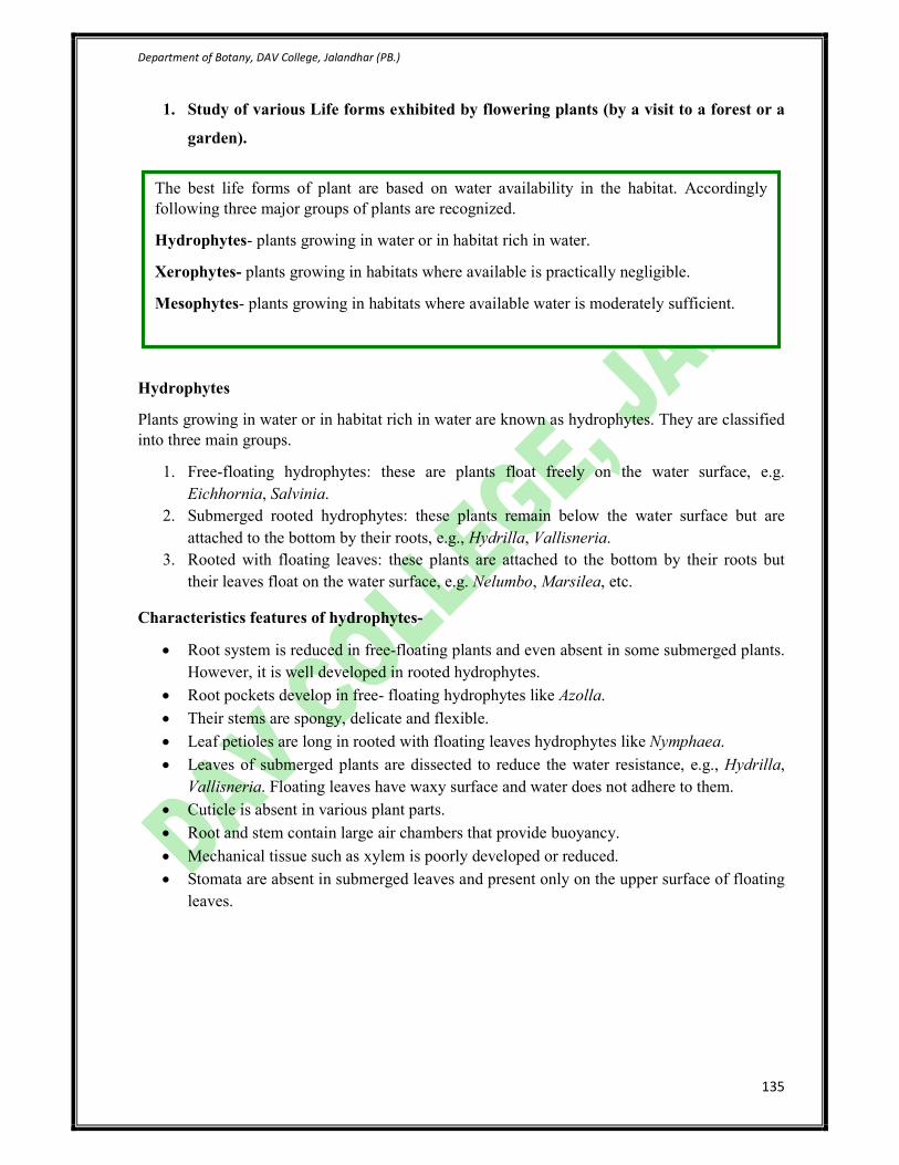
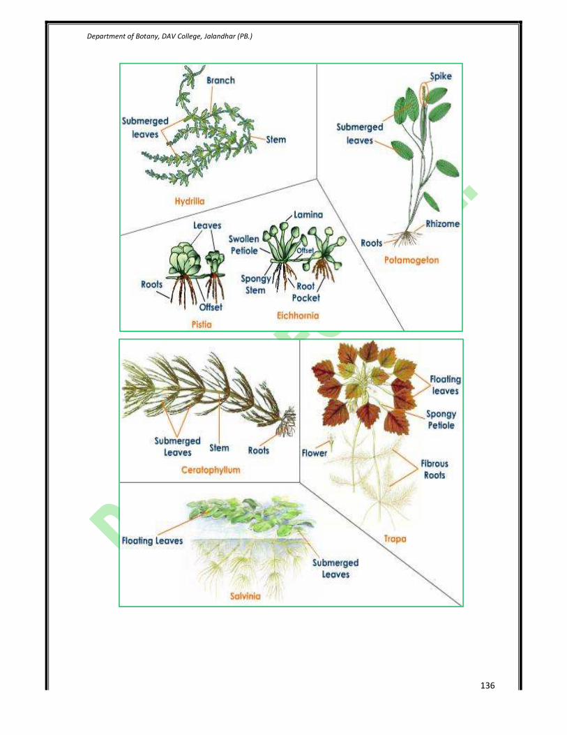
Hydrophytes
Plants growing in water or in habitat rich in water are known as hydrophytes. They are classified
into three main groups.
1. Free-floating hydrophytes: these are plants float freely on the water surface, e.g.
Eichhornia, Salvinia.
2. Submerged rooted hydrophytes: these plants remain below the water surface but are
attached to the bottom by their roots, e.g., Hydrilla, Vallisneria.
3. Rooted with floating leaves: these plants are attached to the bottom by their roots but
their leaves float on the water surface, e.g. Nelumbo, Marsilea, etc.
Characteristics features of hydrophytes-
• Root system is reduced in free-floating plants and even absent in some submerged plants.
However, it is well developed in rooted hydrophytes.
• Root pockets develop in free- floating hydrophytes like Azolla.
• Their stems are spongy, delicate and flexible.
• Leaf petioles are long in rooted with floating leaves hydrophytes like Nymphaea.
• Leaves of submerged plants are dissected to reduce the water resistance, e.g., Hydrilla,
Vallisneria. Floating leaves have waxy surface and water does not adhere to them.
• Cuticle is absent in various plant parts.
• Root and stem contain large air chambers that provide buoyancy.
• Mechanical tissue such as xylem is poorly developed or reduced.
• Stomata are absent in submerged leaves and present only on the upper surface of floating
leaves.
The best life forms of plant are based on water availability in the habitat. Accordingly
following three major groups of plants are recognized.
Hydrophytes- plants growing in water or in habitat rich in water.
Xerophytes- plants growing in habitats where available is practically negligible.
Mesophytes- plants growing in habitats where available water is moderately sufficient.
Department of Botany, DAV College, Jalandhar (PB.)
136

Department of Botany, DAV College, Jalandhar (PB.)
137
Plants growing in hot and dry conditions are known as xerophytes. These are two main groups of
such plants.
(i) Succulent Xerophytes.
(ii) Non-succulent xerophytes.
Characteristic feature of succulent xerophytes:
• These plants posses fleshy and stunted stem.
• Their cells contain large quantity of mucilage.
• They can store water in this tissue during the brief rainy season.
• The stem becomes thick green leaf-like structure to perform photosynthesis.
• Their leaves are small, deciduous or modified into spines to cut the rate of water loss
through transpiration.
• They posses shallow root system spreading horizontally.
• Epidermal cells of their leaves and stems are thick and highly cuticularized.
• Their stomata remain closed during hot days and open at night.

Department of Botany, DAV College, Jalandhar (PB.)
138
Characteristic features of non-succulent xerophytes:
• Their root system is well developed and fast growing.
• Their taproot system grows deep into the soil and reaches the water table.
• Their stem is generally hard and woody, covered with wax, silica, hair, etc.
• Their leaves are reduced or modified to cut the rate of water loss.
• Epidermal cells of the leaves are thick walled.
• The number of stomata per unit area is very less.
• Stomata are mostly sunken.
• The amount of mechanical tissue is higher.
• Presence of double or multiple epidermises to reduce transpiration.
Acacia sp Calotropis procera

Department of Botany, DAV College, Jalandhar (PB.)
139
MESOPHYES are those terrestrial plants which grow in moderately moist habitats and need
well-aerated soils. Broad-leaved trees growing in wet depressions, along lakes and rivers, are
mesophytes. They stand between hydrophytes and xerophytes and lack specific adaptations of
them. Some of the significant morpho-anatomical features of mesophytes are as follows:
1. Root system is well developed. Roots are fairly branched and contain root caps and root-
hairs.
2. Stems are generally aerial, solid and fairly branched.
3. Leaves are generally large, broad, and thin and varied in shapes. They are green and lack
hairy or waxy coatings.
4. In all aerial parts, cuticle is moderately developed.
5. Epidermis is well developed and has no chloroplasts.
6. Stomata generally present on both the surfaces of leaves.
7. Mesophyll in leaves is differentiated into palisade and spongy parenchyma, with many
intercellular spaces.
8. Well developed vascular and mechanical tissues are well differentiated.
9. Mesophytes may exhibit temporary wilting during noon hours.
Examples: Grass, Corn, Clover, Field crops, Goldenrod.

Department of Botany, DAV College, Jalandhar (PB.)
140
3. Study of tree like habit in cycads, bamboos, banana, and yucca and their comparison
with true trees as exemplified by conifers and dicotyledons.
Tree like habit in cycads
• Cycads look like a small tree.
• The stem is woody and hard enough but dwarf.
• Plant is symmetrical which supports a crown of
shiny dark leaves.
• Leaves are deep, semiglossy green about 50 to 150
cm long.
• Leaflets are long, strongly recuured with spiny
edges. The petiole has small protective barbs.
• Cycas posses apogeotropic branches of the lateral
roots, which are thick, blue, and repeatedly
branched. These coralloid roots posses anabaena in
them and do not show secondary growth.
• Plant is dioecious with separate male and female
plants.
• The microsporophylls are arranged in a compact
spiral around the woody central axis of male cone.
• The microsporophyll is woody, flattened wedge
shaped.
• The fertile part of microsporophyll bears numerous
microspores (pollen grains).
• The megasporophylls arise in spiral arrangement
towards the apex of female tree.
• Each megasporophyll is an orange-brown structure
and consists of a lower fertile portion bearing 1-3
pairs of ovules laterally.
• The whole megasporophyll is densely covered with
brown wooly hairs.
• The ovule is orange red; erect structure consisting of
mass of parenchyma called nucellus, covered by a
single integument differentiated into fleshy, middle
stony and inner fleshy layers.
• The internal structure of root, stem and leaf resembles
with the dicot trees.

Department of Botany, DAV College, Jalandhar (PB.)
141
Tree like habit in banana
• The banana plant is largest herbaceous
flowering plant.
• Banana has a stout central axis (false stem)
which grows up to a height of 10-15 feet.
• The stem is formed by the stiff sheathing
petioles which are rolled round one another.
• The true stem grows through the sheath and
forms the large compound spadix.
• In banana, the formation of inflorescence marks
the end of its growth.
• When the parent plant dies, the underground
suckers get separated and grow into new plants.
• The stem in banana is underground rhizome; the
aerial psuedoaerial stem (shaft) is composed of
long stiff leaf sheaths rolled round each other
due to which the plant of banana gives tree like
appearance.
• The leafs in banana are radical, simple, large,
petiole, alternate and have unicostate parallel
venation.
• Inflorescence in banana is represented by
compound spadix, covered with large violet pink
coloured bracts called spathe. The male flowers are
arranged towards the apex and the female towards
the base of spadix.
• The whole plant of banana represents a tree like
habit.
Department of Botany, DAV College, Jalandhar (PB.)
142
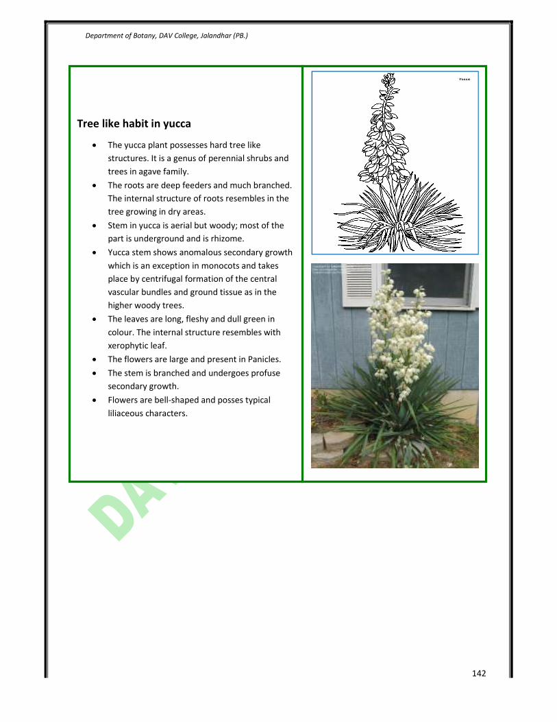
Tree like habit in yucca
• The yucca plant possesses hard tree like
structures. It is a genus of perennial shrubs and
trees in agave family.
• The roots are deep feeders and much branched.
The internal structure of roots resembles in the
tree growing in dry areas.
• Stem in yucca is aerial but woody; most of the
part is underground and is rhizome.
• Yucca stem shows anomalous secondary growth
which is an exception in monocots and takes
place by centrifugal formation of the central
vascular bundles and ground tissue as in the
higher woody trees.
• The leaves are long, fleshy and dull green in
colour. The internal structure resembles with
xerophytic leaf.
• The flowers are large and present in Panicles.
• The stem is branched and undergoes profuse
secondary growth.
• Flowers are bell-shaped and posses typical
liliaceous characters.
Department of Botany, DAV College, Jalandhar (PB.)
143
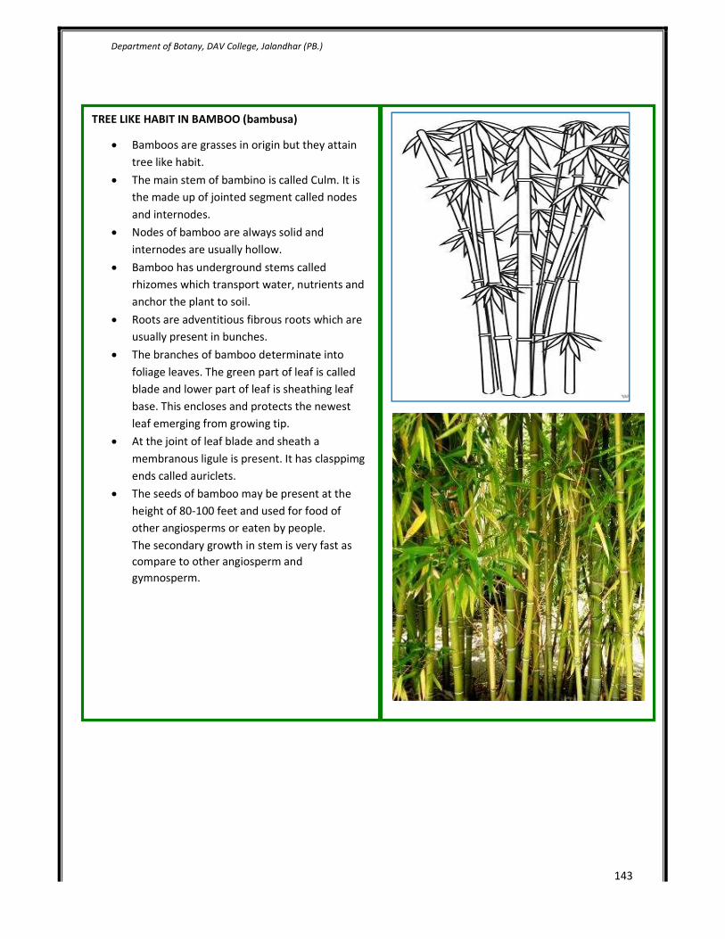
TREE LIKE HABIT IN BAMBOO (bambusa)
• Bamboos are grasses in origin but they attain
tree like habit.
• The main stem of bambino is called Culm. It is
the made up of jointed segment called nodes
and internodes.
• Nodes of bamboo are always solid and
internodes are usually hollow.
• Bamboo has underground stems called
rhizomes which transport water, nutrients and
anchor the plant to soil.
• Roots are adventitious fibrous roots which are
usually present in bunches.
• The branches of bamboo determinate into
foliage leaves. The green part of leaf is called
blade and lower part of leaf is sheathing leaf
base. This encloses and protects the newest
leaf emerging from growing tip.
• At the joint of leaf blade and sheath a
membranous ligule is present. It has clasppimg
ends called auriclets.
• The seeds of bamboo may be present at the
height of 80-100 feet and used for food of
other angiosperms or eaten by people.
The secondary growth in stem is very fast as
compare to other angiosperm and
gymnosperm.

Department of Botany, DAV College, Jalandhar (PB.)
144
4. L.S. shoot tip to study the cytohistological zonation and origin of leaf primordia.
Apical meristems are located at the tips of stems and at the tips of roots just behind the root cap.
The plant tissues that result from primary growth are called primary tissues. During periods of
growth, the cells of apical meristems divide and continually add more cells to the tips of a
seedling’s body. Thus, the seedling lengthens. Primary growth in plants is brought about by the
apical meristems. The elongation of the root and stem forms what is known as the primary plant
body, which is made up of primary tissues. The primary plant body comprises the young, soft
shoots and roots of a tree or shrub, or the entire plant body in some herbaceous plants. Both root
and shoot apical meristems are composed of delicate cells that need protection. The root apical
meristem is protected from the time it emerges by the root cap. Root cap cells are produced by
the root meristem and are sloughed off and replaced as the root moves through the soil. A variety
of adaptive mechanisms protect shoot apical meristem during germination (figure 38.4). The
epicotyl or hypocotyl (“stemlike” tissue above or below the cotyledons) may bend as the
seedling emerges to minimize the force on the shoot tip. In the monocots (a late evolving group
of angiosperms) there is often a coleoptile (sheath of tissue) that forms a protective tube around
the emerging shoot. Later in development, the leaf primordia cover the shoot apical meristem
which is particularly susceptible to desiccation. The apical meristem gives rise to three types of
embryonic tissue systems called primary meristems. Cell division continues in these partly
differentiated tissues as they develop into the primary tissues of the plant body. The three
primary meristems are the protoderm, which forms the epidermis; the procambium, which
produces primary vascular tissues (primary xylem and primary phloem); and the ground
meristem, which differentiates further into ground tissue, which is composed of parenchyma
cells. In some plants, such as horsetails and corn, intercalary meristems arise in stem internodes,
adding to the internode lengths. If you walk through a corn field (when the corn is about knee
high) on a quiet summer night, you may hear a soft popping sound. This is caused by the rapid
growth of intercalary meristems. The amount of stem elongation that occurs in a very short time
is quite surprising.

Department of Botany, DAV College, Jalandhar (PB.)
145
L.S of shoot tip
Department of Botany, DAV College, Jalandhar (PB.)
146
5. To study monopodial and sympodial types of branching in stems (especially
rhizomes)
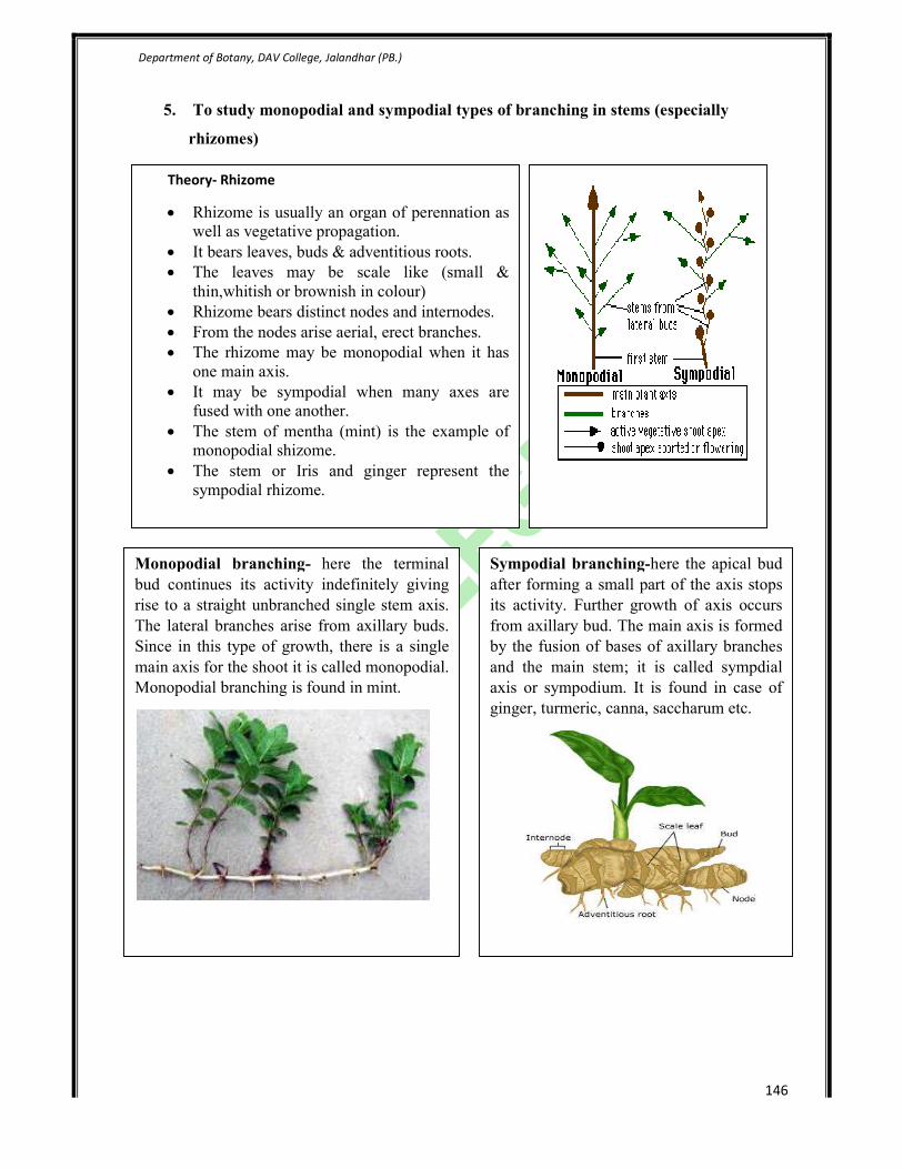
Theory- Rhizome
• Rhizome is usually an organ of perennation as
well as vegetative propagation.
• It bears leaves, buds & adventitious roots.
• The leaves may be scale like (small &
thin,whitish or brownish in colour)
• Rhizome bears distinct nodes and internodes.
• From the nodes arise aerial, erect branches.
• The rhizome may be monopodial when it has
one main axis.
• It may be sympodial when many axes are
fused with one another.
• The stem of mentha (mint) is the example of
monopodial shizome.
• The stem or Iris and ginger represent the
sympodial rhizome.
Monopodial branching- here the terminal
bud continues its activity indefinitely giving
rise to a straight unbranched single stem axis.
The lateral branches arise from axillary buds.
Since in this type of growth, there is a single
main axis for the shoot it is called monopodial.
Monopodial branching is found in mint.
Sympodial branching-here the apical bud
after forming a small part of the axis stops
its activity. Further growth of axis occurs
from axillary bud. The main axis is formed
by the fusion of bases of axillary branches
and the main stem; it is called sympdial
axis or sympodium. It is found in case of
ginger, turmeric, canna, saccharum etc.
Department of Botany, DAV College, Jalandhar (
6. Anatomy of primary and secondary growth in monocots and dicots using free hand
razor technique (Solanum
and Maize) hand sections or prepared slides.
Study of primary growth in a dicot
Anomalous structures
Study of anatomy of Nyctanthus arbortristis
1. Epidermis -consists of parenchymatous cells ,
layered, compctly arranged, interrupted by multicellular
hair ,which is the extension of the epidermal cells , it is
covered above by the layer of cuticle, stomata are also
prsent at intervals.
2. Cortex – this region is divisible into three distinc
parts
(a) Hypodermis- situsted below the epidermis,
consist of 4 to 5 layers of collenchyma.
(b) Cortex- consisting of large rounded, oval, thin,
walled intercellular spaces situated below the
hypodermis. Some isolated resin ducts are also
present in the general cortex.
(c) Endodermis- innermost layer or cortex having
barrel-shaped cells. Endodermis is single layered
having barrel shaped cells without casparian
strips. Cells are composed of single layered
parenchyma.
3. Pericycle is heterogenous having both
sclerenchymatous and parenchymatous cells.
Sclerenchymatous patch is present opposite to vascular
bundles and parenchymatous patch is present in between
the two vascular bundles and performs the storage
function.
4. Vascular bundles are present in the form of a ri
and called as the eustele, the bundles are conjoint,
collateral, endarch and open having well developed pith
on the inner side.
5. Pith cells are parenchymatous and are having storage
function.
College, Jalandhar (PB.)
Anatomy of primary and secondary growth in monocots and dicots using free hand
Solanum, Boerhaavia, Helianthus, Mirabilis, Nyctanthus
and Maize) hand sections or prepared slides.
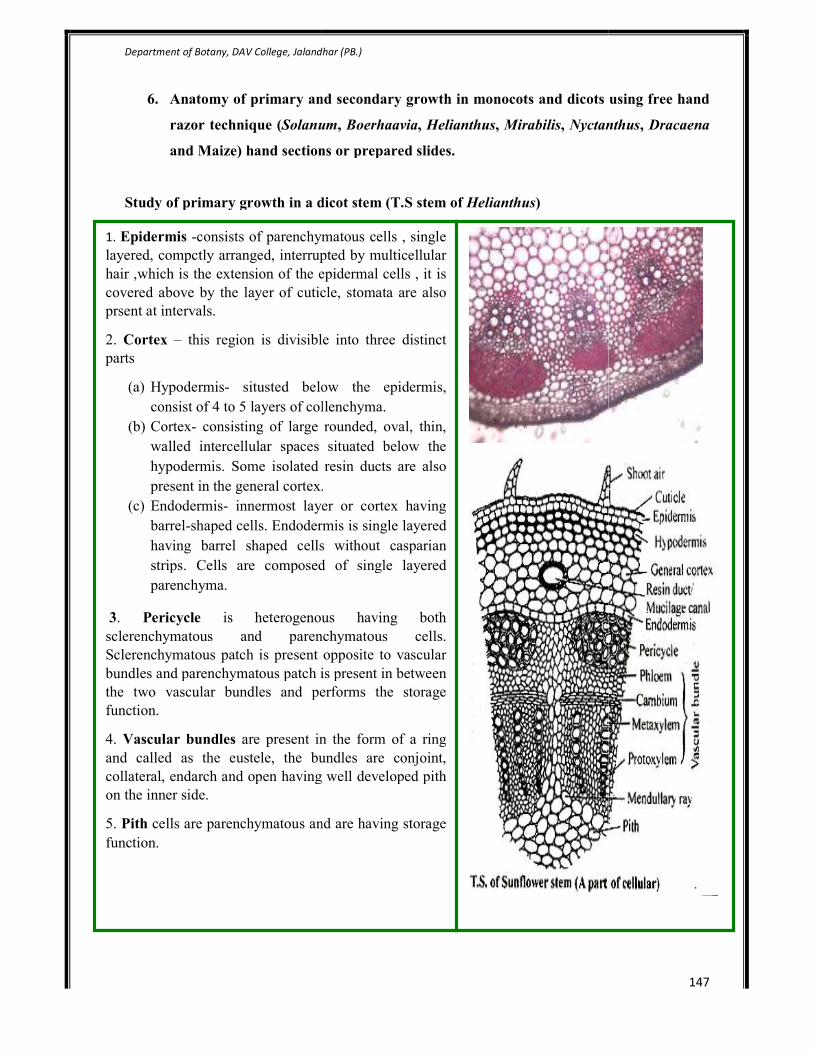
Study of primary growth in a dicot stem (T.S stem of Helianthus)
Study of anatomy of Nyctanthus arbortristis
consists of parenchymatous cells , single
layered, compctly arranged, interrupted by multicellular
hair ,which is the extension of the epidermal cells , it is
covered above by the layer of cuticle, stomata are also
this region is divisible into three distinct
situsted below the epidermis,
consist of 4 to 5 layers of collenchyma.
consisting of large rounded, oval, thin,
walled intercellular spaces situated below the
hypodermis. Some isolated resin ducts are also
cortex.
innermost layer or cortex having
shaped cells. Endodermis is single layered
having barrel shaped cells without casparian
strips. Cells are composed of single layered
is heterogenous having both
hymatous and parenchymatous cells.
Sclerenchymatous patch is present opposite to vascular
bundles and parenchymatous patch is present in between
the two vascular bundles and performs the storage
are present in the form of a ring
and called as the eustele, the bundles are conjoint,
collateral, endarch and open having well developed pith
cells are parenchymatous and are having storage
147
Anatomy of primary and secondary growth in monocots and dicots using free hand
Nyctanthus, Dracaena

Department of Botany, DAV College, Jalandhar (PB.)
148
To study the anatomy of primary growth in monocot stem (T.S. stem of maize)
To study the anatomy of Mirabilis jalapa
Epidermis
It is an outermost single layer of rectangular cells.
The cells of the epidermis are thickly cuticularised.
A few are present which lead into a sub-stomatal cavity
below
Ground tissue
All the tissues inside the epidermis form ground tissue.
It covers most of the section.
A few celled deep sclerenchymatous zones occur just
below the epidermis. It is interrupted at regular intervals
by patches of chlorenchyma.
The patches of chlorenchyma are bounded by
sclerenchyma on their sides and lower faces.
The stomata open only in thin walled parenchyma with
many intercellular spaces.
Vascular tissue system
Vascular tissue system is represented by numerous
vascular bundles.These are arranged in two series.
The bundles of the peripheral series are smaller than the
bundles of the inner series.
The bundles of the peripheral series are mostly
embedded in the sclerenchymatous patch situated below
the epidermis.
Vascular bundles are conjoint,collateral,endarch &
closed.
Each vascular bundles is almost completely enclosed by
a band of sclerenchyma.Bundle sheath is prominent at
the upper and the lower extremities of the vascular
bundle.
The xylem elements are arranged in almost Y-shaped
organisation which occupies the lower region of the
vascular bundle.
Metxylem elements are large and the smaller
protoxylem elements are situated near the inner face of
the vascular bundle
The phloem occurs in the peripheral region of the
bundle. It consists of sieve tubes and companion cells.
There is a hollow cylinder in the centre of the axis.

Department of Botany, DAV College, Jalandhar (PB.)
149
Internal structure of Stem of Boerhaavia diffusa
1. In transverse section, the stem shows a wavy outline.
Outermost layer is epidermis composed of single
layered compactly arranged parenchymatous cells with
no intercellular spaces. Many epidermal cells bear
multicellular hair, which are not the outgrowth of
epidermal cells.
2. Next to epidermis is cortex which is differentiated
into two zones, next to epidermis is collenchymatous
cells, next to collenchyma cells lies the zone of
chlorenchyma cells. It is made up of 4-6 layers of cells.
The are circular, oval or even polygonal and have
abundant chloroplasts.
3. Innermost layer of cortex constitutes the
endodermis. It is clearly distinguishable. It is made up
of thick walled tubular cells with no intercellular
spaces.
4. Next to endodermis lies the zone of parenchymatous
cells of pericycle which are interrupted with
sclerenchymatous cells in between in the form of
patches.
5. Vascular bundles are present in three rings. The
outermost ring have 15-20 small bundles, this ring
surrounds a middle ring of 6-14 vascular bundles.
These are smaller in size and oval or rounded in shape.
In the innermost ring are present two larger vascular
bundles which lie in pith, these are called das
medullary bundles. Of all the bundles these are the
largest in size and are oval in shape. These bundles are
fully developed. The central bundles are enveloped in a
thin walled sheath and lie opposite to each other with
xylem facing towards the center and phloem facing
outwards. The vascular bundles are conjoint, collateral,
endarch and open.
Department of Botany, DAV College, Jalandhar (PB.)
150
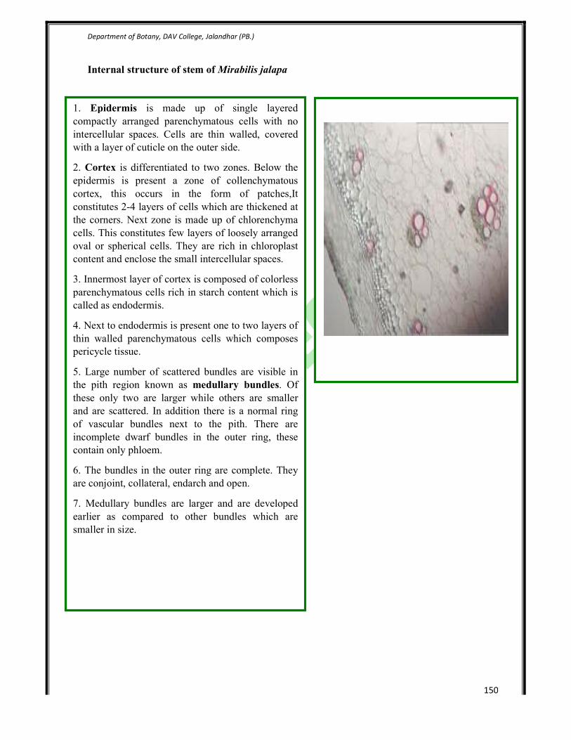
Internal structure of stem of Mirabilis jalapa
1. Epidermis is made up of single layered
compactly arranged parenchymatous cells with no
intercellular spaces. Cells are thin walled, covered
with a layer of cuticle on the outer side.
2. Cortex is differentiated to two zones. Below the
epidermis is present a zone of collenchymatous
cortex, this occurs in the form of patches,It
constitutes 2-4 layers of cells which are thickened at
the corners. Next zone is made up of chlorenchyma
cells. This constitutes few layers of loosely arranged
oval or spherical cells. They are rich in chloroplast
content and enclose the small intercellular spaces.
3. Innermost layer of cortex is composed of colorless
parenchymatous cells rich in starch content which is
called as endodermis.
4. Next to endodermis is present one to two layers of
thin walled parenchymatous cells which composes
pericycle tissue.
5. Large number of scattered bundles are visible in
the pith region known as medullary bundles. Of
these only two are larger while others are smaller
and are scattered. In addition there is a normal ring
of vascular bundles next to the pith. There are
incomplete dwarf bundles in the outer ring, these
contain only phloem.
6. The bundles in the outer ring are complete. They
are conjoint, collateral, endarch and open.
7. Medullary bundles are larger and are developed
earlier as compared to other bundles which are
smaller in size.

Department of Botany, DAV College, Jalandhar (PB.)
151
Internal structure of stem of Nyctanthes arbor-tristis
The stem of this plant has prominent angles and reveals a
quadrangular outline in a transverse section.
1. Epidermis is single layered parenchymatous with a
compact arrangement. The cells are covered by a
continuous layer of thick cuticle. Multicellular hair
arises from the epidermal tissue.
2. Cortex is followed by epidermis having a few
layered collenchymatous tissue towards outside and
oval, rounded cells on the inner side. The main function
of cortex is storage.
3. Endodermis and pericycle are not distinct.
4. Normal vascular bundles occur in the center in the
form of a ring, the bundles are conjoint, collateral,
endarch and open, in addition to the normal ring of
vascular bundles there is present four inversely oriented
vascular bundles in the cortex region at the four corners
of the stem.
5. These cortical bundles always get restricted to the
four prominent angles of the stem. The phloem in such
bundles is restricted towards the inner side and xylem
towards the outer side. The bundles have the exarch
condition.
6. The cambium present in the cortical bundles adds a
small amount of secondary vascular tissue sin a normal
manner.
7. The cambium in normal ring in the center also
functions in a normal manner and produces secondary
phloem towards outer side and secondary xylem
towards inner side.
8. Pith- In the center of the stem there is broad pith
which is composed of thin walled cells.
Department of Botany, DAV College, Jalandhar (PB.)
152
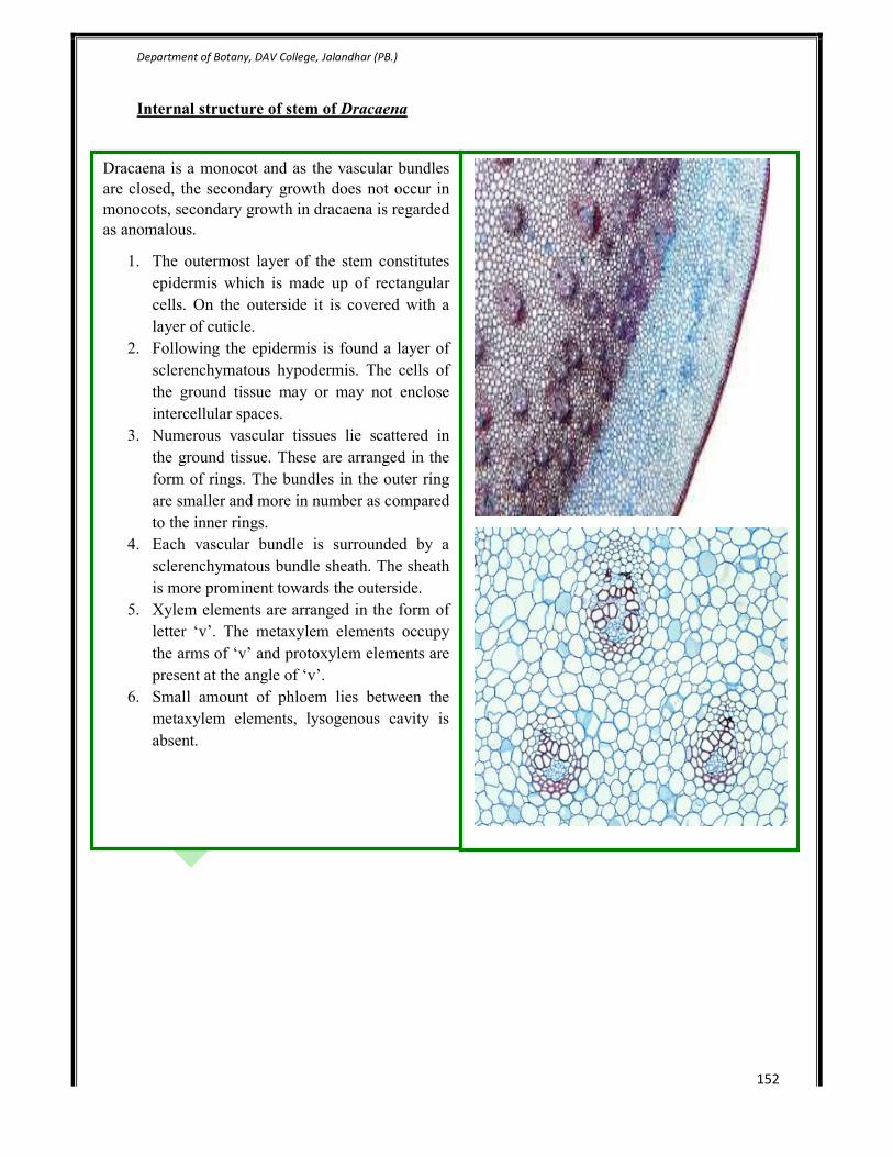
Internal structure of stem of Dracaena
Dracaena is a monocot and as the vascular bundles
are closed, the secondary growth does not occur in
monocots, secondary growth in dracaena is regarded
as anomalous.
1. The outermost layer of the stem constitutes
epidermis which is made up of rectangular
cells. On the outerside it is covered with a
layer of cuticle.
2. Following the epidermis is found a layer of
sclerenchymatous hypodermis. The cells of
the ground tissue may or may not enclose
intercellular spaces.
3. Numerous vascular tissues lie scattered in
the ground tissue. These are arranged in the
form of rings. The bundles in the outer ring
are smaller and more in number as compared
to the inner rings.
4. Each vascular bundle is surrounded by a
sclerenchymatous bundle sheath. The sheath
is more prominent towards the outerside.
5. Xylem elements are arranged in the form of
letter ‘v’. The metaxylem elements occupy
the arms of ‘v’ and protoxylem elements are
present at the angle of ‘v’.
6. Small amount of phloem lies between the
metaxylem elements, lysogenous cavity is
absent.

Department of Botany, DAV College, Jalandhar (PB.)
153

Department of Botany, DAV College, Jalandhar (PB.)
154
7. Structure of secondary xylem and phloem.
XYLEM
Xylem tissue functions in both water transport and mechanical support. In non-angiosperm
tracheophytes, tracheids serve both purposes; in most angiosperms, the xylem contains both
vessel elements, which have a larger diameter and are specialized for water transport, and fibers
for mechanical strength.
Xylem cells commonly have cell walls impregnated with lignin and reinforced with spiral or
ring-like thickenings that project into the lumen of the cell. Both features reinforce the cells for
mechanical support.
Xylem cells are dead and empty of cell contents at maturity and essentially form tubes for water
transport. However, plants have no pumps to move water through these hollow tubes. Thus water
molecules are pulled in long, hydrogen-bonded chains from rhizome to leaf. If the chain breaks,
for example if a bubble forms in a xylem cell, the involved cells lose their function and cannot be
repaired. Since xylem can be modeled as physical pipes following hydrodynamic principles, the
water-transport ability of ancient plants can be easily calculated. Parenchyma cells are often
present in xylem tissue, where they help maintain water balance and carry out metabolism within
the tissue. Because more than one cell type is present in xylem, it is called a complex tissue.
Fibres and fibre-tracheids- FIBRE is defined as ‘a general term of convenience in wood
anatomy for any long, narrow cell of wood or bast other than vessels sieve tubes and
parenchyma. Note: often further qualified as wood fibres or bast fibres; the former including
both the tracheids of gymnosperms (softwoods) and the libriform wood fibres and fibre-tracheids
of woody angiosperms (hardwoods). Also used loosely for wood elements in general’.
Department of Botany, DAV College, Jalandhar (PB.)
155
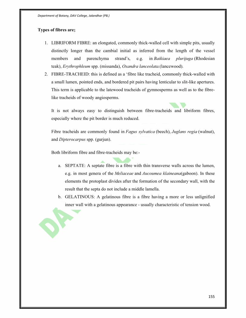
Types of fibres are;
1. LIBRIFORM FIBRE: an elongated, commonly thick-walled cell with simple pits, usually
distinctly longer than the cambial initial as inferred from the length of the vessel
members and parenchyma strand’s, e.g. in Baikiaea plurijuga (Rhodesian
teak), Erythrophleum spp. (missanda), Oxandra lanceolata.(lancewood).
2. FIBRE-TRACHEID: this is defined as a ‘fibre like tracheid, commonly thick-walled with
a small lumen, pointed ends, and bordered pit pairs having lenticular to slit-like apertures.
This term is applicable to the latewood tracheids of gymnosperms as well as to the fibre-
like tracheids of woody angiosperms.
It is not always easy to distinguish between fibre-tracheids and libriform fibres,
especially where the pit border is much reduced.
Fibre tracheids are commonly found in Fagus sylvatica (beech), Juglans regia (walnut),
and Dipterocarpus spp. (gurjun).
Both libriform fibre and fibre-tracheids may be:-
a. SEPTATE: A septate fibre is a fibre with thin transverse walls across the lumen,
e.g. in most genera of the Meliaceae and Aucoumea klaineana(gaboon). In these
elements the protoplast divides after the formation of the secondary wall, with the
result that the septa do not include a middle lamella.
b. GELATINOUS: A gelatinous fibre is a fibre having a more or less unlignified
inner wall with a gelatinous appearance - usually characteristic of tension wood.
Department of Botany, DAV College, Jalandhar (PB.)
156
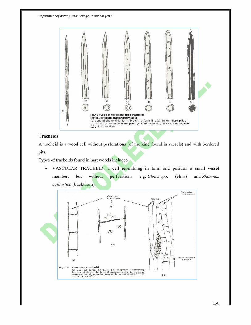
Tracheids
A tracheid is a wood cell without perforations (of the kind found in vessels) and with bordered
pits.
Types of tracheids found in hardwoods include:-
• VASCULAR TRACHEID: a cell resembling in form and position a small vessel
member, but without perforations e.g. Ulmus spp. (elms) and Rhamnus
cathartica (buckthorn).

Department of Botany, DAV College, Jalandhar (PB.)
157
• VASICENTRIC TRACHEID: a short, irregularly-formed tracheid in the immediate
proximity of a vessel and not forming part of a definite axial row, e.g. commonly found
in Quercus spp. (oaks), Castanea sativa (sweet chestnut). Eucalyptus spp.
and Calophyllum spp. (bitangor).

Department of Botany, DAV College, Jalandhar (PB.)
158
PHLOEM
Phloem tissue transports photosynthetic products, other organic molecules (e.g., plant hormones
and waste products), and soluble nutrients throughout the plant. Unlike xylem, phloem is alive at
maturity, but usually with a much reduced cell contents and no nucleus. This is logical because
movement of material through phloem tissue relies on solute gradients and some active transport
that require the activity of living cells. In non-angiosperm seed plants phloem elements consist
mostly of sieve cells (Figure 1.3), while angiosperms have sieve tube cells in association with
parenchymatous companion cells. Phloem fibers also provide some mechanical support. Phloem
cells are commonly unlignified so they do not preserve as readily as xylem.
Growth rings in wood.
Wood is secondary xylem produced by growth of the vascular cambium tissue. At the very
center is the pith. In some trees, this is much softer and possibly a different color than the
surrounding heartwood. Heartwood is made up of dead cells that no longer serve any purpose
except to support the tree. Next is the sapwood, which carries water, minerals, and plant sugars
between the roots and the leaves. This is often lighter in color than the heartwood. Outside the
sapwood, close to the surface, is the cambium, a thin layer of living cells. These cells

Department of Botany, DAV College, Jalandhar (PB.)
159
manufacture the wood as they grow. The cambium is covered by a protective layer of bark. The
cambium grows rapidly at the beginning of each growing season, creating light
colored springwood. As the climate warms, it slows down and produces darker summerwood.
This later growth is somewhat denser and harder than the early springwood. As the weather turns
cold, the cambium becomes dormant until the next spring. This cycle produces
distinctive growth rings. The number of annual rings corresponds to the age of plant.
Dendrochronology is the branch of anatomy which deals with determining the age of plant.
Growth rings vary in width as a result of differing climatic conditions; in temperate climates, a
ring is equivalent to one year's growth. Certain conducting cells form rays that carry water and
dissolved substances radially across the xylem. Bark comprises the tissues outside the vascular
cambium, including secondary phloem (which transports food made in the leaves to the rest of
the tree), cork-producing cells (cork cambium), and cork cells. The outer bark, composed of dead
tissue, protects the inner region from injury, disease, and desiccation.
Cross section of a tree trunk.
Department of Botany, DAV College, Jalandhar (PB.)
160
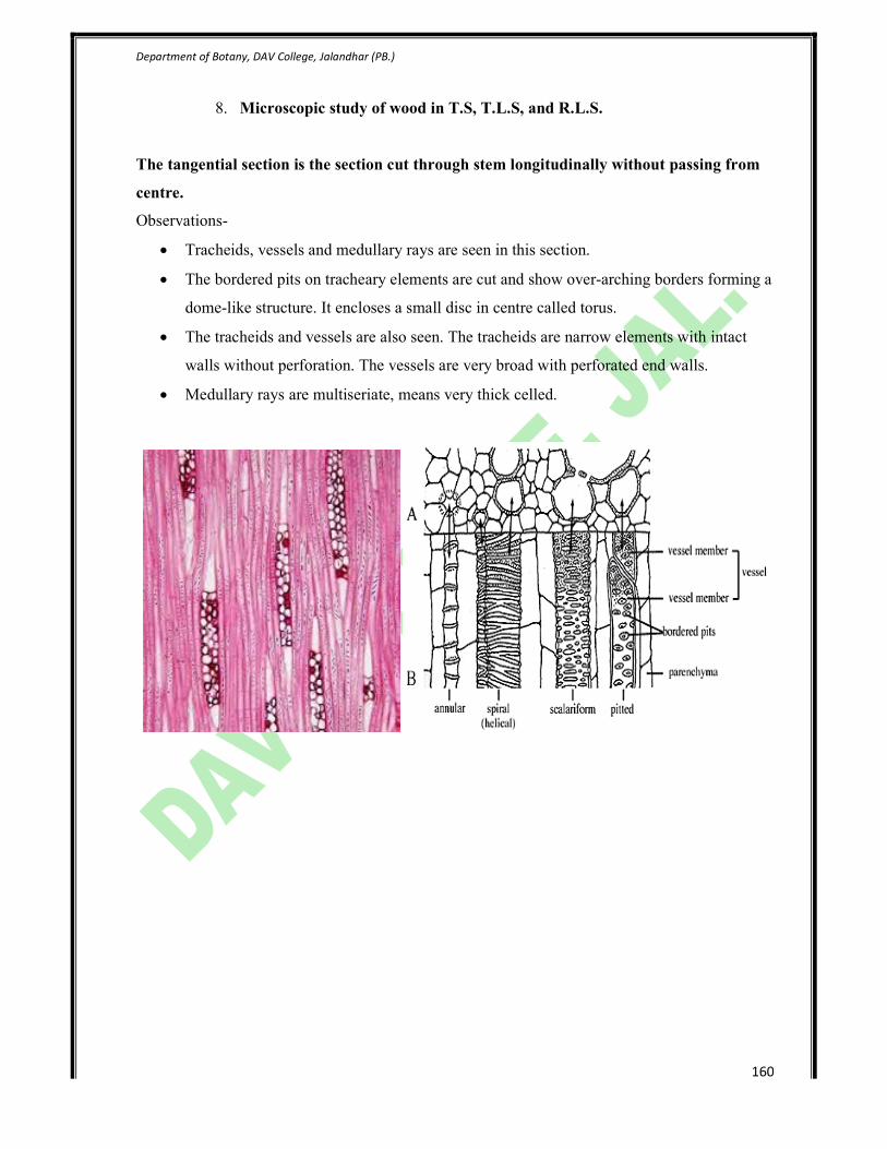
8. Microscopic study of wood in T.S, T.L.S, and R.L.S.
The tangential section is the section cut through stem longitudinally without passing from
centre.
Observations-
• Tracheids, vessels and medullary rays are seen in this section.
• The bordered pits on tracheary elements are cut and show over-arching borders forming a
dome-like structure. It encloses a small disc in centre called torus.
• The tracheids and vessels are also seen. The tracheids are narrow elements with intact
walls without perforation. The vessels are very broad with perforated end walls.
• Medullary rays are multiseriate, means very thick celled.

Department of Botany, DAV College, Jalandhar (PB.)
161
The radial longitudinal section is obtained by cutting stem in such away so that the
longitudinal cut pass through centre. (RLS)
Observations-
• It shows the presence of secondary xylem consisting of tracheids, vessels and medullary
rays.
• Tracheids are narrow with closed end walls but the radial walls show bordered pits.
• Bordered pits are circular as surrounded by special cellulose thickenings.
• Vessels are distinguishable from tracheids being broader and perforated end walls.
• Medullary rays are multiserate made up of ray tracheids and ray parenchyma.
• Ray tracheids are present on both sides of medullary ray cells.
• The cells of the remaining tissue of xylem are called ray parenchyma which is thin, broad
and mostly living.

Department of Botany, DAV College, Jalandhar (
Portions of pinewood sections: (1) cross section, (2) radial section, (3) tangential section
(a) edge ot annual ring, (b) summerwood, (c) Springwood, (d) new series of tracheids, (e)
heterogeneous medullary ray composed of ray
tracheids (f) with small bordered pits, and (g) composed ofparenchyma cells with large
windowlike pits, (h) resin canal (epithelial cells lining it clearly visible), (i) cells of parenchyma
surroundi-ng resin canal, (j)bordered pits, (k) medullary ray with horizo
College, Jalandhar (PB.)
Portions of pinewood sections: (1) cross section, (2) radial section, (3) tangential section
(a) edge ot annual ring, (b) summerwood, (c) Springwood, (d) new series of tracheids, (e)
heterogeneous medullary ray composed of ray
(f) with small bordered pits, and (g) composed ofparenchyma cells with large
windowlike pits, (h) resin canal (epithelial cells lining it clearly visible), (i) cells of parenchyma
ng resin canal, (j)bordered pits, (k) medullary ray with horizontal resin canal.
162
Portions of pinewood sections: (1) cross section, (2) radial section, (3) tangential section
(a) edge ot annual ring, (b) summerwood, (c) Springwood, (d) new series of tracheids, (e)
(f) with small bordered pits, and (g) composed ofparenchyma cells with large
windowlike pits, (h) resin canal (epithelial cells lining it clearly visible), (i) cells of parenchyma
ntal resin canal.

Department of Botany, DAV College, Jalandhar (PB.)
163
9. Field study of diversity in the leaf shape, size, thickness, surface properties.
Morphology of leaf

Department of Botany, DAV College, Jalandhar (PB.)
164

Department of Botany, DAV College, Jalandhar (PB.)
165
VENATION-

Department of Botany, DAV College, Jalandhar (PB.)
166

Department of Botany, DAV College, Jalandhar (PB.)
167
Types of leaf-

Department of Botany, DAV College, Jalandhar (PB.)
168

Department of Botany, DAV College, Jalandhar (PB.)
169

Department of Botany, DAV College, Jalandhar (PB.)
170

Department of Botany, DAV College, Jalandhar (
College, Jalandhar (PB.)
171

Department of Botany, DAV College, Jalandhar (PB.)
172
10. Internal structure of leaf.
Internal structure of leaf of Mangifera indica
Epidermis-
• Lower and upper epidermis is single layered.
• The cells are barrel-shaped and compactly arranged.
• Upper epidermis has a thick cuticle and lacks stomata.
• Lower epidermis has thin cuticle and stomata are present.
Mesophyll-
• It is differentiated into spongy and palisade parenchyma.
• Palisade occurs below upper epidermis in two layers, with parenchyma near the larger
vascular bundle. The cells are compactly arranged, long and tubular and chloroplasts are
present.
• Spongy parenchyma forms rest of the tissue. The cells are small, varied in shapes and sizes,
loosely arranged and enclose small air spaces.
• A few air spaces lead to the stomata openings which form sub-stomatal cavity. Numerous
chloroplasts are present near the walls.
Vascular tissue-
• It consists of one large vascular bundle in the midrib and numerous small vascular bundles in
the wings.
• Each bundle is conjoint, collateral and closed and surrounded by a parenchymatous bundle
sheath. Larger vascular bundle has an extensive bundle sheath that extends both toward
lower and upper epidermis.
• Metaxylem is situated towards the lower epidermis and protoxylem towards the upper
epidermis.
• Phloem of the vascular bundle is directed towards lower epidermis.

Department of Botany, DAV College, Jalandhar (PB.)
173
T.S of leaf of Nerium (xerophytic leaf)
• In transverse section, the leaf of Nerium shows multilayered upper and lower epidermis. The
outer layers of epidermis consist of thick walled cells and covered externally by thick cuticle.
The lower two layers can be regarded as hypodermis.
• Stomata is present only on the lower epidermis .these cavities also bear multicellular hairs or
trichomes which protect the stomata.
• Mesophyll is differentiated into multilayered palisade and spongy parenchyma.
• The vascular bundle of midrib region is larger in comparison to the vascular bundle of the
wings. They are collateral and closed with xylem towards upper epidermis and phloem
towards lower epidermis. Each vascular bundle is surrounded by parenchymatous sheath.
•

Department of Botany, DAV College, Jalandhar (
Study of internal structure of monocot leaf
(isobilateral leaf)
Epidermis-Leaf is bounded by lower and upper
epidermal layers.both layers are thickly
cuticularised.Stomata are present in both epidermal
layers.A few large, empty and colourless bullif
cells occur in upper epidermis.
Mesophyll-It is not differentiated into palisade and
spongy parenchyma.it occurs between upper and
lower epidermis.The cells are isodiametricand
containnumerous chloroplasts. These are compactly
arranged and leave only a few intercellular spaces.
Vascular tissue-There are numerous vascular
bundles of variable sizes arranged in a parallel series.
Each bundle is collateral and closed. There is distinct
parenchymatous bundle sheath. The cells of the
sheath posses plastids and starch grains.
A patch of sclerenchyma each is present above and
below the larger vascular bundles and extends up to
the upper and lower epidermal layers respectively.
Large vascular bundles have distinct and more
College, Jalandhar (PB.)
Study of internal structure of monocot leaf
Leaf is bounded by lower and upper
epidermal layers.both layers are thickly
cuticularised.Stomata are present in both epidermal
layers.A few large, empty and colourless bulliform
It is not differentiated into palisade and
spongy parenchyma.it occurs between upper and
lower epidermis.The cells are isodiametricand
containnumerous chloroplasts. These are compactly
few intercellular spaces.
There are numerous vascular
bundles of variable sizes arranged in a parallel series.
Each bundle is collateral and closed. There is distinct
parenchymatous bundle sheath. The cells of the
d starch grains.
A patch of sclerenchyma each is present above and
below the larger vascular bundles and extends up to
the upper and lower epidermal layers respectively.
Large vascular bundles have distinct and more
174
Department of Botany, DAV College, Jalandhar (PB.)
175
11. Structure and development of stomata (using epidermal peel of leaf).
Materials- Leaves of Tagetes, Tridax, Brassica, Ocimum, Tradescantia, cover slips, microscope,
water, safranin, glycerine, needles, forceps etc.
Method-
• Tear the leaf suddenly and with force with lower epidermis upwards.
• A thin membranous lower epidermis gets separated near the broken edges. Pull this
into astrip with forceps or fingers.
• The strip is stained with 1% aqueous safranin, washed in water and then mounted in
glycerine.
Observations-
Stomata- they are the part of epidermal tissue system and present on upper epidermis or lower
epidermis or both surfaces.
Structure- the size of stomata ranges between 7-38um in length and 3-12um in breath. Each
stoma has a pore bounded by two small specialized green nucleated living epidermal cells called
guard cells. The guard cells contain chloroplast, nucleus, mitochondria, vacuoles, starch, ER,
ribosome, microbodies. They are connected with adjacent epidermal cells through
plasmodesmata. Because of their much small size, they are rapidly influenced by turgor changes.
They may be kidney shaped or dumbbell shaped. Kidney shaped guard cells is semilunar like and
thick on inner (concave) side and thin on convex side. In dumbbell shaped, guard cell is linear
dumbell like. Their expanded (bulged) ends are thin walled while the middle part is highly thick
walled.
Types of stomata-
(A) On the basis of the number and orientation of subsidiary cells, 7 types of stomata are
there:-
• Anomocytic type- subsidiary cells absent and the cells surrounding two guard cells are
few and similar to epidermal cells. Eg. Ranunculaceae, Papeveraceae,Nyctaginaceae.
• Anisocytic type-two guard cells of each stoma are surrounded by 3 subsidiary cells (one
is smaller than the other two).eg. Cruciferae, Solanaceae, Umbelliferae.
• Paracytic type- two guard cells of each stoma are surrounded by two or more subsidisry
cells which lie parallel to the guard cells. Eg. Magnoliaceae, Rubiaceae.
• Diacytic type- the two subsidiary cells lie at right angle to the guard cells.eg.
Magnoliaceae, Labiateae, Acanthaceae.
• Actinocytic type- subsidiary cells are 4 or more elongated radially to stoma.

Department of Botany, DAV College, Jalandhar (PB.)
176
• Cyclocytic type- subsidiary cells are 4 or more, arranged in a close ring around the
stoma.
• Graminaceous type-in monocots, stomata are dumbell shaped, surrounded by subsidiary
cells lying parallel to the long axis of guard cells.
(B)-On the basis of development of guard cells and subsidiary cells, stomata fall into two
categories-
• Haplochellic type- guard cells are derived from stomatal initial cell but subsidiary cells
are formed by the modifications of adjacent epidermal cells.eg. Tobacco.
• Syndetochellic type- here both guard cells and subsidiary cells are formed from a
common stomatal initial.eg. Sugarcane.
Department of Botany, DAV College, Jalandhar (PB.)
177
12. Study of anatomy of root. Primary and secondary structure.
Study of Internal structure of Dicot root
Study of internal structure of monocot root
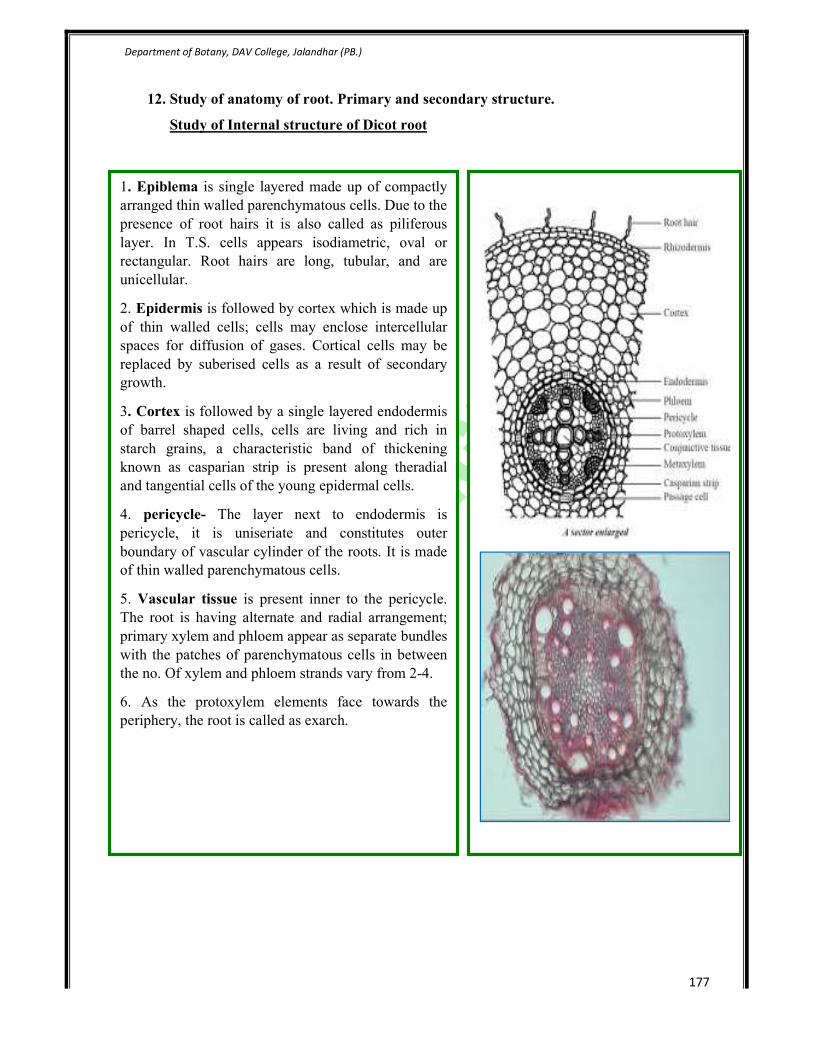
1. Epiblema is single layered made up of compactly
arranged thin walled parenchymatous cells. Due to the
presence of root hairs it is also called as piliferous
layer. In T.S. cells appears isodiametric, oval or
rectangular. Root hairs are long, tubular, and are
unicellular.
2. Epidermis is followed by cortex which is made up
of thin walled cells; cells may enclose intercellular
spaces for diffusion of gases. Cortical cells may be
replaced by suberised cells as a result of secondary
growth.
3. Cortex is followed by a single layered endodermis
of barrel shaped cells, cells are living and rich in
starch grains, a characteristic band of thickening
known as casparian strip is present along theradial
and tangential cells of the young epidermal cells.
4. pericycle- The layer next to endodermis is
pericycle, it is uniseriate and constitutes outer
boundary of vascular cylinder of the roots. It is made
of thin walled parenchymatous cells.
5. Vascular tissue is present inner to the pericycle.
The root is having alternate and radial arrangement;
primary xylem and phloem appear as separate bundles
with the patches of parenchymatous cells in between
the no. Of xylem and phloem strands vary from 2-4.
6. As the protoxylem elements face towards the
periphery, the root is called as exarch.

Department of Botany, DAV College, Jalandhar (
Study of Internal structure of
Epidermis
Made up of thin walled parenchymatous cells
arranged without intercellular spaces.
Cortex-shows three sub zones
Exodermis- composed of one to two layers of
thick walled, dead, suberised cells. It helps in
preventing the exit of water from the root tissues.
General cortex- made up of several rows of thin
walled parenchyma showing intercel
The cells of cortex help in the storage of food
material.
Endodermis-Barrel shaped cells arranged
compactly in single layer without leaving any
intercellular spaces. The radial and transverse
walls are wrapped by casparian bands.
Stele-the stele shows three sub zones.
Pericycle-the cells are thin walled,
parenchymatous and compact without
intercellular spaces.
Vascular Tissue-Primary strands of xylem and
phloem are found separately on different radii,
known as “radial” vascular bundles.The xyl
exarch and usually in polyarch condition.
Pith-Made up of thin walled parenchyma, which
primarily helps in the storage of food.
College, Jalandhar (PB.)
Study of Internal structure of Monocot root
Made up of thin walled parenchymatous cells
arranged without intercellular spaces.
composed of one to two layers of
thick walled, dead, suberised cells. It helps in
preventing the exit of water from the root tissues.
made up of several rows of thin
walled parenchyma showing intercellular spaces.
The cells of cortex help in the storage of food
Barrel shaped cells arranged
compactly in single layer without leaving any
intercellular spaces. The radial and transverse
walls are wrapped by casparian bands.
ele shows three sub zones.
the cells are thin walled,
parenchymatous and compact without
Primary strands of xylem and
phloem are found separately on different radii,
known as “radial” vascular bundles.The xylem is
exarch and usually in polyarch condition.
Made up of thin walled parenchyma, which
primarily helps in the storage of food.
178
Department of Botany, DAV College, Jalandhar (PB.)
179
13. Examination of a wide range of flowers available in the locality and methods of their
pollination.
Pollination is the process by which pollen is transferred from the anther (male part) to the
stigma (female part) of the plant, thereby enabling fertilization and reproduction.This takes
place in the angiosperms, the flower bearing plants.A successful angiosperm pollen grain
(gametophyte) containing the male gametes gets transported to the stigma, where it
germinates and its pollen tube grows down the style to the ovary. Its two gametes travel
down the tube to where the gametophyte containing the female gametes are held within the
carpel. One nucleus fuses with the polar bodies to produce the endosperm tissues, and the
other with the ovule to produce the embryo, Hence the term: "double fertilization".The
receptive part of the carpel is called a stigma in the flowers of angiosperms. The receptive
part of the gymnosperm ovule is called the micropyle. Pollination is a necessary step in the
reproduction of flowering plants, resulting in the production of offspring that are genetically
diverse.
Generally pollen grains have a tough protective coat which prevents them from drying up.
Since pollen grains are light, they can be carried by wind or water. Insects visit flowers and
carry away pollen on their bodies. Some of the pollen lands on the stigma of a flower of the
same kind. The transfer of pollen from the anther to the stigma of a flower is
called pollination. If the pollen lands on the stigma of the same flower it is calledself-
pollination. When the pollen of a flower lands on the stigma of another flower of the same
plant, or that of a different plant of the same kind, it is called cross-pollination.
1) Anemophily:
-Wind pollination is called anemophily, and flower is called anemophilous flower, e.g. Monocot
grass, Maize, Jowar etc.
-It is most primitive type of pollination. There is more wastage of pollen grains in anemophily.
Adaptations in anemophilous flowers:

Department of Botany, DAV College, Jalandhar (PB.)
180
Flowers are small and inconspicuous.
Flowers are colourless, odourless and nectarless.
Flowers are well exposed on plants.
They produce a large number of dry, light and smooth walled pollen grains.
The anthers are versatile.
Stigma is feathery and branched
Style and stigma is long.

Department of Botany, DAV College, Jalandhar (PB.)
181
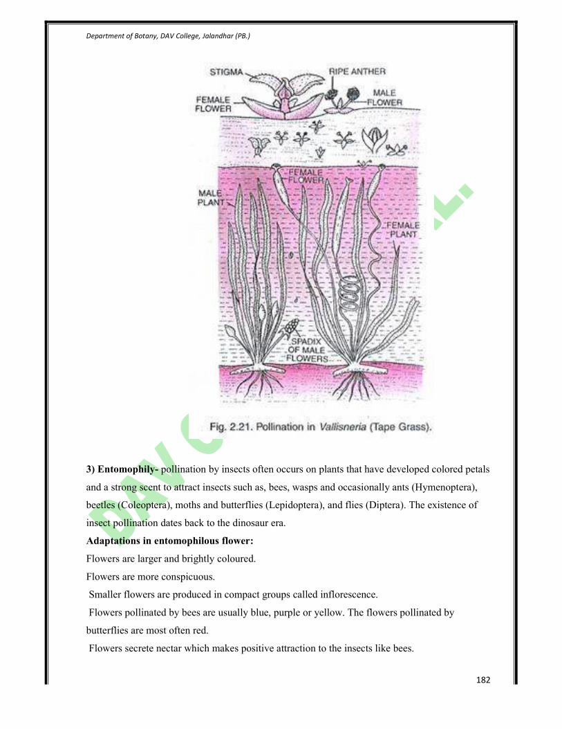
2) Hydrophily:
-Pollination brought about by water is called hydrophily and flower is called hydrophilous
flower.
- It is found in aquatic plants like Vallisneria, Zostera, Ceratophyllum, Hydrilla etc.
Type of hydrophily:
a)Epihydrophily:
-Pollination takes place on the surface of water,is called epihydrophily.
- Vallisneria is a dioecious plant and pollination occurs on the surface of water.
The male flowers get detached from the' plant and float on the water surface.
Female flowers are produced on long coiled pedicel and projecting above the water surface.
Male flowers come in contact with the stigmas cause pollination.
After pollination long pedicel of female flower coils and brings it back to lower level of water
where the fruit is formed.
b) Hypohydrophily:
Pollination takes place below the water surface is called hypohydrophily. It occurs completely
submerged plants like ceratophyllum and zostera.
Plant bears elongated, needle like pollen grains without exine. pollen grains have the same
specific gravity as that of water. Therefore pollens can float below the surface of water.When
pollens reach the stigma; they coiled around it and germinate.
Adaptations in hydrophilous flower:
Pollen grains are light in weight and covered with wax.
The flowers are inconspicuous.
Flower is without bright colours, fragrance and nectar.
Perianth and other parts of flower are unwettable, and covered by mucilage.
Stigma is long and sticky.
The pollen grains are produced in large number and without exine.
Flowers are generally unisexual.
Department of Botany, DAV College, Jalandhar (PB.)
182
3) Entomophily- pollination by insects often occurs on plants that have developed colored petals
and a strong scent to attract insects such as, bees, wasps and occasionally ants (Hymenoptera),
beetles (Coleoptera), moths and butterflies (Lepidoptera), and flies (Diptera). The existence of
insect pollination dates back to the dinosaur era.
Adaptations in entomophilous flower:
Flowers are larger and brightly coloured.
Flowers are more conspicuous.
Smaller flowers are produced in compact groups called inflorescence.
Flowers pollinated by bees are usually blue, purple or yellow. The flowers pollinated by
butterflies are most often red.
Flowers secrete nectar which makes positive attraction to the insects like bees.
Department of Botany, DAV College, Jalandhar (PB.)
183
The pollens of insect pollinating flowers are sticky and spiny (echinulate).
Stigma is short, rough and sticky. In passion flower, staminal tube gives out distinct lobes called
the corona.
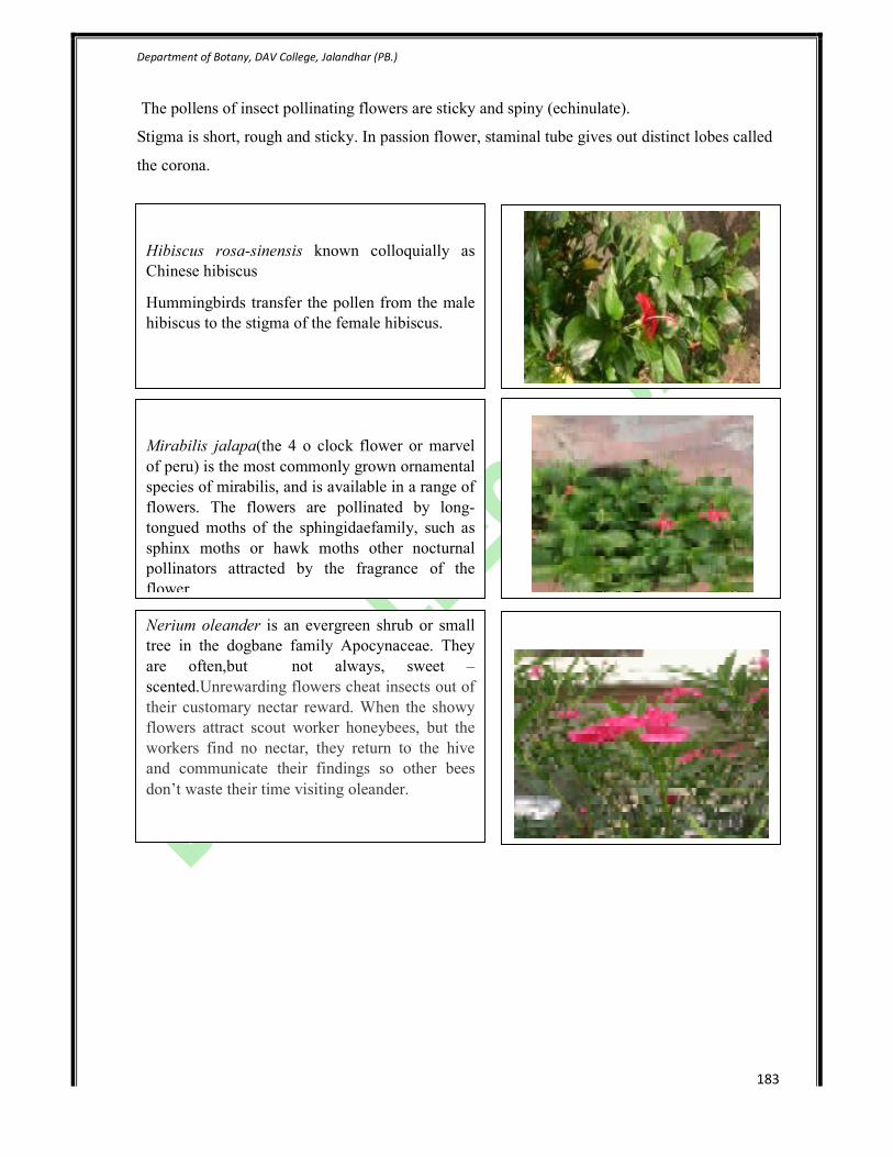
Hibiscus rosa-sinensis known colloquially as
Chinese hibiscus
Hummingbirds transfer the pollen from the male
hibiscus to the stigma of the female hibiscus.
Mirabilis jalapa(the 4 o clock flower or marvel
of peru) is the most commonly grown ornamental
species of mirabilis, and is available in a range of
flowers. The flowers are pollinated by long-
tongued moths of the sphingidaefamily, such as
sphinx moths or hawk moths other nocturnal
pollinators attracted by the fragrance of the
flower.
Nerium oleander is an evergreen shrub or small
tree in the dogbane family Apocynaceae. They
are often,but not always, sweet –
scented.Unrewarding flowers cheat insects out of
their customary nectar reward. When the showy
flowers attract scout worker honeybees, but the
workers find no nectar, they return to the hive
and communicate their findings so other bees
don’t waste their time visiting oleander.

Department of Botany, DAV College, Jalandhar (PB.)
184
Salvia officinalis (sage, also called garden sage or common sage) is a member of the family Lamiaceae.
The defining characteristic of the genus Salvia is the unusual pollination mechanism. It consists of
two stamens (instead of the typical four found in other members of the tribe Menthaceae) and the
two thecae on each stamen are separated by an elongate connective. It is the elongation of the
connective that enables the formation of the lever mechanism. When a pollinator probes a male stage
flower for nectar, (pushing the posterior anther theca) the lever causes the stamens to move and
the pollen to be deposited on the pollinator. When the pollinator withdraws from the flower, the lever
returns the stamens to their original position. In older, female stage flowers, the stigma is bent down in a
general location that corresponds to where the pollen was deposited on the pollinator's body. The lever
of most Salvia species is not specialized for a single pollinator, but is generic and selected to be easily
released by many bird and bee pollinators of varying shapes and sizes. The lever arm can be specialized
to be different lengths so that the pollen is deposited on different parts of the pollinator’s body. For
example, if a bee went to one flower and pollen was deposited on the far back of her body, but then it
flew to another flower where the stigma was more forward (anterior), pollination could not take place.

Department of Botany, DAV College, Jalandhar (PB.)
185
Calotropis procera is a species of flowering plant in
the dogbane family Asclepiadaceae. This plant plays
host to a variety of insects and butterflies. Calotropis
is an example of entomophily pollination (pollination
by insects) and pollination is achieved with the help
of bees. In Calotropis, gynostegium is present
(formed by the fusion of stigma and androecium). The
pollen is arranged in a structure named pollinia which
are attached to a glandular, adhesive disc at the
stigmatic angle (Translator Mechanism). These sticky
discs get attached to the legs of visiting bees so that
pollinia are pulled out when the bee moves away.
When such a bee visits another flower, this flower
gets pollinated by the sticky pollinium.

Department of Botany, DAV College, Jalandhar (PB.)
186
4) Ornithophily- Cross pollination which takes place with the help of birds is called
Ornithophily and flowers are called ornithophilous flowers. E.g. Bignonia, Bottle brush, Butea,
Bombax etc.
Adaptations in ornithophilous flower:
Flowers are large and beautifully coloured.
They produce copious mucilaginous nectar.
Flowers are generally scentless.
Some flowers may have edible parts. More commonly the birds with long narrow breaks are
involved in Ornithophily e.g. Humming, Sunbirds, Honey birds, Crow, Bulbul.
The pollen grains are sticky.
Flower produces thick and fleshy floral parts.
Callistemon viminalis, also known as weeping
bottle brush, is a shrub or small tree in the family
myrtaceae. The bright red flower spikes,which are
4-10cm in length and about 3-6 in diameter occur
between spring and summer.Bottlebrush
displays inflorescences composed of many bright
red flowers which are grouped together. The
larger group of flowers make it extremely
attractive to birds, which have good eyesight and
are very sensitive to the colour red. The
infloresences are also found at the apex of
branches, making them more accessible for birds.
Petunia hybrida is one of the best summer flower
belongs to the family solanaceae. The color range
is huge, with varieties available in every color
except orange. The basic petunia is funnel
shaped,but hybridizers have created many
variations including singles and doubles with
petals that have wavy or fringed margins. Bees
are attracted to flowers that have broad petals, so
they can easily alight on the petals. For this
reason, they are less likely to hover near slim-
petaled petunias. Honeybees are attracted to blue
and purple flowers.

Department of Botany, DAV College, Jalandhar (PB.)
187
Chiropterophily:
Pollination occurs through a bat is called chiropterophily and flower is called chiropterophilous
flower. It is seen in flowers of Adansonia, Kigellia, Bauhinia, Anthocephalus etc.
Adaptations in chiropterophilous flower:
Flowers open during evening or night.
Flowers are large, dull coloured and have a strong scent like that of rotting fruits.
Flowers produce abundant pollen grains.
Flowers produce copious nectar.
Flowers are tough so that bats can hold on the flowers.
Thevetia peruviana is a poisonous plant.
Thevetia is an evergreen tropical shrub or small
tree. Its large showy and tubular flowers make
an ideal mechanism for birds to visit and reach
for its nectar thereby pollinating the flower.
Kigelia is a genus of flowering plants in the
family Bignoniaceae. . Flowers are produced in
panicles; they are bell-shaped (similar to those of
the African Tulip tree but darker and more
waxy), orange to reddish or purplish green, and
about 10 cm wide. Individual flowers do not
hang down but are oriented horizontally. Some
birds are attracted to these flowers and the strong
stems of each flower make ideal footholds. Their
scent is most notable at night indicating that they
are adapted to pollination by bats, which visit
them for pollen and nectar. They also remain
open by day however, and are freely visited by
many insect pollinators, particularly large species
such as carpenter bees.

Department of Botany, DAV College, Jalandhar (PB.)
188
14. Structure of anther, microsporogenesis (using slides) and pollen grains (using whole
mounts).
Structure of young anther
• The section appears slightly lobed
• The outermost is a single layered epidermis. The cells are cuticularised.
• At four corners of the anther, the derivatives formed as a result of Archesporial cells are
present.
• Of these, wall layers are situated below the epidermis and mass of sporogenous cells near
the centre of the lobe.
• The epidermis is followed by alayer or two of parenchymatous wall layers. The innermost
wall layer is called tapetum. It is nutritive in function.
• The sporogenous cells lie inside the wall. These act as pollen or microspore mother cells
and divide meoitically.
• In the middle of the anther lobe,procambial strand is present.
Structure of mature anther
• An organized anther is four chambered in
atransection.
• The wall consists of an outer epidermis,
an endothecium, one to three middle
layers and an innermost tapetum.
• The tapetum at maturity is multinucleate
and contains dense cytoplasm which is
finally used up by the developing
microspores.
• Prior to dehiscence, the tapetum and also
the middle layers degenerate. The cells of
the endothecium are radially elongated
and exhibit, characteristic fibrous
thickenings.
• The microspores or pollen grains are at
first arranged in tetrads, (as a result of
reduction division of the microspore
mother cells). Later, these separate and
occur as individual pollen grains,
dispersed throughout the chamber. Each
shows characteristic shape, size and
structure.

Department of Botany, DAV College, Jalandhar (PB.)
189
Study of development of male gametophyte from the permanent slides under the
microscope.
The pollen grains/microspores germinate on the stigma of the same or different flower, as a
result of which there is development/formation of a structure called male gametophyte.
Development:
1. Each stamen consists of a lobed anther,
containing the microsporangia and
supported by a thin filament.
2. Meiosis of the diploid microspore
mother cells in the anther produces four
haploid microspores.
3. Each of these develops into a pollen
grain consisting of a larger vegetative
cell (also called the tube cell) inside of
which is a smaller germ cell (also
called the generative cell).
4. At some point, depending on the
species, the germ cell divides by mitosis
to produce 2 sperm cells.
5. At the time of pollen dispersal it may
be at 2 cell stage or 3 cell stages
depending upon the species.
6. When generative cell divides after
reaching the stigmatic surface it gives
rise to two male gametes.
7. The pollen tube comes out of the germ
pores present in the pollen grain. The
tube elongates and enters through the
style into the ovule.
8. This structure, a germinated pollen
grain carrying a long pollen tube and
two male gametes inside it, is termed as
a mature male gametophyte.

Department of Botany, DAV College, Jalandhar (PB.)
190
To study the structure of pollen grains using whole mounts
The pollen grain is the male gametophyte in gymnosperms and angiosperms, i.e. the structure that
produces the male gametes and transfers them to the female part. The grains derive from the meiotic
process of the pollen mother cells and at maturity usually consist of a bi or trinucleate cell surrounded by a
wall that has the important function of protecting the microgamethophyte in its journey between male and
female flowers. The pollen grain wall is very resistant to water loss and environmental injuries, primarily
to avoid damage and desiccation during the aerial journey.
Each species elaborates a distinctive sculpture on the surface of the pollen grains, and there are also many
other morphological characteristics that are useful for the pollen analyst in the classification of the pollen.
TYPES OF APERTURES IN MICROSPORES
The first characteristic to be considered when identifying pollen grains are the apertures. An
aperture is a thin or missing part of the exine, which is independent of the patterning of the exine.
Two different types of apertures can be distinguished: pores and fissures (colpi). The latter are
more primitive, they are elongated with pointed ends. Pores are usually isodiametric. They can
also be slightly elongated but, in contrast to colpi, they have rounded ends. In some pollen
grains, the exine around the apertures is either thicker or thinner. In pores this border is termed
annulus (typical in grass pollen) and in colpi margo (e.g. in Hedera helix). Pollen grains with
pores are porate and those with colpi are colpate. If both pore and colpus are combined in the
same aperture, the pollen grain is colporate.
Microspores can be divided into groups according to the number, position and type of apertures.
This classification is simple and consistent. The number of apertures is indicated by the prefixes
mono-, di-, tri- tetra-, penta-, hexa- and poly- with the above terms porate, colpate oder
colporate. Usually three pores and/or colpi are present that are regularly spaced around either the
edge or the equator of the pollen grain, depending on whether the pollen grain is seen from the
polar or equatorial view. If more than three apertures are present, they can either be regularly

Department of Botany, DAV College, Jalandhar (PB.)
191
spaced around the edge, or equator respectively, (zonoporate/zonocolpate), or over the entire
surface (pantoporate/ pantocolpate).
The so-called syncolpate pollen grains have two or more colpi that are fused at the ends. These
colpi can sometimes form a spiral. Fenestrate pollen grains form a further group. They have large
window-like spaces, where the tectum is missing. (See chapter 2.3), the Asteraceae
(Cichorioideae), among others, belong to this group. Inaperturate pollen is rare microspores
without apertures (Juniperus). The Cyperaceae, for example, have more or less round apertures
in the ectexine, but, unlike pores, these do not have a clear margin and are termed lacunae (sing.
lacuna). In some pollen grains, e.g. birch and alder, the two layers of the exine are separated
around the pores and form a chamber between the inner and outer exine (vestibulum). Finally
there are the so-called bisaccate (pollen grains with "air-bags", e.g. the conifers spruce, pine,
Pinus cembra and fir.
The cell-wall structure of fern- and moss-spores differs from that of pollen grains and the spores
do not have pores or colpi. They merely have a fissure in the sexine. Manolete spores only have a
single fissure; trilete spores have three fissures that form a 'Y' ("Mercedes star").

Department of Botany, DAV College, Jalandhar (PB.)
192
Department of Botany, DAV College, Jalandhar (PB.)
193
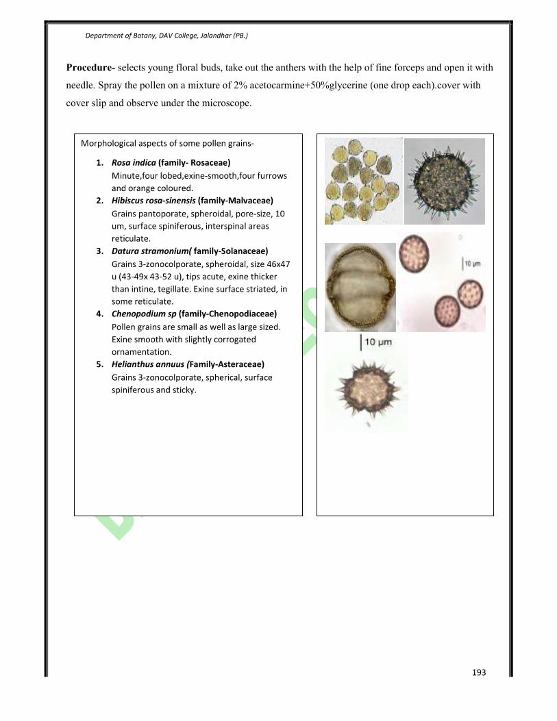
Procedure- selects young floral buds, take out the anthers with the help of fine forceps and open it with
needle. Spray the pollen on a mixture of 2% acetocarmine+50%glycerine (one drop each).cover with
cover slip and observe under the microscope.
Morphological aspects of some pollen grains-
1. Rosa indica (family- Rosaceae)
Minute,four lobed,exine-smooth,four furrows
and orange coloured.
2. Hibiscus rosa-sinensis (family-Malvaceae)
Grains pantoporate, spheroidal, pore-size, 10
um, surface spiniferous, interspinal areas
reticulate.
3. Datura stramonium( family-Solanaceae)
Grains 3-zonocolporate, spheroidal, size 46x47
u (43-49x 43-52 u), tips acute, exine thicker
than intine, tegillate. Exine surface striated, in
some reticulate.
4. Chenopodium sp (family-Chenopodiaceae)
Pollen grains are small as well as large sized.
Exine smooth with slightly corrogated
ornamentation.
5. Helianthus annuus (Family-Asteraceae)
Grains 3-zonocolporate, spherical, surface
spiniferous and sticky.
Department of Botany, DAV College, Jalandhar (
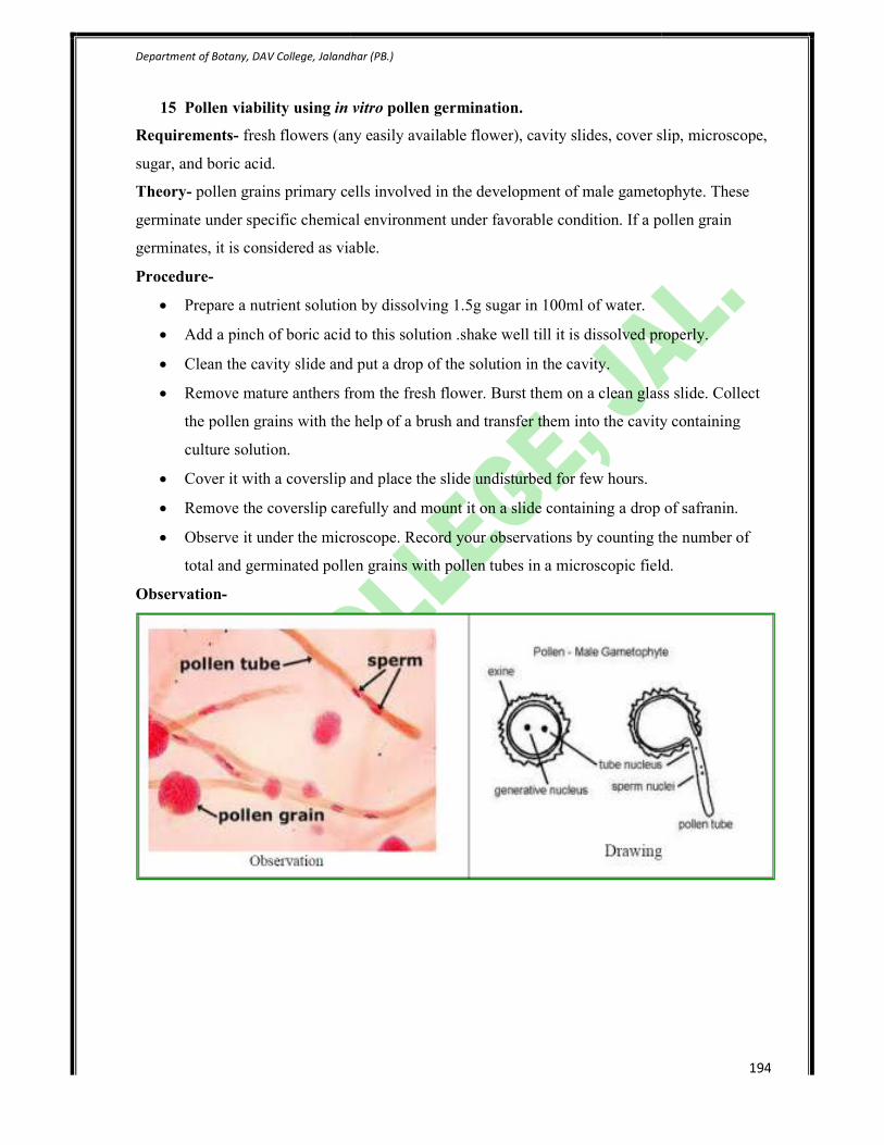
15 Pollen viability using in vitro
Requirements- fresh flowers (any easily available flower), cavity slides, cover slip, microscope,
sugar, and boric acid.
Theory- pollen grains primary cells involved in the development of male gametophyte. These
germinate under specific chemical environment under favorable condition. If a pollen grain
germinates, it is considered as viable.
Procedure-
• Prepare a nutrient solution
• Add a pinch of boric acid to this solution .shake well till it is dissolved properly.
• Clean the cavity slide and put a drop of the solution in the cavity.
• Remove mature anthers from the fresh flower. Burst them o
the pollen grains with the help of a brush and transfer them into the cavity containing
culture solution.
• Cover it with a coverslip and place the slide undisturbed for few hours.
• Remove the coverslip carefully and mount it on
• Observe it under the microscope. Record your observations by counting the number of
total and germinated pollen grains with pollen tubes in a microscopic field.
Observation-
College, Jalandhar (PB.)
in vitro pollen germination.
fresh flowers (any easily available flower), cavity slides, cover slip, microscope,
pollen grains primary cells involved in the development of male gametophyte. These
germinate under specific chemical environment under favorable condition. If a pollen grain
germinates, it is considered as viable.
Prepare a nutrient solution by dissolving 1.5g sugar in 100ml of water.
Add a pinch of boric acid to this solution .shake well till it is dissolved properly.
Clean the cavity slide and put a drop of the solution in the cavity.
Remove mature anthers from the fresh flower. Burst them on a clean glass slide. Collect
the pollen grains with the help of a brush and transfer them into the cavity containing
Cover it with a coverslip and place the slide undisturbed for few hours.
Remove the coverslip carefully and mount it on a slide containing a drop of safranin.
Observe it under the microscope. Record your observations by counting the number of
total and germinated pollen grains with pollen tubes in a microscopic field.
194
fresh flowers (any easily available flower), cavity slides, cover slip, microscope,
pollen grains primary cells involved in the development of male gametophyte. These
germinate under specific chemical environment under favorable condition. If a pollen grain
Add a pinch of boric acid to this solution .shake well till it is dissolved properly.
n a clean glass slide. Collect
the pollen grains with the help of a brush and transfer them into the cavity containing
a slide containing a drop of safranin.
Observe it under the microscope. Record your observations by counting the number of
total and germinated pollen grains with pollen tubes in a microscopic field.

Department of Botany, DAV College, Jalandhar (PB.)
195
Calculation: �������������� ������������� ����
��� ������������������� ����� ���
Precautions –
1. Flowers should be freshly pluckedand should have mature anthers.
2. Allow pollen grains to germinate for sufficient time.
3. Allow pollen germination under closed environment i.e. by using cavity slide.
4. Use clean slide to observe the pollen germination.
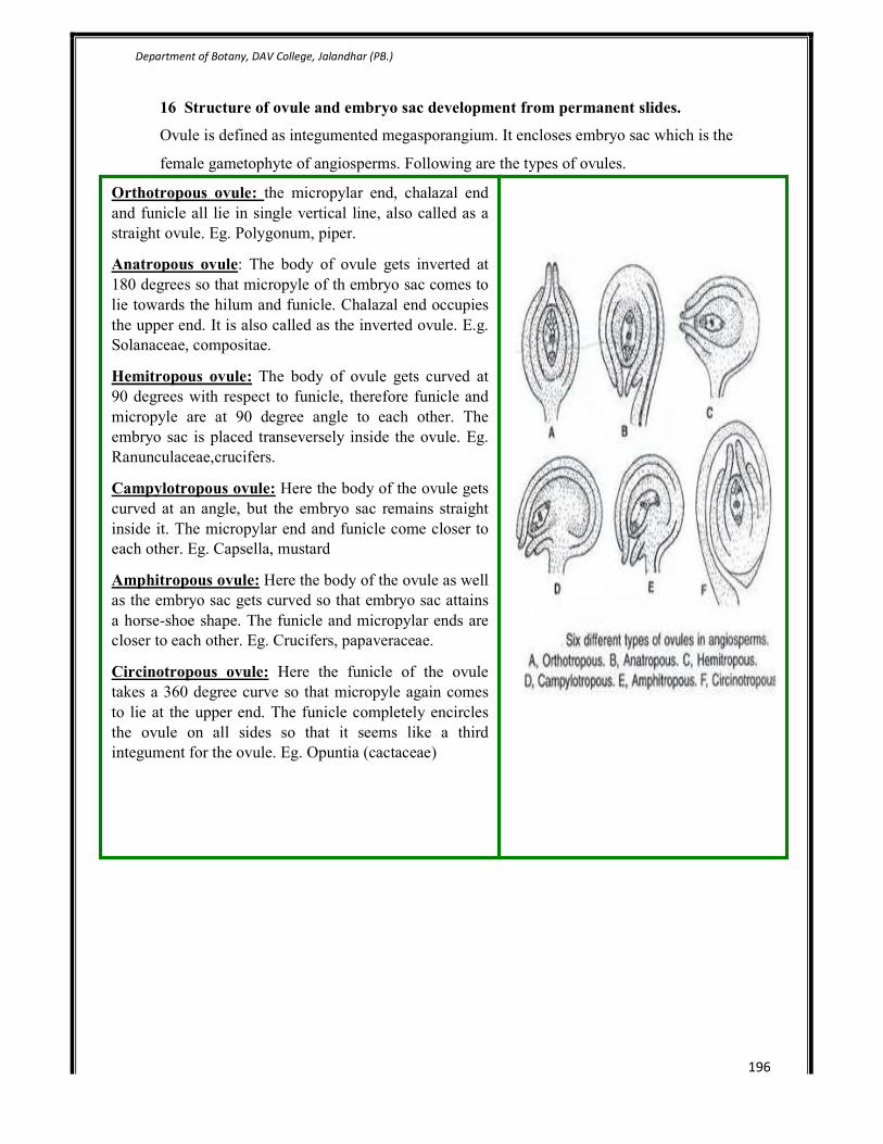
Department of Botany, DAV College, Jalandhar (PB.)
196
16 Structure of ovule and embryo sac development from permanent slides.
Ovule is defined as integumented megasporangium. It encloses embryo sac which is the
female gametophyte of angiosperms. Following are the types of ovules.
Structure of Female Gametophyte of angiosperms
Orthotropous ovule: the micropylar end, chalazal end
and funicle all lie in single vertical line, also called as a
straight ovule. Eg. Polygonum, piper.
Anatropous ovule: The body of ovule gets inverted at
180 degrees so that micropyle of th embryo sac comes to
lie towards the hilum and funicle. Chalazal end occupies
the upper end. It is also called as the inverted ovule. E.g.
Solanaceae, compositae.
Hemitropous ovule: The body of ovule gets curved at
90 degrees with respect to funicle, therefore funicle and
micropyle are at 90 degree angle to each other. The
embryo sac is placed transeversely inside the ovule. Eg.
Ranunculaceae,crucifers.
Campylotropous ovule: Here the body of the ovule gets
curved at an angle, but the embryo sac remains straight
inside it. The micropylar end and funicle come closer to
each other. Eg. Capsella, mustard
Amphitropous ovule: Here the body of the ovule as well
as the embryo sac gets curved so that embryo sac attains
a horse-shoe shape. The funicle and micropylar ends are
closer to each other. Eg. Crucifers, papaveraceae.
Circinotropous ovule: Here the funicle of the ovule
takes a 360 degree curve so that micropyle again comes
to lie at the upper end. The funicle completely encircles
the ovule on all sides so that it seems like a third
integument for the ovule. Eg. Opuntia (cactaceae)

Department of Botany, DAV College, Jalandhar (PB.)
197
Structure of embryo sac/female gametophyte:
1. Embryo sac/female gametophye is an oval
structure within an ovule of an angiosperm
that contains the egg. Together with the
fertilized egg, it develops into a seed. The
upper end where egg apparatus lies is called as
the micropylar end from where the pollen tube
enters the ovule.
2. The embryo sac is the female gametophyte of
angiosperms, consisting of eight nuclei: the
egg and two adjacent and short-lived
synergids that are near the micropyle .the
synergids are also called potential cells.
3. Two polar nuclie in the central cell. These
fuse with one of the male nucleus to form
primary endosperm nucleus in future, to give
rise to endosperm tissue after the syngamy has
taken place.
4. Three antipodal nuclei at the chalazal end of
the embryo sac opposite the micropyle. Like
the synergids, these nuclei degenerate at or
shortly after fertilization. They pass on all the
nourishment they get from nucellus to the
potential egg cell.
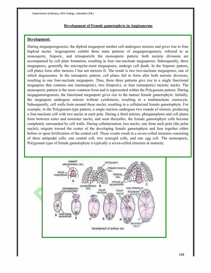
Department of Botany, DAV College, Jalandhar (PB.)
198
Development of Female gametophyte in Angiosperms
Development:
During megasporogenesis, the diploid megaspore mother cell undergoes meiosis and gives rise to four
haploid nuclei. Angiosperms exhibit three main patterns of megasporogenesis, referred to as
monosporic, bisporic, and tetrasporicIn the monosporic pattern, both meiotic divisions are
accompanied by cell plate formation, resulting in four one-nucleate megaspores. Subsequently, three
megaspores, generally the micropylar-most megaspores, undergo cell death. In the bisporic pattern,
cell plates form after meiosis I but not meiosis II. The result is two two-nucleate megaspores, one of
which degenerates. In the tetrasporic pattern, cell plates fail to form after both meiotic divisions,
resulting in one four-nucleate megaspore. Thus, these three patterns give rise to a single functional
megaspore that contains one (monosporic), two (bisporic), or four (tetrasporic) meiotic nuclei. The
monosporic pattern is the most common form and is represented within the Polygonum pattern. During
megagametogenesis, the functional megaspore gives rise to the mature female gametophyte. Initially,
the megaspore undergoes mitosis without cytokinesis, resulting in a multinucleate coenocyte.
Subsequently, cell walls form around these nuclei, resulting in a cellularized female gametophyte. For
example, in the Polygonum-type pattern, a single nucleus undergoes two rounds of mitosis, producing
a four-nucleate cell with two nuclei at each pole. During a third mitosis, phragmoplasts and cell plates
form between sister and nonsister nuclei, and soon thereafter, the female gametophyte cells become
completely surrounded by cell walls. During cellularization, two nuclei, one from each pole (the polar
nuclei), migrate toward the center of the developing female gametophyte and fuse together either
before or upon fertilization of the central cell. These events result in a seven-celled structure consisting
of three antipodal cells, one central cell, two synergid cells, and one egg cell. The monosporic,
Polygonum type of female gametophyte is typically a seven-celled structure at maturity.

Department of Botany, DAV College, Jalandhar (PB.)
199
17 Nuclear and cellular endosperm. Embryo development in monocots and dicots
(using permanent slides/dissection).
Endosperm and Endosperm Formation:
Endosperm is the name of food laden tissue which is meant for nourishing the embryo in seed
plants. In gymnosperms; it represents the female gametophyte. In angiosperms, the endosperm is a
special tissue which is formed as a result of vegetative fertilization or fusion of a male gamete with
diploid secondary nucleus of the central cell.
The endosperm of angiosperms establishes a physiological compatibility with the developing hybrid
embryo, since it has the genome of the male gamete also within it. Vegetative fertilization gives rise
to a primary endosperm cell having a triploid endosperm nucleus.
Depending upon the mode of its formation, angiospermic endosperm is of the following types:
1. Nuclear Endosperm:
In this type, the first division of the primary endosperm cell and several of the following ones are
unaccompanied by cell wall formation. The nuclei may either remain free or in later stages they
become separated by walls, e.g., many Archichlamydeae and Monocots.
2. Cellular Endosperm:
In the cellular type, the first, and most of the subsequent division are accompanied by wall
formation so that the embryo sac becomes divided into several chambers, e.g., most gamopetalae.
3. Helobial Endosperm:
In the helobial type (so called because of its frequent occurrence in the order Helobiales) the first
division of primary endosperm nucleus is followed by cytokinesis to form two cells, micropylar and
chalazal. Further development in both the cells occur like that of nuclear endosperm i.e.,
multinucleate stage followed by wall formation, e.g. Helobiales.
During its growth the endosperm crushes the nucellus. It is in turn eaten by growing embryo. The
endosperm may persist in the seed when the latter is called endospermic or albuminous (e.g.,
Castor). In others, the endosperm is completely absorbed by the growing embryo and the food
reserve gets stored in the cotyledons. Such seeds are called non-endospermic or exalbuminous, e.g.,
Cucurbita.

Department of Botany, DAV College, Jalandhar (PB.)
200
Embryo development in dicots
Embryogeny (Embryo Formation):
Embryogeny is the sum total of changes that occur during the development of a mature embryo
from a zygote or oospore. In a typical dicot, the zygote elongates and then divides by a
transverse wall into two unequal cells.
The larger basal cell is called suspensor cell. The other towards the antipodal end is termed as
terminal cell or embryo cell. The suspensor cell divides transversely a few times to produce a
filamentous suspensor of 6—10 cells.
The suspensor helps in pushing the embryo in the endosperm. The first cell of the suspensor
towards the micropylar end becomes swollen and functions as a haustorium. The haustorium has
wall ingrowths similar to transfer cells. The last cell of the suspensor at the end adjacent to the
embryo is known as hypophysis. Hypophysis later gives rise to the radicle.
The embyro cell undergoes two vertical divisions and one transverse division to form eight cells
arranged in two tiers—epibasal (terminal) and hypobasal (near the suspensor). The epibasal
cells eventually form the two cotyledons and the plumule. Only one cotyledon is produced in
monocots (e.g. Wheat, Maize, Onion, and Palm). The hypobasal cells produce the hypocotyl
except its tip.
The eight embryonic cells or octants divide periclinally to produce an outer layer of protoderm
or dermatogen. The inner cells differentiate further into procambium and ground meristem.
Protoderm forms epidermis, procambium gives rise to steal or vascular strand and ground
meristem produces cortex and pith.
Initially the embryo is globular and undifferentiated. Early embryo with radial symmetry is
called proembryo. It is transformed into embryo with the development of radicle, plumule and
cotyledon. Two cotyledons differentiate from the sides with a faint plumule in the centre.
At this time the embryo becomes heart-shaped. The rate of growth of the cotyledons is very high
so that they elongate tremendously while the plumule remains as a small mound of
undifferentiated tissue. Subsequently, the embryo undergoes rest as the ovule gets transformed
into seed. In some plants the embryo remains in the globular or spherical form even at the time
of seed shedding without showing any distinction of plumule, radicle and cotyledons, e.g.,
Orobanche, Orchids, Utricularia.
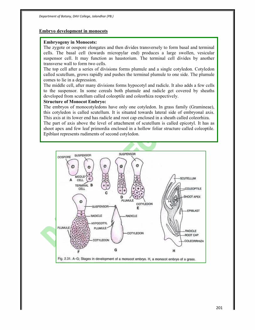
Department of Botany, DAV College, Jalandhar (PB.)
201
Embryo development in monocots
Embryogeny in Monocots:
The zygote or oospore elongates and then divides transversely to form basal and terminal
cells. The basal cell (towards micropylar end) produces a large swollen, vesicular
suspensor cell. It may function as haustorium. The terminal cell divides by another
transverse wall to form two cells.
The top cell after a series of divisions forms plumule and a single cotyledon. Cotyledon
called scutellum, grows rapidly and pushes the terminal plumule to one side. The plumule
comes to lie in a depression.
The middle cell, after many divisions forms hypocotyl and radicle. It also adds a few cells
to the suspensor. In some cereals both plumule and radicle get covered by sheaths
developed from scutellum called coleoptile and coleorhiza respectively.
Structure of Monocot Embryo:
The embryos of monocotyledons have only one cotyledon. In grass family (Gramineae),
this cotyledon is called scutellum. It is situated towards lateral side of embryonal axis.
This axis at its lower end has radicle and root cap enclosed in a sheath called coleorhiza.
The part of axis above the level of attachment of scutellum is called epicotyl. It has as
shoot apex and few leaf primordia enclosed in a hollow foliar structure called coleoptile.
Epiblast represents rudiments of second cotyledon.

Department of Botany, DAV College, Jalandhar (PB.)
202
18 Simple experiments to show vegetative propagation (leaf cuttings in bryophyllum,
begonia; stem cuttings in rose, money plant, sugarcane and bougainvillea).
Introduction-
Vegetative propagation produces the next generation that is genetically identical to the parent.
Such an organism that is genetically identical to the parent is called a clone. In case of plants
with advantageous characteristics, the characteristics can be preserved by producing clones. This
is particularly useful to agriculturists and horticulturists in order to get the best crop and uniform
yield every time.
There are various ways of carrying out artificial propagation of plants. Cutting, layering, grafting
and budding are some of the traditional methods whereas tissue culture is a recent technology.
Cutting
Cutting involves removing a piece of the parent plant - stem, root or leaf, and planting it in a
suitable medium. At first roots are produced and then the shoot with the leaves. If a stem is
taken, it must contain the nodal region. In some cases, rooting hormone may be required to
initiate root formation.
For example:
• Stem cutting is commonly done for rose, sugarcane, banana, geranium, etc.,
• Root cutting is done for dahlia
• Leaf cuttings are used for African violets.
Layering
Layering is the method of inducing certain branches of the parent plant to produce roots by
bending and pegging them to the ground around the parent plant leaving the tips exposed. Once
the roots develop the branch is then cut off from the parent body.
The branch that produces the roots is called the layer. It is a natural method in plants such as
black raspberries. However, it is induced in plants like Jasminum, Rhododendron, strawberries,
Magnolia, etc.
Grafting
It is the transfer of a part of one plant to the stump of another plant. The part taken from a plant
is a portion of the stem with many buds. This portion is called scion and is selected for the

Department of Botany, DAV College, Jalandhar (PB.)
203
quality of its fruit. The stump to which the scion is attached is called the stock. Stock is selected
for qualities such as disease resistance and hardiness.
Grafting by Stem Cutting
The cut ends of both the scion and stock are shaped such that they complement each other and
their cambial tissues are close to each other. The two cut ends are brought together and covered
with grafting wax. After some time, the tissues of the scion and the stock become continuous.
The plant bears flowers and fruits characteristic of the scion.It is commonly practised on apple
trees and on such plants which either do not produce viable seeds or the seeds which have a lot
of variation.
Budding or Bud Grafting
It is a variation of the grafting method explained above. In this method, the scion is a bud along
with some bark. A 'T'-shaped cut is made on the stock into which the scion is inserted and bound
with a tape.

Department of Botany, DAV College, Jalandhar (PB.)
204
Three Stages of Bud Grafting - Method of Cleft Grafting-The bud, once fixed, gives rise to new
branches. For example, bud grafting is done on roses, plums, peaches, pears, citrus, etc.
Stem cuttings-
Stem cuttings are the most important and greatest used type of cutting. In general, they can be
divided into 4 groups, the hardwood, semi hardwood, softwood or herbaceous and the cane
cutting. When preparing a stem cutting a section of stem tissue with lateral or terminal buds is
obtained, typically, stem cuttings are made from the terminal ends of shoots, generally 8-13cm
long and are removed from the parent plant at a ppoint just below a healthy lea. As a general
rule,the stem cuttings( except hardwood and cane types) should have 3 to 4 leaves for quickest
rooting. The leaves from the basal 1/3 the stem cutting should be removed.
Hardwood cuttings-
These cuttings are prepared during the dormant season using wood from the previous season’s
growth, although in a few cases, older wood can be used. The length of hard wood cutting vary

Department of Botany, DAV College, Jalandhar (PB.)
205
from 10-30 cm. older wood can be as long as 0.6-0.9 m. in most cases, cuttings of pencil
thickness, i.e. 0.6-1.2 cm in diameter with atleast 2 nodes are selected depending upon the
species.
Three types of hardwood cuttings can be prepared, the straight, the heel and the mallet cutting.
The heel hardwood cutting has attached at its base a small portion of older wood, while the
mallet type hardwood cutting includes short sections of older stem wood. The straight hardwood
cutting is entirely composed of 1 year wood.

Department of Botany, DAV College, Jalandhar (PB.)
206
Vegetative propagation in Saccharum officinarum by stem cutting
• Select healthy seed material. It should be free from pests and diseases.
• High viability is essential for establishment of the crop. The top one third to half portion
of a cane being and is best to be used as seeds.
• Collect sets from well-matured erect and healthy cones.
• Before planting the sets leaves of the canes talk are removed without damaging the buds.
• Cut these stalks into 3 buds sets each, usually 30-50 cm long.
• Soak the sets in water for four to six hours before sowing.
• To prevent the sets from rot and pineapple disease, dip the sets in suitable fungicide such
as 0.25% mercuric acid or 0.25% aretan or agallol solution. Agallol along with gamma
BHC has been recommended for northern India.
• Plant the sets in farm using appropriate planting irrigation and manuring practices.

Department of Botany, DAV College, Jalandhar (PB.)
207
Softwood cutting
Vegetative propagation in bougainvillea by stem cuttings
• Cut 7.5-10 cm long pieces of shoot with about 4-6 nodes and several leaves on
the terminal end.
• Remove the leaves from basal portion of stem cuttings.
• Give a cut at the base just below the node.
• Collect cuttings during cooler part of the day i.e. early in the morning.
• Treat the stem cuttings with optimum strength of 0.4% IBA depending on the
cultivar and time of the year. The basal cut and exposed part of stem is dipped
in the solution of IBA.
• Place the treated stem cutting with optimum temperature in the pot.
• Also provide intermittent mist. Bougainvillea do not tolerate wet environment.
Vegetative propagation in Rosa indica by stem cutting
When cuttings are prepared from the soft, succulent, herbaceous growth they are
termed softwood, herbaceous or green wood cuttings.an extremely wide range of
horticultural plants are propagated in this manner including chrysanthemums, fuschia
and geraniums among the greenhouse crops and roses, salix, moneyplant and
bougainvillea among the landscape plants.
The stem cuttings are generally 8-13 cm in length with leaves retained on the upper 2/3
to 1/2 of the cuttings. the herbaceous or softwood cutting should have 2or more nodes
and if the leaves are large, they may be cut in order to reduce water loss through
transpiration and save space.

Department of Botany, DAV College, Jalandhar (PB.)
208
Leaf cuttings- whole leaf cuttings are prepared from leaves with or without petioles. When
petioles are not present the leaf is broken or cut off the plant and the basal or proximal edge of
the leaf is inserted into the rooting medium. Plants without petioles include sedum, jade and
cactus. Roots and leaves will eventually forn at base of the leaf. The petiole should not be more
than 4 cm long and should be set deep enough into the medium to keep the cuttings erect.
In addition, leaf cuttings can be prepared by taking leaf sections when propagating plants are like
Rex Begonia and snake plant (sanseveria sp.).
Vegetative propagation in begonia-
• Cut well matured leaves into triangular
sections in such a way that the two sides of
two triangular wedges are bordered by leaf
vein.
• Discard the outer thin section of leaf.
• Insert the medium leaf section upright in
sand.
• Place the pot in warm and humid conditions.
• Allow 5-6 weeks for plants to appear.
• The plantlets appear on the cut surface of the
large leaf veins near the leaf base.

Department of Botany, DAV College, Jalandhar (PB.)
209
Vegetative propagation in Bryophyllum-
• The foliar embryos from leaf margins are
carefully removed.
• These are placed in rooting medium.
• Plantlets grow from foliar embryos or leaf
sections placed in rooting medium.

Department of Botany, DAV College, Jalandhar (PB.)
210
19 Germination of dormant and non dormant seeds.
Theory- dormancy is defined as phenomenon of non-germination of seeds due to internal factors
under favourable condition.
Material required- xanthium seeds, petridish, beaker, conc. Sulphuric acid, cotton, blade,
distilled water etc.
Procedure-
• Take three sets of xanthium seeds each.
• Make splits in hard coat of Xanthium seeds of first set (5 seeds) and put these
seeds in a petridish containing well wet pad of cotton.
• Put seeds of second batch in a beaker containing conc. Sulphuric acid for few
hours. After some time, take out the seeds and wash well in running water to clear
out the sulphuric acid. Place these seeds for germination in a petridish with a wet
cotton seed.
• Place third set of seeds as such in a petridish for germination.
• Put the above three petridishes under same conditions of temperature, air and
light.
• Take care that cotton may not get dry.
• Note the time taken for germination by the seeds in the above three petridishes.

Department of Botany, DAV College, Jalandhar (PB.)
211
Observations- the seeds of 3rd set did not germinate at all, while the seeds in 1
st and 2
nd set
germinate well.
Result- seeds didn’t germinate due to seed dormancy because of seed coat but acid and
mechanical treatment helps in breaking the dormancy.
Precautions-
1. Cotton pads should not get dry during germination study.
2. Acid should be washed completely for germination study.
3. Time taken for germination should be noted down carefully.