Lab FetalPig Circulatory
24
he circulatory (or cardiovascular) system is responsi- ble for transporting nutrients, gases, hormones and metabolic wastes to and from the individual cells of an organism. Mammals are far too large for all of their in- dividual cells to exchange nutrients, wastes and gases with the external world by simple diffusion. Most cells are buried too deep inside the body for this method to be effec- tive. Thus, some system must be in p ace to efficiently ex- change these products b tween the outside world and every cell in the organism's body. For this reason, the circulatory system is a highly-branched network of vessels that spreads throughout the entire organism. In general the circu atory system represents a series of vessels that diverge from the heart (arteries) to supply blood to the tissues and a conflu- ence of vessels draining blood from the tissues and return- ing it to the heart (veins). Despite the extensive network of arteries and veins throughout the body, no actual exchange of water, nutrients, wastes or gases occurs in arteries or veins. Their walls are too thick to permit diffusion. Extensive networks of capillary beds connecting branches of arteries and veins exist throughout the body to transfer these dis- solved substances between the blood tream and the tissues. To simpl fy the identification of the numerous arteries and veins, there are two general principles you should remember: (1) arteries and veins tend to be paired, especially when the organs they supply or drain are paired, and (2) a continuous vessel often undergoes several name changes along it length as it passes through different body regions. Therefore to successfully identify arteries and veins it is necessary to trace them along their enti e length (typically from the heart outward).
-
Upload
corygunther -
Category
Documents
-
view
219 -
download
0
Transcript of Lab FetalPig Circulatory

8/6/2019 Lab FetalPig Circulatory
http://slidepdf.com/reader/full/lab-fetalpig-circulatory 1/24
he circulatory (or cardiovascular) system is responsi-ble for transporting nutrients, gases, hormones and
metabolic wastes to and from the individual cells ofan organism. Mammals are far too large for all of their in-dividual cells to exchange nutrients, wastes and gases withthe external world by simple diffusion. Most cells areburied too deep inside the body for this method to be effec-tive. Thus, some system must be in place to efficiently ex-change these products between the outside world and everycell in the organism's body. For this reason, the circulatorysystem is a highly-branched network of vessels that spreadsthroughout the entire organism. In general the circulatorysystem represents a series of vessels that diverge from theheart (arteries) to supply blood to the tissues and a conflu-
ence of vessels draining blood from the tissues and return-ing it to the heart (veins). Despite the extensive network ofarteries and veins throughout the body, no actual exchangeof water, nutrients, wastes or gases occurs in arteries orveins. Their walls are too thick to permit diffusion. Extensivenetworks of capillary beds connecting branches of arteriesand veins exist throughout the body to transfer these dis-solved substances between the bloodstream and the tissues.
To simplify the identification of the numerous arteries and veins, there are two general principles you shouldremember: (1) arteries and veins tend to be paired, especially when the organs they supply or drain are paired,and (2) a continuous vessel often undergoes several name changes along its length as it passes through differentbody regions. Therefore to successfully identify arteries and veins it is necessary to trace them along their entirelength (typically from the heart outward).
8/6/2019 Lab FetalPig Circulatory
http://slidepdf.com/reader/full/lab-fetalpig-circulatory 2/24
THORACIC CAVITY AND NECK REGION
Notice the thin pericardial membrane surrounding the heart(Figure S.lA). This protective sac contains a small amountof lubricating fluid to protect the heart and cushion its
movements. A portion of the relatively large thymus sits onthe outside of the pericardial membrane and partially ob-scures the heart from view. The thymus should not be con-fused with neighboring lung tissue or with the atria of theheart (which lie inside the pericardium). Make a mentalnote of the position, color and consistency of the thymus.We will return to the thymus along with the other endocrineglands in Chapter 9.
T h e H e a rt: E x te rn a l A n a to m y
The driving force behind the entire circulatory system ofmammals is the heavily muscled heart. Numerous vesselsemanate from the cranial aspect of the heart and radiate
outward in all directions (Figure 5.2). For the moment,will concentrate only on the heart and the major vessels thaoriginate from it. Later we will trace the paths of these majovessels as they diverge throughout the body. Mammals possess a four-chambered heart that delivers blood through twomajor circulatory pathways-the pulmonary circuit (from thheart to the lungs and back) and the systemic circuit (from
the heart through the rest of the body and back). A hallmarkof the mammalian heart is that its internal design keeps thblood from these two circuits entirely separate, thus keepingoxygen-depleted blood from mixing with oxygen-rich blood.
NOTE: Throughout this chapter, we use the terms atrium andauricle to refer to slightly different regions of the heart. The termauricle is used to describe the small , outer, flap-like region thatcovers a portion of the atrial chamber, while the term atrium isused to refer to the entire open space (or actual chamber) insidethat collects the blood. The reason for this distinction isthat partof each atrial chamber extends well beyond the boundaries ofeach flap-like auricle. This will be more evident when you view theinterior of the heart later.
Identify the four chambers of the heart. Caudally therare two large, thick-walled ventricles, the right ventricle andthe left ventricle (Figure S.lB). These chambers pump bloodout of the heart to the lungs and to the rest of the body, respectively. Cranial to the ventricles and somewhat darker icolor are the right and left auricles. Chambers within thright and left auricles receive blood from the body and thlungs, respectively, and pass it to the ventricles. Runningalong the surface of the heart itself, the small coronary arteries (Figure S.2A) should be evident. These vessels suppblood to the heart muscle, insuring that it too receives nutri-
ents and oxygen. Notice the large veins entering the heart onthe right side. These are the cranial and caudal vena cava(Figure S.2A). They bring deoxygenated blood to the rigatrium from the upper and lower portions of the body. Onthe dorsal surface of the heart, adjacent to the juncture othe vena cavae and the right atrium, there is a small sac-likeregion of the heart known as the coronary sinus (FigurS.2E). This sinus is responsible for returning deoxygenatedblood from the wall of the heart to the right atrium.
On the ventral surface of the heart, locate the large pulmonary artery (Figure S.2D) emanating from the right vtricle. In the adult, this artery channels blood from the righ
ventricle through the right and left pulmonary arteries tthe lungs. Follow the pulmonary artery behind the hearand see where it branches into the right and left pulmonaryartery. Notice that at the base of the pulmonary arterythere is a connection between it and the aorta (the large artery leaving the cranial aspect of the left ventricle). Thiconnection is called the ductus arteriosus (Figure 5.2C),short, temporary linkage found only in the fetus. Lying adjacent to the pulmonary arteries are the pulmonary veinsthe vessels that, in the adult, return oxygenated blood tthe left atrium of the heart.

8/6/2019 Lab FetalPig Circulatory
http://slidepdf.com/reader/full/lab-fetalpig-circulatory 3/24
Thy ro id g la nd
Pericardium
Diaphragm
A ortic arch
Pulmonaryartery
Le ft au ri cl e
L eft lu ng
C oro nary aand vein
Lef t ventr ic l
II FIGURE 5 .1 H e a rt su rro u nd ed b y p er ie ard ia l m e m b r an e (a ), h ea rt w i th p e r ie ard ia l m e m b rsh ow in g h is to lo gy o j a n a rte ry a nd v e in (e ) .
Lung
C ranial vena cava
Righ t au ri cl e
R igh t vent ri cl e
m

8/6/2019 Lab FetalPig Circulatory
http://slidepdf.com/reader/full/lab-fetalpig-circulatory 4/24
Vag us n erv e
R ig ht c os to -Le ft ph ren
Cranial nervec er vic al v ei n vena cav a
RightBrachiocep
Cranial Right trunkv en a cav a auricle auricle
Left subclaBranches Pulmonary arteryof the arterycoronary Duc tu s a rt
Pulmonary artery Rightv ein and artery ventricle
Le ft co ronRightP hre nic n erv e ventricle artery
HeartLe ft au ri clCaudal reflected
A cc esso ry lo be v en a ca va laterally reflected
of right lung LeftDiaphragm ventricle
II m
Phrenic
R ight i nt er na lnerve
th ora cic v ein Le ft costo-(cut) cervical
artery Cranial L ef t c os to -
Cranial an d v ein vena cava c er vi ca l v e
vena cava Left Vag us n ervAorta internal Pulmonary
thoracic artery A ortic arcDuctus artery
arteriosus P hre nic n eBrachlo-
Pulmonary cephalic P ulm on aryartery trunk
Coronary Left Bronchusvessels subclavian Accessory
Le ft au ri cl eartery lobe of the
L eft lu ngrig ht lu ng
P hre nic n eterm in atin gth e d ia ph ra
m
R ig ht lu ng
C audal vena cava
Le ft au ri cl e
H ea rt re fle cte d c ra nia lly
R ig ht p hrenic nerv e
C oron ary sinu s
Left pulm onary vein
A cce sso ry lo beof the right lung
II I FIGURE 5.2 C o m po site p ho to grap h o j h ea r t an ato m y, c ra nia l an d ca ud al v en a c av ae {a l, r ig ht aao r ta , p u lm on a ry tru nk , a n d d uc tu s a r te r io su s {c l, p ulm ona ry a r te r ie s a n d v e in s {d l an d co ro n a

8/6/2019 Lab FetalPig Circulatory
http://slidepdf.com/reader/full/lab-fetalpig-circulatory 5/24
Fe ta l v. Adult C ir cu la tio n
There are three major differences in the circulatory systemof a fetal and adult pig (Figure 5.3). The most obvious isthe connection between the fetus and the mother throughthe umbilical cord (Figure 5.3C). This collection of tubesallows oxygen and nutrients to pass from the mother to
the fetus while transporting carbon dioxide and metabolicwastes away from the fetus. The other two major differ-ences lie in the structure of the heart. Since the fetus is notbreathing with its lungs, the lungs do not oxygenate bloodthat passes through them. In fact, the most oxygen-rich
C ranial vena cava
Duc tu s a rt er io sus
C ranial vena cava
Toand fromupper extremity
Umbilic al v ein
Toand fromlower extremity
Caudal
At birth
Umb il ic al a rt er ie s A lla nto ic s ta lk
Umbilical vein U rogenital o
FIGURE 5.3 Schema tic illu str atio n s o f th e fe ta l c ir cu la to ry p a thway (a ) a nd th e compa ra tiv e p aw ith in se t p ho to gra ph o f umbilic al c or d (c ).

8/6/2019 Lab FetalPig Circulatory
http://slidepdf.com/reader/full/lab-fetalpig-circulatory 6/24
blood in the fetal circulatory system is that returning to thefetus through the umbilical vein. As a result, only a smallfraction of the blood leaving the right ventricle travels
through the pulmonary arteries to the lungs. The majorityis redirected through the ductus arteriosus, a connectionbetween the pulmonary artery and the aorta that channels
blood into the aorta (Figure 5.2C). Another structure inside
the heart of the fetus, called the foramen ovale, also aids inrerouting blood to bypass the lungs. This opening in theseptum between the right and left atria allows blood pass-ing into the right atrium to be channeled into the left
atrium and away from the lungs. Both of these adaptations
In tern al ju gu lar v ein
T hyro id g lan d
Rightbrachlocephallc
vein
Internalth oracic v ein
R igh t ven tr ic le
F IGURE 5 .4 Ve in s o j th e th o ra c ic re g io n .
ensure that the majority of oxygenated blood arriving viathe umbilical vein is passed through the fetal circulatorysystem via the aorta, while permitting enough blood toreach the lungs to allow the lung tissue to develop.
Ve in s o j th e T ho ra cic R eg io n
The largest veins in the thoracic region are the cranial venacava and caudal vena cava, which converge at the entranceto the right atrium (Figures 5.4-5.5). In the adult, these twothin-walled veins bring deoxygenated blood back to the
heart from all parts of the body. Trace one cranial vena
E xtern al ju gu lar v einC ep halic v ein (cu t)
Ax il la ry a rtand vein
S ub cla via n v
Costocervicaltrunk
Cranialv en a cav a
Left ventr ic le

8/6/2019 Lab FetalPig Circulatory
http://slidepdf.com/reader/full/lab-fetalpig-circulatory 7/24
cava cranially to its first major branch. This short branch,known as the brachia cephalic vein (or trunk) represents theconfluence of four veins: the internal jugular vein, the ex-ternal jugular vein, the cephalic vein, and the subclavianvein. Remember, blood is flowing back toward the heartthrough these vessels. Identify the internal thoracic veinleading from the arm pit toward the vena cava and heartat a ninety degree angle. The left and right axillary veinsalso lead toward the vena cava at ninety degree angles andbring blood from the forelimbs of the pig. The subscapularvein leading from the arm pit and the axillary vein come
L inguof ac ia l v ein
Ma xilla ry v ein
E xtern al ju gu lar v ein
R ig ht su bclav ian v ein
R ig ht b rach io cep halic v ein
C ranial vena cava
Caudal vena cava
FIGURE 5.5 Ve in s o j th e th ora c ic re gio n.
together to form the subclavian vein which dumps bdirectly into the cranial vena cava. The third vein returnblood from each forelimb is the cephalic vein, the mostnial of the three.
The external jugular veins lead down into the venafrom the neck region, along with the internal jugular vrunning medially alongside the trachea from the headward the heart. Follow the external jugular vein craniallythe point where it bifurcates into the linguofacial veinthe maxillary vein. These veins return blood from theporal portion of the face and lower jaw, respectively.
In tern al ju gu lar v ein
C ep ha lic v ein
Subscapu
L on g th oracic
C os to ce rv ic al v ein
Left ventr ic le
Diaphragm
8/6/2019 Lab FetalPig Circulatory
http://slidepdf.com/reader/full/lab-fetalpig-circulatory 8/24
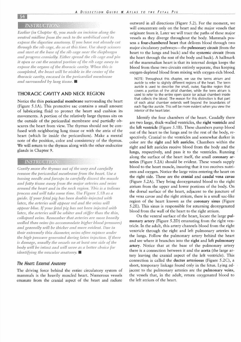
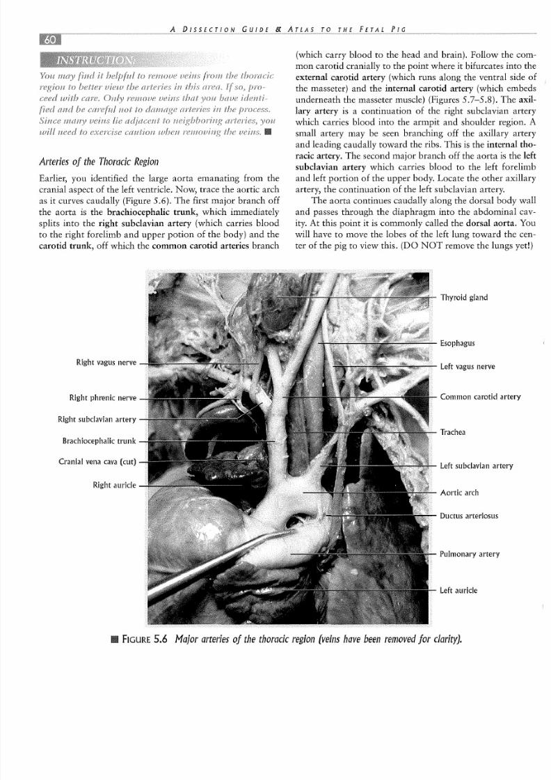
A rte rie s o j th e T ho ra cic R eg io n
Earlier, you identified the large aorta emanating from thecranial aspect of the left ventricle. Now, trace the aortic archas it curves caudally (Figure 5.6). The first major branch offthe aorta is the brachiocephalic trunk, which immediatelysplits into the right subclavian artery (which carries bloodto the right forelimb and upper potion of the body) and thecarotid trunk, off which the common carotid arteries branch
R ig ht v ag us n erv e
R ight phren ic nerve
R ig ht su bc la via n a rte ry
B ra ch io ce ph ali c tr unk
C ranial vena cava (cut)
R ig h t a uri cl e
(which carry blood to the head and brain). Follow the common carotid cranially to the point where it bifurcates into thexternal carotid artery (which runs along the ventral side othe masseter) and the internal carotid artery (which embedsunderneath the masseter muscle) (Figures 5.7-5.8). The axillary artery is a continuation of the right subclavian arterywhich carries blood into the armpit and shoulder region. Asmall artery may be seen branching off the axillary arteryand leading caudally toward the ribs. This is the internal thoracic artery. The second major branch off the aorta is the lefsubclavian artery which carries blood to the left forelimband left portion of the upper body. Locate the other axillaryartery, the continuation of the left subclavian artery.
The aorta continues caudally along the dorsal body waland passes through the diaphragm into the abdominal cavity. At this point it is commonly called the dorsal aorta. Yowill have to move the lobes of the left lung toward the center of the pig to view this. (DO NOT remove the lungs yet!)
T hyro id g lan d
Esophagus
L eft v ag us n erv e
C ommon caro tid art
Trachea
L eft s ub cla via n a rte ry
A ortic arch
Duc tu s a rt er io sus
P ulmon ary a rte ry
Le ft au ri cl e
FIGURE 5 .6 M a jo r a rte rie s o j th e th ora cic re gio n (v ein s h av e b een re m ov ed fo r

8/6/2019 Lab FetalPig Circulatory
http://slidepdf.com/reader/full/lab-fetalpig-circulatory 9/24
I nt er na l c ar ot id a rte ry
R ig ht s ub cl av ia n a rt er y
E x te rn al t ho ra ci c a rt er y---,L-l,l':"""'_''+--II---
I nte rn al t ho ra ci c a rt er y - -_ J.- -H~ r- -Z
B r ach io c ephal ic t ru nk
C ran ial v en a cav a (cu t)
L eft c oro nary a rtery
C audal vena cava
FIGURE 5 .8 A rte r ie s o j th e th ora cic re gio n (v ein s h av e b ee n o m itted io r c
Ex te rn al c ar oti d a
A rterial b ran ch esth e th yro id g lan d
Thy ro ce rv ic al t ru n
Ax il la ry a rt er y
L ef t s ub cl av ia n a r
Co s to c er vi ca l a rt e
A rch of aorta
P ulmon ar y a rte ry
Left ven tr ic le
Diaphragm
8/6/2019 Lab FetalPig Circulatory
http://slidepdf.com/reader/full/lab-fetalpig-circulatory 10/24
T h e H e a rt: In te rn a l A n a to m y
Notice that inside the chambers of the heart there arevalves to prevent blood from flowing backwards (Figures5.9-5.10). As blood enters the right atrium, it immediatelyflows into the right ventricle. Very little blood is actuallypumped by the right atrium into the right ventricle. At the
L eft common carotid artery
juncture of the right atrium and right ventricle is the tricus-pid valve. As the right ventricle contracts and pushes bloodout to the lungs, the blood is forced back up against the tricuspid valve, closing the leaflets of the valve and preventingretrograde flow into the right atrium. Upon entering the pul
monary trunk, the blood also passes through the pulmonarysemilunar valve, which prevents backflow into the rightventricle. The blood returns from the lungs into the lefatrium via the pulmonary veins and then flows into the lefventricle through the bicuspid (or mitral) valve. Blood leav-ing the left ventricle into the aorta is regulated by the aorticsemilunar valve. The valves of the heart are prevented frombeing pushed too far backward (a condition known as "pro-lapse") by small stringlike attachments called chordae ten-dineae. You may be able to see these structures on yourfrontal section of the heart.
Internal jugular vein (cut) V agus nerve
Left t ho racervicaltrunk
R ig ht c ommoncar ot id a rt er y
Le ft ax il lR ight thoraco- arteryc er vi ca l a rt er y brachial
plexus
Righ t ax il la ryartery and nerve
LeftR ight e x te rn al subclaviant ho ra ci c a rt er y
artery
R ight i nt er na lth ora cic a rte ry
Righ t subc lav ianartery
Brachiocephalic Le ft vent rtrunk
FIGURE 5 .7 A rte rie s o j th e th ora cic reg io n (v ein s h av e b ee n re m ov ed jo r c la ri

8/6/2019 Lab FetalPig Circulatory
http://slidepdf.com/reader/full/lab-fetalpig-circulatory 11/24
---- Left subclavian artery
Aorta
Cranial -----1
ven a cav a
Rightauricle
Pulmonaryartery
Rightventricle
Coronaryvessels
Caudalv ena cava
S em ilu na r v alv e
veins
Le ft au ri cl e
C oro nary sin us
--- Left ventricle
Cranven a
Righpulmartery
Righatrium
Righpulmvein
Caudvena
--- R ighventr
© Michael Schenk O J
O pening to coronary arteryB ra ch io ce ph ali c a rt er y
Pulmonary trunk Ductus arteriosus atof aorta and pulm onarytrunk
Le ft a tr ium
A zy go s v ein
C h ord ae te nd in a
P ap illa ry mu sc le---- Left ventricle
Lef t ventr ic le
FIGURE 5.9 I l lu str a tio ns o f h ea rt d ep ic tin g v en tro la te ra l v ie w (a J, d orsa l v ie w (b J a nd in te rh e a r t(c a n d d J .

8/6/2019 Lab FetalPig Circulatory
http://slidepdf.com/reader/full/lab-fetalpig-circulatory 12/24
Left subclavian Costocervical
Brachio-cephalic
trunk Aorta
Cranial Cranial
ven a cav aven a cav a
A zy gos vein
Pulmonary Rightartery pulmonary
R ight a ur ic leartery
Le ft au ri cl e
Le ft au ri cl ePulmonary
veins
RightCoronary
ventriclesinus
R ight a tr iumLeft co ronary Caudal
artery andvein ve na cav a
Lef t ventr ic le Rightcoronary
arteryand vein
Lef t ventr ic le
Aorta
R igh t ven tr ic le
L eft s ub cla via n a rte
B ra ch io ceph al ic t ru nk
P ulmon ary a rte ryP ulmon ary a rte ry
Duc tus a rt er io sus
Opening to leftc oro na ry a rte ry
Tri cu sp id v al ve
R ight a tr ium
L eft c oron ary arte ry
B ic us pi d v al ve
Righ t vent ri cl e
FIGURE 5.10 P h oto g ra ph s o f h ea rt d ep ic tin g v en tra l v iew (a ), d o rs a l v iew (b ) a nd in te r io ro f h ea rt (e ) .
8/6/2019 Lab FetalPig Circulatory
http://slidepdf.com/reader/full/lab-fetalpig-circulatory 13/24
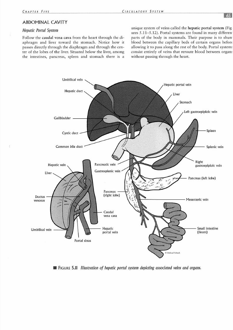
ABDOMINAL CAVITY
H e p a tic P o rta l S ys te m
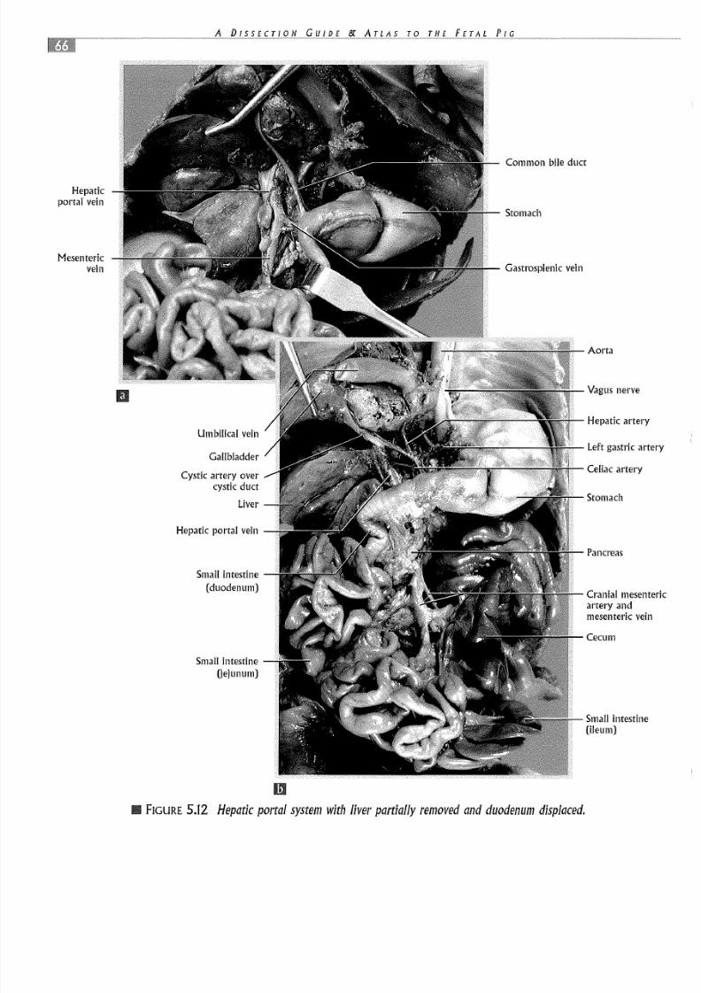
Follow the caudal vena cava from the heart through the di-aphragm and liver toward the stomach. Notice how itpasses directly through the diaphragm and through the cen-ter of the lobes of the liver. Situated below the liver, amongthe intestines, pancreas, spleen and stomach there is a
G allbladder ---
C ys tic d uc t
Common bile duct
H epatic v ein
Ductusvenosus
Pancreas ---+.-1-'0
(rig ht lo be )
Caudalv ena c ava
Umbilic al v ein ,.---- Hepaticpo rta l v ein
P orta l s in us
unique system of veins called the hepatic portal systemures 5.11-5.12). Portal systems are found in many diffeparts of the body in mammals. Their purpose is toblood between the capillary beds of certain organs ballowing it to pass along the rest of the body. Portal sysconsist entirely of veins that reroute blood between orwithout passing through the heart.
H epatic portal vein
L eft g as tro ep ip lo ic v e
Spleen
Spl en ic
Right
g as tro ep ip lo ic
--- Small intestin(ileum)
• FIGURE 5 . 1 1 I l lu s t ra tio n o j h ep a tic p o rta l s ys te m d ep ic tin g a ss oc ia te d v ein s
8/6/2019 Lab FetalPig Circulatory
http://slidepdf.com/reader/full/lab-fetalpig-circulatory 14/24
Hepaticp ortal v ein
Comm on bile duct
Mesentericvein G as tr os ple nic v ein
Umbilic al v ein
Gallbladder
C ystic artery overc ys tic d uc t
H epatic portal vein
Sma ll i nt es ti ne(duodenum)
Small i nt es ti ne(jejunum)
m
FIGURE 5 . 1 2 H e pa tic p or ta l s y ste m w ith liv er p a r tia lly r e m ov ed a nd d u od en um d
Aorta
Va gus n erve
H ep atic a rte ry
L eft g as tric a rt
Ce li ac a rt er y
Stomach
Pancreas
C rani al me sentartery andmes en te ric v ei
Cecum
Small i nt es ti ne(ileum)
8/6/2019 Lab FetalPig Circulatory
http://slidepdf.com/reader/full/lab-fetalpig-circulatory 15/24
In the case of the hepatic portal system, blood flowsfrom the capillary beds of the small and large intestines, thespleen, the pancreas and the stomach into the hepatic portalvein (Figure 5.12) and then into the capillary beds of theliver, before entering the caudal vena cava. This extra stepallows blood from the stomach and intestines that containslarge amounts of sugars and possibly toxins to be filtered by
the liver before the blood is sent to the rest of the body. Also,hormones produced by the pancreas (e.g., insulin, glucagon,somatostatin) can be directed to their target organ, the liver,without the delay and diluting effect of traveling through the
Umbilica l v ein (c ut)
G e nita l v es se ls(spermatic-male,ovarian-female)
M e dia n s ac ra lartery and vein
© M i ch ae l S ch en k
Umb il ic al a rt er ie sUrinarybladder
entire circulatory system first. Depending on the typeamount of hormone released by the pancreas, the liverstore the sugar (as glycogen) or release it into the bstream immediately. Through this regulatory mechanism,liver maintains nearly constant blood glucose levels.
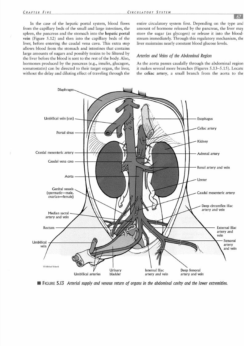
A rte r ie s a nd Vein s o j th e A bd om in al R eAs the aorta passes caudally through the abdominal reit makes several more branches (Figures 5.13-5.15).the celiac artery, a small branch from the aorta to
-+--"1--- R enal artery and v
C au dal m es en te ric
D ee p c irc um fle xartery and vein
E x te rn aartery
FIGURE 5.13 A rte r ia l su pp ly a nd v en ou s r e tu rn o j o rg an s in th e a bd om in al c a vity a n

8/6/2019 Lab FetalPig Circulatory
http://slidepdf.com/reader/full/lab-fetalpig-circulatory 16/24
stomach, pancreas and spleen. Next find the cranial mesen-
teric artery (Figure 5.14A), which has branches that supplythe jejunum, ileum and colon. Embedded in the intestinalmesentery are the arterial arcades (Figure 5.14B), numerous
branches of the mesenteric artery that provide nutrientsand oxygen to the tissues of the intestinal tract. Further
caudally, two short branches of the dorsal aorta lead into
the kidneys. These are the renal arteries. Lying next to therenal arteries, the thinner-walled renal veins are present.
These vessels collect filtered blood from the kidneys. Thecaudal mesenteric artery is a single vessel that suppliesblood to the colon and rectum (Figure 5.15A). Caudal to
this vessel, the genital arteries (Figure 5 .15B) are visiblebranching off the dorsal aorta and leading to the ovaries or
Spiralcolon
A dre nal gla ndPancreas
C ra nia l m ese nte ric arte ryand m esenteric vein
Renal artery and vein
Smallintestine(ileum)
Caudal vena cava
Sp ir al c ol on
Cranialmesenteric
artery
Mesentericlymp h n ode s
Sma ll i nt es ti ne(jejunum)
A rte ria l a rc ado f th e m ese nte( fr om b rancheo f c ra ni almesentericartery)
Sma ll i nt es ti ne(ileum)
m
II FIGURE 5 . 1 4 C r an ia l m e se nte ric a rte ry (a ), a rte ria l a rc ad es o j th e c ra nia l m e se nte ric a rtem e se nte ric , c elia c , a nd c au d al m e se nte ric a rte rie s a nd th eir b ra nc he s(c ) ,
8/6/2019 Lab FetalPig Circulatory
http://slidepdf.com/reader/full/lab-fetalpig-circulatory 17/24
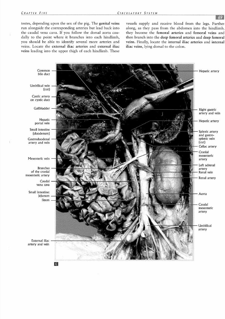
testes, depending upon the sex of the pig. The genital veinsrun alongside the corresponding arteries but lead back intothe caudal vena cava. If you follow the dorsal aorta cau-dally to the point where it branches into each hindlimb,you should be able to identify several more arteries andveins. Locate the external iliac arteries and external iliacveins leading into the upper thigh of each hindlimb. These
Common Hepa ti cb ile d uc t
Umbilic al v ein(cut)
Cy st ic a rt er yon cystic duct
Gallbladder R ight gartery a
Hepatic Hepa ti cp orta l v ein
Sma ll i nt es ti neSp le ni c
(duodenum) a nd g asGastroduodenal s ple nicartery and vein (cut)
Ce li ac a
Cranialmesenter
M e sen te ric ve in artery
Le ft ad rBranches artery
of the cranial R en al vm e se nte ric a rte ry
Rena l aCaudal
ve na c av a
Small i nt es ti ne : Aortajejunum
ileum
Caudalmesenterartery
vessels supply and receive blood from the legs. Furalong, as they pass from the abdomen into the hindlthey become the femoral arteries and femoral veinsthen branch into the deep femoral arteries and deep femveins. Finally, locate the internal iliac arteries and inteiliac veins, lying dorsal to the colon.
Umbilicaartery
E xte rn al ilia c ----artery and vein

8/6/2019 Lab FetalPig Circulatory
http://slidepdf.com/reader/full/lab-fetalpig-circulatory 18/24
U rete r (c ut)
Aorta
Caudalvena cava
Testicularartery
and vein
Umbilicalarteries
Medians ac ra l a rt er y
Femorala rt er y, v ei nand nerve
Deepfemoralartery
and vein
Pe lv is ( cu t)and colonremoved
Caudal
Colon
B ran che s of ca ud alme sent er ic a rt er ys up ply in g d is ta l
colon and rectum
C audal m esenteric artery (cut)
D eep circum flex artery and vein
Common iliac vein
E xternal iliac artery and vein
Internal iliac artery and vein
R en al
Rena l
Kidney
Ureter
Aorta
CaudalmesentarteryOvariaartery
Ex te rnartery
Deepfemoraartery
Femorartery
I I I FIGURE 5 . 1 5 C a ud al m e sen te ric a rte ry (a ) a nd g en ita l, i l ia c, a nd fe m ora l a rte r ie s { b) j re cture m ov ed fo r c la ri ty .

8/6/2019 Lab FetalPig Circulatory
http://slidepdf.com/reader/full/lab-fetalpig-circulatory 19/24
Um b i lic a l C o rd
In placental mammals, the unborn young are attached tothe placenta of the mother via the umbilical cord (Figure5.16). This cord serves as the lifeline for the fetus, trans-porting nutrients and oxygen to the growing fetus, and pro-viding a channel for carbon dioxide and excess metabolicwastes to be eliminated from the fetus. Identify the singleumbilical vein. Unlike most veins, this vessel actually car-ries oxygen- and nutrient-rich blood to the fetus from thefetal side of the placenta. The maternal blood is restrictedto the maternal side of the placenta and never mixes with
fetal blood under normal circumstances. Exchange of gases,nutrients and wastes is accomplished by diffusion acrossthe placental barrier. You should see two smaller umbilicalarteries (Figures 5.16-5.17). These carry blood from thefetus to the placenta. While the fetus remains in its mother'suterus, metabolic waste (urine) that collects in the bladderpasses through the allantoic duct and into the allantois for
Umb il ic al a rt er y
B ran ch es o f
umb il ic al a rt er ys up ply in g th eu rin ary b la dd er
Ureter
storage. Thus the urinary bladder remains connected toumbilical cord until the fetus is born (Figure 5.17). Abirth, the connection to the umbilical cord deterioratesthe urinary bladder drains into the urethra.
Umbilical arteries U rogenital ope
FIGURE 5 . 1 6 D is ta l e nd o j u m b ilic al c or
Urinarybladder
III FIGURE 5 . 1 7 C lo se -u p o j u m b ilic a l a rte rie s a nd u rin ary b la dd er .

8/6/2019 Lab FetalPig Circulatory
http://slidepdf.com/reader/full/lab-fetalpig-circulatory 20/24
The Sp le en
The spleen is a vascular, ductless organ that plays a criticalrole in the circulatory system of vertebrates (Figure 5.18).Since mammalian red blood cells do not contain nuclei,they cannot undergo cell division and thus have a finite lifespan. New red blood cells are continuously produced in the
bone marrow and delivered to the spleen. The spleen also
manufactures white blood cells (lymphocytes) to fend ofdiseases, and destroys and recycles worn-out blood cellsThe spleen stores these cells along with excess blood plasmato be released into the bloodstream as needed. Through thimechanism the spleen regulates the body's total blood vol
ume and the concentration of red blood cells in the blood.
StomachSplenic artery and vein
Sma ll i nt es ti ne
III FIGURE 5 . 1 8 Sp le en (d is pla ce d to th e sid e)w i th accompany ing h is to logy pho tog raph. m Sp l e e n . 250X

8/6/2019 Lab FetalPig Circulatory
http://slidepdf.com/reader/full/lab-fetalpig-circulatory 21/24
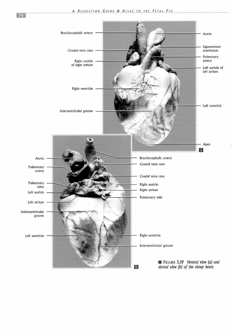
THE SHEEP HEART
NOTE: Due to the popularity of the sheep heart in manylaboratory courses, the following section will concentrateon the anatomy of the sheep heart as a model for studyingthe "typical" mammalian heart. Much of this informationwas covered previously with the fetal pig heart. Check with
your instructor to see if you will be completing this sectionand to determine what level of depth you will be requiredto know.
Identify the four chambers of the heart. Caudally thereare two large, thick-walled ventricles, the right ventricleand the left ventricle (Figure 5.19). These chambers pumpblood out of the heart to the lungs and to the rest of thebody, respectively. In the sheep heart, there is a superficiallandmark separating these two chambers known as the in-terventricular groove that runs obliquely down the ventralsurface of the heart toward the apex, but runs more longi-tudinally on the dorsal surface of the heart. Cranial to the
ventricles and somewhat darker in color are the right andleft auricles. Chambers within the right and left auricles re-ceive blood from the body and the lungs, respectively, andpass it to the ventricles. Running along the surface of theheart itself are the small coronary arteries and veins. Thecoronary arteries supply blood to the heart muscle, ensuringthat it too receives nutrients and oxygen to maintain an en-ergy supply to support its continuous, methodical beatingthroughout the entire life of the animal. In the sheep, thecoronary vessels are typically buried under dense fat on thesurface of the heart.
Notice remnants of the large veins entering the heart on theright side. These are the cranial and caudal vena cavae (Fig-ure 5.19). They bring deoxygenated blood to the rightatrium from the upper and lower portions of the body. Themost visible artery from the ventral surface leaving theheart is the large pulmonary artery (pulmonary trunk) ema-nating from the right ventricle. This artery channels bloodfrom the right ventricle through the right and left pul-monary arteries to the lungs. Also notice the large, thiclc-walled aorta (aortic arch) leaving the heart from the cranialaspect of the left ventricle. The aorta and pulmonary arteryare connected externally for a short distance by remnanttissue of the ductus arteriosus that diverted blood from the
pulmonary artery to the aorta during fetal developmeThis band of solid connective tissue now joining thesevessels is known as the ligamentum arteriosum. Insheep heart, the large brachiocephalic artery will alsovisible as the first branch off the aorta. On the dorsalface of the heart you should be able to identify themonary veins leading back to the left auricle.
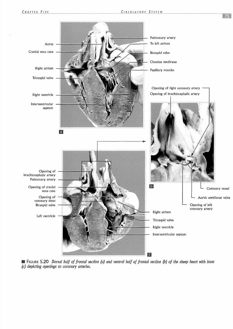
Notice that the atrial chambers extend well beyondboundaries of each auricle (as mentioned earlier) and tthe walls of the atria are much thinner than the walls ofventricles. Since the ventricles are responsible for pumpiblood much greater distances, they have evolved into heily muscularized chambers capable of generating masspressure to force blood out of the heart and throughbody. Notice that inside the chambers of the heart therevalves to prevent blood from flowing backwards (Fig5.20). As blood enters the right atrium, it immediat
flows into the right ventricle. Very little blood is actuapumped by the right atrium into the right ventricle. Atjuncture of the right atrium and right ventricle there is acuspid valve. As the right ventricle contracts and pusblood out to the lungs, some blood is forced back up agaithe tricuspid valve, slamming its leaflets shut and preveing retrograde flow into the right atrium. Upon enteringpulmonary trunk, the blood also passes through the pmonary semilunar valve, which prevents backflow intoright ventricle as it relaxes and receives more blood frthe right atrium. Fully oxygenated blood returns fromlungs into the left atrium via the pulmonary veins and thflows into the left ventricle through the bicuspid valvemitral valve). Blood leaving the left ventricle into the aopasses through the aortic semilunar valve, another valveprevent baclcflow as this ventricle relaxes. The bicuspid atricuspid valves are prevented from being pushed toobackward (a condition known as "prolapse") by smstringlike attachments of connective tissue called chordtendineae which have small muscular attachments (paplary muscles) to the inner wall of the heart (Figure 5.20).
8/6/2019 Lab FetalPig Circulatory
http://slidepdf.com/reader/full/lab-fetalpig-circulatory 22/24
8/6/2019 Lab FetalPig Circulatory
http://slidepdf.com/reader/full/lab-fetalpig-circulatory 23/24
Aorta
C ranial vena cava
R ight a tr ium
Tri cu sp id v al ve
Righ t vent ri cl e
Interventricularseptum
Opening ofb ra ch lo ceph al lc a rt er y
P ulmon ary a rte ry
O pening of cranialve na c av a
Opening ofc oron ary sin usB ic us pid v al ve
Lef t ventr ic le
P ulmon ar y a rte ry
To le ft a trium
B icus pi d v al ve
Cho rd ae te nd in ea e
Pap il la ry musc le s
O pening of right coronary artery
O pening of brachlocephallc artery
C oron ar
A ortic sem ilu
R ight a tr ium
O pening of leftc oro na ry a rte ry
Tri cu sp id v al ve
Righ t vent ri cl e
I nt er ventr ic ul ar s ep tum
FIGURE 5.20 D orsal half of frontal section (a) and ventra l half of fron tal section (b) of the sheep( c) depic ti ng open ings t o co ronary a rte ri es .

8/6/2019 Lab FetalPig Circulatory
http://slidepdf.com/reader/full/lab-fetalpig-circulatory 24/24