l enta l i n ic lOp Journal of Clinical & Experimental ht ... · Research Article Open Access...
4
Volume 4 • Issue 2 • 1000271 J Clin Exp Ophthalmol ISSN:2155-9570 JCEO an open access journal Open Access Research Article Panse et al., J Clin Exp Ophthalmol 2013, 4:2 DOI: 10.4172/2155-9570.1000271 *Corresponding author: Dr. Nikhil Panse, Vimal Niwas, Sudarshan Society, Near Model Colony Post Office, Shivajinagar, Pune 16, India, Tel: 91 9422314809; E-mail: [email protected] Received March 07, 2013; Accepted March 12, 2013; Published March 18, 2013 Citation: Panse N, Sambhus M, Sahasrabudhe P, Deodhar A (2013) The Tarsoconjunctival Flap for Lower Lid Reconstruction-Review of Literature and Case Series. J Clin Exp Ophthalmol 4: 271. doi:10.4172/2155-9570.1000271 Copyright: © 2013 Panse N, et al. This is an open-access article distributed under the terms of the Creative Commons Attribution License, which permits unrestricted use, distribution, and reproduction in any medium, provided the original author and source are credited. The Tarsoconjunctival Flap for Lower Lid Reconstruction-Review of Literature and Case Series Nikhil Panse 1 *, Mahesh Sambhus 2 , Parag Sahasrabudhe 1 and Avinash Deodhar 1 1 Plastic Surgery, B.J Medical College and Sassoon Hospital, Pune, India 2 Consultant Oncosurgeon–Deenanath Mangeshkar Hospital, Pune, India Keywords: Lower lid defects; Tarsoconjunctival flap; Early division Introduction e eyelid region is one of the most common sites for nonmelanoma skin cancers. In fact, skin cancers of the eyelid, including basal cell carcinoma (BCC), squamous cell carcinoma (SCC), and melanoma, account for five to ten percent of all skin cancers. Ninety five percent of these tumors are basal cell carcinomas or squamous cell carcinomas [1,2]. A lower-eyelid reconstruction aſter cancer extirpation or trauma is a challenging undertaking. Numerous options are available for lower lid reconstruction depending on the extent of the defect. We have used the Tarsoconjunctival flap for total and near total lower lid reconstruction, and this is an attempt to share our experience with the Tarsoconjunctival flap. Materials and Methods Nine patients of lower lid tumors were managed by wide local excision and reconstruction using the Tarsoconjunctival flap for posterior lamella recreation, and the cheek advancement flap for the anterior lamella creation. Surgical technique e surgical technique used was as follows. All the cases were operated under Local anesthesia with sedation by the first author. Wide local excision of the tumor was done with a 5 mm margin from the area of induration. e margins of the specimen were tagged for identification and sent for histopathological examination. All the histopathology reports suggested that margins were free of tumor. e defect that resulted was a full thickness defect of the lower eyelid comprising of the conjunctiva, tarsus, orbicularis and skin in all the cases. Vertically, the entire tarsus was deficient in all cases, and transversely there was a total or near total (More than 3/4th) eyelid loss. e upper eyelid was everted using a desmarres retractor (Figures 1 and 2). Infiltration was done with 2% lignocaine adrenaline solution. Markings were done on the everted conjunctiva in such a manner that the distal incision on the conjunctival side of the upper lid was Abstract Background: Recent literature on the use of Hughes Tarsoconjunctival flap for lower lid reconstruction is sparse. Aim: To study the use of the Hughes Tarsoconjunctival flap for total lower lid reconstruction. Materials and methods: We have analyzed the use of Hughes flap for the posterior lamella recreation after post malignancy defects in nine patients with certain modifications in the existing techniques. Anterior lamella was reconstructed using the cheek skin. All flaps were divided at two weeks. Result and conclusion: All the flaps did well. There was post operative ectropion and tarsal fracture in one patient each. The Tarsoconjunctival flap is based on the principle of like for like and probably gives the best possible functional and aesthetic outcomes in lower lid reconstruction. The evolutions of the Tarsoconjunctival flap over the years with important modifications and our experience with lower lid reconstruction using this technique have been outlined. at least 4mm away from the lid margin. A 4 mm margin was kept so as to prevent the donor site tarsal fracture. Incision was made in an inverted U shaped manner over the upper lid conjunctiva to reach the Figure 1: Basal cell carcinoma of lower lid. Figure 2: Total lower lid defect with everted upper lid. Journal of Clinical & Experimental Ophthalmology J o ur n a l o f C l i n ic a l & E x pe r i m e n t a l O p h t h a l m o lo g y ISSN: 2155-9570
Transcript of l enta l i n ic lOp Journal of Clinical & Experimental ht ... · Research Article Open Access...
Research Article Open Access
Volume 4 • Issue 2 • 1000271J Clin Exp OphthalmolISSN:2155-9570 JCEO an open access journal
Open AccessResearch Article
Panse et al., J Clin Exp Ophthalmol 2013, 4:2 DOI: 10.4172/2155-9570.1000271
*Corresponding author: Dr. Nikhil Panse, Vimal Niwas, Sudarshan Society, Near Model Colony Post Office, Shivajinagar, Pune 16, India, Tel: 91 9422314809; E-mail: [email protected]
Received March 07, 2013; Accepted March 12, 2013; Published March 18, 2013
Citation: Panse N, Sambhus M, Sahasrabudhe P, Deodhar A (2013) The Tarsoconjunctival Flap for Lower Lid Reconstruction-Review of Literature and Case Series. J Clin Exp Ophthalmol 4: 271. doi:10.4172/2155-9570.1000271
Copyright: © 2013 Panse N, et al. This is an open-access article distributed under the terms of the Creative Commons Attribution License, which permits unrestricted use, distribution, and reproduction in any medium, provided the original author and source are credited.
The Tarsoconjunctival Flap for Lower Lid Reconstruction-Review of Literature and Case SeriesNikhil Panse1*, Mahesh Sambhus2, Parag Sahasrabudhe1 and Avinash Deodhar1
1Plastic Surgery, B.J Medical College and Sassoon Hospital, Pune, India2Consultant Oncosurgeon–Deenanath Mangeshkar Hospital, Pune, India
Keywords: Lower lid defects; Tarsoconjunctival flap; Early division
Introduction The eyelid region is one of the most common sites for nonmelanoma
skin cancers. In fact, skin cancers of the eyelid, including basal cell carcinoma (BCC), squamous cell carcinoma (SCC), and melanoma, account for five to ten percent of all skin cancers. Ninety five percent of these tumors are basal cell carcinomas or squamous cell carcinomas [1,2]. A lower-eyelid reconstruction after cancer extirpation or trauma is a challenging undertaking. Numerous options are available for lower lid reconstruction depending on the extent of the defect. We have used the Tarsoconjunctival flap for total and near total lower lid reconstruction, and this is an attempt to share our experience with the Tarsoconjunctival flap.
Materials and Methods Nine patients of lower lid tumors were managed by wide local
excision and reconstruction using the Tarsoconjunctival flap for posterior lamella recreation, and the cheek advancement flap for the anterior lamella creation.
Surgical technique
The surgical technique used was as follows.
All the cases were operated under Local anesthesia with sedation by the first author.
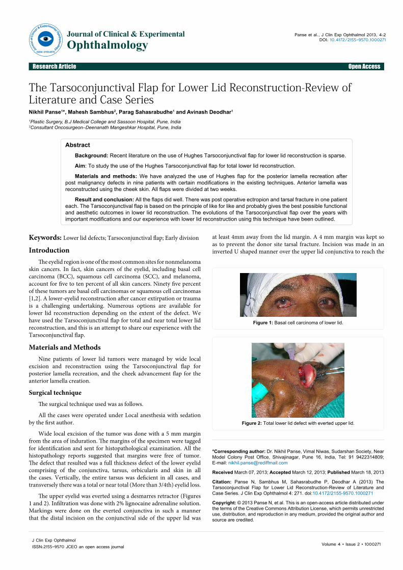
Wide local excision of the tumor was done with a 5 mm margin from the area of induration. The margins of the specimen were tagged for identification and sent for histopathological examination. All the histopathology reports suggested that margins were free of tumor. The defect that resulted was a full thickness defect of the lower eyelid comprising of the conjunctiva, tarsus, orbicularis and skin in all the cases. Vertically, the entire tarsus was deficient in all cases, and transversely there was a total or near total (More than 3/4th) eyelid loss.
The upper eyelid was everted using a desmarres retractor (Figures 1 and 2). Infiltration was done with 2% lignocaine adrenaline solution. Markings were done on the everted conjunctiva in such a manner that the distal incision on the conjunctival side of the upper lid was
AbstractBackground: Recent literature on the use of Hughes Tarsoconjunctival flap for lower lid reconstruction is sparse.
Aim: To study the use of the Hughes Tarsoconjunctival flap for total lower lid reconstruction.
Materials and methods: We have analyzed the use of Hughes flap for the posterior lamella recreation after post malignancy defects in nine patients with certain modifications in the existing techniques. Anterior lamella was reconstructed using the cheek skin. All flaps were divided at two weeks.
Result and conclusion: All the flaps did well. There was post operative ectropion and tarsal fracture in one patient each. The Tarsoconjunctival flap is based on the principle of like for like and probably gives the best possible functional and aesthetic outcomes in lower lid reconstruction. The evolutions of the Tarsoconjunctival flap over the years with important modifications and our experience with lower lid reconstruction using this technique have been outlined.
at least 4mm away from the lid margin. A 4 mm margin was kept so as to prevent the donor site tarsal fracture. Incision was made in an inverted U shaped manner over the upper lid conjunctiva to reach the
Figure 1: Basal cell carcinoma of lower lid.
Figure 2: Total lower lid defect with everted upper lid.
Journal of Clinical & Experimental OphthalmologyJo
urna
l of C
linica
l & Experimental Ophthalmology
ISSN: 2155-9570

Citation: Panse N, Sambhus M, Sahasrabudhe P, Deodhar A (2013) The Tarsoconjunctival Flap for Lower Lid Reconstruction-Review of Literature and Case Series. J Clin Exp Ophthalmol 4: 271. doi:10.4172/2155-9570.1000271
Page 2 of 4
Volume 4 • Issue 2 • 1000271J Clin Exp OphthalmolISSN:2155-9570 JCEO an open access journal
flaps did well. None of the patients had evidence of recurrence at the time of follow up. There were no cases of flap ischemia, or necrosis after division. Postoperative complications included lower eyelid retraction with epiphora in 1 patients and upper eyelid contour deformity due to tarsal fracture in one patient. The patient who had lower lid retraction was relatively younger (age 49 yrs) and the retraction can be attributed to the relatively lesser laxity of the cheek skin. The patient with tarsal fracture and contour deformity did not have any functional problems. None of the other patients had any functional problems (Table 1).
DiscussionLower lid reconstruction is a complex reconstructive challenge.
There are different techniques for total or partial lower eyelid reconstruction, such as the method described by Mustarde [3], the Hughes transposition flap with its modifications and evolution [4,5], the eyelid cutaneous rim graft [6], the hard palate graft covered by an orbicularis oculis myocutaneous advancement flap [7], the Tripier [8] flap and more complex approaches, such as the pre-expansion mucosa-lined tongue flap [9], the use of a cellular human dermis [10,11], the cheek flap supported by fascia lata [12], the island Tarsoconjunctival mucochondrocutaneous flap [13,14] and the use of an expanded forehead Fricke flap [15]. All of these techniques are useful when reconstruction of the lower eyelid is required; however, some of these procedures are complex and expensive.
The Hughes flap has evolved since the time is was first described in the 1937’s and several modifications have been described. However recent literature on the use of Hughes Tarsoconjunctival flap is sparse. Nevertheless, it is the only technique used in entire lower lid reconstruction where like is replaced by like. To achieve both excellent functional and aesthetic results, the layers of the lower eyelid must be
sub tarsal plane. The sub tarsal plane is an avascular plane, and the flap can be harvested without any difficulty. The Tarsoconjunctival flap is reasonably thick and fleshy flap. The flap was harvested till the fibers of the levator palpebrae superioris are identified inserting on the tarsus (Figure 3). This much length of the flap was sufficient in all our cases to comfortably reconstruct the defect.
After the flap elevation was complete, the flap was inset over the defect to recreate the posterior lamella of the lower lid. The Tarsoconjunctival flap was sutured in situ, taking care that the knots of the sutures were outside to prevent irritation to the cornea (Figure 4). A rectangular skin flap was then advanced from the cheek skin for recreation of the anterior lamella (Figure 4). While suturing due care was taken to take very few and superficial bites through the tarsus to prevent retraction of the cheek skin.
Neosporin eye ointment was applied and a bulky dressing was given.
Flap division was undertaken at two weeks under local anesthesia. The flap was divided approximately 2 mm above the area of the skin inset and the margin of the skin and conjunctiva was revised. It was done so as to give the maximum tarsal support to the lower lid and prevent post operative retraction and ectropion. The upper part of the tarsus was relocated to its original position by few sutures. The key step which we followed here was the sustained traction given on the cut edges of the Tarsoconjunctival flap of the lower lid for a couple of minutes with the help of skin hook to allow for stretch of the flap. Conventionally the Hughes flap was divided at 6 weeks to allow for stretch of the flap. Since we divided the flap at two weeks, we gave traction on the cut edges so as to compensate for the stretch of the flap. We do feel that few minutes of traction does not compensate for the stretch over the period of couple of weeks, but it definitely does stretch the flap to some extent to make it more lax and pliable.
Post operative massage of the upper and lower lids was initiated on day three.
Few clinical series are shown (Figures 5 and 6).
Results The Hughes Tarsoconjunctival flap was successfully executed in
nine patients over a period of four years. There were five female and four male patients. All the patients except one were above the age of 60 years. The younger patient was 49 years of age. Of the nine patients, five had basal cell carcinomas, two had squamous cell carcinomas, one had sebaceous carcinoma and one had basosquamous carcinoma. The post excisional defect in all the patients resulted in total or near total loss of the lower eyelid including the full thickness loss of the lower tarsus. The follow up ranged from four months to two years. All the
Figure 3: Harvested Tarsoconjunctival flap.
Figure 4: Tarsoconjunctival flap in situ.
Figure 5: Post op functional and aesthetic outcomes.
Figure 6: Clinical outcome.

Citation: Panse N, Sambhus M, Sahasrabudhe P, Deodhar A (2013) The Tarsoconjunctival Flap for Lower Lid Reconstruction-Review of Literature and Case Series. J Clin Exp Ophthalmol 4: 271. doi:10.4172/2155-9570.1000271
Page 3 of 4
Volume 4 • Issue 2 • 1000271J Clin Exp OphthalmolISSN:2155-9570 JCEO an open access journal
successfully reconstructed. These layers include the posterior lamella, consisting of the conjunctiva and the tarsal plate, and the anterior lamella, consisting of the pretarsal orbicularis oculi muscle and the lower eyelid skin.
The original Hughes flap included a Tarsoconjunctival flap fashioned from the ipsilateral upper eyelid which was designed based on the superior conjunctiva, and it was advanced inferiorly after splitting into two into the lower eyelid to replace the absent posterior lamella. A second stage at 4 weeks was required for free transplantation of the upper lid eyelashes, and a third stage (after an additional 12 weeks) was required for division and inset of the flap. The main problem with his original technique was postoperative donor-site morbidity. The upper lid frequently underwent retraction and entropion after dividing the pedicle [16]. Hughes again published a detailed account of his flap for lower-lid reconstruction [17]. There were essentially no modifications from his original method.
Macomber et al. [18] used a full-thickness skin graft to cover the exposed tarsal plate. Macomber et al. also recognized that the eyelashes were more anatomically functional on the upper lid for blink reflex and less important on the lower lid. After 6 weeks, they divided the lid.
Hughes published further technical details and revisions, which he had subsequently developed for lower lid reconstruction, 40 years after his first article [4]. Hughes made an oblique cut through the tarsus at the margin of the lid to preserve eyelash root bulbs and create a thinner flap.
Cies and Bartlett [19] and Pollock et al. [20] to left the inferior portion of the upper eyelid tarsal plate in situ by placing the incision above the lid margin. These authors argued that this maneuver preserved upper eyelid support and decreased postoperative upper eyelid retraction, entropion, and trichiasis.
The most important modification of the Hughes flap was described by McCord and Nunery [21]. They stated that the horizontal inferior edge of the Hughes flap must be at least 4 mm away from the lid margin for sufficient tarsal plate to remain in the upper eyelid donor site, thereby preventing postoperative deformity.
Doxanas [22] modified the Hughes procedure by adding orbicularis oculi muscle mobilization. He noted that placing a full-thickness skin graft over the Tarsoconjunctival flap on the lower lid forced blood destined for the graft to diffuse from the conjunctiva through the essentially avascular tarsal plate to reach the graft. Doxanas believed this contributed to a rigid reconstruction. To avoid this, he mobilized the remaining lower lid preseptal orbicularis oculi muscle over the tarsoconjunctival flap by leaving it bipedicled at the medial and lateral
canthi to provide a vascular bed for the full-thickness skin graft. With this additional vascularity, all of his grafts were softer and more mobile. Lowry et al. [23] demonstrated that this modification yielded electromyographic activity during voluntary orbicularis contraction postoperatively in the lower lid, thereby potentially enhancing the functional results of the reconstruction.
Leibsohn et al. [24] studied the effect of intentionally buttonholing the Hughes flap. Overall, the authors believed that this maneuver did not jeopardize flap viability, and it permitted postoperative inspection of the eye.
Hughes initially left his flap pedicle for 3 months [16,17]. Cies and Bartlett [19] reported dividing the flap between 3 and 4 weeks without complications, and McCord and Nunery [21] waited 6 to 8 weeks before division. Leibovitch et al. [25] divided the flap at seven days and found that it did not compromise the blood supply of the flap, and they had good aesthetic and functional results after early division of the flap.
Bartley and Putterman [26], divided the Tarsoconjunctival pedicle flush with the lower lid and allowed spontaneous granulation. This permitted the mucocutaneous junction to form through secondary intention and alleviated postoperative hyperemia.
We have used combination of the various modifications of the Hughes’s flap for total eyelid reconstruction. We would like to stress upon certain key points to achieve an aesthetically and functionally pleasing and stable reconstruction. We believe
1. The harvestation of the Tarsoconjunctival flap must be initiated 4 mm away from the lid margin to provide adequate support and minimize donor site morbidity and tarsal fracture.
2. In our series, we never had the need to dissect the mullers muscle or the levator palpebrae superioris (LPS) tendon from the tarsal plate. Adequate mobilization of the flap is achieved by dissection the flap up to the LPS insertion on the tarsal plate.
3. We divided all our flaps at 15 days. Sustained traction with skin hooks was provided on the transected edges of the Tarsoconjunctival flaps, which lead to minimal stretching of the flap. Stretching of the tarsus gives a soft feel to the reconstructed lids.
4. We have used the cheek advancement flap for the anterior lamella reconstruction in all our patients. However the elasticity of the cheek skin must be verified before considering this reconstructive option. Inadequate laxity of cheek skin may lead to lid retraction. If there is inadequate cheek laxity, full thickness grafts can be considered for anterior lamella reconstruction.
5. Post operative massage is important in softening the scar and giving the patient a supple reconstruction. Using these principles will yield successful lower eyelid reconstructions with the Hughes flap technique. Common complications, such as an uneven lower eyelid, bulky reconstruction, entropion, ectropion, or trichiasis will be minimized.
In spite of all the attempts to recreate the eyelid, some lacunae do exist. Secondary procedures like scar revision, eyelash implants can be undertaken to improve the aesthetic outcomes of the procedure. However in case of malignancies, tumor removal and functional restoration are the main priorities, and none of our patients demanded revision surgeries.
Sr.No Age Sex Type of malignancy Complication1. 65 F BCC ------------
2. 69 F Sebaceous Carcinoma Tarsal fracture
3. 63 M SCC --------------
4. 72 M BCC --------------
5. 68 F Basosquamous Carcinoma ------------
6. 49 M BCC Ectropion
7. 78 F SCC --------------
8. 70 F BCC --------------
9. 66 M BCC --------------
BCC: Basal Cell Carcinoma; SCC: Squamous Cell Carcinoma
Table 1: Patient distribution and tumor types.

Citation: Panse N, Sambhus M, Sahasrabudhe P, Deodhar A (2013) The Tarsoconjunctival Flap for Lower Lid Reconstruction-Review of Literature and Case Series. J Clin Exp Ophthalmol 4: 271. doi:10.4172/2155-9570.1000271
Page 4 of 4
Volume 4 • Issue 2 • 1000271J Clin Exp OphthalmolISSN:2155-9570 JCEO an open access journal
Conclusion In spite of all the advances, the Tarsoconjunctival flap till date
remains the only option for lower lid reconstruction where like is replaced by like. Tarsoconjunctival flaps are easy to execute and give good aesthetic and functional outcomes, and must be considered as one of the primary options for post oncologic reconstruction of total and near total lower lid defects.
References
1. Cook BE Jr, Bartley GB (2001) Treatment options and future prospects for the management of eyelid malignancies: an evidence-based update. Ophthalmology 108: 2088-2098.
2. Abraham JC, Jabaley ME, Hoopes JE (1973) Basal cell carcinoma of the medial canthal region. Am J Surg 126: 492-495.
3. Mustarde JC (1980) Repair and reconstruction of the orbital region. (2ndedn) Edinburgh: Churchill Livingstone, USA.
4. Hughes WL (1976) Total lower lid reconstruction: technical details. Trans Am Ophthalmol Soc 74: 321-329.
5. Rohrich RJ, Zbar RI (1999) The evolution of the Hughes Tarsoconjunctival flap for the lower eyelid reconstruction. Plast Reconstr Surg 104: 518-522.
6. O’Donnell BA (2002) The cutaneomarginal eyelid graft. Clin Experiment Ophthalmol 30: 136-139.
7. Lalonde DH, Osei-Tutu KB (2005) Functional reconstruction of unilateral, subtotal, full-thickness upper and lower eyelid defects with a single hard palate graft covered with advancement orbicularis myocutaneous flaps. Plast Reconstr Surg 115: 1696-1700.
8. Herman AR, Bennet RG (2005) Reconstruction of a large surgical defect on the lower eyelid and infraorbital cheek. Dermatol Surg 31: 689-691.
9. Miyawaki T, Hisako A, Suzuki H, Kurihara K, Jackson IT (2005) Pre-expansion of mucosa-lined flap for lower eyelid reconstruction. Plast Reconstr Surg 116: 76e-82e.
10. Li TG, Shorr N, Goldberg RA (2005) Comparison of the efficacy of hard palate grafts with acellular human dermis grafts in lower eyelid surgery. Plast Reconstr Surg 116: 873-878.
11. Taban M, Douglas R, Li T, Goldberg RA, Shorr N (2005) Efficacy of “thick” acellular human dermis (AlloDerm) for lower eyelid reconstruction: comparison with hard palate and thin AlloDerm grafts. Arch Facial Plast Surg 7: 38-44.
12. Matsumoto K, Nakanishi H, Urano Y, Kubo Y, Nagae H (1999) Lower eyelid reconstruction with a cheek flap supported by fascia lata. Plast Reconstr Surg 103: 1650-1654.
13. Porfiris E, Christopoulos A, Sandris P, Georgiou P, Ioannidis A, et al. (1999) Upper eyelid orbicularis oculi flap with tarsoconjunctival island for reconstruction of full-thickness lower lid defects. Plast Reconstr Surg 103: 186-191.
14. Porfiris E, Georgiou P, Harkiolakis G, Popa CV, Sandris P, et al. (1997) Island mucochrondrocutaneous flap for reconstruction of total loss of the lower eyelid. Plast Reconstr Surg 100: 104-107.
15. Salomon J, Bieniek A, Baran E, Szepietowski JC (2004) Basal cell carcinoma on the eyelids: own experience. Dermatol Surg 30: 257-263.
16. Hughes WL (1937) A new method for rebuilding a lower lid: Report of a case. Arch Ophthalmol 17: 1008-1017.
17. Hughes WL (1945) Reconstruction of the lids. Am J Ophthalmol 28: 1203-1211.
18. Macomber WB, Wang MK, Gottlieb E (1954) Epithelial tumors of the eyelids. Surg Gynecol Obstet 98: 331-342.
19. Cies WA, Bartlett RE (1975) Modification of the Mustardé and Hughes methods of reconstructing the lower lid. Ann Ophthalmol 7: 1497-1502.
20. Pollock WJ, Colon GA, Ryan RF (1972) Reconstruction of the lower eyelid by a different lid-splitting operation: case report. Plast Reconstr Surg 50: 184-187.
21. McCord CD Jr, Nunery WR (1981) Reconstructive Procedures of the Lower Eyelid and Outer Canthus. In: McCord CD Jr, (Ed), Oculoplastic Surgery. Raven Press, New York, pp: 194-198.
22. Doxanas MT (1986) Orbicularis muscle mobilization in eyelid reconstruction. Arch Ophthalmol 104: 910-914.
23. Lowry JC, Bartley GB, Litchy WJ (1995) Electromyographic studies of the reconstructed lower eyelid after a modified Hughes procedure. Am J Ophthalmol 119: 225-228.
24. Leibsohn JM, Dryden R, Ross J (1993) Intentional buttonholing of the Hughes’ flap. Ophthal Plast Reconstr Surg 9: 135-138.
25. Leibovitch I, Selva D (2004) Modified Hughes flap: division at 7 days. Ophthalmology 111: 2164-2167.
26. Bartley GB, Putterman AM (1995) A minor modification of the Hughes’ operation for lower eyelid reconstruction. Am J Ophthalmol 119: 96-97.