L 7 ct physics
23
INTRODUCTION TO CT PHYSICS Shahid Younas Lecture 7
-
Upload
shahid-younas -
Category
Healthcare
-
view
52 -
download
0
Transcript of L 7 ct physics

INTRODUCTION TO CT PHYSICS
Shahid Younas Lecture 7

Slice Thickness: Multiple Detector Array Scanners
Shahid Younas Lecture 7
The slice thickness of multiple detector array CT scanners is determined not by the collimation but by the width of the detectors in the slice thickness dimension.

Slice Thickness: Multiple Detector Array Scanners
Shahid Younas Lecture 7

Slice Thickness: Multiple Detector Array Scanners
Shahid Younas Lecture 7
The width of the detectors is changed by binning different
numbers of individual detector elements together.
The electronic signals generated by adjacent detector
elements are electronically summed.
Detector Pitch & Collimator Pitch
Shahid Younas Lecture 7
Pitch is a parameter that comes to play when helical scan protocols are used.
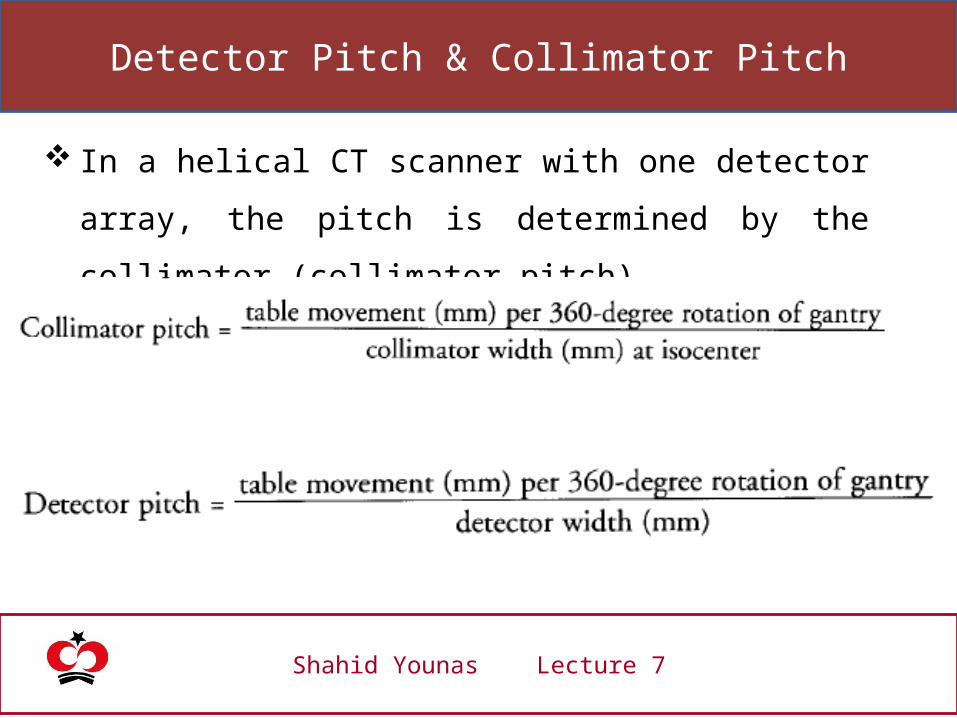
In a helical CT scanner with one detector array, the pitch is determined by the collimator (collimator pitch).
Single and Multiple detector arrays scanner have different definitions.
Detector Pitch & Collimator Pitch
Shahid Younas Lecture 7
In a helical CT scanner with one detector array, the pitch is
determined by the collimator (collimator pitch),
Detector Pitch & Collimator Pitch
Shahid Younas Lecture 7
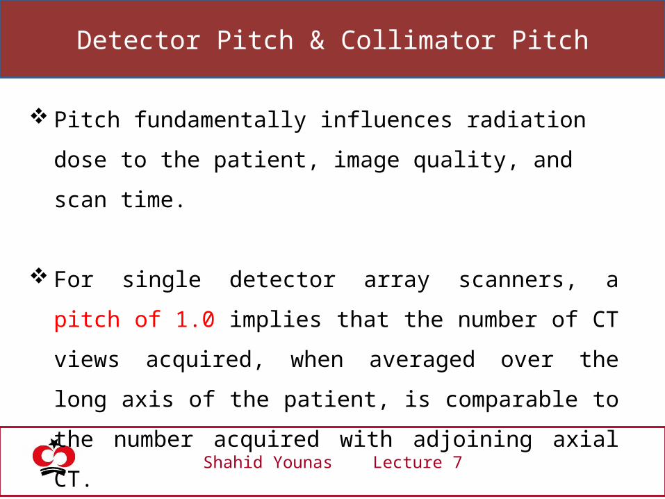
Pitch fundamentally influences radiation dose to the patient,
image quality, and scan time.
For single detector array scanners, a pitch of 1.0 implies that
the number of CT views acquired, when averaged over the
long axis of the patient, is comparable to the number acquired
with adjoining axial CT.
Detector Pitch & Collimator Pitch
Shahid Younas Lecture 7
A pitch of less than 1.0 involves over-scanning may result in
some slight improvement in image quality and a higher
radiation dose to the patient.
pitches greater than 1.0, and pitches up to 1.5 are commonly
used.

Detector Pitch & Collimator Pitch
Shahid Younas Lecture 7
Collimator Pitch = T / C
Detector width = T / D
For a multiple detector array CT scanner with four detector arrays, a collimator pitch of 1.0 is equal to a detector pitch of 4.0.

Detector Pitch & Collimator Pitch
Shahid Younas Lecture 7
Scanners that have multiple detector arrays require a different
definition of pitch.
N = Number of detectors used

Detector Pitch & Collimator Pitch
Shahid Younas Lecture 7
For scanners with four detector arrays, detector pitches running
from 3 to 6 are used. A detector pitch of 3 for a four-detector
array scanner is equivalent to a collimator pitch of 0.75 (3/4), and
a detector pitch of 6 corresponds to a collimator pitch of 1.5
(6/4).

CT Number or Hounsfield Units
Shahid Younas Lecture 7
Each pixel in the image is represented by a high-precision
floating point number that is useful for computation but less
useful for display.
Consequently, after CT reconstruction, but before storing and
displaying, CT images are normalized and truncated to integer
values.

CT Number or Hounsfield Units
Shahid Younas Lecture 7
where µ(x,y) is the floating point number of the (x,y) pixel before conversion
CT numbers are quantitative, and this property leads to more accurate diagnosis in some clinical settings.

CT Dose Measurement
Shahid Younas Lecture 7
Compton scattering is the principal interaction mechanism
in CT, so the radiation dose attributable to scattered
radiation is considerable, and it can be higher than the
radiation dose from the primary beam.
The multiple scan average dose (MSAD) is the standard
for determining radiation dose in CT.

CT Dose Measurement Methods
Shahid Younas Lecture 7
CT Dose Measurement
Shahid Younas Lecture 7
The MSAD is the dose to tissue that includes the dose
attributable to scattered radiation emanating from all
adjacent slices.
The MSAD is defined as the average dose, at a particular
depth from the surface, resulting from a large series of CT
slices.

CT Dose Measurement
Shahid Younas Lecture 7
Anther way is the CT Dose Index (CTDI) to estimate MSAD.
The CTDI is defined by the U.S. Food and Drug Agency as
the radiation dose to any point in the patient including the
scattered radiation contribution from 7 CT slices in both
directions, for a total of 14 slices.

CT Dose Measurement Methods
Shahid Younas Lecture 7
TLD Dosimeters
Pencil Ionization
Chambers

CT Dose Measurement Methods
Same as previous but a correction factor is needed,
CT Artifacts
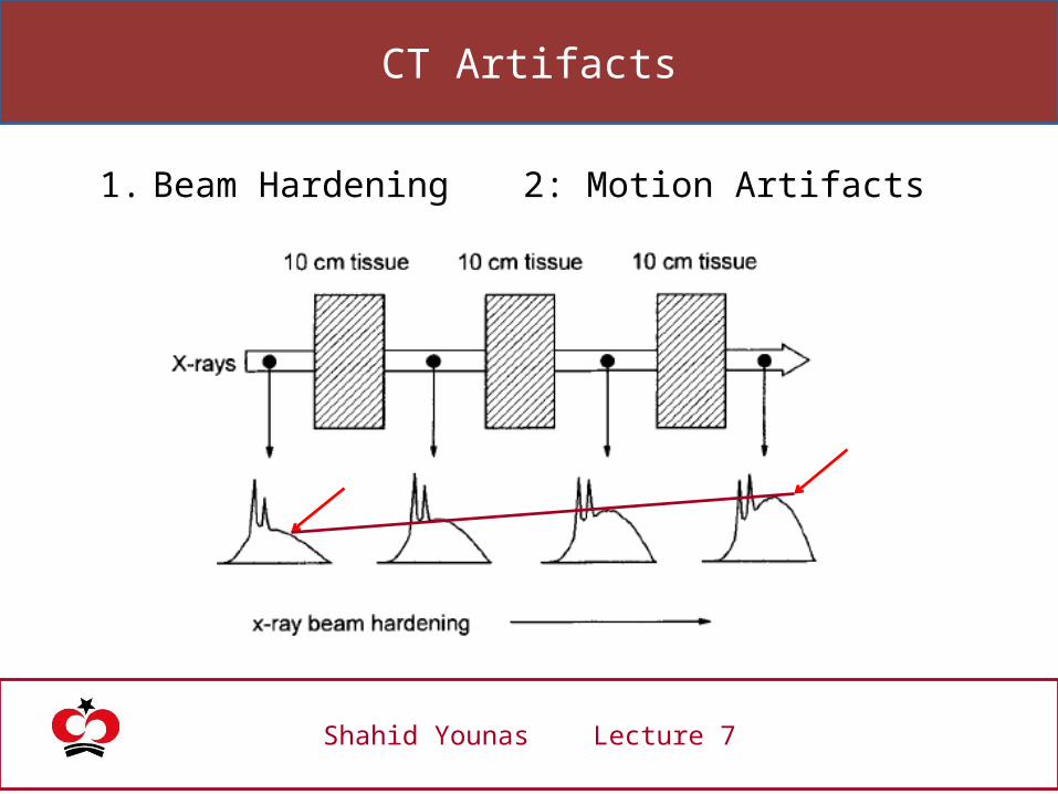
Shahid Younas Lecture 7
1. Beam Hardening 2: Motion Artifacts
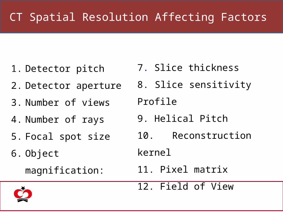
CT Spatial Resolution Affecting Factors
1. Detector pitch
2. Detector aperture
3. Number of views
4. Number of rays
5. Focal spot size
6. Object magnification:
7. Slice thickness
8. Slice sensitivity Profile
9. Helical Pitch
10. Reconstruction kernel
11. Pixel matrix
12. Field of View

Practical Sessions for Medical Physics
Dose Calibrator and sealed sources used in Nuclear Medicine
March, 10, 20144 to 5 pm
Hot Lab of Nuclear Medicine

Future Books