
KOH SMEAR D. M. M. Lab.. KOH Smear Aim of the test Treatment of KOH allows rapid observation of...
17
KOH SMEAR D. M. M. Lab .
-
Upload
job-fitzgerald -
Category
Documents
-
view
215 -
download
1
Transcript of KOH SMEAR D. M. M. Lab.. KOH Smear Aim of the test Treatment of KOH allows rapid observation of...

KOH SMEARD. M. M. Lab.

KOH Smear
Aim of the test
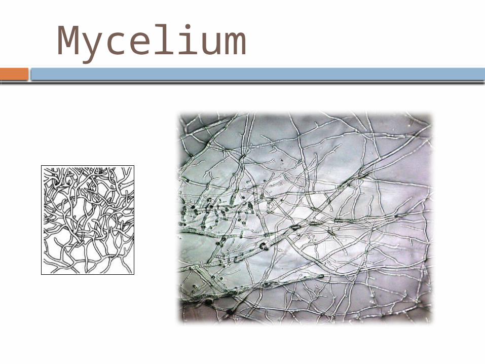
Treatment of KOH allows rapid observation of fungal elements because it digests protein debris and clears keratinized tissue so fungi present in specimen can be seen more readily.
Because of several variable factors, a KOH preparation may not reveal fungi even when they are present, the collection of the specimen by the physician, the selection of the portion of the specimen to be examined by the laboratory, and the size and number of the organism are extremely important.
The test is useful as a rapid screening a specimen for fungal elements (hyphae and spores)

Types of SpecimenSuperficial Mycoses:
I. Skin - scrapings collected with either a scalpel or the edge of a glass slide from the outer area of the lesion. Sent to the laboratory in folded black paper or any sterile container.

Types of SpecimenSuperficial Mycoses:
II. Hair - specimens scraped from the scalp with a scalpel or hairs plucked with forceps. Infected hairs are easily removed with their stubs.

Types of Specimen
III. Nail - any damaged, discolored or brittle parts of the nail are sampled by clipping the full thickness. Where a nail is grossly thickened scrapings can be taken from underneath the nail to add to the clippings.
Superficial Mycoses:

Types of Specimen
IV. Mucous membranes - scrapings from the mouth or vagina are better than swabs if the material is to be processed immediately. However swabs are commonly used and as yeast’s quickly lose viability on drying a transport swab is preferred.
Superficial Mycoses:

Types of Specimen
I. Scrapings/crusts - can be used for microscopy and culture but bacterial contamination may be a problem. Cleaning of the site with 70 percent alcohol prior to taking the sample may help.
II. Pus - should be aspirated and sent with any associated granules to the laboratory in a sterile container rather than a swab.
III. Biopsy specimens - transported to the laboratory in a sterile container
Subcutaneous Mycoses.

Pre Specimen ProcessingSpecimen collection
See types of specimens
Who will collect the specimenDepending on the type of the specimen
Quantity of specimenSee types of specimens
Time relapse before processing the sample
As soon as possible to prevent overgrowth of contaminating bacteria or fungi, and storage at room temperature.

Specimen Processing
1. Place a drop of (10-20)% KOH in the center of a clean microscopic slide.
2. Place a fragment of tissue, purulent materials or scraping in KOH drop.
3. Tease the materials well enough with corner of a coverslip to give a thin preparation or break up the materials with a sterile loop.
4. Mount with coverslip.
5. Allow preparation to digest for approximately 10 minutes or longer depending on the tested materials.
Procedure:

Procedure continue..…
6. Gently warm the slide (do not overheat) Gently press on the slide to help disperse tissue materials.
7. Screen under low power objective,
8. Then Use high (40 X) power magnification to verify the presence of fungal elements.


Hyphae

Hyphae

Yeast

Yeast

Post Specimen ProcessingResult reporting:Report any fungal element (Description of the hyphae is important, e.g. septated or non septated)
Turn around time:The results is expected 2 hours after specimen reception.