Knee OA ppt 2017 - Home Page - LECOM Education System · 2017-08-17 · 1 Presentation, Diagnosis,...
31
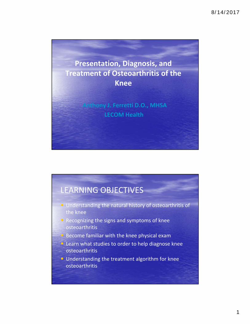
8/14/2017 1 Presentation, Diagnosis, and Treatment of Osteoarthritis of the Knee Anthony J. Ferretti D.O., MHSA LECOM Health LEARNING OBJECTIVES • Understanding the natural history of osteoarthritis of the knee • Recognizing the signs and symptoms of knee osteoarthritis • Become familiar with the knee physical exam • Learn what studies to order to help diagnose knee osteoarthritis • Understanding the treatment algorithm for knee osteoarthritis
-
Upload
nguyennhan -
Category
Documents
-
view
214 -
download
0
Transcript of Knee OA ppt 2017 - Home Page - LECOM Education System · 2017-08-17 · 1 Presentation, Diagnosis,...
8/14/2017
1
Presentation, Diagnosis, and Treatment of Osteoarthritis of the
Knee
Anthony J. Ferretti D.O., MHSA
LECOM Health
LEARNING OBJECTIVES
• Understanding the natural history of osteoarthritis of the knee
• Recognizing the signs and symptoms of knee osteoarthritis
• Become familiar with the knee physical exam
• Learn what studies to order to help diagnose knee osteoarthritis
• Understanding the treatment algorithm for knee osteoarthritis
8/14/2017
2
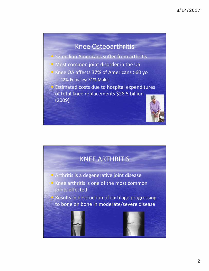
Knee Osteoarthritis
• 52 million Americans suffer from arthritis
• Most common joint disorder in the US
• Knee OA affects 37% of Americans >60 yo
– 42% Females: 31% Males
• Estimated costs due to hospital expenditures of total knee replacements $28.5 billion (2009)
KNEE ARTHRITIS
• Arthritis is a degenerative joint disease
• Knee arthritis is one of the most common joints effected
• Results in destruction of cartilage progressing to bone on bone in moderate/severe disease

8/14/2017
3
General Principles
• Knee is composed of three joint compartments
– Medial, lateral and patellofemoral compartments
• Normal knee functions as a complex hinge allowing
– Flexion, extension, rotation, and gliding
• Weight distribution across the knee with normal alignment
– 60% through medial compartment
– 40% through lateral compartment
Zones of Articular Cartilage
• Superficial (tangential or zone I) – Forms the gliding surface– Collagen fibers parallel to the articular surface
• Middle (transitional or zone II) – Thicker with oblique collagen fibers– Constitutes most of the cartilage depth
• Deep (radial or zone III)– Collagen fibers perpendicular to articular surface
• Calcified cartilage (zone IV) – Radially aligned collagen fibers
8/14/2017
4
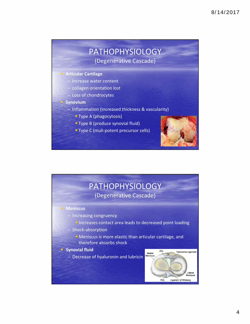
PATHOPHYSIOLOGY (Degenerative Cascade)
• Articular Cartilage
– Increase water content
– collagen orientation lost
– Loss of chondrocytes
• Synovium
– Inflammation (increased thickness & vascularity)
• Type A (phagocytosis)• Type B (produce synovial fluid)• Type C (muli‐potent precursor cells)
PATHOPHYSIOLOGY (Degenerative Cascade)
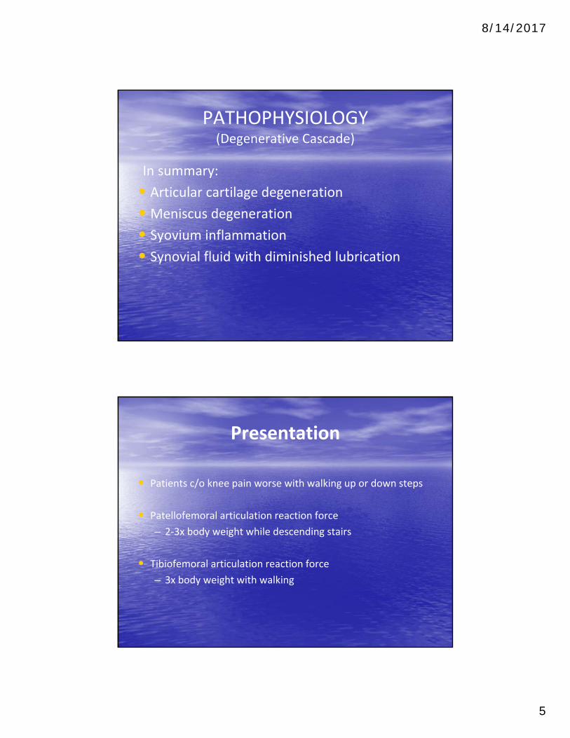
• Meniscus
– Increasing congruency
• Increases contact area leads to decreased point loading– Shock‐absorption
•Meniscus is more elastic than articular cartilage, and therefore absorbs shock
• Synovial fluid
– Decrease of hyaluronin and lubricin
8/14/2017
5
PATHOPHYSIOLOGY (Degenerative Cascade)
In summary:
• Articular cartilage degeneration
• Meniscus degeneration
• Syovium inflammation
• Synovial fluid with diminished lubrication
Presentation
• Patients c/o knee pain worse with walking up or down steps
• Patellofemoral articulation reaction force
– 2‐3x body weight while descending stairs
• Tibiofemoral articulation reaction force
– 3x body weight with walking

8/14/2017
6
Presentation
• Symptoms may wax & wane often in correlation with recent activities or body stressors (illness)
• Not uncommon for OA exacerbation to occur during hospital admission for unrelated event – Surgery, CHF, COPD, pneumonia, viral illness
Physical Examination
• Joint line tenderness to palpation
– Degenerative compartment will often correlate to overall alignment
– Varus deformity = medial joint space narrowing
– Valgus deformity = lateral joint space narrowing
• Effusion
– Persistent large/tense effusion may represent degenerative meniscus tear (without specific event)
8/14/2017
7
Physical Examination
• McMurray’s test
– Flex knee & place one hand on medial side of knee
– Gently externally rotate leg & bring knee into extension
– Palpable click is a positive test (medial meniscus tear)
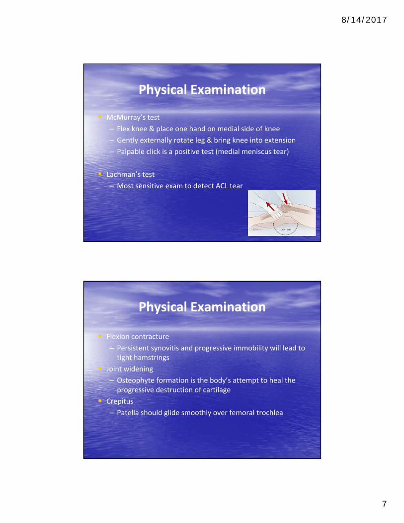
• Lachman’s test
– Most sensitive exam to detect ACL tear
Physical Examination
• Flexion contracture
– Persistent synovitis and progressive immobility will lead to tight hamstrings
• Joint widening
– Osteophyte formation is the body’s attempt to heal the progressive destruction of cartilage
• Crepitus
– Patella should glide smoothly over femoral trochlea
8/14/2017
8
Radiographs
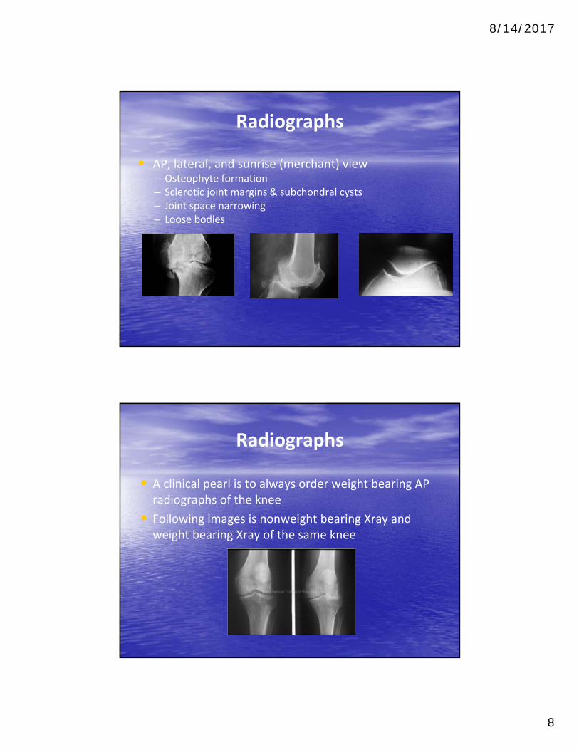
• AP, lateral, and sunrise (merchant) view– Osteophyte formation– Sclerotic joint margins & subchondral cysts– Joint space narrowing– Loose bodies
Radiographs
• A clinical pearl is to always order weight bearing AP radiographs of the knee
• Following images is nonweight bearing Xray and weight bearing Xray of the same knee

8/14/2017
9
MRI
• MRI can be useful in the workup for osteoarthritis of the knee if a degenerative meniscus tear is suspected
MRI
• Degenerative tears in older patients are most commonly found in the posterior horn of the medial meniscus
• Correlation to physical exam findings and/or mechanical symptoms is critical to confirm diagnosis

8/14/2017
10
MRI
• MRI has been shown to find asymptomatic degenerative meniscus tears in over 60% of patients > 65 yo
• Diagnosis of symptomatic meniscus tear becomes difficult in the setting of concomitant OA
• Articular cartilage destruction may be the root cause of the patients symptoms
MRI
• Adjacent bone marrow lesions can be identified in osteoarthritis
• Representing bone marrow edema of subchondral bone
8/14/2017
11
Nonoperative Treatment
• Weight Loss
– Indications: symptomatic OA and BMI > 25
– Improvement in joint pain and function
– Reducing the risk of progression of OA
– Each pound of weight loss results in a fourfold reduction in the load exerted on the knee per step during daily activities
Nonoperative Treatment
• Exercise / Physical therapy
– First line treatment for all patients with symptomatic arthritis
– Low impact aerobic exercise
• Swimming
•Bicycling
– Improving flexibility and strengthening muscles improve functional outcome and pain scores

8/14/2017
12
Nonoperative Treatment
• Exercise / Physical therapy
– Quadriceps strengthening
• Improve stability of joints and lessens pain
– Hamstring stretching
•Prevention of flexion contracture
– Combination of supervised exercises and home program show the best results
– Benefits often lost after 6 months if exercises are stopped
Nonoperative Treatment
• Viscosupplement intra‐articular injections
– Hyaluronic acid (HA) forms the backbone of aggrecans
– The macromolecule that makes up cartilage matrix
– HA at low load speeds acts as a lubricant and faster movements as a shock absorber
– In OA the concentration of HA is reduced by half to one third of normal

8/14/2017
13
Hyaluronic Acid vs Corticosteroid Injections
• Meta‐analysis, Randomized trial
• Reported effects of intra‐articular hyaluronic acid vs corticosteroids on knee osteoarthritis
• 7 eligible trials included 606 patients
• 0 – 4 weeks: – Intraarticular corticosteroids appear to be more effective for pain than
intraarticular hyaluronic acid
• 4 – 8 weeks:– The 2 approaches have equal efficacy
• > 8 weeks:– Hyaluronic acid has greater efficacy
Pharmacologic Treatment
• NSAIDS
– First line treatment for all patients with symptomatic arthritis
– Risk factors for adverse reaction
• Age > 60
•Multiple medical comorbidities
•H/o PUD•H/o GI bleeding• Concurrent corticosteroid use
• Anticoagulant use
8/14/2017
14
Pharmacologic Treatment
• NSAIDS
– Cox‐2 inhibitors limit inflammation without interfering with normal production of protective prostaglandins and thromboxane
•Decrease the potential gastric toxicity of NSAIDs
– Cox‐2 inhibitors along with all NSAIDs may cause cardiovascular and renal side effects to varying degrees
Pharmacologic Treatment
• Acetaminophen at doses of up to 4 g per day have demonstrated to be superior to placebo in relief of pain resulting form OA
• Acetaminophen less effective than NSAIDs
• Tramadol
– Strongly recommended by AAOS
8/14/2017
15
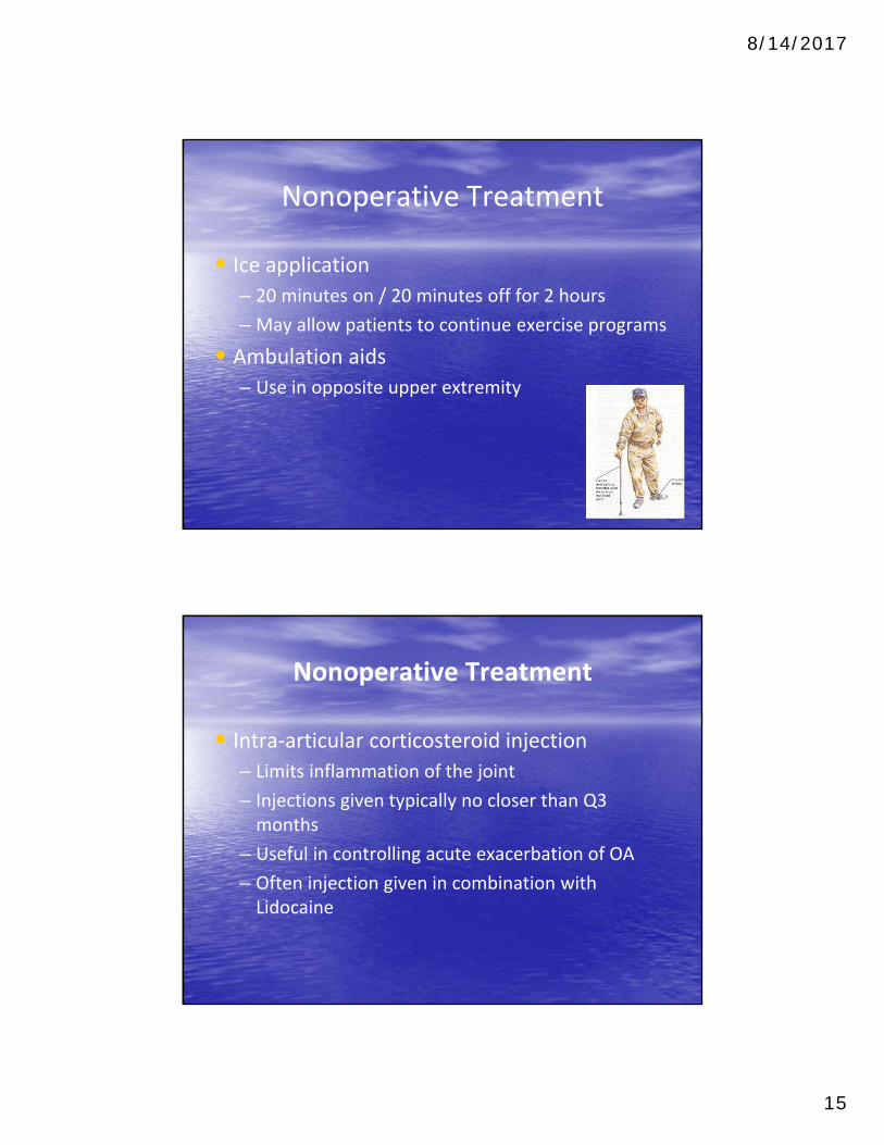
Nonoperative Treatment
• Ice application
– 20 minutes on / 20 minutes off for 2 hours
– May allow patients to continue exercise programs
• Ambulation aids
– Use in opposite upper extremity
Nonoperative Treatment
• Intra‐articular corticosteroid injection
– Limits inflammation of the joint
– Injections given typically no closer than Q3 months
– Useful in controlling acute exacerbation of OA
– Often injection given in combination with Lidocaine

8/14/2017
16
Nonoperative Treatment
• Unloader brace
– Used less frequently
– Designed to reduce reactive forces in involved compartment
– Provides 3 point bending force
– $ 800‐1000
Orthotics
• Padded shoe inserts
– Decrease in joint impact forces to joints
– $ 8‐22
• Varus knee deformity
– Lateral heel wedges

8/14/2017
17
Arthroscopy
• Debridement
– Synovectomy (plica removal)
– Removal of loose bodies
– Chondroplasty
– Resection of torn/damaged meniscus
Arthroscopy
• Direct Visualization of articular cartilage
Outerbridge Arthroscopic Grading System
Grade 0 Normal cartilage
Grade I Softening and swelling
Grade II Partial thickness defect, fissures < 1.5cm diameter
Grade III Fissures down to subchondral bone, diameter > 1.5cm
Grade IV Exposed subchondral bone
8/14/2017
18
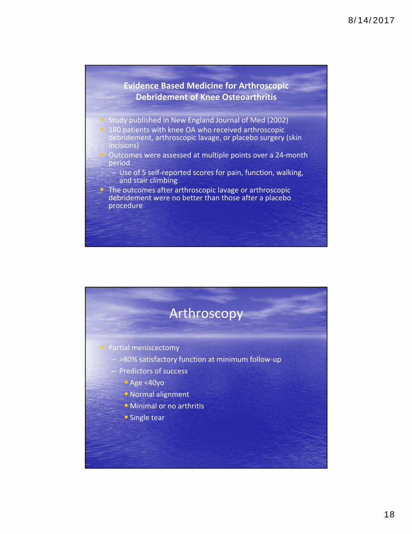
Evidence Based Medicine for Arthroscopic Debridement of Knee Osteoarthritis
• Study published in New England Journal of Med (2002)• 180 patients with knee OA who received arthroscopic
débridement, arthroscopic lavage, or placebo surgery (skin incisions)
• Outcomes were assessed at multiple points over a 24‐month period– Use of 5 self‐reported scores for pain, function, walking, and stair climbing
• The outcomes after arthroscopic lavage or arthroscopic debridement were no better than those after a placebo procedure
Arthroscopy
• Partial meniscectomy
– >80% satisfactory function at minimum follow‐up
– Predictors of success
•Age <40yo•Normal alignment
•Minimal or no arthritis
• Single tear

8/14/2017
19
Arthroscopy
• Total meniscectomy
– 70% have arthritic Xray changes 3 years after surgery
– 100% have arthrosis at 20 years
– Severity of degenerative changes is proportional to percent of the meniscus removed
Unicompartmental Knee Arthroplasty
• Indications– Isolated unicompartmental noninflammatory arthritis
– Deformity of less than 10 degrees
– Intact anterior cruciate ligament (ACL)
– Little or no joint subluxation
– Little or no patellofemoral disease
– Weight < 90 kg
8/14/2017
20
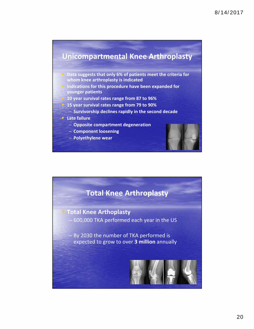
Unicompartmental Knee Arthroplasty
• Data suggests that only 6% of patients meet the criteria for whom knee arthroplasty is indicated
• Indications for this procedure have been expanded for younger patients
• 10 year survival rates range from 87 to 96%
• 15 year survival rates range from 79 to 90%
– Survivorship declines rapidly in the second decade
• Late failure
– Opposite compartment degeneration
– Component loosening
– Polyethylene wear
Total Knee Arthroplasty
• Total Knee Arthoplasty– 600,000 TKA performed each year in the US
– By 2030 the number of TKA performed is expected to grow to over 3 million annually
8/14/2017
21
Total Knee Arthroplasty
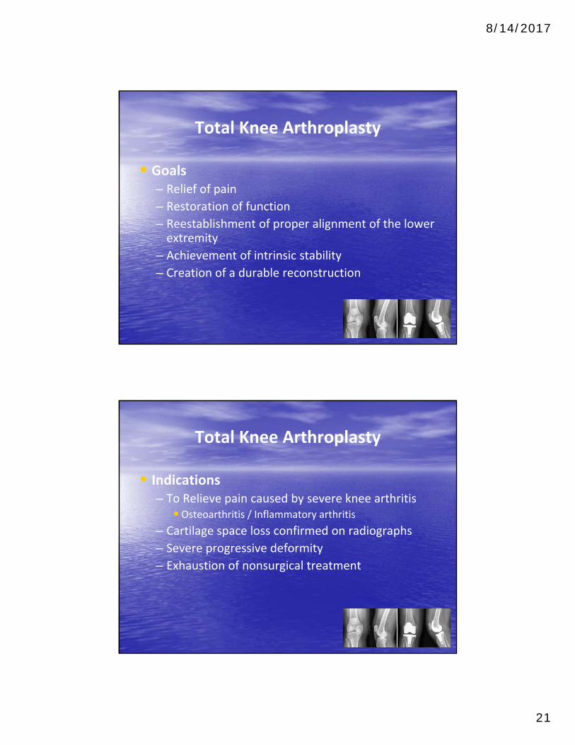
• Goals– Relief of pain
– Restoration of function
– Reestablishment of proper alignment of the lower extremity
– Achievement of intrinsic stability
– Creation of a durable reconstruction
Total Knee Arthroplasty
• Indications– To Relieve pain caused by severe knee arthritis
•Osteoarthritis / Inflammatory arthritis
– Cartilage space loss confirmed on radiographs
– Severe progressive deformity
– Exhaustion of nonsurgical treatment
8/14/2017
22
Total Knee Arthroplasty
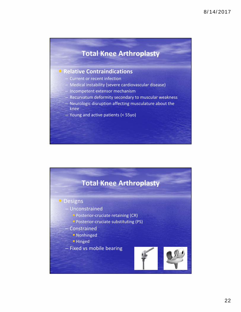
• Relative Contraindications– Current or recent infection
– Medical instability (severe cardiovascular disease)
– Incompetent extensor mechanism
– Recurvatum deformity secondary to muscular weakness
– Neurologic disruption affecting musculature about the knee
– Young and active patients (< 55yo)
Total Knee Arthroplasty
• Designs– Unconstrained
•Posterior‐cruciate retaining (CR)• Posterior‐cruciate substituting (PS)
– Constrained•Nonhinged•Hinged
– Fixed vs mobile bearing
8/14/2017
23
Total Knee Arthroplasty
• Survival rates range from 91 to 96% at 15 year follow up
– Survival rate for cemented CR ranges from 96‐97% at 10 year follow up
– Survival rate for cemented PS ranges is 97% at 10 year follow up and 94% at 13 year follow up
– Survival rate for cementless TKA ranges from 95 to 97% at 10 to 12 year follow up
Total Knee Arthroplasty
• Complications– Symptomatic instability occurs in 1‐2%
•Ligament instability accounts for 6% of revision TKA
– Patellofemoral maltracking
•Most common cause of revision TKA (8‐35%)
– Vascular injury (rare)
– Nerve palsy
• Incidence of nerve injury reported at 0.3%– Infection
• Incidence of infection reported at 1‐2%
8/14/2017
24
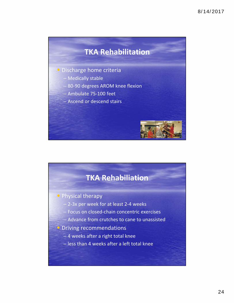
TKA Rehabilitation
• Discharge home criteria
– Medically stable
– 80‐90 degrees AROM knee flexion
– Ambulate 75‐100 feet
– Ascend or descend stairs
TKA Rehabiliation
• Physical therapy
– 2‐3x per week for at least 2‐4 weeks
– Focus on closed‐chain concentric exercises
– Advance from crutches to cane to unassisted
• Driving recommendations
– 4 weeks after a right total knee
– less than 4 weeks after a left total knee

8/14/2017
25
Question 1
Which of the following non‐operative treatments for osteoarthritis has the best evidence to support its use?
• 1. Combination of supervised and home exercise programs
• 2. Hyaluronic acid injections
• 3. Lateral heel wedge
• 4. Acetaminophen
• 5. Glucosamine
Question 1
Which of the following non‐operative treatments for osteoarthritis has the best evidence to support its use?
• 1. Combination of supervised and home exercise programs
• 2. Hyaluronic acid injections
• 3. Lateral heel wedge
• 4. Acetaminophen
• 5. Glucosamine

8/14/2017
26
Question 2A 62‐year‐old female undergoes an uncomplicated primary total knee replacement. Her knee range‐of‐motion pre‐operatively was 0‐135 degrees of flexion. Which of the following is true regarding the immediate post‐operative use of a continuous passive motion machine in this patient?
• 1. Reduced risk of venous thromboembolism
• 2. No long‐term difference in ROM compared to patients not using CPM
• 3. Increased passive knee flexion at 6 months
• 4. Increased length of hospitalization
• 5. Decreased risk of surgical site infection

8/14/2017
27
Question 3
The following are risk factors for developing knee osteoarthritis EXCEPT:
• 1. Knee articular trauma
• 2. Metabolic syndrome
• 3. Female gender
• 4. Increased age
• 5. Participating in physical fitness
Question 3
The following are risk factors for developing knee osteoarthritis EXCEPT:
• 1. Knee articular trauma
• 2. Metabolic syndrome
• 3. Female gender
• 4. Increased age
• 5. Participating in physical fitness
8/14/2017
28
Question 4
• All the following are common complaints associated with knee osteoarthritis EXCEPT?
• 1. Knee pain at night
• 2. Knee pain while climbing stairs
• 3. Knee stiffness
• 4. Instability, clicking, or locking sensation
• 5. Numbness in the ankle or foot
Question 4
• All the following are common complaints associated with knee osteoarthritis EXCEPT?
• 1. Knee pain at night
• 2. Knee pain while climbing stairs
• 3. Knee stiffness
• 4. Instability, clicking, or locking sensation
• 5. Numbness in the ankle or foot

8/14/2017
29
Question 5
Which radiographic images are most commonly used to identify the degree of degenerative joint disease caused by knee osteoarthritis?
1. Knee MRI to identify meniscal pathology
2. Knee CT scan
3. X‐ray images of knee with patient lying down
4. Ultrasound images of the knee joint
5. X‐rays: Standing AP, lateral, and sunrise views of the knee

8/14/2017
30
THE END
References
• Bannuru,R., Natov, N., Obadan, I., Price, L, Schmid, C., McAlindon, T. (2009). Therapeutic trajectory of hyaluronic acid versus corticosteroids in the treatment of knee osteoarthritis: a systematic review and meta‐analysis. Journal of Arthritis Rheumatology. Dec 15;61(12):1704‐11
• Boyer, M. (2014) Primary Knee Arthroplasty. Wellman, S., Bolognesi, M. AAOS Comprehensive Orthopaedic Review (pp 1289‐1304) Rosemont, IL: American Academy of Orthopaedic Surgeons.
• Fischgrund, J. (2008). Knee Reconstruction and Replacement. Peters, C., Crofoot, C. Orthopaedic Knowledge Update (pp 457‐472). Rosemont, IL: American Academy of Orthopaedic Surgeons.
• Hoshino, A., Wallace, W. (1987). Impact‐absorbing properties of the human knee. Journal of Bone Joint Surgery British, 69(5), 807‐811.
• Miller, M., Thompson, S., Hart, J. (2012). Total Knee Arthroplasty. Review of Orthopaedics (pp 394‐398). Philadelphia, PA: Saunders Elsevier.
8/14/2017
31
References
• Moore, D. Orthobullets: Basic Science. Linage Medical. 2015. March, 29. www.orthobullets.com.
• Moore, D. Orthobullets: Adult Reconstruction. Linage Medical. 2015. March, 29. www.orthobullets.com.
• Moore, D. Orthobullets: Sports. Linage Medical. 2015. March, 29. www.orthobullets.com.
• Moseley, J., O'Malley, K., Petersen, N., Menke, T., Brody, B., Kuykendall, D., Hollingsworth, J., Ashton, C., Wray, N. (2002). A controlled trial of arthroscopic surgery for osteoarthritis of the knee. New England Journal of Medicine: 347(2):81‐8
• Sheth, N., Lonner, J. (2009). Total Knee Arthroplasty. Pill, S. Gowned and Gloved Orthopaedics (pp 251‐267). Philadelphia, PA: Saunders Elsevier