Klaas(N) M.A. Bax, MD, PhD, FRCS(Ed) - Biliary · PDF fileJejunum for bridging long-gap...
6
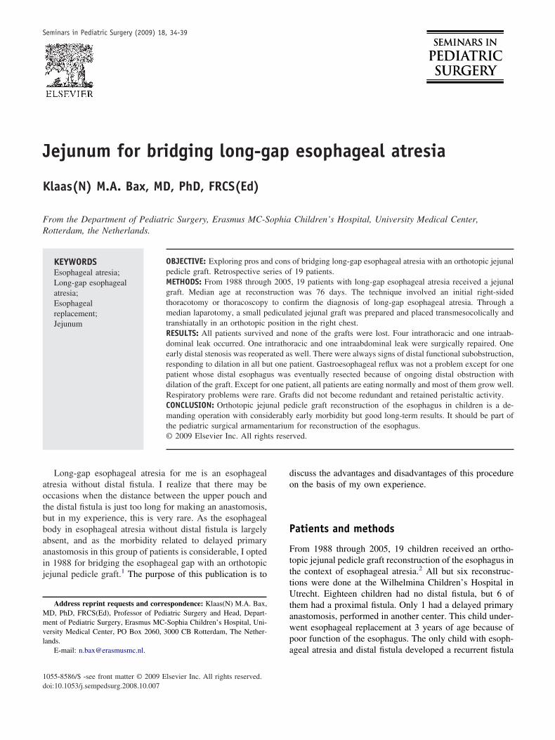
Jejunum for bridging long-gap esophageal atresia Klaas(N) M.A. Bax, MD, PhD, FRCS(Ed) From the Department of Pediatric Surgery, Erasmus MC-Sophia Children’s Hospital, University Medical Center, Rotterdam, the Netherlands. OBJECTIVE: Exploring pros and cons of bridging long-gap esophageal atresia with an orthotopic jejunal pedicle graft. Retrospective series of 19 patients. METHODS: From 1988 through 2005, 19 patients with long-gap esophageal atresia received a jejunal graft. Median age at reconstruction was 76 days. The technique involved an initial right-sided thoracotomy or thoracoscopy to confirm the diagnosis of long-gap esophageal atresia. Through a median laparotomy, a small pediculated jejunal graft was prepared and placed transmesocolically and transhiatally in an orthotopic position in the right chest. RESULTS: All patients survived and none of the grafts were lost. Four intrathoracic and one intraab- dominal leak occurred. One intrathoracic and one intraabdominal leak were surgically repaired. One early distal stenosis was reoperated as well. There were always signs of distal functional subobstruction, responding to dilation in all but one patient. Gastroesophageal reflux was not a problem except for one patient whose distal esophagus was eventually resected because of ongoing distal obstruction with dilation of the graft. Except for one patient, all patients are eating normally and most of them grow well. Respiratory problems were rare. Grafts did not become redundant and retained peristaltic activity. CONCLUSION: Orthotopic jejunal pedicle graft reconstruction of the esophagus in children is a de- manding operation with considerably early morbidity but good long-term results. It should be part of the pediatric surgical armamentarium for reconstruction of the esophagus. © 2009 Elsevier Inc. All rights reserved. KEYWORDS Esophageal atresia; Long-gap esophageal atresia; Esophageal replacement; Jejunum Long-gap esophageal atresia for me is an esophageal atresia without distal fistula. I realize that there may be occasions when the distance between the upper pouch and the distal fistula is just too long for making an anastomosis, but in my experience, this is very rare. As the esophageal body in esophageal atresia without distal fistula is largely absent, and as the morbidity related to delayed primary anastomosis in this group of patients is considerable, I opted in 1988 for bridging the esophageal gap with an orthotopic jejunal pedicle graft. 1 The purpose of this publication is to discuss the advantages and disadvantages of this procedure on the basis of my own experience. Patients and methods From 1988 through 2005, 19 children received an ortho- topic jejunal pedicle graft reconstruction of the esophagus in the context of esophageal atresia. 2 All but six reconstruc- tions were done at the Wilhelmina Children’s Hospital in Utrecht. Eighteen children had no distal fistula, but 6 of them had a proximal fistula. Only 1 had a delayed primary anastomosis, performed in another center. This child under- went esophageal replacement at 3 years of age because of poor function of the esophagus. The only child with esoph- ageal atresia and distal fistula developed a recurrent fistula Address reprint requests and correspondence: Klaas(N) M.A. Bax, MD, PhD, FRCS(Ed), Professor of Pediatric Surgery and Head, Depart- ment of Pediatric Surgery, Erasmus MC-Sophia Children’s Hospital, Uni- versity Medical Center, PO Box 2060, 3000 CB Rotterdam, The Nether- lands. E-mail: [email protected]. 1055-8586/$ -see front matter © 2009 Elsevier Inc. All rights reserved. doi:10.1053/j.sempedsurg.2008.10.007 Seminars in Pediatric Surgery (2009) 18, 34-39
-
Upload
phamnguyet -
Category
Documents
-
view
218 -
download
1
Transcript of Klaas(N) M.A. Bax, MD, PhD, FRCS(Ed) - Biliary · PDF fileJejunum for bridging long-gap...
J
K
FR
aotbbaaij
Mmvl
1d
Seminars in Pediatric Surgery (2009) 18, 34-39
ejunum for bridging long-gap esophageal atresia
laas(N) M.A. Bax, MD, PhD, FRCS(Ed)
rom the Department of Pediatric Surgery, Erasmus MC-Sophia Children’s Hospital, University Medical Center,
otterdam, the Netherlands.OBJECTIVE: Exploring pros and cons of bridging long-gap esophageal atresia with an orthotopic jejunalpedicle graft. Retrospective series of 19 patients.METHODS: From 1988 through 2005, 19 patients with long-gap esophageal atresia received a jejunalgraft. Median age at reconstruction was 76 days. The technique involved an initial right-sidedthoracotomy or thoracoscopy to confirm the diagnosis of long-gap esophageal atresia. Through amedian laparotomy, a small pediculated jejunal graft was prepared and placed transmesocolically andtranshiatally in an orthotopic position in the right chest.RESULTS: All patients survived and none of the grafts were lost. Four intrathoracic and one intraab-dominal leak occurred. One intrathoracic and one intraabdominal leak were surgically repaired. Oneearly distal stenosis was reoperated as well. There were always signs of distal functional subobstruction,responding to dilation in all but one patient. Gastroesophageal reflux was not a problem except for onepatient whose distal esophagus was eventually resected because of ongoing distal obstruction withdilation of the graft. Except for one patient, all patients are eating normally and most of them grow well.Respiratory problems were rare. Grafts did not become redundant and retained peristaltic activity.CONCLUSION: Orthotopic jejunal pedicle graft reconstruction of the esophagus in children is a de-manding operation with considerably early morbidity but good long-term results. It should be part ofthe pediatric surgical armamentarium for reconstruction of the esophagus.© 2009 Elsevier Inc. All rights reserved.
KEYWORDSEsophageal atresia;Long-gap esophagealatresia;Esophagealreplacement;Jejunum
do
P
FtttUtawp
Long-gap esophageal atresia for me is an esophagealtresia without distal fistula. I realize that there may beccasions when the distance between the upper pouch andhe distal fistula is just too long for making an anastomosis,ut in my experience, this is very rare. As the esophagealody in esophageal atresia without distal fistula is largelybsent, and as the morbidity related to delayed primarynastomosis in this group of patients is considerable, I optedn 1988 for bridging the esophageal gap with an orthotopicejunal pedicle graft.1 The purpose of this publication is to
Address reprint requests and correspondence: Klaas(N) M.A. Bax,D, PhD, FRCS(Ed), Professor of Pediatric Surgery and Head, Depart-ent of Pediatric Surgery, Erasmus MC-Sophia Children’s Hospital, Uni-
ersity Medical Center, PO Box 2060, 3000 CB Rotterdam, The Nether-ands.
aE-mail: [email protected].
055-8586/$ -see front matter © 2009 Elsevier Inc. All rights reserved.oi:10.1053/j.sempedsurg.2008.10.007
iscuss the advantages and disadvantages of this proceduren the basis of my own experience.
atients and methods
rom 1988 through 2005, 19 children received an ortho-opic jejunal pedicle graft reconstruction of the esophagus inhe context of esophageal atresia.2 All but six reconstruc-ions were done at the Wilhelmina Children’s Hospital intrecht. Eighteen children had no distal fistula, but 6 of
hem had a proximal fistula. Only 1 had a delayed primarynastomosis, performed in another center. This child under-ent esophageal replacement at 3 years of age because ofoor function of the esophagus. The only child with esoph-
geal atresia and distal fistula developed a recurrent fistula
actciolcd
T
Tb
tRfwewempt
liptli
d
amctdww
mfsopa
oarlrtt
lnepmti
tgidstid
enc
c
pgpffw
R
G
N
S
E
n
dytt
acfinect
35Bax Jejunum for Bridging Long-Gap Esophageal Atresia
nd long stenosis after the primary repair. Four of the 19hildren had trisomy 21, and 1 had the CHARGE associa-ion. Two more children were mentally retarded, 1 due toerebral hemorrhage complicated by hydrocephalus, 1 on andiopathic basis. One child had a duodenal atresia and an-ther child a rectovestibular fistula. There were no otherife-threatening associated anomalies. Only 1 child had aervical esophagostomy. Median age at replacement was 76ays (range 16-1080).
echnique
he technique has been described in detail before but isriefly repeated here.2
In the waiting period between birth and the jejunal in-erposition, the upper esophagus is emptied by means ofeplogle catheter suction. A gastrostomy is created for
eeding. As the incidence of proximal fistula in patientsithout distal fistula is high, a proximal fistula is excluded
arly.3 It is also wise to exclude intestinal malrotation,hich has a higher incidence in esophageal atresia.4,5 In the
vent of malrotation, the vascular supply of the jejunumay preclude its use for pedicle graft interposition, as hap-
ened in one of our patients. In this patient, not included inhis series, a gastric pull-up was performed.
Timing of the jejunal interposition depends on how prob-ematic the conservative approach is. In the event of repet-tive lung problems, the interposition procedure is not post-oned; otherwise, the jejunal interposition is performed at 8o 12 weeks. The day before surgery, antegrade whole-bowelavage is performed to leave all options open. In a patient notncluded in this series, a pedicle ileal graft was used.6
If a cervical esophagostomy had been created, it is takenown first.
I prefer a right-sided posterolateral thoracotomy, whichvoids interference with the aortic arch and allows for aore straight posterior mediastinal position of the graft. A
lassic but large posterolateral thoracotomy is performedhrough the bed of the 6th or 7th rib. After verification of theiagnosis of long-gap esophageal atresia, the skin is closedith a running nylon suture. Verification in the last patientas done thoracoscopically.Next, the patient is placed in a supine position and a
idline laparotomy is performed. The fundus is detachedrom the diaphragm, and the upper short gastric vessels areevered. The left crus is freed and the posterior hiatus ispened. Access is gained behind the distal esophagealouch into the right pleural cavity. The tunnel from thebdomen into the right chest is dilated with Hegars.
The creation of the pedicle graft is the most critical partf the operation. The first two or three mesenterial vesselsre divided between ligatures close to the main mesentericoute (Figure 1). The jejunum is transected close to Treitzigament, leaving enough proximal jejunal length for resto-ation of continuity. The jejunum is severed again oppositehe level of the third mesenteric branch. Only jejunum is
aken, not the arcade. The jejunum thus isolated is far too long. No more than maybe the 5 proximal centimeters areeeded. The rest therefore is removed, starting at the distalnd of the graft. Bowel continuity is restored behind theedicle of the graft. The graft is passed through the leftesocolon, behind the remaining short gastric vessels, and
hrough the posterior hiatus behind the esophageal remnantnto the right chest.
It is best to close the abdomen temporarily, to repositionhe child and to quickly access the right chest to check theraft. The distal esophageal pouch is opened longitudinallyn the posterolateral area. One should make sure that theistal part is widely patent. In one of our patients, the distalegment was stenotic over about 2 cm, which went unno-iced, however, until the graft had been trimmed, resultingn an anastomosis under tension and postoperative leakeveloped. Next, the upper pouch is opened.
An anastomosis is made between the upper and lowersophagus and the graft. Before finishing the anastomosis, aasogastric tube is passed through the graft into the stomach. Ahest drain is inserted and the thoracotomy closed in layers.
Finally, a gastrostomy is recreated and the abdomenlosed.
The patient is weaned from the ventilator when appro-riate. Gastrostomy feedings are started when there are noastric retentions. The drain is removed after 5 to 6 days,rovided a contrast study shows that there are no leaks. Oraleeding is then started. It is important to monitor the patientor stenosis. In case of distal stenosis, the graft will dilate,hich must be avoided at all times.
esults
eneral
one of the patients died and none of the grafts were lost.
pecific results
arly resultsGaining enough jejunal length was not a problem, even
ot in the patient with an esophagostomy.The median duration of endotracheal intubation was 5
ays (range 1-43). One child was excluded from this anal-sis because of iatrogenic stenosis of the trachea and long-erm intubation as a result. Three patients developed ARDS,wo in connection with leakage.
Five leaks occurred, four in the chest, and one in thebdomen. The leak in the abdomen as well as one leak in thehest were treated surgically. In one patient, a proximalstula was missed and this required ligation through theeck in a second operative session. One patient developedarly distal graft anastomotic occlusion requiring surgicalorrection. Dilation in this patient could not be performed ashe nasogastric tube had been removed prematurely and no
umen could be seen at esophagoscopy.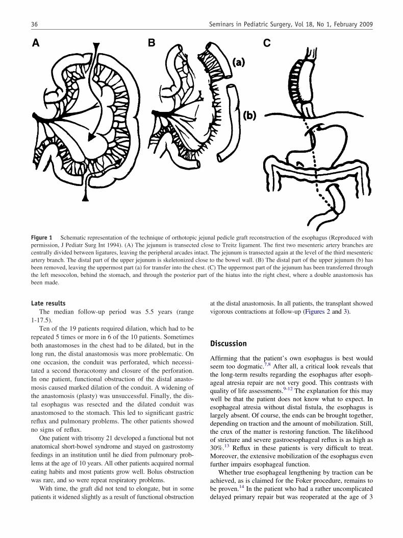
L
1
rblotImttarn
aflew
p
av
D
Astaqweldto3Mf
ab
Fpcabtb
36 Seminars in Pediatric Surgery, Vol 18, No 1, February 2009
ate resultsThe median follow-up period was 5.5 years (range
-17.5).Ten of the 19 patients required dilation, which had to be
epeated 5 times or more in 6 of the 10 patients. Sometimesoth anastomoses in the chest had to be dilated, but in theong run, the distal anastomosis was more problematic. Onne occasion, the conduit was perforated, which necessi-ated a second thoracotomy and closure of the perforation.n one patient, functional obstruction of the distal anasto-osis caused marked dilation of the conduit. A widening of
he anastomosis (plasty) was unsuccessful. Finally, the dis-al esophagus was resected and the dilated conduit wasnastomosed to the stomach. This led to significant gastriceflux and pulmonary problems. The other patients showedo signs of reflux.
One patient with trisomy 21 developed a functional but notnatomical short-bowel syndrome and stayed on gastrostomyeedings in an institution until he died from pulmonary prob-ems at the age of 10 years. All other patients acquired normalating habits and most patients grow well. Bolus obstructionas rare, and so were repeat respiratory problems.With time, the graft did not tend to elongate, but in some
igure 1 Schematic representation of the technique of orthotopiermission, J Pediatr Surg Int 1994). (A) The jejunum is transecteentrally divided between ligatures, leaving the peripheral arcadesrtery branch. The distal part of the upper jejunum is skeletonizedeen removed, leaving the uppermost part (a) for transfer into the che left mesocolon, behind the stomach, and through the posterioreen made.
atients it widened slightly as a result of functional obstruction d
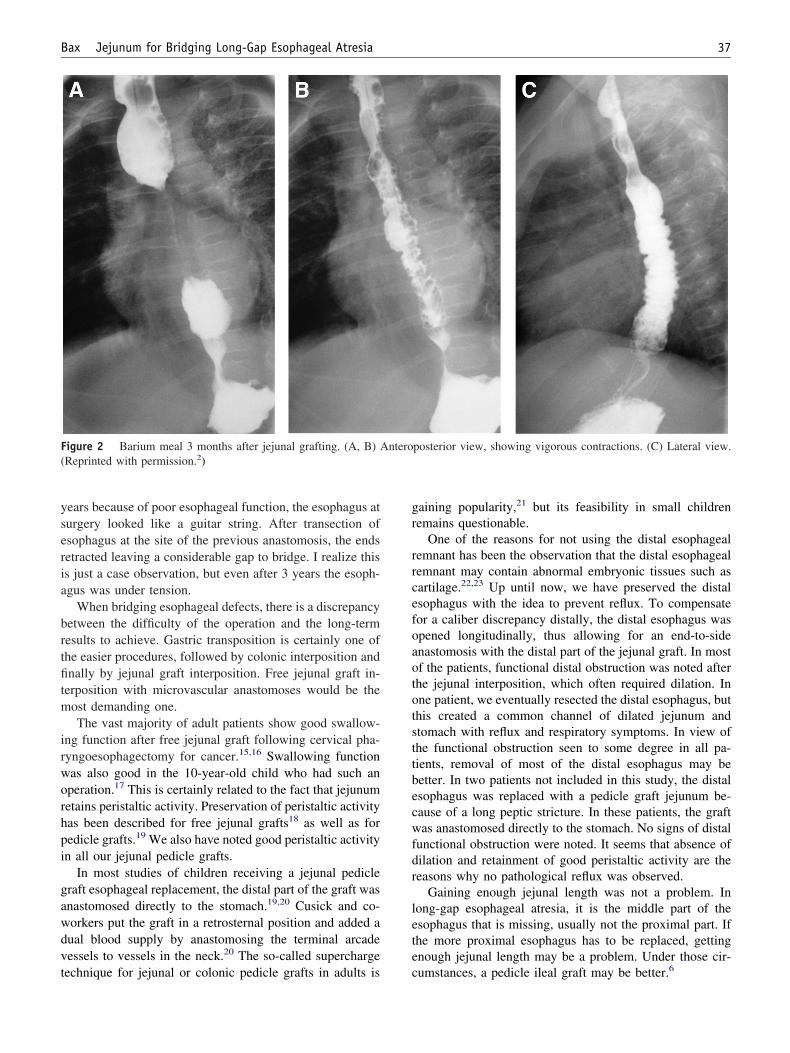
t the distal anastomosis. In all patients, the transplant showedigorous contractions at follow-up (Figures 2 and 3).
iscussion
ffirming that the patient’s own esophagus is best wouldeem too dogmatic.7,8 After all, a critical look reveals thathe long-term results regarding the esophagus after esoph-geal atresia repair are not very good. This contrasts withuality of life assessments.9-12 The explanation for this mayell be that the patient does not know what to expect. In
sophageal atresia without distal fistula, the esophagus isargely absent. Of course, the ends can be brought together,epending on traction and the amount of mobilization. Still,he crux of the matter is restoring function. The likelihoodf stricture and severe gastroesophageal reflux is as high as0%.13 Reflux in these patients is very difficult to treat.oreover, the extensive mobilization of the esophagus even
urther impairs esophageal function.Whether true esophageal lengthening by traction can be
chieved, as is claimed for the Foker procedure, remains toe proven.14 In the patient who had a rather uncomplicated
al pedicle graft reconstruction of the esophagus (Reproduced withe to Treitz ligament. The first two mesenteric artery branches areThe jejunum is transected again at the level of the third mesenterico the bowel wall. (B) The distal part of the upper jejunum (b) has) The uppermost part of the jejunum has been transferred through
f the hiatus into the right chest, where a double anastomosis has
c jejund clos
intact.close thest. (Cpart o
elayed primary repair but was reoperated at the age of 3
yseria
brtfitm
irworhpi
gawdvt
gr
rrcefoaototsttbecwfdr
lete
F(
37Bax Jejunum for Bridging Long-Gap Esophageal Atresia
ears because of poor esophageal function, the esophagus aturgery looked like a guitar string. After transection ofsophagus at the site of the previous anastomosis, the endsetracted leaving a considerable gap to bridge. I realize thiss just a case observation, but even after 3 years the esoph-gus was under tension.
When bridging esophageal defects, there is a discrepancyetween the difficulty of the operation and the long-termesults to achieve. Gastric transposition is certainly one ofhe easier procedures, followed by colonic interposition andnally by jejunal graft interposition. Free jejunal graft in-
erposition with microvascular anastomoses would be theost demanding one.The vast majority of adult patients show good swallow-
ng function after free jejunal graft following cervical pha-yngoesophagectomy for cancer.15,16 Swallowing functionas also good in the 10-year-old child who had such anperation.17 This is certainly related to the fact that jejunumetains peristaltic activity. Preservation of peristaltic activityas been described for free jejunal grafts18 as well as foredicle grafts.19 We also have noted good peristaltic activityn all our jejunal pedicle grafts.
In most studies of children receiving a jejunal pedicleraft esophageal replacement, the distal part of the graft wasnastomosed directly to the stomach.19,20 Cusick and co-orkers put the graft in a retrosternal position and added aual blood supply by anastomosing the terminal arcadeessels to vessels in the neck.20 The so-called supercharge
igure 2 Barium meal 3 months after jejunal grafting. (A, B) AReprinted with permission.2)
echnique for jejunal or colonic pedicle grafts in adults is c
aining popularity,21 but its feasibility in small childrenemains questionable.
One of the reasons for not using the distal esophagealemnant has been the observation that the distal esophagealemnant may contain abnormal embryonic tissues such asartilage.22,23 Up until now, we have preserved the distalsophagus with the idea to prevent reflux. To compensateor a caliber discrepancy distally, the distal esophagus waspened longitudinally, thus allowing for an end-to-sidenastomosis with the distal part of the jejunal graft. In mostf the patients, functional distal obstruction was noted afterhe jejunal interposition, which often required dilation. Inne patient, we eventually resected the distal esophagus, buthis created a common channel of dilated jejunum andtomach with reflux and respiratory symptoms. In view ofhe functional obstruction seen to some degree in all pa-ients, removal of most of the distal esophagus may beetter. In two patients not included in this study, the distalsophagus was replaced with a pedicle graft jejunum be-ause of a long peptic stricture. In these patients, the graftas anastomosed directly to the stomach. No signs of distal
unctional obstruction were noted. It seems that absence ofilation and retainment of good peristaltic activity are theeasons why no pathological reflux was observed.
Gaining enough jejunal length was not a problem. Inong-gap esophageal atresia, it is the middle part of thesophagus that is missing, usually not the proximal part. Ifhe more proximal esophagus has to be replaced, gettingnough jejunal length may be a problem. Under those cir-
posterior view, showing vigorous contractions. (C) Lateral view.
nteroumstances, a pedicle ileal graft may be better.6
dtwba
R1
1
1
1
1
1
Fp
38 Seminars in Pediatric Surgery, Vol 18, No 1, February 2009
Esophageal replacement with jejunum in children is aemanding operation with considerable morbidity, but inhis series, there was no mortality and none of the graftsere lost. The technique of jejunal pedicle grafting shoulde part of the pediatric surgical armamentarium for esoph-gus reconstruction.
eferences
1. Bax NMA, Rövekamp MH, Pull ter Gunne AJ, et al. Early one-stageorthotopic jejunal pedicle graft interposition in long-gap esophagealatresia. Pediatr Surg Int 1994;9:483-5.
2. Bax NM, van der Zee DC. Jejunal pedicle grafts for reconstruction ofthe esophagus in children. J Pediatr Surg 2007;42:363-9.
3. Bax KN, Roskott AM, van der Zee DC. Esophageal atresia withoutdistal fistula: high incidence of proximal fistula. J Pediatr Surg 2008;43:522-5.
4. Cieri MV, Arnold GL, Torfs CP. Malrotation in conjunction withesophageal atresia/tracheo-esophageal fistula. Teratology 1999;60:114-6.
5. Upadhyay V, Hea CM, Matthews RD. Oesophageal atresia: a hand-shake with malrotation. Eur J Pediatr Surg 2001;11:368-70.
6. Bax NM, Van Renterghem KM. Ileal pedicle grafting for esophageal
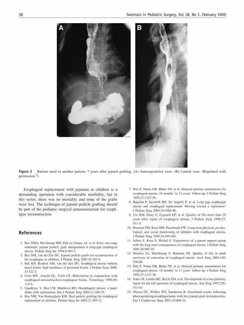
igure 3 Barium meal in another patient, 7 years after jejunalermission.2)
replacement in children. Pediatr Surg Int 2005;21:369-72.
7. Puri P, Ninan GK, Blake NS, et al. Delayed primary anastomosis foresophageal atresia: 18 months’ to 11 years’ follow-up. J Pediatr Surg1992;27:1127-30.
8. Bagolan P, Iacobelli BD, De Angelis P, et al. Long gap esophaegalatresia and esophageal replacement. Moving toward a separation?J Pediatr Surg 2004;39:1084-90.
9. Ure BM, Slany E, Eypasch EP, et al. Quality of life more than 20years after repair of esophageal atresia. J Pediatr Surg 1998;33:511-5.
0. Bouman NH, Koot HM, Hazebroek FW. Long-term physical, psycho-logical, and social functioning of children with esophageal atresia.J Pediatr Surg 1999;34:399-404.
1. Schier F, Korn S, Michel E. Experiences of a parent support groupwith the long-term consequences of esophageal atresia. J Pediatr Surg2001;36:605-10.
2. Deurloo JA, Ekkelkamp S, Hartman EE. Quality of life in adultsurvivors of correction of esophageal atresia. Arch Surg 2005;140:976-80.
3. Puri P, Ninan GK, Blake NS, et al. Delayed primary anastomosis foresophageal atresia: 18 months’ to 11 years’ follow-up. J Pediatr Surg1992;27:1127-30.
4. Foker JE, Linden BC, Boyle EM, et al. Development of a true prtimaryrepair for the full spectrum of esophageal atresia. Ann Surg 1997;226:533-43.
5. Oniscu GC, Walker WS, Sanderson R. Functional results followingpharyngolaryngoesophagectomy with free jejunal graft reconstruction.
ng. (A) Anteroposterior view. (B) Lateral view. (Reprinted with
graftiEur J Cardiovasc Surg 2001;19:406-10.
1
1
1
1
2
2
2
2
39Bax Jejunum for Bridging Long-Gap Esophageal Atresia
6. Shirakawa Y, Naomoto Y, Noma K, et al. Free jejunal graft forhypopharyngeal and esophageal reconstruction. Langenbecks ArchSurg 2004;389:387-90.
7. Saitua F, Madrid A, Capdeville F, et al. Pharyngo-esophageal recon-struction by free jejunal graft and microvascular anastomosis in a10-year-old girl. J Pediatr Surg 2004;39:e10-12.
8. Weyers WC, Seigler HF, Hanks JB, et al. Postoperative function offree jejunal transplants for replacement of the cervical esophagus. AnnSurg 1980;192:439-50.
9. Saeki M, Tsuchida Y, Ogata T, et al. Long-term results of jejunal
replacement of the esophagus. J Pediatr Surg 1988;23:483-9.0. Cusick EL, Batchelor AA, Spicer RD. Development of a technique forjejunal interposition in long gap esophageal atresia. J Pediatr Surg1993;28:990-4.
1. Sekido M, Yamamoto Y, Sasaki S, et al. Use of the “supercharge”technique in esophageal ands pharyngeal reconstruction to augmentmicrovascular blood flow. Surgery 2003;134:420-4.
2. Shamberger RC, Eraklis AJ, Kozakewich HPW, et al. Fate of the distalesophageal remnant following esophageal replacement. J Pediatr Surg1988;23:1210-14.
3. Raffensperger JG, Luck SR, Reynolds M, et al. Intestinal bypass of the
esophagus. J Pediatr Surg 1996;31:38-46, discussion 46-7.