Kinney JP , Trivedi MK , Branton A , Trivedi D , Like MG ...
6
Citation: Kinney JP, Trivedi MK, Branton A, Trivedi D, Nayak G, et al. Effect of Biofield Energy Treated Vitamin D3 on the Proliferation of Osteoblast-Like MG-63 Human Osteosarcoma Cells in vitro. J Microbiol Microb Technol 2018; 2(1): 6. J Microbiol Microb Technol June 2018 Volume:2, Issue:1 © All rights are reserved by Kinney, et al. Effect of Biofield Energy Treated Vitamin D 3 on the Proliferation of Osteoblast- Like MG-63 Human Osteo- sarcoma Cells in vitro Keywords: The Trivedi effect ® ; Biofield energy healing treatment; Osteosarcoma cells (MG-63); Vitamin D 3 deficiency; Osteoporosis; Low bone density Abstract The study objective was to evaluate the effect of Biofield Treated vitamin D 3 and DMEM on bone health. The Test Items (TI) were divided into two parts. One part of each sample was received Consciousness Energy Healing Treatment by Janice Patricia Kinney and those samples were labeled as Biofield Energy Treated (BT) samples, while other parts of each sample were denoted as Untreated Test items (UT). The cell viability showed test samples were found as safe in the tested concentrations. ALP was significantly increased by 212.57% and 134.55% in UT-DMEM + BT-TI and BT-DMEM + BT-TI groups, respectively at 50 µg/ mL, while 242.77% increased in BT-DMEM + UT-TI at 10 µg/mL compared to UT-DMEM + UT-TI group. Collagen was significantly increased by 172.04%, 293.55%, and 248.39% in UT-DMEM + BT-TI, BT-DMEM + UT-TI, and BT-DMEM + BT-TI groups, respectively at 0.1 µg/mL compared to untreated group. Moreover, collagen was significantly increased by 99.25% and 172.93% in UT-DMEM + BT-TI and BT-DMEM + BT-TI groups, respectively at 1µg/mL, while increased by 156.38% and 145.64% in UT-DMEM + BT-TI and BT-DMEM + BT-TI groups, respectively at 10 µg/ mL than untreated group. Besides, percent of bone mineralization was distinctly increased by 198.62%, 111.72%, and 168.28% in UT-DMEM + BT- TI, BT-DMEM + UT-TI, and BT-DMEM + BT-TI groups, respectively at 0.1µg/ mL compared to untreated group. Overall, Biofield Treated vitamin D 3 was remarkably improved the bone health parameters and could be beneficial against bone-related disorders (osteoporosis, osteomalacia etc.), stress, inflammatory and autoimmune disorders. Abbreviations MG-63: Human Bone Osteosarcoma Cells; ALP: Alkaline phosphatase; CAM: Complementary and Alternative Medicine; NHIS: National Health Interview Survey; NCCIH: National Center of Complementary and Integrative Health; DMEM: Dulbecco’s Modified Eagle’s Medium; FBS: Fetal Bovine Serum; ATCC: American Type Culture Collection; UT: Untreated; BT: Biofield Energy Treated; TI: Test item Introduction Vitamin D has multiple effects, which regulate the functions in different organs viz. brain, liver, lungs, heart, kidneys, skeletal, immune and reproductive systems. Moreover, it has significant anti-inflammatory, anti-aging, anti-stress, anti-arthritic, anti- osteoporosis, anti-apoptotic, wound healing, anti-cancer, anti- psychotic and anti-fibrotic actions [1]. Vitamin D receptors are widely distributed in most of the body organs viz. brain, liver, heart, lungs, kidney, pancreas, large and small intestines, muscles, reproductive, nervous system, etc. Vitamin D receptors influence cell-to-cell communication, normal cell growth, cell differentiation, cell cycling and proliferation, hormonal balance, neurotransmission process, skin health, immune and cardiovascular functions. In any living vertebrates, vitamin D plays an important role in maintaining a healthy skeletal structure and is essential for bone health. Naturally, it is synthesized in the presence of sunlight in the skin [2]. Most foods do not contain any vitamin D, additionally now-a-days due to aging, use of sunscreen, and change of zenith angle of sun the production of vitamin D 3 has reduced [3]. Increasing age is not only related to a decrease in bone marrow depression and muscle strength but is also associated with marked changes in the immune and inflammatory responses [4]. Deficiency of vitamin D 3 causes metabolic bone diseases like osteomalacia and exacerbate osteoporosis, etc. [5]. e quality of life for menopausal women is one of the mostcritical health problem in the today world. Metabolic bone disorders like osteoporosis are mainly prevalent in post-menopausal women. Hormonal factors and rapid bone loss in post-menopausal women leads to an increased risk of fractures [6]. Hence, the serum calcium and Alkaline Phosphatase (ALP) levels in post-menopausal women are the main two vital biochemical markers of bone metabolism. However, bone-specific ALP is the most important marker for osteoblast differentiation [7]. Further, it is generally accepted that an increased calcium intake along with an adequate source of vitamin D is important for maintaining good bone health. Vitamin D also plays an important role in maintaining an adequate level of serum calcium and phosphorus. erefore, vitamin D has a great impact in forming and maintaining strong bones [8,9]. Bone strength depends on the quality, geometry, shape, micro architecture, turnover, mineral content, and the collagen content. Collagen is the major structural protein responsible for bone calcification. In the aging state, the mechanical properties of the bones become impaired and the bones get fragile, that causes various clinical disorders associated with bone collagen abnormalities and bone fragility, such as osteogenesis imperfect a and osteoporosis [10,11]. In recent years, several scientific reports and clinical trials have revealed the useful effects of Biofield Energy Treatments, which have Kinney JP 1 , Trivedi MK 1 , Branton A 1 , Trivedi D 1 , Nayak G 1 , Mondal SC 2 and Jana S 2 * 1 Trivedi Global Inc, USA 2 Trivedi Science Research Laboratory Pvt. Ltd., India *Address for Correspondence: Jana S, Trivedi Science Research Laboratory Pvt. Ltd., Bhopal, India; Email: [email protected] Submission: 23 April, 2018 Accepted: 22 June, 2018 Published: 27 June, 2018 Copyright: © 2018 Kinney JP, et al. This is an open access article distributed under the Creative Commons Attribution License, which permits unrestricted use, distribution, and reproduction in any medium, provided the original work is properly cited. Research Article Open Access Journal of Microbiology & Microbial Technology Avens Publishing Group Invi ting Innovations
Transcript of Kinney JP , Trivedi MK , Branton A , Trivedi D , Like MG ...
Citation: Kinney JP, Trivedi MK, Branton A, Trivedi D, Nayak G, et al. Effect of Biofield Energy Treated Vitamin D3 on the Proliferation of Osteoblast-Like MG-63 Human Osteosarcoma Cells in vitro. J Microbiol Microb Technol 2018; 2(1): 6.
J Microbiol Microb Technol June 2018 Volume:2, Issue:1© All rights are reserved by Kinney, et al.
Effect of Biofield Energy Treated Vitamin D3 on the Proliferation of Osteoblast-Like MG-63 Human Osteo-sarcoma Cells in vitro
Keywords: The Trivedi effect®; Biofield energy healing treatment; Osteosarcoma cells (MG-63); Vitamin D3 deficiency; Osteoporosis; Low bone density
Abstract
The study objective was to evaluate the effect of Biofield Treated vitamin D3 and DMEM on bone health. The Test Items (TI) were divided into two parts. One part of each sample was received Consciousness Energy Healing Treatment by Janice Patricia Kinney and those samples were labeled as Biofield Energy Treated (BT) samples, while other parts of each sample were denoted as Untreated Test items (UT). The cell viability showed test samples were found as safe in the tested concentrations. ALP was significantly increased by 212.57% and 134.55% in UT-DMEM + BT-TI and BT-DMEM + BT-TI groups, respectively at 50 µg/mL, while 242.77% increased in BT-DMEM + UT-TI at 10 µg/mL compared to UT-DMEM + UT-TI group. Collagen was significantly increased by 172.04%, 293.55%, and 248.39% in UT-DMEM + BT-TI, BT-DMEM + UT-TI, and BT-DMEM + BT-TI groups, respectively at 0.1 µg/mL compared to untreated group. Moreover, collagen was significantly increased by 99.25% and 172.93% in UT-DMEM + BT-TI and BT-DMEM + BT-TI groups, respectively at 1µg/mL, while increased by 156.38% and 145.64% in UT-DMEM + BT-TI and BT-DMEM + BT-TI groups, respectively at 10 µg/mL than untreated group. Besides, percent of bone mineralization was distinctly increased by 198.62%, 111.72%, and 168.28% in UT-DMEM + BT-TI, BT-DMEM + UT-TI, and BT-DMEM + BT-TI groups, respectively at 0.1µg/mL compared to untreated group. Overall, Biofield Treated vitamin D3 was remarkably improved the bone health parameters and could be beneficial against bone-related disorders (osteoporosis, osteomalacia etc.), stress, inflammatory and autoimmune disorders.
AbbreviationsMG-63: Human Bone Osteosarcoma Cells; ALP: Alkaline
phosphatase; CAM: Complementary and Alternative Medicine; NHIS: National Health Interview Survey; NCCIH: National Center of Complementary and Integrative Health; DMEM: Dulbecco’s Modified Eagle’s Medium; FBS: Fetal Bovine Serum; ATCC: American Type Culture Collection; UT: Untreated; BT: Biofield Energy Treated; TI: Test item
Introduction
Vitamin D has multiple effects, which regulate the functions in different organs viz. brain, liver, lungs, heart, kidneys, skeletal, immune and reproductive systems. Moreover, it has significant anti-inflammatory, anti-aging, anti-stress, anti-arthritic, anti-osteoporosis, anti-apoptotic, wound healing, anti-cancer, anti-psychotic and anti-fibrotic actions [1]. Vitamin D receptors are widely distributed in most of the body organs viz. brain, liver, heart, lungs, kidney, pancreas, large and small intestines, muscles, reproductive, nervous system, etc. Vitamin D receptors influence
cell-to-cell communication, normal cell growth, cell differentiation, cell cycling and proliferation, hormonal balance, neurotransmission process, skin health, immune and cardiovascular functions. In any living vertebrates, vitamin D plays an important role in maintaining a healthy skeletal structure and is essential for bone health. Naturally, it is synthesized in the presence of sunlight in the skin [2]. Most foods do not contain any vitamin D, additionally now-a-days due to aging, use of sunscreen, and change of zenith angle of sun the production of vitamin D3 has reduced [3]. Increasing age is not only related to a decrease in bone marrow depression and muscle strength but is also associated with marked changes in the immune and inflammatory responses [4]. Deficiency of vitamin D3 causes metabolic bone diseases like osteomalacia and exacerbate osteoporosis, etc. [5]. The quality of life for menopausal women is one of the mostcritical health problem in the today world. Metabolic bone disorders like osteoporosis are mainly prevalent in post-menopausal women. Hormonal factors and rapid bone loss in post-menopausal women leads to an increased risk of fractures [6]. Hence, the serum calcium and Alkaline Phosphatase (ALP) levels in post-menopausal women are the main two vital biochemical markers of bone metabolism. However, bone-specific ALP is the most important marker for osteoblast differentiation [7]. Further, it is generally accepted that an increased calcium intake along with an adequate source of vitamin D is important for maintaining good bone health. Vitamin D also plays an important role in maintaining an adequate level of serum calcium and phosphorus. Therefore, vitamin D has a great impact in forming and maintaining strong bones [8,9]. Bone strength depends on the quality, geometry, shape, micro architecture, turnover, mineral content, and the collagen content. Collagen is the major structural protein responsible for bone calcification. In the aging state, the mechanical properties of the bones become impaired and the bones get fragile, that causes various clinical disorders associated with bone collagen abnormalities and bone fragility, such as osteogenesis imperfect a and osteoporosis [10,11].
In recent years, several scientific reports and clinical trials have revealed the useful effects of Biofield Energy Treatments, which have
Kinney JP1, Trivedi MK1, Branton A1, Trivedi D1, Nayak G1, Mondal SC2 and Jana S2*1Trivedi Global Inc, USA2Trivedi Science Research Laboratory Pvt. Ltd., India*Address for Correspondence:Jana S, Trivedi Science Research Laboratory Pvt. Ltd., Bhopal, India; Email: [email protected]
Submission: 23 April, 2018Accepted: 22 June, 2018Published: 27 June, 2018
Copyright: © 2018 Kinney JP, et al. This is an open access article distributed under the Creative Commons Attribution License, which permits unrestricted use, distribution, and reproduction in any medium, provided the original work is properly cited.
Research ArticleOpen Access
Journal of
Microbiology & Microbial Technology
Avens Publishing GroupInviting Innovations
Avens Publishing GroupInviting Innovations
Citation: Kinney JP, Trivedi MK, Branton A, Trivedi D, Nayak G, et al. Effect of Biofield Energy Treated Vitamin D3 on the Proliferation of Osteoblast-Like MG-63 Human Osteosarcoma Cells in vitro. J Microbiol Microb Technol 2018; 2(1): 6.
J Microbiol Microb Technol 2(1): 6 (2018) Page - 02
ISSN: 2474-4530
shown to enhance immune function in cases of cervical cancer patients via therapeutic touch [12], massage therapy [13], etc. Complementary and Alternative Medicine (CAM) therapies are now rising as preferred models of treatment, among which Biofield Therapy (or Healing Modalities) is one approach that has been reported to have several benefits to enhance physical, mental and emotional human wellness. However, as per the data of 2012 from the National Health Interview Survey (NHIS), which indicated that the highest percentage of the Americans used dietary supplements as a complementary health approach as compared with other practices in past years. The National Center of Complementary and Integrative Health (NCCIH) has recognized and accepted Biofield Energy Healing as a CAM health care approach in addition to other therapies, medicines and practices such as natural products, deep breathing, yoga, Tai Chi, Qi Gong, chiropractic/osteopathic manipulation, meditation, massage, special diets, homeopathy, progressive relaxation, guided imagery, acupressure, acupuncture, relaxation techniques, hypnotherapy, healing touch, movement therapy, pilates, rolfing structural integration, mindfulness, Ayurvedic medicine, traditional Chinese herbs and medicines, naturopathy, essential oils, aromatherapy, Reiki, and cranial sacral therapy.
The human biofield is the energetic matrix that surrounds the human body [14]. It directly links with the cellular function that allows the DNA to communicate faster than light and maintain intelligence in the organisms [15]. It can be measured with the help of Electromyography (EMG), Electrocardiography (ECG) and Electroencephalogram (EEG) [16]. Thus, a human has the capability to harness energy from environment/Universe and can transmit into any object (living or non-living) around the Globe. The object(s) always receive the energy and responded into useful way that is called ‘Biofield Energy’. This process is known as ‘Biofield Treatment’. Biofield Energy Treatment (The Trivedi Effect®) has been published in numerous peer-reviewed science journals with significant outcomes in many scientific fields such as cancer research [17,18], microbiology [19-22], biotechnology [23,24], pharmaceutical science [25-28], agricultural science [29-32], materials science [33-36], nutraceuticals [37,38], skin health, human health and wellness. Based on the literature information and importance of vitamin D3 on bone health, the authors sought to evaluate the impact of the Biofield Energy Treatment (The Trivedi Effect®) on the test samples (vitamin D3 and DMEM medium) for bone health activity with respect to the assessment of different bone health parameters like ALP, collagen content, and bone mineralization using standard assay in MG-63 cells.
Materials and MethodsChemicals and reagents
Fetal Bovine Serum (FBS) and Dulbecco’s Modified Eagle’s Medium (DMEM) were purchased from Life Technology, USA. Rutin hydrate was purchased from TCI, Japan, while vitamin D3 and L-ascorbic acid were obtained from Sigma-Aldrich, USA. Antibiotic solution (penicillin-streptomycin) was procured from HiMedia, India, while 3-(4,5-dimethyl-2-thiazolyl)-2,5-diphenyl-2H-tetrazolium) (MTT), Direct Red 80, and Ethylenediaminetetraacetic Acid (EDTA) were purchased from Sigma, USA. All the other chemicals used in this experiment were analytical grade procured from India.
Cell culture
The human bone osteosarcoma cell line, MG-63(ATCC® CRL-1427™) obtained from Sigma, India, was used as the test system in the present study. The MG-63 cell line was maintained under the DMEM growth medium for routine culture and supplemented with 10% FBS. Growth conditions were maintained as 37 °C, 5% CO2 and 95% humidity and subculture by trypsinisation followed by splitting the cell suspension into fresh flasks and supplementing with fresh cell growth medium. Three days before the start of the experiment (i.e., day -3), the growth medium of near-confluent cells was replaced with fresh phenol-free DMEM, supplemented with 10% charcoal-dextran stripped FBS (CD-FBS) and 1% penicillin-streptomycin [39].
Experimental design
The experimental groups consisted of untreated cells group (baseline control), vehicle control groups (0.05% DMSO with Biofield Energy Treated and untreated DMEM), a positive control group (rutin hydrate) and experimental test groups. Experimental groups included the combination of the Biofield Energy Treated and untreated vitamin D3/DMEM. It consisted of four major treatment groups on specified cells with UT-DMEM + UT-Test item, UT-DMEM + Biofield Energy Treated test item (BT- test item), BT-DMEM + UT- test item, and BT-DMEM + BT- test item.
Consciousness energy healing treatment strategies
The test item (vitamin D3) and DMEM were divided into two parts. One part each of the test item and DMEM were treated with the Biofield Energy (also known as The Trivedi Effect®) and coded as the Biofield Energy Treated items, while the second part did not receive any sort of treatment and was defined as the untreated samples. This Biofield Energy Healing Treatment was provided by Janice Patricia Kinney, who participated in this study and performed the Biofield Energy Treatment remotely for ~5 minutes through healer’s unique Energy Transmission process under laboratory conditions. Janice Patricia Kinney was remotely located in the USA, while the test samples were located in the research laboratory (Dabur Research Foundation, New Delhi, India). Healer’in this study, never visited the laboratory in person, nor had any contact with the test item and medium. Further, the untreated (control) group was treated with a sham healer for comparative purposes. The sham healer did not have any knowledge about the Biofield Energy Treatment. After that, the Biofield Energy Treated and untreated samples were kept in similar sealed conditions for experimental study.
Determination of non-cytotoxic concentration
The cell viability test was performed by MTT assay in the human bone osteosarcoma cell line (MG-63). The cells were counted and plated in 96-well plates at the density corresponding to 5 X 10³ to 10 X 103 cells/well/180 µL of cell growth medium. The above cells were incubated overnight under growth conditions and allowed cell recovery and exponential growth and then they were subjected to serum stripping or starvation. The cells were treated with the test item, DMEM, and the positive control. The untreated cells served as baseline control. The cells in the above plate(s) were incubated for a time point ranging from 24 to 72 hours in a CO2 incubator at 37 °C, 5% CO2, and 95% humidity. Following incubation, the plates were taken out and 20 µL of 5 mg/mL of MTT solution was added to all the
Citation: Kinney JP, Trivedi MK, Branton A, Trivedi D, Nayak G, et al. Effect of Biofield Energy Treated Vitamin D3 on the Proliferation of Osteoblast-Like MG-63 Human Osteosarcoma Cells in vitro. J Microbiol Microb Technol 2018; 2(1): 6.
J Microbiol Microb Technol 2(1): 6 (2018) Page - 03
ISSN: 2474-4530
wells followed by an additional incubation for 3 hours at 37°C. The supernatant was aspirated and 150 µL of DMSO and was added to each well to dissolve formazan crystals. The absorbance of each well was read at 540 nm using a Synergy HT microplate reader, BioTek, USA. The percentage cytotoxicity at each tested concentration of the test substance was calculated using Equation (1):
% Cytotoxicity = {(1-X)/R}*100-------------------------------(1)
Where, X = Absorbance of treated cells; R = Absorbance of untreated cells
The percentage cell viability corresponding to each treatment was then be obtained using Equation (2):
% Cell Viability = 100 - % Cytotoxicity -----------------------(2)
The concentration exhibiting ≥ 70% cell viability was considered as non-cytotoxic and safe [40].
Assessment of alkaline phosphatase (ALP) activity
The cells were counted using a hemocytometer and plated in a 24-well plate at the density corresponding 1 X 10⁴ cells/well in phenol-free DMEM supplemented with 10% CD-FBS. Following the respective treatments, the cells in the above plate were incubated for 48 hours in a CO2 incubator at 37 °C, 5% CO2, and 95% humidity. After 48 hours of incubation, the plate was taken out and processed for the measurement of ALP enzyme activity. The cells were washed with 1X PBS and lysed by freeze-thaw method i.e., incubation at -80 °C for 20 minutes followed by incubation at 37 °C for 10 minutes. To the lysed cells, 50 µL of substrate solution i.e., 5 mM of p-Nitrophenyl Phosphate (pNPP) in 1M diethanolamine and 0.24 mM magnesium chloride (MgCl2) solution (pH 10.4) was added to all the wells followed by incubation for 1 hour at 37 °C. The absorbance of the above solution was read at 405 nm using Synergy HT microplate reader (Biotek, USA). The absorbance values obtained were normalized with substrate blank (pNPP solution alone) absorbance values. The percentage increase in ALP enzyme activity with respect to the untreated cells (baseline group) was calculated using Equation (3):
% Increase in ALP = {(X-R)/R}*100---------------------------(3)
Where,
X = Absorbance of cells corresponding to positive control and test groups
R = Absorbance of cells corresponding to baseline group (untreated cells)
Assessment of collagen synthesis
The MG-63 cells were counted using a hemocytometer and plated in a 24-well plate at the density corresponding to 10 X 103 cells/well in phenol-free DMEM supplemented with 10% CD-FBS. Following the respective treatments, the cells in the above plate were incubated for 48 hours in a CO2 incubator at 37 °C, 5% CO2 and 95% humidity. After 48 hours of incubation, the plate was taken out and the amount of collagen accumulated in MG-63 cells corresponding to each treatment was measured by Direct Sirius red dye binding assay. In brief, the cell layers were washed with PBS and fixed in Bouin’s solution (5% acetic acid, 9% formaldehyde and 0.9% picric acid)
for 1 hour at Room Temperature (RT). After 1 hour of incubation, the above wells were washed with milliQ water and air dried. The cells were then stained with Sirius red dye solution for 1 hour at RT followed by washing in 0.01 N HCl to remove unbound dye. The collagen dye complex obtained in the above step was dissolved in 0.1 N NaOH and absorbance was read at 540 nm using Biotek Synergy HT microplate reader. The level of collagen was extrapolated using standard curve obtained from purified Calf Collagen Bornstein and Traub Type I (Sigma Type III). The percentage increase in collagen level with respect to the untreated cells (baseline group) was calculated using Equation (4):
% Increase in collagen levels = {(X-R)/R}*100-----------------(4)
Where,
X = Collagen levels in cells corresponding to positive control and test groups
R = Collagen levels in cells corresponding to baseline group (untreated cells)
Assessment of bone mineralization by alizarin red S staining
The MG-63 cells were counted using a hemocytometer and plated in a 24-well plate at the density corresponding to 10 X 103 cells/well in phenol-free DMEM supplemented with 10% CD-FBS. Following the respective treatments, the cells in the above plate were incubated for 48 hours in a CO2 incubator at 37 °C, 5% CO2 and 95% humidity to allow cell recovery and exponential growth. Following overnight incubation, the above cells were subjected to serum stripping for 24 hours. The cells were then treated with non-cytotoxic concentrations of the test samples and positive control. Following 3-7 days of incubation with the test samples and positive control, the plates were taken out, cell layers processed further by staining with Alizarin Red S dye. The cells were fixed in 70% ethanol for 1 hour, after which Alizarin Red solution (40 µm; pH 4.2) was added to the samples for 20 minutes with shaking. The cells were washed with distilled water to remove unbound dye. For quantitative analysis by absorbance evaluation, nodules were solubilized with 10% cetylpyridinium chloride for 15 minutes with shaking. Absorbance was measured at 562 nm using Biotek Synergy HT microplate reader. The percentage increase in bone mineralization with respect to the untreated cells (baseline group) was calculated using Equation (5):
% Increase = {(X-R)/R}*100------------------------------------(5)
Where,
X = Absorbance in cells corresponding to positive control or test groups; R = Absorbance in cells corresponding to baseline (untreated) group.
Statistical analysis
All the values were represented as percentage of the respective parameters.
Results and DiscussionMTT assay
The cell viability data of the Biofield Energy Treated vitamin D3
Citation: Kinney JP, Trivedi MK, Branton A, Trivedi D, Nayak G, et al. Effect of Biofield Energy Treated Vitamin D3 on the Proliferation of Osteoblast-Like MG-63 Human Osteosarcoma Cells in vitro. J Microbiol Microb Technol 2018; 2(1): 6.
J Microbiol Microb Technol 2(1): 6 (2018) Page - 04
ISSN: 2474-4530
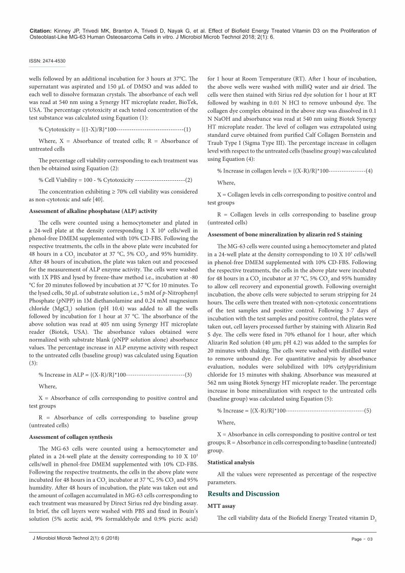
and DMEM by MTT assay in MG-63 cells are depicted in Figure 1. The data showed that the test samples in combination did not exhibit any cytotoxicity (as evidence of cell viability approximately greater than 73%) across all the tested concentrations up to 100 µg/mL. Hence, the same concentrations were assessed further to see the effect of the test samples on the levels of Alkaline Phosphatase (ALP) activity, collagen synthesis, and bone mineralization in MG-63 cells.
Alkaline phosphatase (ALP) activity
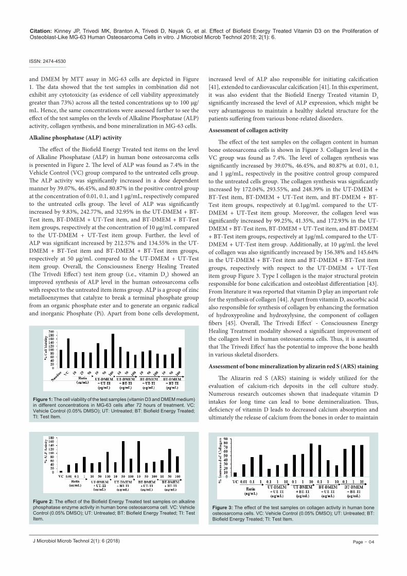
The effect of the Biofield Energy Treated test items on the level of Alkaline Phosphatase (ALP) in human bone osteosarcoma cells is presented in Figure 2. The level of ALP was found as 7.4% in the Vehicle Control (VC) group compared to the untreated cells group. The ALP activity was significantly increased in a dose dependent manner by 39.07%, 46.45%, and 80.87% in the positive control group at the concentration of 0.01, 0.1, and 1 µg/mL, respectively compared to the untreated cells group. The level of ALP was significantly increased by 9.83%, 242.77%, and 32.95% in the UT-DMEM + BT-Test item, BT-DMEM + UT-Test item, and BT-DMEM + BT-Test item groups, respectively at the concentration of 10 µg/mL compared to the UT-DMEM + UT-Test item group. Further, the level of ALP was significant increased by 212.57% and 134.55% in the UT-DMEM + BT-Test item and BT-DMEM + BT-Test item groups, respectively at 50 µg/mL compared to the UT-DMEM + UT-Test item group. Overall, the Consciousness Energy Healing Treated (The Trivedi Effect®) test item group (i.e., vitamin D3) showed an improved synthesis of ALP level in the human osteosarcoma cells with respect to the untreated item items group. ALP is a group of zinc metalloenzymes that catalyze to break a terminal phosphate group from an organic phosphate ester and to generate an organic radical and inorganic Phosphate (Pi). Apart from bone cells development,
increased level of ALP also responsible for initiating calcification [41], extended to cardiovascular calcification [41]. In this experiment, it was also evident that the Biofield Energy Treated vitamin D3 significantly increased the level of ALP expression, which might be very advantageous to maintain a healthy skeletal structure for the patients suffering from various bone-related disorders.
Assessment of collagen activity
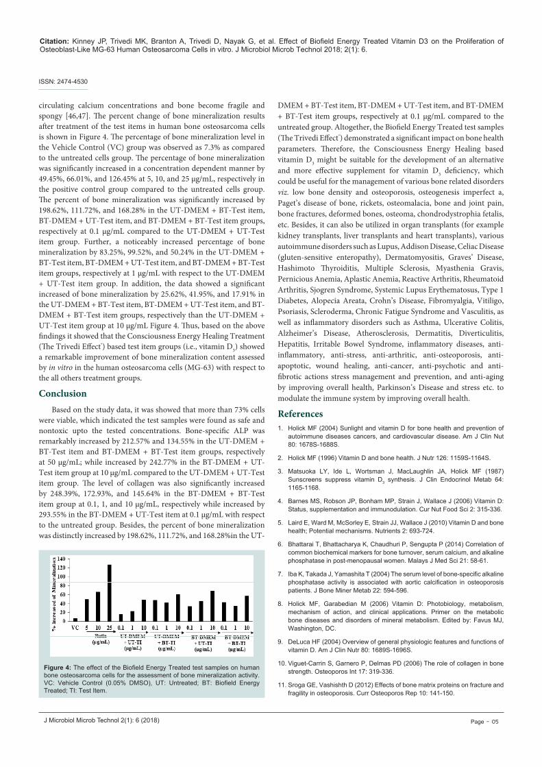
The effect of the test samples on the collagen content in human bone osteosarcoma cells is shown in Figure 3. Collagen level in the VC group was found as 7.4%. The level of collagen synthesis was significantly increased by 39.07%, 46.45%, and 80.87% at 0.01, 0.1, and 1 µg/mL, respectively in the positive control group compared to the untreated cells group. The collagen synthesis was significantly increased by 172.04%, 293.55%, and 248.39% in the UT-DMEM + BT-Test item, BT-DMEM + UT-Test item, and BT-DMEM + BT-Test item groups, respectively at 0.1µg/mL compared to the UT-DMEM + UT-Test item group. Moreover, the collagen level was significantly increased by 99.25%, 41.35%, and 172.93% in the UT-DMEM + BT-Test item, BT-DMEM + UT-Test item, and BT-DMEM + BT-Test item groups, respectively at 1µg/mL compared to the UT-DMEM + UT-Test item group. Additionally, at 10 μg/mL the level of collagen was also significantly increased by 156.38% and 145.64% in the UT-DMEM + BT-Test item and BT-DMEM + BT-Test item groups, respectively with respect to the UT-DMEM + UT-Test item group Figure 3. Type I collagen is the major structural protein responsible for bone calcification and osteoblast differentiation [43]. From literature it was reported that vitamin D play an important role for the synthesis of collagen [44]. Apart from vitamin D, ascorbic acid also responsible for synthesis of collagen by enhancing the formation of hydroxyproline and hydroxylysine, the component of collagen fibers [45]. Overall, The Trivedi Effect® - Consciousness Energy Healing Treatment modality showed a significant improvement of the collagen level in human osteosarcoma cells. Thus, it is assumed that The Trivedi Effect® has the potential to improve the bone health in various skeletal disorders.
Assessment of bone mineralization by alizarin red S (ARS) staining
The Alizarin red S (ARS) staining is widely utilized for the evaluation of calcium-rich deposits in the cell culture study. Numerous research outcomes shown that inadequate vitamin D intakes for long time can lead to bone demineralization. Thus, deficiency of vitamin D leads to decreased calcium absorption and ultimately the release of calcium from the bones in order to maintain
Figure 1: The cell viability of the test samples (vitamin D3 and DMEM medium) in different concentrations in MG-63 cells after 72 hours of treatment. VC: Vehicle Control (0.05% DMSO); UT: Untreated; BT: Biofield Energy Treated; TI: Test Item.
Figure 2: The effect of the Biofield Energy Treated test samples on alkaline phosphatase enzyme activity in human bone osteosarcoma cell. VC: Vehicle Control (0.05% DMSO); UT: Untreated; BT: Biofield Energy Treated; TI: Test Item.
Figure 3: The effect of the test samples on collagen activity in human bone osteosarcoma cells. VC: Vehicle Control (0.05% DMSO); UT: Untreated; BT: Biofield Energy Treated; TI: Test Item.
Citation: Kinney JP, Trivedi MK, Branton A, Trivedi D, Nayak G, et al. Effect of Biofield Energy Treated Vitamin D3 on the Proliferation of Osteoblast-Like MG-63 Human Osteosarcoma Cells in vitro. J Microbiol Microb Technol 2018; 2(1): 6.
J Microbiol Microb Technol 2(1): 6 (2018) Page - 05
ISSN: 2474-4530
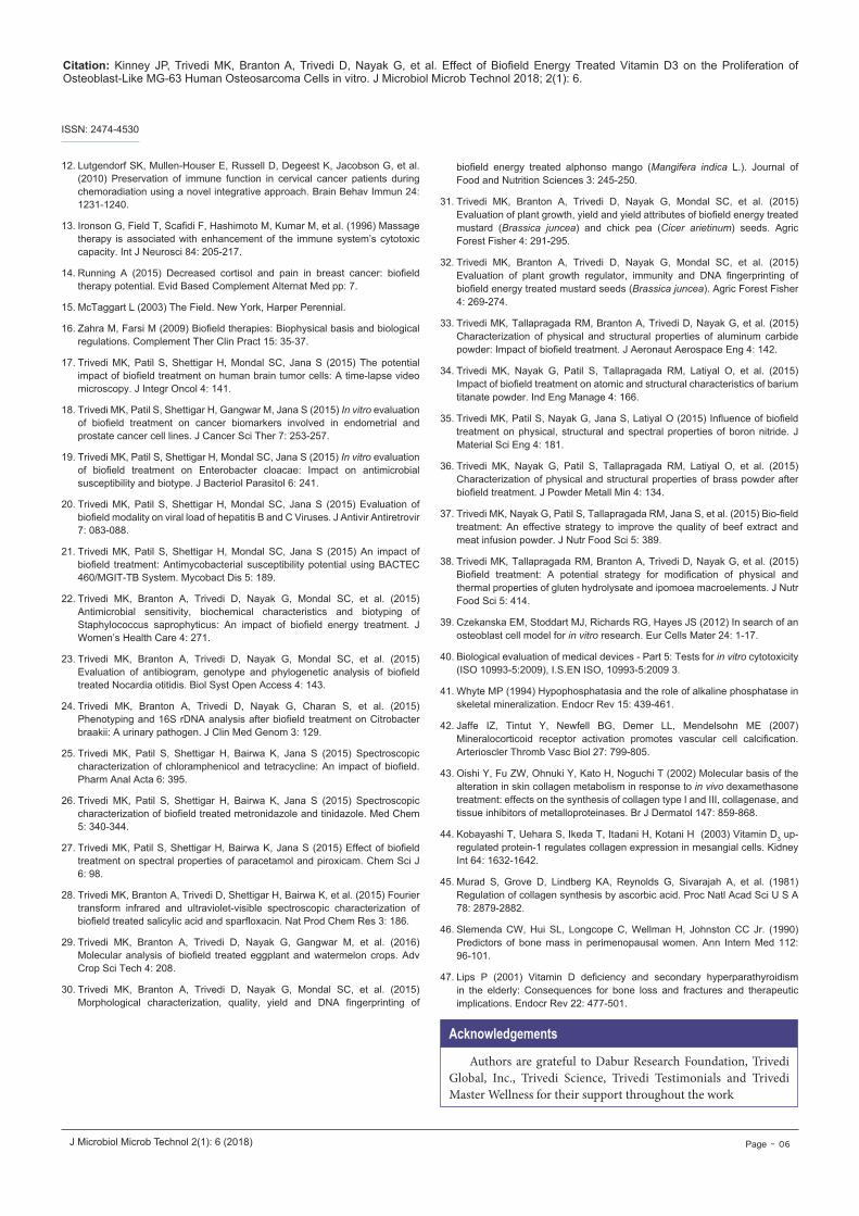
circulating calcium concentrations and bone become fragile and spongy [46,47]. The percent change of bone mineralization results after treatment of the test items in human bone osteosarcoma cells is shown in Figure 4. The percentage of bone mineralization level in the Vehicle Control (VC) group was observed as 7.3% as compared to the untreated cells group. The percentage of bone mineralization was significantly increased in a concentration dependent manner by 49.45%, 66.01%, and 126.45% at 5, 10, and 25 µg/mL, respectively in the positive control group compared to the untreated cells group. The percent of bone mineralization was significantly increased by 198.62%, 111.72%, and 168.28% in the UT-DMEM + BT-Test item, BT-DMEM + UT-Test item, and BT-DMEM + BT-Test item groups, respectively at 0.1 µg/mL compared to the UT-DMEM + UT-Test item group. Further, a noticeably increased percentage of bone mineralization by 83.25%, 99.52%, and 50.24% in the UT-DMEM + BT-Test item, BT-DMEM + UT-Test item, and BT-DMEM + BT-Test item groups, respectively at 1 µg/mL with respect to the UT-DMEM + UT-Test item group. In addition, the data showed a significant increased of bone mineralization by 25.62%, 41.95%, and 17.91% in the UT-DMEM + BT-Test item, BT-DMEM + UT-Test item, and BT-DMEM + BT-Test item groups, respectively than the UT-DMEM + UT-Test item group at 10 µg/mL Figure 4. Thus, based on the above findings it showed that the Consciousness Energy Healing Treatment (The Trivedi Effect®) based test item groups (i.e., vitamin D3) showed a remarkable improvement of bone mineralization content assessed by in vitro in the human osteosarcoma cells (MG-63) with respect to the all others treatment groups.
ConclusionBased on the study data, it was showed that more than 73% cells
were viable, which indicated the test samples were found as safe and nontoxic upto the tested concentrations. Bone-specific ALP was remarkably increased by 212.57% and 134.55% in the UT-DMEM + BT-Test item and BT-DMEM + BT-Test item groups, respectively at 50 µg/mL; while increased by 242.77% in the BT-DMEM + UT-Test item group at 10 µg/mL compared to the UT-DMEM + UT-Test item group. The level of collagen was also significantly increased by 248.39%, 172.93%, and 145.64% in the BT-DMEM + BT-Test item group at 0.1, 1, and 10 µg/mL, respectively while increased by 293.55% in the BT-DMEM + UT-Test item at 0.1 µg/mL with respect to the untreated group. Besides, the percent of bone mineralization was distinctly increased by 198.62%, 111.72%, and 168.28%in the UT-
DMEM + BT-Test item, BT-DMEM + UT-Test item, and BT-DMEM + BT-Test item groups, respectively at 0.1 µg/mL compared to the untreated group. Altogether, the Biofield Energy Treated test samples (The Trivedi Effect®) demonstrated a significant impact on bone health parameters. Therefore, the Consciousness Energy Healing based vitamin D3 might be suitable for the development of an alternative and more effective supplement for vitamin D3 deficiency, which could be useful for the management of various bone related disorders viz. low bone density and osteoporosis, osteogenesis imperfect a, Paget’s disease of bone, rickets, osteomalacia, bone and joint pain, bone fractures, deformed bones, osteoma, chondrodystrophia fetalis, etc. Besides, it can also be utilized in organ transplants (for example kidney transplants, liver transplants and heart transplants), various autoimmune disorders such as Lupus, Addison Disease, Celiac Disease (gluten-sensitive enteropathy), Dermatomyositis, Graves’ Disease, Hashimoto Thyroiditis, Multiple Sclerosis, Myasthenia Gravis, Pernicious Anemia, Aplastic Anemia, Reactive Arthritis, Rheumatoid Arthritis, Sjogren Syndrome, Systemic Lupus Erythematosus, Type 1 Diabetes, Alopecia Areata, Crohn’s Disease, Fibromyalgia, Vitiligo, Psoriasis, Scleroderma, Chronic Fatigue Syndrome and Vasculitis, as well as inflammatory disorders such as Asthma, Ulcerative Colitis, Alzheimer’s Disease, Atherosclerosis, Dermatitis, Diverticulitis, Hepatitis, Irritable Bowel Syndrome, inflammatory diseases, anti-inflammatory, anti-stress, anti-arthritic, anti-osteoporosis, anti-apoptotic, wound healing, anti-cancer, anti-psychotic and anti-fibrotic actions stress management and prevention, and anti-aging by improving overall health, Parkinson’s Disease and stress etc. to modulate the immune system by improving overall health.
References1. Holick MF (2004) Sunlight and vitamin D for bone health and prevention of
autoimmune diseases cancers, and cardiovascular disease. Am J Clin Nut 80: 1678S-1688S.
2. Holick MF (1996) Vitamin D and bone health. J Nutr 126: 1159S-1164S.
3. Matsuoka LY, Ide L, Wortsman J, MacLaughlin JA, Holick MF (1987) Sunscreens suppress vitamin D3 synthesis. J Clin Endocrinol Metab 64: 1165-1168.
4. Barnes MS, Robson JP, Bonham MP, Strain J, Wallace J (2006) Vitamin D: Status, supplementation and immunodulation. Cur Nut Food Sci 2: 315-336.
5. Laird E, Ward M, McSorley E, Strain JJ, Wallace J (2010) Vitamin D and bone health; Potential mechanisms. Nutrients 2: 693-724.
6. Bhattarai T, Bhattacharya K, Chaudhuri P, Sengupta P (2014) Correlation of common biochemical markers for bone turnover, serum calcium, and alkaline phosphatase in post-menopausal women. Malays J Med Sci 21: 58-61.
7. Iba K, Takada J, Yamashita T (2004) The serum level of bone-specific alkaline phosphatase activity is associated with aortic calcification in osteoporosis patients. J Bone Miner Metab 22: 594-596.
8. Holick MF, Garabedian M (2006) Vitamin D: Photobiology, metabolism, mechanism of action, and clinical applications. Primer on the metabolic bone diseases and disorders of mineral metabolism. Edited by: Favus MJ, Washington, DC.
9. DeLuca HF (2004) Overview of general physiologic features and functions of vitamin D. Am J Clin Nutr 80: 1689S-1696S.
10. Viguet-Carrin S, Garnero P, Delmas PD (2006) The role of collagen in bone strength. Osteoporos Int 17: 319-336.
11. Sroga GE, Vashishth D (2012) Effects of bone matrix proteins on fracture and fragility in osteoporosis. Curr Osteoporos Rep 10: 141-150.
Figure 4: The effect of the Biofield Energy Treated test samples on human bone osteosarcoma cells for the assessment of bone mineralization activity. VC: Vehicle Control (0.05% DMSO), UT: Untreated; BT: Biofield Energy Treated; TI: Test Item.
Citation: Kinney JP, Trivedi MK, Branton A, Trivedi D, Nayak G, et al. Effect of Biofield Energy Treated Vitamin D3 on the Proliferation of Osteoblast-Like MG-63 Human Osteosarcoma Cells in vitro. J Microbiol Microb Technol 2018; 2(1): 6.
J Microbiol Microb Technol 2(1): 6 (2018) Page - 06
ISSN: 2474-4530
12. Lutgendorf SK, Mullen-Houser E, Russell D, Degeest K, Jacobson G, et al. (2010) Preservation of immune function in cervical cancer patients during chemoradiation using a novel integrative approach. Brain Behav Immun 24: 1231-1240.
13. Ironson G, Field T, Scafidi F, Hashimoto M, Kumar M, et al. (1996) Massage therapy is associated with enhancement of the immune system’s cytotoxic capacity. Int J Neurosci 84: 205-217.
14. Running A (2015) Decreased cortisol and pain in breast cancer: biofield therapy potential. Evid Based Complement Alternat Med pp: 7.
15. McTaggart L (2003) The Field. New York, Harper Perennial.
16. Zahra M, Farsi M (2009) Biofield therapies: Biophysical basis and biological regulations. Complement Ther Clin Pract 15: 35-37.
17. Trivedi MK, Patil S, Shettigar H, Mondal SC, Jana S (2015) The potential impact of biofield treatment on human brain tumor cells: A time-lapse video microscopy. J Integr Oncol 4: 141.
18. Trivedi MK, Patil S, Shettigar H, Gangwar M, Jana S (2015) In vitro evaluation of biofield treatment on cancer biomarkers involved in endometrial and prostate cancer cell lines. J Cancer Sci Ther 7: 253-257.
19. Trivedi MK, Patil S, Shettigar H, Mondal SC, Jana S (2015) In vitro evaluation of biofield treatment on Enterobacter cloacae: Impact on antimicrobial susceptibility and biotype. J Bacteriol Parasitol 6: 241.
20. Trivedi MK, Patil S, Shettigar H, Mondal SC, Jana S (2015) Evaluation of biofield modality on viral load of hepatitis B and C Viruses. J Antivir Antiretrovir 7: 083-088.
21. Trivedi MK, Patil S, Shettigar H, Mondal SC, Jana S (2015) An impact of biofield treatment: Antimycobacterial susceptibility potential using BACTEC 460/MGIT-TB System. Mycobact Dis 5: 189.
22. Trivedi MK, Branton A, Trivedi D, Nayak G, Mondal SC, et al. (2015) Antimicrobial sensitivity, biochemical characteristics and biotyping of Staphylococcus saprophyticus: An impact of biofield energy treatment. J Women’s Health Care 4: 271.
23. Trivedi MK, Branton A, Trivedi D, Nayak G, Mondal SC, et al. (2015) Evaluation of antibiogram, genotype and phylogenetic analysis of biofield treated Nocardia otitidis. Biol Syst Open Access 4: 143.
24. Trivedi MK, Branton A, Trivedi D, Nayak G, Charan S, et al. (2015) Phenotyping and 16S rDNA analysis after biofield treatment on Citrobacter braakii: A urinary pathogen. J Clin Med Genom 3: 129.
25. Trivedi MK, Patil S, Shettigar H, Bairwa K, Jana S (2015) Spectroscopic characterization of chloramphenicol and tetracycline: An impact of biofield. Pharm Anal Acta 6: 395.
26. Trivedi MK, Patil S, Shettigar H, Bairwa K, Jana S (2015) Spectroscopic characterization of biofield treated metronidazole and tinidazole. Med Chem 5: 340-344.
27. Trivedi MK, Patil S, Shettigar H, Bairwa K, Jana S (2015) Effect of biofield treatment on spectral properties of paracetamol and piroxicam. Chem Sci J 6: 98.
28. Trivedi MK, Branton A, Trivedi D, Shettigar H, Bairwa K, et al. (2015) Fourier transform infrared and ultraviolet-visible spectroscopic characterization of biofield treated salicylic acid and sparfloxacin. Nat Prod Chem Res 3: 186.
29. Trivedi MK, Branton A, Trivedi D, Nayak G, Gangwar M, et al. (2016) Molecular analysis of biofield treated eggplant and watermelon crops. Adv Crop Sci Tech 4: 208.
30. Trivedi MK, Branton A, Trivedi D, Nayak G, Mondal SC, et al. (2015) Morphological characterization, quality, yield and DNA fingerprinting of
biofield energy treated alphonso mango (Mangifera indica L.). Journal of Food and Nutrition Sciences 3: 245-250.
31. Trivedi MK, Branton A, Trivedi D, Nayak G, Mondal SC, et al. (2015) Evaluation of plant growth, yield and yield attributes of biofield energy treated mustard (Brassica juncea) and chick pea (Cicer arietinum) seeds. Agric Forest Fisher 4: 291-295.
32. Trivedi MK, Branton A, Trivedi D, Nayak G, Mondal SC, et al. (2015) Evaluation of plant growth regulator, immunity and DNA fingerprinting of biofield energy treated mustard seeds (Brassica juncea). Agric Forest Fisher 4: 269-274.
33. Trivedi MK, Tallapragada RM, Branton A, Trivedi D, Nayak G, et al. (2015) Characterization of physical and structural properties of aluminum carbide powder: Impact of biofield treatment. J Aeronaut Aerospace Eng 4: 142.
34. Trivedi MK, Nayak G, Patil S, Tallapragada RM, Latiyal O, et al. (2015) Impact of biofield treatment on atomic and structural characteristics of barium titanate powder. Ind Eng Manage 4: 166.
35. Trivedi MK, Patil S, Nayak G, Jana S, Latiyal O (2015) Influence of biofield treatment on physical, structural and spectral properties of boron nitride. J Material Sci Eng 4: 181.
36. Trivedi MK, Nayak G, Patil S, Tallapragada RM, Latiyal O, et al. (2015) Characterization of physical and structural properties of brass powder after biofield treatment. J Powder Metall Min 4: 134.
37. Trivedi MK, Nayak G, Patil S, Tallapragada RM, Jana S, et al. (2015) Bio-field treatment: An effective strategy to improve the quality of beef extract and meat infusion powder. J Nutr Food Sci 5: 389.
38. Trivedi MK, Tallapragada RM, Branton A, Trivedi D, Nayak G, et al. (2015) Biofield treatment: A potential strategy for modification of physical and thermal properties of gluten hydrolysate and ipomoea macroelements. J Nutr Food Sci 5: 414.
39. Czekanska EM, Stoddart MJ, Richards RG, Hayes JS (2012) In search of an osteoblast cell model for in vitro research. Eur Cells Mater 24: 1-17.
40. Biological evaluation of medical devices - Part 5: Tests for in vitro cytotoxicity (ISO 10993-5:2009), I.S.EN ISO, 10993-5:2009 3.
41. Whyte MP (1994) Hypophosphatasia and the role of alkaline phosphatase in skeletal mineralization. Endocr Rev 15: 439-461.
42. Jaffe IZ, Tintut Y, Newfell BG, Demer LL, Mendelsohn ME (2007) Mineralocorticoid receptor activation promotes vascular cell calcification. Arterioscler Thromb Vasc Biol 27: 799-805.
43. Oishi Y, Fu ZW, Ohnuki Y, Kato H, Noguchi T (2002) Molecular basis of the alteration in skin collagen metabolism in response to in vivo dexamethasone treatment: effects on the synthesis of collagen type I and III, collagenase, and tissue inhibitors of metalloproteinases. Br J Dermatol 147: 859-868.
44. Kobayashi T, Uehara S, Ikeda T, Itadani H, Kotani H (2003) Vitamin D3 up-regulated protein-1 regulates collagen expression in mesangial cells. Kidney Int 64: 1632-1642.
45. Murad S, Grove D, Lindberg KA, Reynolds G, Sivarajah A, et al. (1981) Regulation of collagen synthesis by ascorbic acid. Proc Natl Acad Sci U S A 78: 2879-2882.
46. Slemenda CW, Hui SL, Longcope C, Wellman H, Johnston CC Jr. (1990) Predictors of bone mass in perimenopausal women. Ann Intern Med 112: 96-101.
47. Lips P (2001) Vitamin D deficiency and secondary hyperparathyroidism in the elderly: Consequences for bone loss and fractures and therapeutic implications. Endocr Rev 22: 477-501.
Authors are grateful to Dabur Research Foundation, Trivedi Global, Inc., Trivedi Science, Trivedi Testimonials and Trivedi Master Wellness for their support throughout the work
Acknowledgements