Keratins determine network stress responsiveness in ...
9
3954 | Soft Matter, 2021, 17, 3954–3962 This journal is © The Royal Society of Chemistry 2021 Cite this: Soft Matter, 2021, 17, 3954 Keratins determine network stress responsiveness in reconstituted actin–keratin filament systems† Iman Elbalasy, ‡* ab Paul Mollenkopf,‡ ac Cary Tutmarc, ac Harald Herrmann de and Jo ¨ rg Schnauß* acf The cytoskeleton is a major determinant of cell mechanics, and alterations in the central mechanical aspects of cells are observed during many pathological situations. Therefore, it is essential to investigate the interplay between the main filament systems of the cytoskeleton in the form of composite networks. Here, we investigate the role of keratin intermediate filaments (IFs) in network strength by studying in vitro reconstituted actin and keratin 8/18 composite filament networks via bulk shear rheology. We co-polymerized these structural proteins in varying ratios and recorded how their relative content affects the overall mechanical response of the various composites. For relatively small deformations, we found that all composites exhibited an intermediate linear viscoelastic behaviour compared to that of the pure networks. In stark contrast, when larger deformations were imposed the composites displayed increasing strain stiffening behaviour with increasing keratin content. The extent of strain stiffening is much more pronounced than in corresponding experiments performed with vimentin IF as a composite network partner for actin. Our results provide new insights into the mechanical interplay between actin and keratin filaments in which keratin provides reinforcement to actin. This interplay may contribute to the overall integrity of cells. Hence, the high keratin 8/18 content of mechanically stressed simple epithelial cell layers, as found in the lung and the intestine, provides an explanation for their exceptional stability. Introduction The complex mechanical behaviour of eukaryotic cells is largely determined by the three principal filament systems of the cytoskeleton, i.e. actin filaments (F-actin), microtubules, and intermediate filaments (IFs) as well as their regulated interplay. 1 In addition to establishing a cell’s specific functional and tissue-related shape, these components and their distinct structures are also crucial for numerous general cellular processes such as stability within tissues as well as cell motility, division, and signal transduction. 2 Alterations of these key constituents are the cause of numerous human diseases. It is somehow perplexing that these very same cytoskeletal components can lead to very static, stable cell conformations, which allow, for instance, the formation of stable tissue layers such as the epithelium, or highly dynamic cells during wound healing and cancer metastasis. 3 Switching between these physically seemingly contradictory cell states highly depends on the ratios of the individual cytoskeletal components. This becomes especially apparent during embryo- genesis when cells need to constantly switch between stable epithelial states and motile mesenchymal states to form complex tissue structures. 4 During these switching events, which are the so-called epithelial to mesenchymal transition (EMT) along with the reverse process called mesenchymal to epithelial transition (MET), the cytoskeletal components are very differently expressed. These different compositions lead to different mechanical and motile behaviours. Besides actin-related structures, crucial elements are keratin IFs, which, with their 37 cytoplasmic members, constitute the largest group of the IF protein-family. 5,6 They are typically expressed in epithelial cells in various combinations and provide crucial cell type-specific structural support upon mechanical stresses. These diverse mechanical properties are realized by expressing various specific keratin pairs as keratins obligate heterodimeric complexes representing parallel coiled coils made from two alpha-helical molecules of two sequence- related classes each. 7 They are also involved in other a Peter-Debye Institute for Soft Matter Physics, Leipzig University, 04103 Leipzig, Germany b Faculty of Science, Cairo University, 12613, Giza, Egypt c Fraunhofer Institute for Cell Therapy and Immunology, 04103 Leipzig, Germany d Molecular Genetics, German Cancer Research Centre, 69120 Heidelberg, Germany e Department of Neuropathology, University Hospital Erlangen, 91054, Erlangen, Germany f Unconventional Computing Lab, Department of Computer Science and Creative Technologies, UWE, Bristol BS16 1QY, UK † Electronic supplementary information (ESI) available. See DOI: 10.1039/ d0sm02261f ‡ These authors contributed equally to this work. Received 24th December 2020, Accepted 4th March 2021 DOI: 10.1039/d0sm02261f rsc.li/soft-matter-journal Soft Matter PAPER Open Access Article. Published on 05 March 2021. Downloaded on 1/15/2022 5:46:24 PM. This article is licensed under a Creative Commons Attribution 3.0 Unported Licence. View Article Online View Journal | View Issue
Transcript of Keratins determine network stress responsiveness in ...

3954 | Soft Matter, 2021, 17, 3954–3962 This journal is © The Royal Society of Chemistry 2021
Cite this: Soft Matter, 2021,
17, 3954
Keratins determine network stress responsivenessin reconstituted actin–keratin filament systems†
Iman Elbalasy, ‡*ab Paul Mollenkopf,‡ac Cary Tutmarc, ac Harald Herrmannde
and Jorg Schnauß*acf
The cytoskeleton is a major determinant of cell mechanics, and alterations in the central mechanical
aspects of cells are observed during many pathological situations. Therefore, it is essential to investigate
the interplay between the main filament systems of the cytoskeleton in the form of composite networks.
Here, we investigate the role of keratin intermediate filaments (IFs) in network strength by studying
in vitro reconstituted actin and keratin 8/18 composite filament networks via bulk shear rheology.
We co-polymerized these structural proteins in varying ratios and recorded how their relative content
affects the overall mechanical response of the various composites. For relatively small deformations, we
found that all composites exhibited an intermediate linear viscoelastic behaviour compared to that of
the pure networks. In stark contrast, when larger deformations were imposed the composites displayed
increasing strain stiffening behaviour with increasing keratin content. The extent of strain stiffening is
much more pronounced than in corresponding experiments performed with vimentin IF as a composite
network partner for actin. Our results provide new insights into the mechanical interplay between actin
and keratin filaments in which keratin provides reinforcement to actin. This interplay may contribute to
the overall integrity of cells. Hence, the high keratin 8/18 content of mechanically stressed simple
epithelial cell layers, as found in the lung and the intestine, provides an explanation for their exceptional
stability.
Introduction
The complex mechanical behaviour of eukaryotic cells is largelydetermined by the three principal filament systems of thecytoskeleton, i.e. actin filaments (F-actin), microtubules, andintermediate filaments (IFs) as well as their regulatedinterplay.1 In addition to establishing a cell’s specific functionaland tissue-related shape, these components and their distinctstructures are also crucial for numerous general cellular processessuch as stability within tissues as well as cell motility, division,and signal transduction.2 Alterations of these key constituents arethe cause of numerous human diseases. It is somehow perplexing
that these very same cytoskeletal components can lead to verystatic, stable cell conformations, which allow, for instance, theformation of stable tissue layers such as the epithelium, or highlydynamic cells during wound healing and cancer metastasis.3
Switching between these physically seemingly contradictory cellstates highly depends on the ratios of the individual cytoskeletalcomponents. This becomes especially apparent during embryo-genesis when cells need to constantly switch between stableepithelial states and motile mesenchymal states to form complextissue structures.4 During these switching events, which are theso-called epithelial to mesenchymal transition (EMT) along withthe reverse process called mesenchymal to epithelial transition(MET), the cytoskeletal components are very differently expressed.These different compositions lead to different mechanical andmotile behaviours. Besides actin-related structures, crucial elementsare keratin IFs, which, with their 37 cytoplasmic members,constitute the largest group of the IF protein-family.5,6 They aretypically expressed in epithelial cells in various combinationsand provide crucial cell type-specific structural support uponmechanical stresses. These diverse mechanical properties arerealized by expressing various specific keratin pairs as keratinsobligate heterodimeric complexes representing parallel coiledcoils made from two alpha-helical molecules of two sequence-related classes each.7 They are also involved in other
a Peter-Debye Institute for Soft Matter Physics, Leipzig University, 04103 Leipzig,
Germanyb Faculty of Science, Cairo University, 12613, Giza, Egyptc Fraunhofer Institute for Cell Therapy and Immunology, 04103 Leipzig, Germanyd Molecular Genetics, German Cancer Research Centre, 69120 Heidelberg, Germanye Department of Neuropathology, University Hospital Erlangen, 91054, Erlangen,
Germanyf Unconventional Computing Lab, Department of Computer Science and Creative
Technologies, UWE, Bristol BS16 1QY, UK
† Electronic supplementary information (ESI) available. See DOI: 10.1039/d0sm02261f‡ These authors contributed equally to this work.
Received 24th December 2020,Accepted 4th March 2021
DOI: 10.1039/d0sm02261f
rsc.li/soft-matter-journal
Soft Matter
PAPER
Ope
n A
cces
s A
rtic
le. P
ublis
hed
on 0
5 M
arch
202
1. D
ownl
oade
d on
1/1
5/20
22 5
:46:
24 P
M.
Thi
s ar
ticle
is li
cens
ed u
nder
a C
reat
ive
Com
mon
s A
ttrib
utio
n 3.
0 U
npor
ted
Lic
ence
.
View Article OnlineView Journal | View Issue
This journal is © The Royal Society of Chemistry 2021 Soft Matter, 2021, 17, 3954–3962 | 3955
non-mechanical functions, including regulation of cell growth,migration, and protection from apoptosis.8,9 Several studieshave highlighted the potential inhibitory role of keratins forcell mobility and thus the need to downregulate them for EMTto increase cellular mobility. For example, it was found thatkeratinocytes with all keratins deleted by gene-targeting aresofter, more deformable, and more invasive compared to thesmall overall effect generated after actin depolymerization.10
They are also able to migrate twice as fast as wild type cells.11
Conversely, keratin re-expression in these studies has theopposite effect. This indicates a direct link between down-regulation of keratins during EMT and loss of stiffness withincreased migration and invasion ability of tumor cells.Although F-actin networks dramatically reorganize as a prere-quisite for morphological and migratory changes during EMTand MET, it is indeed remarkable that the entire IF system isrebuilt by a switch between keratin and vimentin expressions.12
As demonstrated for numerous physiological situations, thecytoskeletal systems act synergistically.13 By coordinating theirfunctions, they affect each other’s mechanics. However, it ischallenging to investigate this mechanical interplay in cells due totheir inherent complexity, which makes it difficult to disentanglemechanical crosstalk and regulatory biochemical interactions.In this respect, reconstituted composite cytoskeletal filamentnetworks have been investigated in vitro by quantitativerheological measurements and theoretical modelling. The resultshave often revealed unexpected mechanical responses. Forinstance, the presence of microtubules was found to induceunexpected local compressibility14 and non-linear strain stiffeningin actin networks.15,16 Concomitantly, actin reinforces microtubulesagainst compressive loads.17
When designing and interpreting such in vitro studies, it isimportant to choose the system parameters carefully asobserved when measuring the mechanical properties ofcomposite filament networks of actin and vimentin. Holdingthe molecular content constant for different mixing ratios ofactin and vimentin has yielded contradictory results for theirmechanical response, namely a synergistic increase in thelinear elasticity18 and formation of stiffer or softer networksdepending on the investigation technique.19,20 In this case,however, vimentin filaments carry roughly twice as manymonomers per unit length than actin filaments, i.e. totalfilament length is half compared to that of F-actin. Consequently,it has been shown that actin–vimentin filament composites withthe same total polymer length and mesh size, can be described bya superposition of the mechanical properties of the underlyingconstituents.21
Due to their intrinsic ability to self-assemble in vitro, theco-polymerization of actin and keratin filaments is possiblewithout additional accessory proteins. Hence, it has beenrecently demonstrated that encapsulation of actin and keratinwithin vesicles leads to the formation of composite filamentnetworks, where F-actin acts as steric resistance for keratin IFspreventing their collapse to bundled clusters.22
Here, we investigate the mechanical properties of in vitroreconstituted F-actin and keratin 8/18 (K8–K18) composite
networks by bulk shear rheology measurements. We aim tocharacterize networks with similar total filament lengths.By systematically varying the relative mass ratios of actin andkeratin, we investigate how their relative presence impacts theoverall mechanical response of the composites. We complementthese rheology measurements with confocal microscopy of thecomposites.
ExperimentalPolymer lengths
The mass per unit length (mL) for actin is 2.66� 10�11 g m�1,23 andfor K8–K18 in Tris-based assembly buffer is 3.16 � 10�11 g m�1.24
At 0.5 mg ml�1 actin monomers (11.9 mM) and 0.6 mg ml�1 K8–18tetramers (11.34 mM), both actin and keratin filament networks willhave similar total filament length. Using these concentrations asour boundary conditions, we can select actin–keratin mixtures withthe same total polymer length per unit volume.
Protein preparation and co-polymerization
G-actin was prepared from rabbit muscle and stored at �80 1Cin G-Buffer (2 mM Tris–HCl pH 7.5, 0.2 mM ATP, 0.1 mM CaCl2,1 mM DTT, 0.01% NaN3, pH 7.8) as described previously.25
Vectors containing keratin genes (pET 24a–K8 and pET 23a–K18) were transformed into E. coli BL21 for protein expression.Recombinant K8 and K18 were isolated and purified aspreviously described.26
For reconstitution, purified K8 and K18 proteins were mixedin equimolar amounts and renatured by stepwise dialysisagainst 8 M urea, 2 mM Tris–HCl, 1 mM DTT, pH 9.0 withstepwise reduction of urea concentration (6 M, 4 M, 2 M, 1 M,and 0 M). Each dialysis step was done for 20 minutes at roomtemperature, then the dialysis was continued overnight against2 mM Tris–HCl, 1 mM DTT, pH 9.0 at 4 1C. The final proteinconcentration was determined by measuring the absorption at280 nm using a DU 530 UV/vis Spectrophotometer (BeckmanCoulter Inc., USA). Composite networks were prepared bymixing actin monomers at 0.5 mg ml�1 and K8–K18 tetramersat 0.6 mg ml�1 in varying mixing ratios. Assembly of pure andmixed networks was initiated by adding 1/10 volume of 10�F-buffer (20 mM Tris–HCl, 1 M KCl, 10 mM MgCl2, 2 mM ATP,10 mM DTT, pH 7.5) to the protein sample.
Shear rheology
Rheology measurements were conducted using a strain-controlled ARES (TA Instruments, USA) and a MCR 502 WESP(Anton Paar, Austria) rheometer using 40 mm plate–plategeometry with 140 mm gap width at 20 1C. In all measurements,F-buffer was mixed with the protein mixture on ice, and asample volume of 200 ml was loaded quickly between the twoplates. F-Buffer with the same conditions as in the sample wasdistributed around the sample to prevent artefacts from inter-facial elasticity and a solvent trap was placed around thesample to prevent evaporation. The sample was given two hoursto equilibrate between the rheometer plates as the filament
Paper Soft Matter
Ope
n A
cces
s A
rtic
le. P
ublis
hed
on 0
5 M
arch
202
1. D
ownl
oade
d on
1/1
5/20
22 5
:46:
24 P
M.
Thi
s ar
ticle
is li
cens
ed u
nder
a C
reat
ive
Com
mon
s A
ttrib
utio
n 3.
0 U
npor
ted
Lic
ence
.View Article Online
3956 | Soft Matter, 2021, 17, 3954–3962 This journal is © The Royal Society of Chemistry 2021
assembly was monitored with a dynamic time sweep for2 hours, one data point per minute at a frequency (o) of 1 Hzand a strain (g) of 2%. The linear viscoelastic response ofequilibrated networks was measured with dynamic frequencysweeps ranging from 0.01 Hz to 80 Hz at a strain of 2% and20 points per decade. Data points plotted in Fig. 2 are the meanof at least five independent rheology measurements. A transientstep rate test with a strain rate of 0.1 s�1 was applied to measurethe strain-dependent stress in the non-linear strain regime.The differential shear modulus (K) was determined with aself-written Python script, calculating the gradient of thesmoothed stress data divided by the strain step width. The lineardifferential shear modulus Klin is given by the first non-negativevalue of the smoothed stress data, whereas negative stress ismeasured due to technical limitations, particularly for smallstrains, lacking every physical relevance. The linear frequencysweeps as well as the nonlinear step rate measurements wereevaluated in the frame of the GWLC model with a self-writtenPython script. At least six independent rheology measurementswere taken into account for the evaluation and comparison withthe model predictions. Details about the model are presented inthe ESI.†
Protein labelling, sample preparation and imaging
We have developed a new method for direct labelling of thewild type K8–K18 without mutation, unlike the techniques usedin previous studies.22,27 The labelling is based on the couplingof Atto-488-NHS-ester (ATTO-TEC, Siegen, Germany) to the freeamine (NH2) residues (lysine or terminus) in K8 tetramers. K8was renatured by stepwise dialysis against the labelling buffer(2 mM sodium phosphate, pH 8.5) as described above. K8tetramers were mixed with Atto-488-NHS-ester solubilized inDMSO at 10 mM at a molar ratio of 5 : 1. The mixture wasincubated in the dark for 1 hour at room temperature. In onestep, the free dye was removed, and labelled K8 was denaturedinto monomers by overnight dialysis against 8 M urea, 2 mMsodium phosphate buffer pH 8.5 at 4 1C. On the second day, thelabelling buffer was exchanged by the dialysis buffer (8 M urea,2 mM Tris–HCl, pH 9.0) by dialysis for 2 hours at roomtemperature. The concentration of labelled K8 was determinedby measuring the absorbance at 280 and 500 nm using a DU530 UV/vis Spectrophotometer (Beckman Coulter Inc., USA).Labelled K8 was then distributed to appropriate aliquots andstored at �80 1C. One day before imaging, equal amounts of20% labelled K8 were mixed with unlabelled K8 and K18 in 8 Murea, 2 mM Tris–HCl, pH 9.0 to have a final sample labellingof 10% by concentration. This mixture was renatured intotetramers by stepwise dialysis against 2 mM Tris–HCl, pH 9.0.
For network visualization, labelled K8–K18 prepared in theprevious step was mixed with unlabelled K8–K18 tetramers in aratio of 1 : 10, with a final protein concentration of 0.6 mg ml�1.Fluorescently labelled actin was prepared by mixing G-actin at5 mM with phalloidin–tetramethylrhodamine B isothio-cyanate(phalloidin–TRITC – Sigma-Aldrich Co.) in a molar ratio of 1 : 1.For composite networks, 10% labelled keratin samples weremixed with labelled actin samples in varying ratios. Networks
were created by adding 1/10 volume fraction of 10� F-buffer tothe sample and immediately pipetting it into an experimentalsample chamber to rest for 2 hours at room temperature beforeimaging as described in ref. 28. Images were captured using aspinning disc confocal microscope (inverted Axio Observer.Z1/Yokogawa CSU-X1A 5000, Carl Zeiss Microscopy GmbH,Germany), 100� oil immersion objective NA 1.40 with aHamamatsu camera at an exposure time of 100 ms.
Results and discussionCo-polymerization and filament length
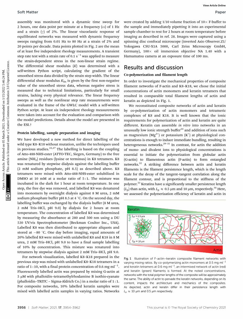
In order to investigate the mechanical properties of compositefilament networks of F-actin and K8–K18, we chose the initialconcentrations of actin monomers and keratin tetramers thatresulted in comparable total filament lengths of actin andkeratin as depicted in Fig. 1.
We reconstituted composite networks of actin and keratinby co-polymerization of actin monomers and tetramericcomplexes of K8 and K18. It is well known that the ionicrequirements for polymerization of actin and keratin are quitedifferent. Keratin can assemble in vitro into networks in anunusually low ionic strength buffer24 and addition of ions suchas magnesium (Mg2+) or potassium (K+) at physiological con-centrations is enough to induce immediate bundling, resulting inheterogeneous networks.29–31 In contrast, for actin the additionof mono- and divalent ions to physiological concentrations isessential to initiate the polymerization from globular actin(G-actin) to filamentous actin (F-actin) to form entanglednetworks.32 A striking difference between actin and keratinfilaments is the filament persistence length, which is the lengthscale for the decay of the tangent–tangent correlation along thefilament contour, and is proportional to the stiffness of thepolymer.33 Keratins have a significantly smaller persistence length(Lp) than actin, with Lp E 0.5 mm and 10 mm, respectively.34 Here,we assessed the polymerization efficiency of keratin and actin in
Fig. 1 Illustration of F-actin–keratin composite filament networks withvarying mixing ratios. By co-polymerizing actin monomers at 0.5 mg ml�1
and keratin tetramers at 0.6 mg ml�1, an intermixed network of actin (red)and keratin (green) filaments is formed. At the noted concentrations,networks with the total polymer lengths of the composite will be approximatelythe same. The ability of actin to pervade the keratin networks, depending on itscontent, impacts the architecture and mechanics of the composites.As depicted, actin and keratin differ in their persistence length withLp E 10 mm and 0.5 mm respectively.
Soft Matter Paper
Ope
n A
cces
s A
rtic
le. P
ublis
hed
on 0
5 M
arch
202
1. D
ownl
oade
d on
1/1
5/20
22 5
:46:
24 P
M.
Thi
s ar
ticle
is li
cens
ed u
nder
a C
reat
ive
Com
mon
s A
ttrib
utio
n 3.
0 U
npor
ted
Lic
ence
.View Article Online

This journal is © The Royal Society of Chemistry 2021 Soft Matter, 2021, 17, 3954–3962 | 3957
different concentrations of MgCl2 (1, 0.5, 0.1 and 0 mM) byhigh-speed sedimentation of protein assemblies followed bySDS-PAGE of pellet and supernatant fractions (data not shown).We found that a lack of Mg2+ ions had no effect on the polymer-ization efficiency of keratin, and soluble keratin tetramers werepolymerized completely into filaments, whereas for actin at least1 mM MgCl2 was required to ensure complete polymerization.Thus, we had to adjust the polymerization buffer (see Materialsand methods) to include a concentration of 1 mM MgCl2 forthe co-polymerization in order to form actin–keratin filamentnetworks.
Linear viscoelasticity of pure actin and keratin filamentnetworks
Using bulk shear rheology, the linear viscoelastic properties ofpolymer solutions can be quantified by the frequency (o)dependent complex shear modulus G*(o) = G0(o) + iG00(o),where G0 and G00 are the elastic and viscous moduli, respectively.Under the selected assembly conditions, pure K8–K18 filamentnetworks at 0.6 mg ml�1 exhibit predominantly elastic behaviourwith G0 being consistently larger than G00 by nearly one order ofmagnitude (Fig. 2A). Accordingly, the loss factor tan(f) = G00/G0
has a small value (tan(f) at 1 Hz = 0.13), indicative of highlyelastic networks. Over the entire frequency range, G0 and G00
show a very weak frequency dependence. A weak power law wasobtained by a linear fit of G0 in the log–log-plot yielding a power-law exponent of a(G0) = 0.07. We observed no G00/G0 crossoverin the measurable frequency range for pure keratin. This isconsistent with results previously reported for K8–K18 networkbulk rheological behaviour.24,35,36 By comparison, G0 and G00 ofpure F-actin filament networks showed a more pronouncedfrequency dependence with a(G0) = 0.23, (Fig. 2E). Accordingly,the crossover between G0 and G00 regimes was observed at lowfrequency. Actin networks have a larger loss factor (tan(f) at1 Hz = 0.52) and, consequently, the elastic contributions are lessdominant than in pure keratin networks. These mechanicalfeatures are similar to those obtained for entangled actinnetworks under comparable conditions.21,37
Composite filament networks exhibit an intermediate linearviscoelastic behaviour
To date, previous in vitro studies have investigated the mechanicalproperties of only one-component systems of either actinfilaments as entangled38–40 and crosslinked28,41–44 networks orkeratin single filaments27 and networks.24,29,35,36,45
In our study, composite networks of F-actin and K8–K18filaments with varying mixing ratios revealed an intermediatelinear viscoelastic behaviour with regard to their composite-specific protein content. With increasing actin/decreasingkeratin contents as shown in Fig. 2B–D, the dependence of G0
and G00 on the frequency increased gradually as indicated by thegradual increase in a values; a = 0.09 in keratin-dominatednetworks (0.45 mg ml�1 K8–K18–0.125 mg ml�1 actin), 0.16 inequal ratio-networks (0.3 mg ml�1 K8–K18–0.25 mg ml�1 actin)and 0.21 in F-actin-dominated networks (0.15 mg ml�1
K8–K18–0.375 mg ml�1 actin). Furthermore, the crossover to
Fig. 2 Linear viscoelasticity of reconstituted filemant networks. By comparison,the keratin network (A) shows more predominant elasticity with a weakerfrequency dependence of both elastic G0 (filled symbols) and viscous G00
(open symbols) moduli than the F-actin network (E); the keratin network ismore elastic (tanfker = 0.13) than the F-actin network (tanfact= 0.52) withno observed G0/G00 crossover, which, in contrast, is a signature of actinnetworks in this frequency range. Actin–keratin composite filamentnetworks with varying mixing ratios of actin and keratin (B–D) showintermediate viscoelastic properties. With increasing actin/decreasing keratincontent, the frequency dependence of G0 and G00 moduli increases gradually,and the networks become less elastic as the loss factor values increase.Consequently, the G0/G00 crossover appears at high frequency in keratin-dominated networks and shifts gradually to lower frequencies with increasingactin content. a represents the slope between 0.1 and 10 Hz of G0. Data pointsin all curves represent the mean of at least five independent measurements anderror bars represent the standard deviation from the mean.
Paper Soft Matter
Ope
n A
cces
s A
rtic
le. P
ublis
hed
on 0
5 M
arch
202
1. D
ownl
oade
d on
1/1
5/20
22 5
:46:
24 P
M.
Thi
s ar
ticle
is li
cens
ed u
nder
a C
reat
ive
Com
mon
s A
ttrib
utio
n 3.
0 U
npor
ted
Lic
ence
.View Article Online

3958 | Soft Matter, 2021, 17, 3954–3962 This journal is © The Royal Society of Chemistry 2021
the predominant viscous regime, observed only in F-actinnetworks, started to appear in keratin-dominated networks athigh frequencies. With increasing actin content, actin contributesmore to the composite’s behaviour, shifting the crossover pointgradually to lower frequencies. The loss factor tan(f) increasedwith increasing actin/decreasing keratin content, which indicatesa smooth transition to less elastic networks, as shown in Fig. 3A.We used a Mann–Whitney U test to investigate the statisticalsignificance of the loss factor values with respect to each other.We found p-values less than 0.05, indicating significantdifference, for all compared distributions except for the purenetworks compared to composites with a respective division of0.75/0.25 (Table S1, ESI†). In Fig. 3B, we show that the elasticmoduli G0 for all networks exhibit only minor variations.
Filament–filament interactions constitute a fundamental causefor mechanical responses
Inter-filament interactions are directly related to microscopicdetails of polymers constituting a network, which, however, aremostly neglected in theoretical approaches. Thus, we havelimited our argumentations to points we could directly controlin the experiments or which were predefined by the polymertype. The transition from keratin-rich to actin-rich networksinvolves a gradual increase of the frequency dependence of theelastic modulus as illustrated in Fig. 3C, showing the slopes ofG0 between 0.1 and 10 Hz for respective composites.
The weak power law behaviour exhibited in actin filamentnetworks can be described by the glassy worm-like chain model(GWLC), where the exponent of this power law depends on the levelof pre-stress and the interaction strength (e).46 This interactionstrength is a phenomenological parameter that is typically neglectedin models of entangled networks such as the tube model.47 Thefundamental idea of the GWLC is an exponential stretching of awormlike chain’s mode relaxation times (tn) according to:
tGWLCn ¼
tWLCn if ln � L
tWLCn eeNn if ln 4L;
(
where ln = L/n is the half wavelength of eigenmodes with modenumber n and Nn = ln/L � 1 the number of interactions per lengthln. Mode relaxation times with ln 4 L, where L describes acharacteristic interaction length, are stretched.46 For the interestedreader, we would like to refer to the ESI† for a more detaileddescription of this model. Recently, we demonstrated that this e canbe interpreted as a polymer-specific stickiness and we showedthat isotropic networks of K8–K18 filaments assembled in low-saltbuffer are much more sticky than F-actin networks assembled inF-buffer.37 We used the GWLC model to investigate the non-specificfilament–filament interactions that are compiled in this parameterby fitting the expected values obtained from the model to ourexperimental data (Fig. 4). Using persistence lengths and contourlengths averaged according to the polymer composition, we
Fig. 3 Mechanical properties of pure and composite filament networks in the linear regime. (A) Mean values of the loss factor tan(f) at f = 1 Hz ofcomposite networks showing a gradual increase in the loss factor values, i.e. the network’s viscosity, with increasing actin/decreasing keratin content. Wefound the distributions to be significantly different except for the constellations of pure actin compared to 0.15 mg ml�1 keratin–0.375 mg ml�1 actin andpure keratin compared to 0.45 mg ml�1 keratin–0.125 mg ml�1 actin respectively. (B) The plateau modulus G0 = G0 (f = 1 Hz) for all networks shows onlyminor variations between networks in the linear regime while in (C) the slopes a show a gradual increase with actin content where we found distributionsdiffering significantly except for the constellation of pure actin compared to 0.15 mg ml�1 keratin–0.375 mg ml�1 actin (p-value = 0.5). In (A)–(C) dotsrepresent single measurements and error bars represent the standard deviation from the mean.
Fig. 4 Attractive filament–filament interactions (captured in the stickinessparameter e) determine the mechanical response of composite filamentnetworks in linear rheology. Normalized storage moduli (G0) versus fre-quency for different actin/keratin compositions evaluated with the glassywormlike chain model. Solid lines represent the measured data and dashedlines represent the fitting curves, yielding values for stickiness e as shown.For pure keratin, the model could not be fitted to the data.
Soft Matter Paper
Ope
n A
cces
s A
rtic
le. P
ublis
hed
on 0
5 M
arch
202
1. D
ownl
oade
d on
1/1
5/20
22 5
:46:
24 P
M.
Thi
s ar
ticle
is li
cens
ed u
nder
a C
reat
ive
Com
mon
s A
ttrib
utio
n 3.
0 U
npor
ted
Lic
ence
.View Article Online

This journal is © The Royal Society of Chemistry 2021 Soft Matter, 2021, 17, 3954–3962 | 3959
obtained good fits for every composite, but not for pure keratinfilament networks.
Fitting the GWLC model to the experimental data yieldsvalues for e that indicate stronger attractive filament–filamentinteractions for increasing keratin contents in composite networks(Fig. 4). While actin networks are considered a model system forentangled networks, keratin networks form sticky clusters due topronounced hydrophobic interactions36,46 and, therefore, purekeratin networks are not readily accessible to the GWLC model.
As the GWLC model is based on the assumption of isotropicnetworks, it cannot capture pure keratin systems due to theinherent bundling under the investigated conditions (Fig. S4,ESI†). Cluster-forming networks may be better described bymodels for crosslinked networks such as an affine model.48
Recently, a detailed model for bundling of keratin was suggestedbased on the interplay between inter-filament electrostatic andhydrophobic interactions. This model predicts that the processof keratin bundling is determined by the electric charge ofthe filaments, the number of hydrophobic residues, and theexclusion of the ions from the bundle interior.49 Compared withactin, the attractive interactions between filaments in purekeratin structures are much stronger than those in entangledactin networks.37 Consequently, the viscous loss modulus inkeratin networks during oscillatory shear is significantly reducedcompared to the viscous dissipation in actin networks.50
Therefore, the viscous modulus G00 of actin networks wasobserved steeply increasing towards the elastic modulus G0,and the crossover point was observed in the linear regime.
This denotes the transition from a regime dominated byfilament interactions within the network to a regime dominatedby single filament behaviour, which is a signature of actinnetworks.51 The resulting parameters for the stickinessparameter e are shown in Fig. 4. For increasing keratin content,we observed a stronger attractive interaction between individualfilaments, reflected in higher e values.
Keratin induces strain stiffening in the non-linear regime
Strain stiffening of biopolymer networks is a feature sharedby various intermediate filament proteins.52 This physicalresponse is of particular physiological significance for keratins,which make up a major portion of structural proteins found inepithelial cells, and provide protection against large-scaledeformation. Interestingly, keratin filament networks exhibitstrain stiffening even in the absence of crosslinkers or divalentcations,24,36 while in actin networks the strain stiffeningdepends on the crosslinks.51,53
These features of keratin and actin filament assemblies arereflected in the differential shear modulus K for the differentcomposite networks. For networks with lower keratin content,we see weak strain-stiffening effects comparable to that of pureactin networks. This is expressed in a maximum value K that istwo orders of magnitude lower than for pure keratin andkeratin-dominated networks (Fig. 5, inset) appearing at stressesshifted to values more than one order of magnitude lower(Fig. 5). Besides distinctly higher values for the differentialmodulus, we see a clear increase in yield stresses for networks
of high keratin content. This becomes evident from the shiftof maxima in stress–strain relations towards higher strains,translating to higher stresses where the differential modulusintersects the x-axis as illustrated in Fig. 5. The onset of thedifferential modulus occurs at stresses/strains that are comparablefor all networks except for the pure actin, where the onset stress isshifted an order of magnitude to higher stresses.
Network architectures
Using spinning disc confocal microscopy, we examined thearchitecture of all filament networks (10% labeled sample)under these assembly conditions. As mentioned above, keratinis known to quickly form networks due to its high self-affinity,and bundled networks if positively charged ions arepresent.24,31 We initiated the assembly at low temperature bymixing the protein solution with the assembly buffer on ice inorder to slow down the assembly process. A dense, hetero-geneous network was obtained as shown in Fig. 6A. Interestingly,a bundled keratin network was also formed even at a very lowprotein concentration (e.g. as low as 0.1 mg ml�1) (Fig. S1A,ESI†). In the absence of salts, keratin can assemble into isotropicnetworks even at high protein concentrations (Fig. S1B, ESI†).By contrast, actin at 0.5 mg ml�1 assembled solely into highlyisotropic entangled networks under selected ionic conditions(Fig. 6B). Actin filaments often associate into bundles andnetworks with diverse structures only in the presence of ‘‘bund-ling factors’’ such as high salt conditions or actin-bindingproteins.54–56 Consequently, bundling of actin was not an issue,and actin filaments completely arranged into isotropic networksunder the investigated conditions.
In composite networks, actin provides steric hindrance,creating ‘‘obstacles’’ in between keratin filaments, therebypreventing or reducing their tight bundling. This observation
Fig. 5 Actin–keratin filament composite networks exhibit strain stiffeningincreasing in manifestation with increasing keratin content. This isexpressed in the differential shear modulus K = ds/dg rescaled by its valuein the linear regime Klin and plotted versus stress s. Solid lines aremeasurement curves and dashed vertical lines indicate the yield stresses.The inset shows K/Klin versus strain.
Paper Soft Matter
Ope
n A
cces
s A
rtic
le. P
ublis
hed
on 0
5 M
arch
202
1. D
ownl
oade
d on
1/1
5/20
22 5
:46:
24 P
M.
Thi
s ar
ticle
is li
cens
ed u
nder
a C
reat
ive
Com
mon
s A
ttrib
utio
n 3.
0 U
npor
ted
Lic
ence
.View Article Online

3960 | Soft Matter, 2021, 17, 3954–3962 This journal is © The Royal Society of Chemistry 2021
is consistent with a previous in vitro study on actin–keratincomposites encapsulated in vesicles.22 However, we observedthat this steric effect became less pronounced in increasinglykeratin-dominated networks (Fig. 6C). Under these conditions,actin and keratin were observed completely co-localized, butmostly appeared as a large cluster. In actin-dominated regimesas well as equal ratio networks, keratin filaments appeared asextended networks or as very small clusters surrounded byhomogenous actin filament networks (Fig. 6D and E). In allcomposites, both keratin and actin networks did not demix, butappeared as interdependent elements.
To visualize keratin networks, we developed a new methodfor labeling the wild type keratin without mutations (describedin Materials and methods). As a control, we tested the bulkmechanical properties of the 10% labeled keratin samples.We found that these labeled samples behave as unlabeled keratin,indicating that the filaments and network properties were notaffected by the presence of 10% labeled keratin (Fig. S2, ESI†).
Conclusions
We investigated the bulk rheology of composite networks madefrom F-actin and K8–K18 filaments at varying relative concen-trations. We show that for small deformations, i.e., the lineardeformation regime, the composites revealed an intermediatelinear viscoelastic behaviour compared to that of the purenetworks. This provides important new information about themechanical coupling between networks of these two structuralproteins within the cell environment. Similar to mixed F-actin/vimentin IF networks,21 the mechanics of composite F-actin/K8–K18 filament networks can be described from their respectivesubstructures as a superposition in the frame of the GWLCmodel.21 This suggests that cells can tune their networkmechanics by varying the relative ratios of actin and keratin.
They may likewise utilize this tunability when changingtheir viscoelastic properties by varying cytoskeletal networkcomponents to meet the mechanical demands in various differentsimple epithelia such as found in lung and small intestine. Thesechanges are especially important for large deformations, i.e.non-linear deformations. The F-actin/K8–K18 filament compo-sites show drastic strain stiffening under larger deformations,which is induced and dominated by the keratin content. Strainstiffening can be especially used to absorb large external forces,ensuring that tissue structures such as the epithelium remainstable. This may provide insights into the mechanical interplaythey might display in a physiological situation, for instance, forcells integrated into an epithelial cell layer when starting to turninto a migrating cell during and after EMT. In the latter situation,cells build up a vimentin IF system, which provides completelydifferent mechanical properties.57–59 Hence, this change of the IFsystem is probably of direct importance to the process as vimentinIF provides a much softer counterpart system to F-actin withstrain stiffening more than one order of magnitude lower thanK8–K18 IF.21
Here, our rheological measurements are in the same linewith the concept that the downregulation of K8–K18 maycontribute to the loss of cell stiffness. This is supported byseveral knockout experiments showing that cells lackingkeratins are more deformable and invasive,10 migrating fasterthan wild type cells.11 These effects are also much morepronounced than the softening effects resulting from actindepolymerization.10 In particular, loss of K8–K18 in epithelialcancer cells was found to increase collective cell migration.60
To study how actin and keratins affect each other’s organi-zation, we have developed a suitable labelling procedure forK8–K18, which enabled us to find that actin sterically hindersand effectively reduces the tight bundling of keratin in somecomposites. This observation highlights the supportive role ofactin within the cell as it enables keratin networks to extend in
Fig. 6 Confocal micrographs of in situ formed filmanet networks. Keratin assembles into a dense heterogeneous network immediately after the additionof F-buffer (A), actin assembles solely into an isotropic entangled network as no accessory proteins or crosslinking factors have been used (B). In keratin-dominated networks, a complete co-localization of F-actin and keratin filament networks culminates in a large cluster (C). In equal ratio and F-actin-dominated networks, the isotropic actin networks provide steric hindrance against tight bundling of keratin, and thus keratin appears as filaments andsmall clusters in these composites (D and E respectively). Scale bar for all images: 10 mm.
Soft Matter Paper
Ope
n A
cces
s A
rtic
le. P
ublis
hed
on 0
5 M
arch
202
1. D
ownl
oade
d on
1/1
5/20
22 5
:46:
24 P
M.
Thi
s ar
ticle
is li
cens
ed u
nder
a C
reat
ive
Com
mon
s A
ttrib
utio
n 3.
0 U
npor
ted
Lic
ence
.View Article Online
This journal is © The Royal Society of Chemistry 2021 Soft Matter, 2021, 17, 3954–3962 | 3961
order to provide protection to the entire cell. Previous reportsdemonstrate that the downregulation of keratins can providespace in the cell periphery to enable F-actin networks toreorganize more freely to form protrusions for migration, whilethe presence of an intact keratin network in the cell periphery,however, slows down actin reorganization.61,62 These resultssupport our observation that this hindrance effect provided byactin diminishes in keratin-dominated networks.
The behaviour of composite cytoskeletal networks in cells isalso highly affected by different crosslinkers. For example,many actin crosslinkers mediate the formation of actin filamentbundles in cells63 and induce strain stiffening in in vitroreconstituted actin filament networks.51,53 In the case of keratin,depletion of keratin-associated plectin, which is able to cross-bridge individual IFs and to connect them to other cytoskeletalcomponents, alters the organization and dynamics of keratin,but does not affect the overall mechanical properties of thecell.64,65 Further studies centering on authentic cellularcytolinkers such as plectin that crossbridge keratin and actinfilaments are necessary to begin to understand the complex anddynamic behaviour these composite networks exhibit in aliving cell.
Conflicts of interest
There are no conflicts to declare.
Acknowledgements
We thank Prof. Dr Josef Kas for fruitful discussions. Weacknowledge funding by the ESF: European Social Fund forI. E. (ESF—100327895), P. M. (ESF—100316844), and C. T.(ESF—100380880). Furthermore, we acknowledge funding bythe European Research Council (ERC-741350) and the GermanResearch Foundation (INST 268/296-1 FUGG & HE 1853/11-1).
References
1 D. A. Fletcher and R. D. Mullins, Nature, 2010, 463, 485–492.2 T. Hohmann and F. Dehghani, Cells, 2019, 8, 362.3 F. Huber, J. Schnauss, S. Ronicke, P. Rauch, K. Muller,
C. Futterer and J. Kas, Adv. Phys., 2013, 62, 1–112.4 D. H. Kim, T. Xing, Z. Yang, R. Dudek, Q. Lu and Y.-H. Chen,
J. Clin. Med., 2018, 7, 1.5 H. Herrmann and U. Aebi, Cold Spring Harbor Perspect. Biol.,
2016, 8, a018242.6 J.-F. Nolting and S. Koster, New J. Phys., 2013, 15, 045025.7 J. T. Jacob, P. A. Coulombe, R. Kwan and M. B. Omary, Cold
Spring Harbor Perspect. Biol., 2018, 10, a018275.8 S. Yoon and R. E. Leube, Essays Biochem., 2019, 63, 521–533.9 X. Pan, R. P. Hobbs and P. A. Coulombe, Curr. Opin. Cell
Biol., 2013, 25, 47–56.10 K. Seltmann, A. W. Fritsch, J. A. Kas and T. M. Magin, Proc.
Natl. Acad. Sci. U. S. A., 2013, 110, 18507–18512.
11 K. Seltmann, W. Roth, C. Kroger, F. Loschke, M. Lederer,S. Huttelmaier and T. M. Magin, J. Invest. Dermatol., 2013,133, 181–190.
12 B. O. Sun, Y. Fang, Z. Li, Z. Chen and J. Xiang, Biomed. Rep.,2015, 3, 603–610.
13 M. Schoumacher, R. D. Goldman, D. Louvard andD. M. Vignjevic, J. Cell Biol., 2010, 189, 541–556.
14 V. Pelletier, N. Gal, P. Fournier and M. L. Kilfoil, Phys. Rev.Lett., 2009, 102, 188303.
15 Y.-C. Lin, G. H. Koenderink, F. C. MacKintosh andD. A. Weitz, Soft Matter, 2011, 7, 902–906.
16 S. N. Ricketts, J. L. Ross and R. M. Robertson-Anderson,Biophys. J., 2018, 115, 1055–1067.
17 C. P. Brangwynne, F. C. MacKintosh, S. Kumar, N. A. Geisse,J. Talbot, L. Mahadevan, K. K. Parker, D. E. Ingber andD. A. Weitz, J. Cell Biol., 2006, 173, 733–741.
18 O. Esue, A. A. Carson, Y. Tseng and D. Wirtz, J. Biol. Chem.,2006, 281, 30393–30399.
19 M. H. Jensen, E. J. Morris, R. D. Goldman and D. A. Weitz,Bioarchitecture, 2014, 4, 138–143.
20 H. Lopez-Menendez and L. Gonzalez-Torres, J. Mech. Phys.Solids, 2019, 127, 208–220.
21 T. Golde, C. Huster, M. Glaser, T. Handler, H. Herrmann,J. A. Kas and J. Schnauss, Soft Matter, 2018, 14, 7970–7978.
22 J. Deek, R. Maan, E. Loiseau and A. R. Bausch, Soft Matter,2018, 14, 1897–1902.
23 U. Aebi, R. Millonig, H. Salvo and A. Engel, Ann. N. Y. Acad.Sci., 1986, 483, 100–119.
24 P. Pawelzyk, H. Herrmann and N. Willenbacher, Soft Matter,2013, 9, 8871.
25 B. Gentry, D. Smith and J. Kas, Phys. Rev. E: Stat., Nonlinear,Soft Matter Phys., 2009, 79, 031916.
26 K. L. A. U. Herrmann Hh, Methods Cell. Biol., 2004, 78, 3–24.27 C. Lorenz, J. Forsting, A. V. Schepers, J. Kraxner, S. Bauch,
H. Witt, S. Klumpp and S. Koster, Phys. Rev. Lett., 2019,123, 188102.
28 D. Strehle, J. Schnauss, C. Heussinger, J. Alvarado, M. Bathe,J. Kas and B. Gentry, Eur. Biophys. J., 2011, 40, 93–101.
29 A. Leitner, T. Paust, O. Marti, P. Walther, H. Herrmann andM. Beil, Biophys. J., 2012, 103, 195–201.
30 I. Martin, M. Moch, T. Neckernuss, S. Paschke,H. Herrmann and O. Marti, Soft Matter, 2016, 12,6964–6974.
31 C. Y. Hemonnot, M. Mauermann, H. Herrmann andS. Koster, Biomacromolecules, 2015, 16, 3313–3321.
32 H. Lodish, A. Berk, S. L. Zipursky, P. Matsudaira,D. Baltimore and J. Darnell, Cell Motility and Shape I:Microfilaments, Molecular Cell Biology, W. H. Freeman, 2000.
33 Q. Wen and P. A. Janmey, Curr. Opin. Solid State Mater. Sci.,2011, 15, 177–182.
34 A. F. Pegoraro, P. Janmey and D. A. Weitz, Cold SpringHarbor Perspect. Biol., 2017, 9, a022038.
35 S. Yamada, D. Wirtz and P. A. Coulombe, Mol. Biol. Cell,2002, 13, 382–391.
36 P. Pawelzyk, N. Mucke, H. Herrmann and N. Willenbacher,PLoS One, 2014, 9.
Paper Soft Matter
Ope
n A
cces
s A
rtic
le. P
ublis
hed
on 0
5 M
arch
202
1. D
ownl
oade
d on
1/1
5/20
22 5
:46:
24 P
M.
Thi
s ar
ticle
is li
cens
ed u
nder
a C
reat
ive
Com
mon
s A
ttrib
utio
n 3.
0 U
npor
ted
Lic
ence
.View Article Online

3962 | Soft Matter, 2021, 17, 3954–3962 This journal is © The Royal Society of Chemistry 2021
37 T. Golde, M. Glaser, C. Tutmarc, I. Elbalasy, C. Huster,G. Busteros, D. M. Smith, H. Herrmann, J. A. Kas andJ. Schnauß, Soft Matter, 2019, 15, 4865–4872.
38 B. J. Gurmessa, N. Bitten, D. T. Nguyen, O. A. Saleh,J. L. Ross, M. Das and R. M. Robertson-Anderson, SoftMatter, 2019, 15, 1335–1344.
39 M. L. Gardel, M. T. Valentine, J. C. Crocker, A. R. Bausch andD. A. Weitz, Phys. Rev. Lett., 2003, 91, 158302.
40 T. T. Falzone and R. M. Robertson-Anderson, ACS MacroLett., 2015, 4, 1194–1199.
41 B. Gurmessa, S. Ricketts and R. M. Robertson-Anderson,Biophys. J., 2017, 113, 1540–1550.
42 M. L. Gardel, J. H. Shin, F. C. MacKintosh, L. Mahadevan,P. Matsudaira and D. A. Weitz, Science, 2004, 304, 1301–1305.
43 D. Strehle, P. Mollenkopf, M. Glaser, T. Golde, C. Schuldt,J. A. Kas and J. Schnauss, Molecules, 2017, 22, 1–11.
44 R. Tharmann, M. M. Claessens and A. R. Bausch, Phys. Rev.Lett., 2007, 98, 088103.
45 S. Yamada, D. Wirtz and P. A. Coulombe, J. Struct. Biol.,2003, 143, 45–55.
46 K. Kroy and J. Glaser, New J. Phys., 2007, 9, 416.47 H. Isambert and A. C. Maggs, Macromolecules, 1996, 29,
1036–1040.48 F. C. MacKintosh, J. Kas and P. A. Janmey, Phys. Rev. Lett.,
1995, 75, 4425–4428.49 E. Haimov, R. Windoffer, R. E. Leube, M. Urbakh and
M. M. Kozlov, Biophys. J., 2020, 119, 65–74.50 O. Lieleg, M. M. Claessens, Y. Luan and A. R. Bausch, Phys.
Rev. Lett., 2008, 101, 108101.51 C. Semmrich, T. Storz, J. Glaser, R. Merkel, A. R. Bausch and
K. Kroy, Proc. Natl. Acad. Sci. U. S. A., 2007, 104, 20199–20203.52 C. Storm, J. J. Pastore, F. C. MacKintosh, T. C. Lubensky and
P. A. Janmey, Nature, 2005, 435, 191–194.
53 M. L. Gardel, J. H. Shin, F. C. MacKintosh, L. Mahadevan,P. Matsudaira and D. A. Weitz, Science, 2004, 304,1301–1305.
54 J. X. Tang and P. A. Janmey, Biol. Bull., 1998, 194, 406–408.55 C. Schuldt, J. Schnauss, T. Handler, M. Glaser, J. Lorenz,
T. Golde, J. A. Kas and D. M. Smith, Phys. Rev. Lett., 2016,117, 197801.
56 J. Schnauß, T. Golde, C. Schuldt, B. S. Schmidt, M. Glaser,D. Strehle, T. Handler, C. Heussinger and J. A. Kas, Phys.Rev. Lett., 2016, 116, 108102.
57 A. E. Patteson, A. Vahabikashi, K. Pogoda, S. A. Adam,K. Mandal, M. Kittisopikul, S. Sivagurunathan,A. Goldman, R. D. Goldman and P. A. Janmey, J. Cell Biol.,2019, 218, 4079–4092.
58 A. Aufderhorst-Roberts and G. H. Koenderink, Soft Matter,2019, 15, 7127–7136.
59 J. Block, H. Witt, A. Candelli, E. J. Peterman, G. J. Wuite,A. Janshoff and S. Koster, Phys. Rev. Lett., 2017, 118, 048101.
60 A.-M. Fortier, E. Asselin and M. Cadrin, J. Biol. Chem., 2013,288, 11555–11571.
61 A. W. Holle, M. Kalafat, A. S. Ramos, T. Seufferlein,R. Kemkemer and J. P. Spatz, Sci. Rep., 2017, 7, 45152.
62 S. Karsch, F. Buchau, T. M. Magin and A. Janshoff, Cell. Mol.Life Sci., 2020, 77, 4397–4411.
63 S. J. Winder and K. R. Ayscough, J. Cell Sci., 2005, 118,651–654.
64 M. Moch, R. Windoffer, N. Schwarz, R. Pohl, A. Omenzetter,U. Schnakenberg, F. Herb, K. Chaisaowong, D. Merhof,L. Ramms, G. Fabris, B. Hoffmann, R. Merkel andR. E. Leube, PLoS One, 2016, 11, e0149106.
65 N. Bonakdar, A. Schilling, M. Sporrer, P. Lennert, A. Mainka,L. Winter, G. Walko, G. Wiche, B. Fabry and W. H.Goldmann, Exp. Cell Res., 2015, 331, 331–337.
Soft Matter Paper
Ope
n A
cces
s A
rtic
le. P
ublis
hed
on 0
5 M
arch
202
1. D
ownl
oade
d on
1/1
5/20
22 5
:46:
24 P
M.
Thi
s ar
ticle
is li
cens
ed u
nder
a C
reat
ive
Com
mon
s A
ttrib
utio
n 3.
0 U
npor
ted
Lic
ence
.View Article Online