Kent Academic Repository fatigue and Central Fatigue... · Pageaux etal. Mental fatigue and central...
13
Kent Academic Repository Full text document (pdf) Copyright & reuse Content in the Kent Academic Repository is made available for research purposes. Unless otherwise stated all content is protected by copyright and in the absence of an open licence (eg Creative Commons), permissions for further reuse of content should be sought from the publisher, author or other copyright holder. Versions of research The version in the Kent Academic Repository may differ from the final published version. Users are advised to check http://kar.kent.ac.uk for the status of the paper. Users should always cite the published version of record. Enquiries For any further enquiries regarding the licence status of this document, please contact: [email protected] If you believe this document infringes copyright then please contact the KAR admin team with the take-down information provided at http://kar.kent.ac.uk/contact.html Citation for published version Pageaux, Benjamin and Marcora, Samuele Maria and Rozand, Vianney and Lepers, Romuald (2015) Mental fatigue induced by prolonged self-regulation does not exacerbate central fatigue during subsequent whole-body endurance exercise. Frontiers in human neuroscience, 9 . p. 67. DOI https://doi.org/10.3389/fnhum.2015.00067 Link to record in KAR http://kar.kent.ac.uk/60791/ Document Version Publisher pdf
Transcript of Kent Academic Repository fatigue and Central Fatigue... · Pageaux etal. Mental fatigue and central...
Kent Academic RepositoryFull text document (pdf)
Copyright & reuse
Content in the Kent Academic Repository is made available for research purposes. Unless otherwise stated all
content is protected by copyright and in the absence of an open licence (eg Creative Commons), permissions
for further reuse of content should be sought from the publisher, author or other copyright holder.
Versions of research
The version in the Kent Academic Repository may differ from the final published version.
Users are advised to check http://kar.kent.ac.uk for the status of the paper. Users should always cite the
published version of record.
Enquiries
For any further enquiries regarding the licence status of this document, please contact:
If you believe this document infringes copyright then please contact the KAR admin team with the take-down
information provided at http://kar.kent.ac.uk/contact.html
Citation for published version
Pageaux, Benjamin and Marcora, Samuele Maria and Rozand, Vianney and Lepers, Romuald (2015) Mental fatigue induced by prolonged self-regulation does not exacerbate central fatigueduring subsequent whole-body endurance exercise. Frontiers in human neuroscience, 9 . p.67.
DOI
https://doi.org/10.3389/fnhum.2015.00067
Link to record in KAR
http://kar.kent.ac.uk/60791/
Document Version
Publisher pdf
ORIGINAL RESEARCH ARTICLEpublished: 25 February 2015
doi: 10.3389/fnhum.2015.00067
Mental fatigue induced by prolonged self-regulation doesnot exacerbate central fatigue during subsequentwhole-body endurance exercise
Benjamin Pageaux 1,2 , Samuele M. Marcora 1*, Vianney Rozand 2 and Romuald Lepers 2
1 Endurance Research Group, School of Sport & Exercise Sciences, University of Kent at Medway, Chatham Maritime, UK2 Laboratoire INSERM U1093, Faculté des Sciences du Sports – UFR Staps, Université de Bourgogne, Dijon, France
Edited by:
Sean P. Mullen, University of Illinois at
Urbana-Champaign, USA
Reviewed by:
Daniel Boullosa, Universidade Católica
de Brasília, Brazil
Alexandre Hideki Okano, Federal
University of Rio Grande do Norte,
Brazil
*Correspondence:
Samuele M. Marcora, Endurance
Research Group, School of Sport &
Exercise Sciences, University of Kent
at Medway, Chatham Maritime,
Kent ME4 4AG, UK
e-mail: [email protected]
It has been shown that the mental fatigue induced by prolonged self-regulation increases
perception of effort and reduces performance during subsequent endurance exercise.
However, the physiological mechanisms underlying these negative effects of mental fatigue
are unclear. The primary aim of this study was to test the hypothesis that mental fatigue
exacerbates central fatigue induced by whole-body endurance exercise. Twelve subjects
performed 30 min of either an incongruent Stroop task to induce a condition of mental
fatigue or a congruent Stroop task (control condition) in a random and counterbalanced
order. Both cognitive tasks (CTs) were followed by a whole-body endurance task (ET)
consisting of 6 min of cycling exercise at 80% of peak power output measured during a
preliminary incremental test. Neuromuscular function of the knee extensors was assessed
before and after CT, and after ET. Rating of perceived exertion (RPE) was measured during
ET. Both CTs did not induce any decrease in maximal voluntary contraction (MVC) torque
(p = 0.194). During ET, mentally fatigued subjects reported higher RPE (mental fatigue
13.9 ± 3.0, control 13.3 ± 3.2, p = 0.044). ET induced a similar decrease in MVC torque
(mental fatigue –17 ± 15%, control –15 ± 11%, p = 0.001), maximal voluntary activation
level (mental fatigue –6 ± 9%, control –6 ± 7%, p = 0.013) and resting twitch (mental
fatigue –30 ± 14%, control –32 ± 10%, p < 0.001) in both conditions. These findings
reject our hypothesis and confirm previous findings that mental fatigue does not reduce
the capacity of the central nervous system to recruit the working muscles. The negative
effect of mental fatigue on perception of effort does not reflect a greater development of
either central or peripheral fatigue. Consequently, mentally fatigued subjects are still able
to perform maximal exercise, but they are experiencing an altered performance during
submaximal exercise due to higher-than-normal perception of effort.
Keywords: muscle fatigue, mental exertion, neuromuscular fatigue, perceived exertion, perception of effort, sense
of effort, Stroop task, response inhibition
INTRODUCTION
Self-regulation is the modulation of thought, affect, behav-
ior, or attention via deliberate or automated use of cognitive
control mechanisms (Karoly, 1993) such as response inhibition
(Ridderinkhof et al., 2004). Although the effect size may be exag-
gerated because of publication bias (Carter and McCullough,
2013), several psychological studies have shown that few minutes
of engagement with cognitive tasks (CTs) requiring self-regulation
(e.g., incongruent Stroop task) can lead to impaired performance
in subsequent tasks also requiring self-regulation, including physi-
cal tasks like sustained handgrip exercise (Hagger et al., 2010). This
phenomenon is often referred to as self-regulatory or ego deple-
tion because the prominent explanation is that self-regulation
relies on a limited resource that, when depleted, leads to impaired
self-regulation (Muraven and Baumeister, 2000).
In the context of whole-body exercise physiology, we and oth-
ers found that prolonged (30–90 min) engagement with CTs
requiring self-regulation impairs endurance performance dur-
ing subsequent running or cycling exercise (Marcora et al., 2009;
MacMahon et al., 2014; Pageaux et al., 2014). In this context, the
prominent explanation for impaired endurance performance is
that prolonged engagement with CTs requiring self-regulation
induces a subjective state of mental fatigue characterized by feel-
ings of tiredness/lack of energy at rest and/or higher-than-normal
perception of effort during subsequent whole-body endurance
exercise. In these studies, no negative effects of mental fatigue
were found on the physiological systems (cardiorespiratory and
metabolic) supporting whole-body endurance exercise. As moti-
vation related to the endurance tasks (ETs) was also unaffected, the
authors ascribed the observed impairment in endurance perfor-
mance to the higher-than-normal perception of effort experienced
by mentally fatigued subjects. Indeed, as stated by the psychobi-
ological model of endurance performance (Marcora et al., 2008;
Marcora and Staiano, 2010), exhaustion is not caused by muscle
fatigue (i.e., by the inability to produce the force/power required
by the ET despite a maximal voluntary effort), but is caused
by the conscious decision to disengage from the ET. In highly
motivated subjects, this effort-based decision is taken when they
Frontiers in Human Neuroscience www.frontiersin.org February 2015 | Volume 9 | Article 67 | 1
Pageaux et al. Mental fatigue and central fatigue
perceive their effort to be maximal and continuation of the ET
seems impossible. During time to exhaustion tests at a fixed work-
load, higher-than-normal perception of effort means that mentally
fatigued subjects reach their maximal perceived effort and dis-
engage from the ET prematurely (Marcora et al., 2009; Pageaux
et al., 2013). During self-paced time trials (Pageaux, 2014), the
psychobiological model correctly predicts that mentally fatigued
subjects consciously reduce the power output/speed in order
to compensate for the higher-than-normal perception of effort
and, thus, avoid premature exhaustion (Marcora, 2010a; Pageaux,
2014).
Although the psychobiological model seems to provide a valid
explanation for the negative effects of mental fatigue on endurance
performance, at present we cannot totally exclude the possibility
that the negative effects of mental fatigue on endurance perfor-
mance may be mediated, at least in part, by the central component
of muscle fatigue: central fatigue [operationally defined as an
exercise-induced decrease in maximal voluntary activation level
(VAL); Gandevia, 2001]. This is relevant because, similarly to men-
tal fatigue, muscle fatigue can also increase perception of effort and
reduce performance during ETs (Marcora et al., 2008; de Mor-
ree and Marcora, 2013). Pageaux et al. (2013) recently assessed
neuromuscular function of the knee extensors before and after
a prolonged CT requiring self-regulation (90-min AX continu-
ous performance task), and after a subsequent ET (submaximal
isometric knee extensor exercise until exhaustion). The authors
found that mental fatigue did not decrease VAL during maximal
voluntary contraction (MVC) of the knee extensors before the ET,
and that mental fatigue did not exacerbate central fatigue induced
by the subsequent ET. Although these findings suggest that mental
fatigue does not reduce the capacity of the central nervous system
(CNS) to recruit the working muscles, it has to be noticed that
neuromuscular function was not assessed for the same duration
of exercise between conditions. Because mental fatigue reduced
time to exhaustion, exercise duration was significantly different
between conditions and it is possible that mental fatigue increased
the rate of central fatigue development compared to the control
condition. Furthermore, it is well-known that muscle fatigue is
task specific (Bigland-Ritchie et al., 1995) and that both neural
control of movement and systemic stress differ between single-
joint and whole-body exercise (Sidhu et al., 2013). Of particular
interest is the fact whole-body endurance exercise is known to
induce homeostatic disturbances within the CNS that may influ-
ence central fatigue (for review see Nybo and Secher, 2004). It
is therefore possible that mental fatigue can interact with these
processes leading to greater central fatigue when neuromuscu-
lar function is measured after the same duration of whole-body
endurance exercise.
The primary aim of this study was to test the hypothesis
that mental fatigue induced by a prolonged CT requiring strong
response inhibition (30-min incongruent Stroop task) exacerbates
central fatigue during subsequent whole-body endurance exercise.
As perception of effort can be increased by muscle fatigue (Mar-
cora et al., 2008; de Morree et al., 2012; de Morree and Marcora,
2013), we examined both central fatigue and peripheral fatigue
(i.e., fatigue produced by changes at or distal to the neuromuscular
junction; Gandevia, 2001) before and after the incongruent Stroop
task. Neuromuscular function was also examined after a whole-
body ET consisting of 6 min of high-intensity cycling exercise in
order to control for the confounding effects of exercise duration.
MATERIALS AND METHODS
SUBJECTS AND ETHICAL APPROVAL
Twelve physically active male adults (mean ± SD; age: 25 ± 4 years,
height: 182 ± 5 cm, weight: 77 ± 11 kg) volunteered to partici-
pate in this study. None of the subjects had any known mental or
somatic disorder. “Active” was defined as taking part in moderate
to high intensity exercise at least twice a week for a minimum of
6 months. Our subjects can be included in the performance level
2 in the classification of subject groups in sport science research
(de Pauw et al., 2013). Each subject gave written informed consent
prior to the study. Experimental protocol and procedures were
approved by the local Ethics Committee of the Faculty of Sport
Sciences, University of Burgundy in Dijon. All subjects were given
written instructions describing all procedures related to the study
but were naive of its aims and hypotheses. At the end of the last
visit, subjects were debriefed and asked not to discuss the real aims
of the study with other participants. The study conformed to the
standards set by the World Medical Association (2013).
EXPERIMENTAL PROTOCOL
Subjects visited the laboratory on three different occasions. Dur-
ing the first visit, a preliminary incremental test (2 min at 50 W
+ 50 W increments every 2 min) was performed until exhaustion
(defined as a cadence below 60 RPM for more than 5 s despite
strong verbal encouragement) on an electromagnetically braked
cycle ergometer (Excalibur Sport, Lode, Groningen, The Nether-
lands) to measure peak power output (303 ± 30 W). The cycle
ergometer was set in hyperbolic mode, which allows the power
output to be regulated independently of cadence over the range
of 30–120 RPM. Before the incremental test the position on the
cycle ergometer was adjusted for each subject, and settings were
recorded and reproduced at each subsequent visit. Thirty min-
utes after the incremental test, subjects were familiarized with all
experimental procedures.
During the second and third visit, subjects performed a 30-min
CT either involving response inhibition (self-regulation task) or
a control task (see Cognitive Tasks) in a randomized and coun-
terbalanced order. After the CTs and a short warm up, subjects
performed 6 min of high intensity cycling exercise at a fixed work-
load (see Whole-Body Endurance Task). Neuromuscular function
of the knee extensors was tested before and after the CTs, and after
the whole-body ET (see Neuromuscular Function Tests). Mood was
assessed before and after the CTs, subjective workload was assessed
after the CTs and after the ET. For more details see Physiological
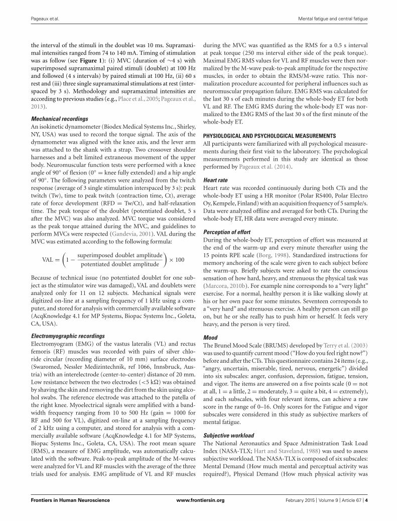
and Psychological Measurements. An overview of the experimental
protocol performed during the second and third visit is presented
in Figure 1. Heart rate (HR) was recorded continuously through-
out the experiment. Each participant completed all three visits
over a period of 2 weeks with a minimum of 48 h recovery period
between visits. All participants were given instructions to sleep for
at least 7 h, refrain from the consumption of alcohol, and not to
practice vigorous physical activity the day before each visit. Partic-
ipants were also instructed not to consume caffeine and nicotine
Frontiers in Human Neuroscience www.frontiersin.org February 2015 | Volume 9 | Article 67 | 2
Pageaux et al. Mental fatigue and central fatigue
FIGURE 1 | Graphical overview of the experimental protocol. Order and timing were the same for each subject and each visit. Q, psychological
questionnaires; PPO, peak power output; MVC, maximal voluntary contraction; ET, whole-body endurance task.
for at least 3 h before testing, and were asked to declare if they had
taken any medication or had any acute illness, injury, or infection.
COGNITIVE TASKS
Both CTs were performed for 30 min, and they are identical to
those used by Pageaux et al. (2014) to reduce self-paced endurance
running performance. An incongruent Stroop task and a congru-
ent Stroop task were used respectively for the self-regulation task
and the control task (Stroop, 1992). A brief description of these
CTs can be found below.
Self-regulation task
The modified incongruent Stroop task used as self-regulation task
consisted of color words (yellow, blue, green, red) printed in a
different ink color (either yellow, blue, green, red) presented on
a computer screen. Subjects were instructed to press one of four
colored buttons on the computer keyboard (yellow, blue, green,
red) with the correct response being the button corresponding to
the ink color (either yellow, blue, green, red) of the word presented
on the computer screen. If however, the ink color was red, the
button to be pressed was the button linked to the real meaning
of the word, not the ink color (e.g., if the word blue appears in
red, the button blue has to be pressed). If the ink color was blue,
green or yellow, then the button pressed matched the ink color.
The word presented and its ink color were randomly selected by
the computer (100% incongruent). Subjects were instructed to
respond as quickly and accurately as possible. Feedback (correct
or incorrect response, reaction time, and response accuracy so far)
was provided on the computer screen after each word. Participants
were also informed that points would be awarded for speed and
accuracy of their responses, and the score for both CTs would be
added to the score for each time trial.
Control task
The congruent version of the Stroop color-word task used as
control task was similar to the modified incongruent version of
the Stroop color-word task. However, all words and their ink
color were matched in order to greatly reduce the extent of self-
regulation required by the CT. Subjects were familiarized with both
CTs during the first visit to the laboratory. Response accuracy
(percentage of correct responses) and reaction time were mea-
sured to monitor cognitive performance. Data were averaged every
5 min and analyzed offline using the E-Prime software (Psychology
Software Tools, Pittsburgh, PA, USA). No filters were applied to
trim the reaction time data.
WHOLE-BODY ENDURANCE TASK
Fifteen minutes after completion of the CT, subjects performed the
whole-body ET on an electromagnetically braked cycle ergometer
(Excalibur Sport, Lode, Groningen, The Netherlands) set in hyper-
bolic mode. After a 3-min warm-up cycling at 40% of peak power
output (121 ± 12 W), subjects cycled at 80% of peak power out-
put (242 ± 23 W) for 6 min. Cadence was freely chosen between
60 and 100 RPM, and a fan was placed in a standardized posi-
tion in front of the subject during the entire duration of the task.
Feedback on elapsed time, cadence, power output, and HR was
not available to the subject. Once the 6 min were elapsed, sub-
jects stopped cycling immediately and were transferred to the
isokinetic dynamometer for the assessment of neuromuscular
function (see Neuromuscular Function Tests). At the end of the
warm-up, and at the end of each minute thereafter, rating of per-
ceived exertion (RPE) and cadence were recorded. Subjects were
familiarized with the whole-body ET during the first visit to the
laboratory.
NEUROMUSCULAR FUNCTION TESTS
All participants were familiarized with all neuromuscular function
tests during their first visit to the laboratory. The neuromuscu-
lar function tests performed in this study are identical as those
performed by Pageaux et al. (2013).
Electrical stimulation
Both single and double (100 Hz frequency) stimulation were used
for assessment of neuromuscular function. All central fatigue
parameters were obtained within 45 s after completion of the
whole-body ET. Transcutaneous electric1ally evoked contractions
of the knee extensor muscles were induced by using a high-voltage
(maximal voltage 400 V) constant-current stimulator (model DS7
modified, Digitimer, Hertfordshire, UK). A monopolar cathode
ball electrode (0.5 cm diameter) pressed into the femoral triangle
by the same experimenter during all tests was used to stimulate
the femoral nerve. To ensure reliability of measurement, the site
of stimulation producing the largest resting twitch amplitude and
compound muscle action potential (M-wave) was marked on the
skin with permanent marker. The anode was a 50 cm2 (10 × 5 cm)
rectangular electrode (Compex SA, Ecublens, Switzerland) located
on the gluteus maximus opposite to the cathode. The stimulus
intensity required to evoke a maximal compound muscle action
potential (Mmax) was determined at rest and during submaxi-
mal isometric knee extensors contractions (50% MVC) before the
experiment on each day. The stimulus duration was 1 ms and
Frontiers in Human Neuroscience www.frontiersin.org February 2015 | Volume 9 | Article 67 | 3
Pageaux et al. Mental fatigue and central fatigue
the interval of the stimuli in the doublet was 10 ms. Supramaxi-
mal intensities ranged from 74 to 140 mA. Timing of stimulation
was as follow (see Figure 1): (i) MVC (duration of ∼4 s) with
superimposed supramaximal paired stimuli (doublet) at 100 Hz
and followed (4 s intervals) by paired stimuli at 100 Hz, (ii) 60 s
rest and (iii) three single supramaximal stimulations at rest (inter-
spaced by 3 s). Methodology and supramaximal intensities are
according to previous studies (e.g., Place et al., 2005; Pageaux et al.,
2013).
Mechanical recordings
An isokinetic dynamometer (Biodex Medical Systems Inc., Shirley,
NY, USA) was used to record the torque signal. The axis of the
dynamometer was aligned with the knee axis, and the lever arm
was attached to the shank with a strap. Two crossover shoulder
harnesses and a belt limited extraneous movement of the upper
body. Neuromuscular function tests were performed with a knee
angle of 90◦ of flexion (0◦ = knee fully extended) and a hip angle
of 90◦. The following parameters were analyzed from the twitch
response (average of 3 single stimulation interspaced by 3 s): peak
twitch (Tw), time to peak twitch (contraction time, Ct), average
rate of force development (RFD = Tw/Ct), and half-relaxation
time. The peak torque of the doublet (potentiated doublet, 5 s
after the MVC) was also analyzed. MVC torque was considered
as the peak torque attained during the MVC, and guidelines to
perform MVCs were respected (Gandevia, 2001). VAL during the
MVC was estimated according to the following formula:
VAL =
(
1 −superimposed doublet amplitude
potentiated doublet amplitude
)
× 100
Because of technical issue (no potentiated doublet for one sub-
ject as the stimulator wire was damaged), VAL and doublets were
analyzed only for 11 on 12 subjects. Mechanical signals were
digitized on-line at a sampling frequency of 1 kHz using a com-
puter, and stored for analysis with commercially available software
(AcqKnowledge 4.1 for MP Systems, Biopac Systems Inc., Goleta,
CA, USA).
Electromyographic recordings
Electromyogram (EMG) of the vastus lateralis (VL) and rectus
femoris (RF) muscles was recorded with pairs of silver chlo-
ride circular (recording diameter of 10 mm) surface electrodes
(Swaromed, Nessler Medizintechnik, ref 1066, Innsbruck, Aus-
tria) with an interelectrode (center-to-center) distance of 20 mm.
Low resistance between the two electrodes (<5 k�) was obtained
by shaving the skin and removing the dirt from the skin using alco-
hol swabs. The reference electrode was attached to the patella of
the right knee. Myoelectrical signals were amplified with a band-
width frequency ranging from 10 to 500 Hz (gain = 1000 for
RF and 500 for VL), digitized on-line at a sampling frequency
of 2 kHz using a computer, and stored for analysis with a com-
mercially available software (AcqKnowledge 4.1 for MP Systems,
Biopac Systems Inc., Goleta, CA, USA). The root mean square
(RMS), a measure of EMG amplitude, was automatically calcu-
lated with the software. Peak-to-peak amplitude of the M-waves
were analyzed for VL and RF muscles with the average of the three
trials used for analysis. EMG amplitude of VL and RF muscles
during the MVC was quantified as the RMS for a 0.5 s interval
at peak torque (250 ms interval either side of the peak torque).
Maximal EMG RMS values for VL and RF muscles were then nor-
malized by the M-wave peak-to-peak amplitude for the respective
muscles, in order to obtain the RMS/M-wave ratio. This nor-
malization procedure accounted for peripheral influences such as
neuromuscular propagation failure. EMG RMS was calculated for
the last 30 s of each minutes during the whole-body ET for both
VL and RF. The EMG RMS during the whole-body ET was nor-
malized to the EMG RMS of the last 30 s of the first minute of the
whole-body ET.
PHYSIOLOGICAL AND PSYCHOLOGICAL MEASUREMENTS
All participants were familiarized with all psychological measure-
ments during their first visit to the laboratory. The psychological
measurements performed in this study are identical as those
performed by Pageaux et al. (2014).
Heart rate
Heart rate was recorded continuously during both CTs and the
whole-body ET using a HR monitor (Polar RS400, Polar Electro
Oy, Kempele, Finland) with an acquisition frequency of 5 sample/s.
Data were analyzed offline and averaged for both CTs. During the
whole-body ET, HR data were averaged every minute.
Perception of effort
During the whole-body ET, perception of effort was measured at
the end of the warm-up and every minute thereafter using the
15 points RPE scale (Borg, 1998). Standardized instructions for
memory anchoring of the scale were given to each subject before
the warm-up. Briefly subjects were asked to rate the conscious
sensation of how hard, heavy, and strenuous the physical task was
(Marcora, 2010b). For example nine corresponds to a “very light”
exercise. For a normal, healthy person it is like walking slowly at
his or her own pace for some minutes. Seventeen corresponds to
a “very hard” and strenuous exercise. A healthy person can still go
on, but he or she really has to push him or herself. It feels very
heavy, and the person is very tired.
Mood
The Brunel Mood Scale (BRUMS) developed by Terry et al. (2003)
was used to quantify current mood (“How do you feel right now?”)
before and after the CTs. This questionnaire contains 24 items (e.g.,
“angry, uncertain, miserable, tired, nervous, energetic”) divided
into six subscales: anger, confusion, depression, fatigue, tension,
and vigor. The items are answered on a five points scale (0 = not
at all, 1 = a little, 2 = moderately, 3 = quite a bit, 4 = extremely),
and each subscales, with four relevant items, can achieve a raw
score in the range of 0–16. Only scores for the Fatigue and vigor
subscales were considered in this study as subjective markers of
mental fatigue.
Subjective workload
The National Aeronautics and Space Administration Task Load
Index (NASA-TLX; Hart and Staveland, 1988) was used to assess
subjective workload. The NASA-TLX is composed of six subscales:
Mental Demand (How much mental and perceptual activity was
required?), Physical Demand (How much physical activity was
Frontiers in Human Neuroscience www.frontiersin.org February 2015 | Volume 9 | Article 67 | 4
Pageaux et al. Mental fatigue and central fatigue
required?), Temporal Demand (How much time pressure did
you feel due to the rate or pace at which the task occurred?),
Performance (How much successful do you think you were in
accomplishing the goals of the task set by the experimenter?),
Effort (How hard did you have to work to accomplish your level of
performance?), and Frustration (How much irritating, annoying
did you perceive the task?). The participants had to score each of
the items on a scale divided into 20 equal intervals anchored by a
bipolar descriptor (e.g., High/Low). This score was multiplied by
5, resulting in a final score between 0 and 100 for each of the six
subscales. Participants completed the NASA-TLX after the CT and
after the whole-body ET.
STATISTICS
All data are presented as means ± standard deviation (SD) unless
stated. Assumptions of statistical tests such as normal distribution
and sphericity of data were checked as appropriate. Lower-Bound
correction to the degrees of freedom was applied when violations
to sphericity were present. Paired t-tests were used to assess the
effect of condition (mental fatigue vs. control) on HR during
both CTs and on NASA-TLX scores after the CTs and after the
whole-body ET. Fully repeated measure 2 × 6 ANOVAs were used
to test the effects of condition and time on response accuracy
and reaction time during the CTs. Fully repeated measure 2 × 2
ANOVAs were used to test the effects of condition and time on
mood before and after the CTs. Fully repeated measure 2 × 3
ANOVAs were used to test the effects of condition and time on
MVC torque, VAL, M-wave parameters for each muscle, RMS/M-
wave ratio, twitch properties, and peak doublet torque before
and after the CTs, and after the whole-body ET. Fully repeated
measure 2 × 6 ANOVAs were used to test the effects of condi-
tion and time on HR, and EMG RMS during the whole-body
ET. Fully repeated measure 2 × 7 ANOVA was used to test the
effects of condition and time on RPE and cadence during the
whole-body ET. Significant main effects of time and significant
interactions were followed up with Bonferroni tests as appropri-
ate. Significance was set at 0.05 (2-tailed) for all analyses, which
were conducted using the Statistical Package for the Social Sci-
ences, version 20 for Mac OS X (SPSS Inc., Chicago, IL, USA).
Cohen’s effects size dz and f(V) were calculated with G*Power
software (version 3.1.6, Universität Düsseldorf, Germany) and
reported.
RESULTS
COGNITIVE TASKS
Mood
Self-reported fatigue was significantly higher [p = 0.009,
f(V) = 0.957] post-CTs (mental fatigue condition 3.7 ± 3.4, con-
trol condition 4.5 ± 3.6) compared to pre-CTs (mental fatigue
condition 1.5 ± 2.0, control condition 1.8 ± 1.5). However,
neither the main effect of condition [p = 0.369, f(V) = 0.951]
nor the interaction [p = 0.401, f(V) = 0.264] were signifi-
cant. Vigor decreased [p = 0.009, f(V) = 0.283] significantly
after the self-regulation task (10.2 ± 3.0 to 8.3 ± 3.9) and
the control task (10.6 ± 4.0 to 7.8 ± 4.7) with no signif-
icant difference between conditions [interaction p = 1.000,
f(V) = 0.032].
Cognitive performance
Response accuracy during CTs did not present any main effect
of condition [p = 0.070, f(V) = 0.605] or time [p = 0.236,
f(V) = 0.378]. Reaction time during both conditions did not
change over time [p = 0.507, f(V) = 0.207] but was significantly
longer during the self-regulation task compared to the control task
[834 ± 109 vs. 597 ± 80 ms, p < 0.001, f(V) = 2.500]. Reaction
time during the self-regulation task was significantly higher for all
subjects.
Heart rate
Heart rate was significantly higher (p < 0.001, dz = 0.577) during
the self-regulation task (65.8 ± 9.3 beats/min) compared to the
control task (62.0 ± 4.5 beats/min).
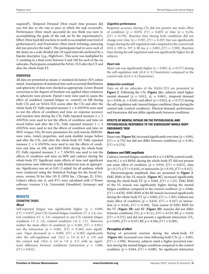
Subjective workload
Data on all six subscales of the NASA-TLX are presented in
Figure 2. Following the CTs (Figure 2A), subjects rated higher
mental demand (p = 0.012, dz = 0.861), temporal demand
(p = 0.050, dz = 0.626) and effort (p = 0.022, dz = 0.772) during
the self-regulation task (mental fatigue condition) than during the
control task (control condition). Physical demand, performance
and frustration did not differ significantly between conditions.
EFFECTS OF MENTAL FATIGUE ON THE PHYSIOLOGICAL AND
PSYCHOLOGICAL RESPONSES TO THE SUBSEQUENT WHOLE-BODY
ENDURANCE TASK
Heart rate
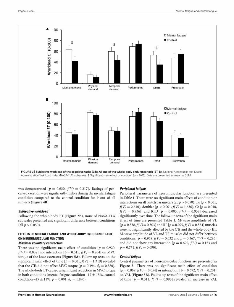
Heart rate (Figure 3A) increased significantly over time [p < 0.001,
f(V) = 4.776] but did not differ between conditions [p = 0.381,
f(V) = 0.274].
Cadence and EMG amplitude
Cadence (mental fatigue condition 84.4 ± 5.4 RPM, control condi-
tion 84.2 ± 6.0 RPM) during the whole-body ET did not present
any main effect of condition [p = 0.919, f(V) = 0.031], time
[p = 0.175, f(V) = 0.418], or interaction [p = 0.101, f(V) = 0.412].
Electromyogram amplitude data are presented in Figure 3.
EMG RMS of the VL muscle (Figure 3C) increased significantly
during the whole-body ET [p = 0.002, f(V) = 1.25]. EMG RMS
of the VL muscle was significantly higher during the mental
fatigue condition compared to the control condition [p = 0.046,
f(V) = 0.678]. EMG RMS of the RF muscle increased significantly
during the whole-body ET [p = 0.002, f(V) = 1.305] without any
main effect of condition [p = 0.610, f(V) = 0.167] or interac-
tion [p = 0.626, f(V) = 0.160]. Time course of EMG RMS for
the VL (Figure 3B) and RF (Figure 3D) muscles did not differ
between conditions [VL, p = 0.111, f(V) = 0.523; RF, p = 0.410,
f(V) = 0.272] and did not present a significant interaction [VL,
p = 0.091, f(V) = 0.557; RF, p = 0.384, f(V) = 0.289].
Perception of effort
Rating of perceived exertion during the whole-body ET
(Figure 4A) increased over time following both CTs [p < 0.001,
f(V) = 3.590]. However, subjects rated a higher perceived exer-
tion during the mental fatigue condition compared to the control
condition [p = 0.044, f(V) = 0.680]. No significant interaction
Frontiers in Human Neuroscience www.frontiersin.org February 2015 | Volume 9 | Article 67 | 5
Pageaux et al. Mental fatigue and central fatigue
FIGURE 2 | Subjective workload of the cognitive tasks (CTs, A) and of the whole-body endurance task (ET, B). National Aeronautics and Space
Administration Task Load Index (NASA-TLX) subscales. $ Significant main effect of condition (p < 0.05). Data are presented as mean ± SEM.
was demonstrated [p = 0.630, f(V) = 0.217]. Ratings of per-
ceived exertion were significantly higher during the mental fatigue
condition compared to the control condition for 9 out of all
subjects (Figure 4B).
Subjective workload
Following the whole-body ET (Figure 2B), none of NASA-TLX
subscales presented any significant difference between conditions
(all p > 0.050).
EFFECTS OF MENTAL FATIGUE AND WHOLE-BODY ENDURANCE TASK
ON NEUROMUSCULAR FUNCTION
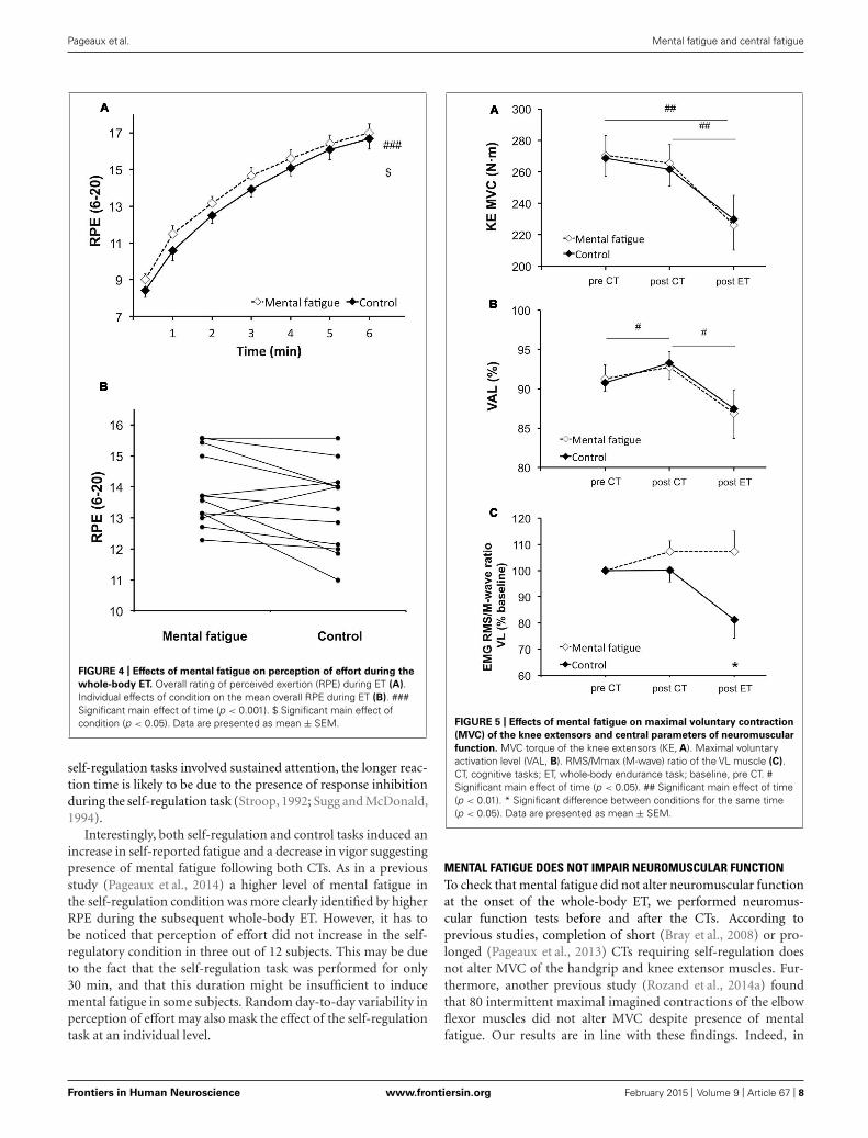
Maximal voluntary contraction
There was no significant main effect of condition [p = 0.920,
f(V) = 0.032] nor interaction [p = 0.515, f(V) = 0.204] on MVC
torque of the knee extensors (Figure 5A). Follow-up tests on the
significant main effect of time [p = 0.001, f(V) = 1.319] revealed
that the CTs did not affect MVC torque [p = 0.194, dz = 0.580].
The whole-body ET caused a significant reduction in MVC torque
in both conditions (mental fatigue condition –17 ± 15%, control
condition –15 ± 11%, p = 0.001, dz = 1.890).
Peripheral fatigue
Peripheral parameters of neuromuscular function are presented
in Table 1. There were no significant main effects of condition or
interactions on all twitch parameters (all p > 0.050). Tw [p < 0.001,
f(V) = 2.610], doublet [p < 0.001, f(V) = 1.636], Ct [p = 0.010,
f(V) = 0.936], and RFD [p = 0.003, f(V) = 0.938] decreased
significantly over time. The follow-up tests of the significant main
effect of time are presented Table 1. M-wave amplitude of VL
[p = 0.338, f(V) = 0.303] and RF [p = 0.079, f(V) = 0.584] muscles
were not significantly affected by the CTs and the whole-body ET.
M-wave amplitude of VL and RF muscles did not differ between
conditions [p = 0.958, f(V) = 0.032 and p = 0.367, f(V) = 0.283]
and did not show any interaction [p = 0.620, f(V) = 0.153 and
p = 0.771, f(V) = 0.090].
Central fatigue
Central parameters of neuromuscular function are presented in
Figure 5. There was no significant main effect of condition
[p = 0.869, f(V) = 0.054] or interaction [p = 0.672, f(V) = 0.201]
on VAL (Figure 5B). Follow-up tests of the significant main effect
of time [p = 0.011, f(V) = 0.990] revealed an increase in VAL
Frontiers in Human Neuroscience www.frontiersin.org February 2015 | Volume 9 | Article 67 | 6
Pageaux et al. Mental fatigue and central fatigue
FIGURE 3 | Effects of mental fatigue on heart rate and
electromyogram (EMG) amplitude of the knee extensors during
the whole-body endurance task (ET). Heart rate (HR) during ET
(A). EMG root mean square (RMS) for the vastus lateralis (VL)
muscle normalized by the first minute of ET (baseline; B). EMG
RMS of the VL muscle during ET (C). EMG RMS for the rectus
femoris (RF) muscle normalized by the first minute of ET (baseline;
D). $ Significant main effect of condition (p < 0.05). ## Significant
main effect of time (p < 0.01). ### Significant main effect of time
(p < 0.001). Data are presented as mean ± SEM.
post-CTs (p = 0.024, dz = 0.438). On the contrary, the whole-
body ET significantly reduced VAL (p = 0.013, dz = 0.880).
RMS/M-wave ratio of the VL muscle (Figure 5C) did not present
any significant main effect of time [p = 0.313, f(V) = 0.318] or
condition [p = 0.279, f(V) = 0.343]. Follow-up tests of the inter-
action [p = 0.021, f(V) = 0.810] revealed that the RMS/M-wave
ratio of the VL muscle decreased only during the control condition
following the whole-body ET (p = 0.038, dz = 0.305). RMS/M-
wave ratio of the RF muscle did not change overtime [p = 0.063,
f(V) = 0.280] and did not present any main effect of condition
[p = 0.915, f(V) = 0.032] or interaction [p = 0.335, f(V) = 0.335].
DISCUSSION
The primary aim of this study was to test the hypothesis that
mental fatigue exacerbates central fatigue induced by whole-body
endurance exercise. The results of the present study do not support
this hypothesis. Furthermore, mental fatigue did not exacerbate
peripheral fatigue induced by whole-body exercise. Therefore,
the higher-than-normal perception of effort experienced by men-
tally fatigued subjects is independent of any central or peripheral
alteration of neuromuscular function.
SELF-REGULATION, MENTAL FATIGUE, AND PERCEPTION OF EFFORT
We used a self-regulation task (incongruent Stroop task) to induce
mental fatigue. The higher HR experienced during the incon-
gruent Stroop task confirms that self-regulation is cognitively
demanding and requires higher effort mobilization compared to
the control task (Richter et al., 2008). The more demanding nature
of the self-regulation task is also supported by higher ratings
of mental demand, temporal demand, and effort compared to
the control task. Moreover, the subjects presented a longer reac-
tion time during the self-regulation task compared to the control
task, confirming the presence of an additional cognitive control
mechanism during the self-regulation task. As both control and
Frontiers in Human Neuroscience www.frontiersin.org February 2015 | Volume 9 | Article 67 | 7
Pageaux et al. Mental fatigue and central fatigue
FIGURE 4 | Effects of mental fatigue on perception of effort during the
whole-body ET. Overall rating of perceived exertion (RPE) during ET (A).
Individual effects of condition on the mean overall RPE during ET (B). ###
Significant main effect of time (p < 0.001). $ Significant main effect of
condition (p < 0.05). Data are presented as mean ± SEM.
self-regulation tasks involved sustained attention, the longer reac-
tion time is likely to be due to the presence of response inhibition
during the self-regulation task (Stroop, 1992; Sugg and McDonald,
1994).
Interestingly, both self-regulation and control tasks induced an
increase in self-reported fatigue and a decrease in vigor suggesting
presence of mental fatigue following both CTs. As in a previous
study (Pageaux et al., 2014) a higher level of mental fatigue in
the self-regulation condition was more clearly identified by higher
RPE during the subsequent whole-body ET. However, it has to
be noticed that perception of effort did not increase in the self-
regulatory condition in three out of 12 subjects. This may be due
to the fact that the self-regulation task was performed for only
30 min, and that this duration might be insufficient to induce
mental fatigue in some subjects. Random day-to-day variability in
perception of effort may also mask the effect of the self-regulation
task at an individual level.
FIGURE 5 | Effects of mental fatigue on maximal voluntary contraction
(MVC) of the knee extensors and central parameters of neuromuscular
function. MVC torque of the knee extensors (KE, A). Maximal voluntary
activation level (VAL, B). RMS/Mmax (M-wave) ratio of the VL muscle (C).
CT, cognitive tasks; ET, whole-body endurance task; baseline, pre CT. #
Significant main effect of time (p < 0.05). ## Significant main effect of time
(p < 0.01). * Significant difference between conditions for the same time
(p < 0.05). Data are presented as mean ± SEM.
MENTAL FATIGUE DOES NOT IMPAIR NEUROMUSCULAR FUNCTION
To check that mental fatigue did not alter neuromuscular function
at the onset of the whole-body ET, we performed neuromus-
cular function tests before and after the CTs. According to
previous studies, completion of short (Bray et al., 2008) or pro-
longed (Pageaux et al., 2013) CTs requiring self-regulation does
not alter MVC of the handgrip and knee extensor muscles. Fur-
thermore, another previous study (Rozand et al., 2014a) found
that 80 intermittent maximal imagined contractions of the elbow
flexor muscles did not alter MVC despite presence of mental
fatigue. Our results are in line with these findings. Indeed, in
Frontiers in Human Neuroscience www.frontiersin.org February 2015 | Volume 9 | Article 67 | 8
Pageaux et al. Mental fatigue and central fatigue
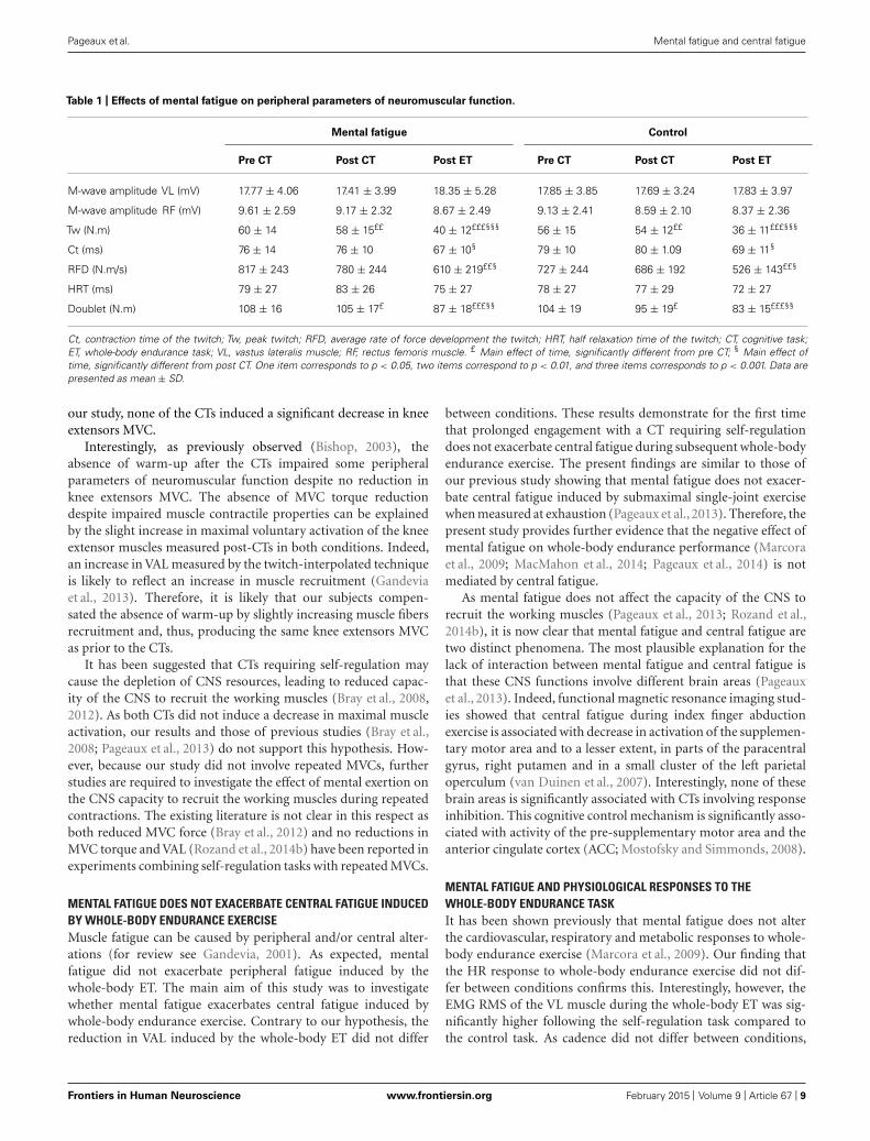
Table 1 | Effects of mental fatigue on peripheral parameters of neuromuscular function.
Mental fatigue Control
Pre CT Post CT Post ET Pre CT Post CT Post ET
M-wave amplitude VL (mV) 17.77 ± 4.06 17.41 ± 3.99 18.35 ± 5.28 17.85 ± 3.85 17.69 ± 3.24 17.83 ± 3.97
M-wave amplitude RF (mV) 9.61 ± 2.59 9.17 ± 2.32 8.67 ± 2.49 9.13 ± 2.41 8.59 ± 2.10 8.37 ± 2.36
Tw (N.m) 60 ± 14 58 ± 15££ 40 ± 12£££§§§ 56 ± 15 54 ± 12££ 36 ± 11£££§§§
Ct (ms) 76 ± 14 76 ± 10 67 ± 10§ 79 ± 10 80 ± 1.09 69 ± 11§
RFD (N.m/s) 817 ± 243 780 ± 244 610 ± 219££§ 727 ± 244 686 ± 192 526 ± 143££§
HRT (ms) 79 ± 27 83 ± 26 75 ± 27 78 ± 27 77 ± 29 72 ± 27
Doublet (N.m) 108 ± 16 105 ± 17£ 87 ± 18£££§§ 104 ± 19 95 ± 19£ 83 ± 15£££§§
Ct, contraction time of the twitch; Tw, peak twitch; RFD, average rate of force development the twitch; HRT, half relaxation time of the twitch; CT, cognitive task;
ET, whole-body endurance task; VL, vastus lateralis muscle; RF, rectus femoris muscle. £ Main effect of time, significantly different from pre CT; § Main effect of
time, significantly different from post CT. One item corresponds to p < 0.05, two items correspond to p < 0.01, and three items corresponds to p < 0.001. Data are
presented as mean ± SD.
our study, none of the CTs induced a significant decrease in knee
extensors MVC.
Interestingly, as previously observed (Bishop, 2003), the
absence of warm-up after the CTs impaired some peripheral
parameters of neuromuscular function despite no reduction in
knee extensors MVC. The absence of MVC torque reduction
despite impaired muscle contractile properties can be explained
by the slight increase in maximal voluntary activation of the knee
extensor muscles measured post-CTs in both conditions. Indeed,
an increase in VAL measured by the twitch-interpolated technique
is likely to reflect an increase in muscle recruitment (Gandevia
et al., 2013). Therefore, it is likely that our subjects compen-
sated the absence of warm-up by slightly increasing muscle fibers
recruitment and, thus, producing the same knee extensors MVC
as prior to the CTs.
It has been suggested that CTs requiring self-regulation may
cause the depletion of CNS resources, leading to reduced capac-
ity of the CNS to recruit the working muscles (Bray et al., 2008,
2012). As both CTs did not induce a decrease in maximal muscle
activation, our results and those of previous studies (Bray et al.,
2008; Pageaux et al., 2013) do not support this hypothesis. How-
ever, because our study did not involve repeated MVCs, further
studies are required to investigate the effect of mental exertion on
the CNS capacity to recruit the working muscles during repeated
contractions. The existing literature is not clear in this respect as
both reduced MVC force (Bray et al., 2012) and no reductions in
MVC torque and VAL (Rozand et al., 2014b) have been reported in
experiments combining self-regulation tasks with repeated MVCs.
MENTAL FATIGUE DOES NOT EXACERBATE CENTRAL FATIGUE INDUCED
BY WHOLE-BODY ENDURANCE EXERCISE
Muscle fatigue can be caused by peripheral and/or central alter-
ations (for review see Gandevia, 2001). As expected, mental
fatigue did not exacerbate peripheral fatigue induced by the
whole-body ET. The main aim of this study was to investigate
whether mental fatigue exacerbates central fatigue induced by
whole-body endurance exercise. Contrary to our hypothesis, the
reduction in VAL induced by the whole-body ET did not differ
between conditions. These results demonstrate for the first time
that prolonged engagement with a CT requiring self-regulation
does not exacerbate central fatigue during subsequent whole-body
endurance exercise. The present findings are similar to those of
our previous study showing that mental fatigue does not exacer-
bate central fatigue induced by submaximal single-joint exercise
when measured at exhaustion (Pageaux et al., 2013). Therefore, the
present study provides further evidence that the negative effect of
mental fatigue on whole-body endurance performance (Marcora
et al., 2009; MacMahon et al., 2014; Pageaux et al., 2014) is not
mediated by central fatigue.
As mental fatigue does not affect the capacity of the CNS to
recruit the working muscles (Pageaux et al., 2013; Rozand et al.,
2014b), it is now clear that mental fatigue and central fatigue are
two distinct phenomena. The most plausible explanation for the
lack of interaction between mental fatigue and central fatigue is
that these CNS functions involve different brain areas (Pageaux
et al., 2013). Indeed, functional magnetic resonance imaging stud-
ies showed that central fatigue during index finger abduction
exercise is associated with decrease in activation of the supplemen-
tary motor area and to a lesser extent, in parts of the paracentral
gyrus, right putamen and in a small cluster of the left parietal
operculum (van Duinen et al., 2007). Interestingly, none of these
brain areas is significantly associated with CTs involving response
inhibition. This cognitive control mechanism is significantly asso-
ciated with activity of the pre-supplementary motor area and the
anterior cingulate cortex (ACC; Mostofsky and Simmonds, 2008).
MENTAL FATIGUE AND PHYSIOLOGICAL RESPONSES TO THE
WHOLE-BODY ENDURANCE TASK
It has been shown previously that mental fatigue does not alter
the cardiovascular, respiratory and metabolic responses to whole-
body endurance exercise (Marcora et al., 2009). Our finding that
the HR response to whole-body endurance exercise did not dif-
fer between conditions confirms this. Interestingly, however, the
EMG RMS of the VL muscle during the whole-body ET was sig-
nificantly higher following the self-regulation task compared to
the control task. As cadence did not differ between conditions,
Frontiers in Human Neuroscience www.frontiersin.org February 2015 | Volume 9 | Article 67 | 9
Pageaux et al. Mental fatigue and central fatigue
this result suggests that prolonged self-regulation induced alter-
ations in muscle recruitment at the onset and throughout the
subsequent whole-body ET. This is not the first report of higher
EMG amplitude during a physical task following a self-regulation
task. In accordance with our results, Bray et al. (2008) mea-
sured higher EMG amplitude during sustained handgrip exercise
following a short (3 min 40 s) engagement with the same incon-
gruent Stroop task used in the present study. Therefore, our
results, combined with those of Bray et al. (2008), suggest that
both prolonged and short engagement with CTs requiring self-
regulation can alter muscle recruitment during a subsequent
physical task.
Because central and peripheral fatigue did not differ between
conditions, higher EMG RMS of the VL muscle during the
whole-body ET in the self-regulation condition cannot represent a
compensatory increase in muscle recruitment. A possible explana-
tion is that this EMG alteration represents an alteration in motor
control in conditions of mental fatigue. This conclusion is sup-
ported by the findings of two recent studies showing that mental
fatigue reduces mechanically induced tremor (Budini et al., 2014)
and has adverse effects in all the three phases of slips (Lew and Qu,
2014). As injury in sport is more likely to occur in the late stage
of an event or a season (e.g., Ekstrand et al., 2011), it seems that
the effects of mental fatigue on motor control during whole-body
physical tasks warrant further investigations.
MENTAL FATIGUE AND PERCEPTION OF EFFORT
The higher-than normal perception of effort experienced by men-
tally fatigued subjects in the present experiment is similar to
that reported in previous studies involving submaximal single-
joint exercise (Pageaux et al., 2013) and whole-body endurance
exercise (Marcora et al., 2009) at a fixed workload, as well as self-
paced whole-body endurance exercise (Brownsberger et al., 2013;
MacMahon et al., 2014; Pageaux et al., 2014). In some of these
studies, the abnormal perception of effort has been associated
with the negative effect of mental fatigue on endurance perfor-
mance. However, despite strong evidences that mental fatigue
increases RPE and impairs performance during endurance exer-
cise, the underlying mechanisms of this alteration in perception
of effort remain unclear.
It is well-accepted that, like any other perceptions, perception
of effort results from the neurocognitive processing of sensory
signals. However, the nature of the sensory signals involved in
perception of effort generation remains debated. Briefly, two dif-
ferent theoretical models suggest that perception of effort reflects
the neurocognitive processing of (i) signals from premotor/motor
to sensory areas of the cortex during voluntary muscle contrac-
tions (corollary discharge model; Marcora, 2009; de Morree et al.,
2012, 2014); or (ii) afferent sensory signals about the physiolog-
ical condition of the body (interoception) and the environment
(afferent feedback model; Hampson et al., 2001). Interestingly, in
our study, mentally fatigued subjects experienced a higher-than-
normal perception of effort despite no significant effects of mental
fatigue on HR and peripheral fatigue. Because sensory signals
from the heart and peripheral muscles are considered primary
sources of afferent feedback for the generation of perception of
effort (Hampson et al., 2001), it is unlikely that the higher RPE
observed in our study reflects an alteration of afferent feedback
induced by mental fatigue. Another possibility is that the higher
than-normal perception of effort observed in mentally fatigued
subjects reflects higher activity of premotor and/or motor areas
of the cortex (i.e., higher central motor command) during whole-
body endurance exercise. Although no direct neurophysiological
measures of central motor command were taken in the present
study, the abnormal EMG RMS of the VL muscle during the
whole-body ET suggests that alterations in motor control may
force mentally fatigued subjects to increase their central motor
command in order to produce the same power output even when
central and peripheral fatigue are not exacerbated. Finally, prelim-
inary evidence that prolonged and demanding cognitive activity
disrupts sensorimotor gating (van der Linden et al., 2006) suggests
that mental fatigue may also affect the neurocognitive processing
of the sensory signals underlying perception of effort. Further
studies are required to investigate whether mental fatigue (i) alters
the neurocognitive processing of the corollary discharges associ-
ated with central motor command, (ii) alters the central motor
command itself, or (iii) alters the neurocognitive processing of
afferent sensory signals.
Despite that we did not measure intrinsic changes in the brain
induced by prolonged self-regulation leading to mental fatigue, it
is possible to speculate on the mechanisms involved based on pre-
vious studies. The ACC is strongly activated during incongruent
Stroop tasks (Bush et al., 1998; Swick and Jovanovic, 2002) and
is also known to be linked with perception of effort (Williamson
et al., 2001, 2002) and effort-based decision-making (Walton et al.,
2006). Furthermore, studies with caffeine suggest an association
between brain adenosine and mental fatigue (Lorist and Tops,
2003). It is therefore plausible that the higher perception of effort
experienced by mentally fatigued subjects is caused by an accumu-
lation of adenosine in the ACC. Indeed, experimental evidences
that neural activity increases extracellular concentration of adeno-
sine (Lovatt et al., 2012) and that brain adenosine accumulation
reduces endurance performance (Davis et al., 2003) support this
hypothesis. Further studies are required to confirm these specula-
tions, and to investigate other cortical areas and neurotransmitters
involved in the negative effects of mental fatigue on perception of
effort and endurance performance.
CONCLUSION
This study was the first to test the hypothesis that mental fatigue
and central fatigue induced by whole-body exercise are causally
related. Contrary to this hypothesis, our findings show that
mental fatigue does not exacerbate central fatigue during sub-
sequent whole-body exercise. However, we must acknowledge
some limitations. Firstly, the whole-body ET had to be per-
formed on a cycle ergometer, inducing a time delay between
the end of exercise and the start of neuromuscular testing
due to the need to transfer the participant from the cycle
ergometer to the isokinetic dynamometer. Therefore, the extent
of muscle fatigue is likely to be underestimated in both exper-
imental conditions. Secondly, the whole-body ET consisted
of 6 min of high-intensity cycling exercise at a fixed work-
load. Future studies should investigate the effects of mental
fatigue on more prolonged low-to-moderate intensity whole-body
Frontiers in Human Neuroscience www.frontiersin.org February 2015 | Volume 9 | Article 67 | 10
Pageaux et al. Mental fatigue and central fatigue
endurance exercise including running where the extent of cen-
tral fatigue may be greater (Millet and Lepers, 2004). The
effects of mental fatigue on central fatigue induced by self-paced
whole-body endurance exercise and repeated sprints also warrant
further investigations given their relevance to both endurance
competitions and team sports. Finally, brain activity during
exercise was not measured in the present study and we can
only speculate, based on previous studies, on the mechanisms
underlying the increase in RPE observed in mentally fatigued
subjects.
Despite these limitations, this study provides further evidences
that mental fatigue does not reduce the capacity of the CNS to
recruit the working muscles. Our results suggest that the neg-
ative effect of mental fatigue on perception of effort does not
reflect a greater development of either central or peripheral fatigue.
Consequently, mentally fatigued subjects are still able to perform
maximal exercise, but they are experiencing an altered perfor-
mance during submaximal exercise due to higher-than-normal
perception of effort. Therefore, further studies should investi-
gate the brain alterations underlying the negative effect of mental
fatigue on perception of effort and endurance performance. A
better understanding of these brain alterations could lead to devel-
opment of novel targeted interventions to decrease perception
of effort and improve endurance performance in athletes, and
reduced exertional fatigue in patients (Macdonald et al., 2012).
REFERENCESBigland-Ritchie, B., Rice, C. L., Garland, S. J., and Walsh, M. L. (1995). “Task-
Dependent factors in fatigue of human voluntary contractions,” in Fatigue, eds S.
Gandevia, R. Enoka, A. Mccomas, D. Stuart, C. Thomas, and P. Pierce (New York,
NY: Springer ), 361–380.
Bishop, D. (2003). Warm up I: potential mechanisms and the effects of passive warm
up on exercise performance. Sports Med. 33, 439–454. doi: 10.2165/00007256-
200333060-00005
Borg, G. (1998). Borg’s Perceived Exertion and Pain Scales. Champaign, IL: Human
Kinetics.
Bray, S. R., Graham, J. D., Martin Ginis, K. A., and Hicks, A. L. (2012). Cognitive
task performance causes impaired maximum force production in human hand
flexor muscles. Biol. Psychol. 89, 195–200. doi: 10.1016/j.biopsycho.2011.10.008
Bray, S. R., Martin Ginis, K. A., Hicks, A. L., and Woodgate, J. (2008).
Effects of self-regulatory strength depletion on muscular performance and
EMG activation. Psychophysiology 45, 337–343. doi: 10.1111/j.1469-8986.2007.
00625.x
Brownsberger, J., Edwards, A., Crowther, R., and Cottrell, D. (2013). Impact of
mental fatigue on self-paced exercise. Int. J. Sports Med. 34, 1029–1036. doi:
10.1055/s-0033-1343402
Budini, F., Lowery, M., Durbaba, R., and De Vito, G. (2014). Effect of mental
fatigue on induced tremor in human knee extensors. J. Electromyogr. Kinesiol. 24,
412–418. doi: 10.1016/j.jelekin.2014.02.003
Bush, G., Whalen, P. J., Rosen, B. R., Jenike, M. A., Mcinerney, S. C., and Rauch,
S. L. (1998). The counting Stroop: an interference task specialized for functional
neuroimaging–validation study with functional MRI. Hum. Brain Mapp. 6, 270–
282. doi: 10.1002/(SICI)1097-0193(1998)6:4<270::AID-HBM6>3.0.CO;2-0
Carter, E. C., and McCullough, M. E. (2013). Is ego depletion too incredible? Evi-
dence for the overestimation of the depletion effect. Behav. Brain Sci. 36, 683–684;
discussion 707–626. doi: 10.1017/S0140525X13000952
Davis, J. M., Zhao, Z., Stock, H. S., Mehl, K. A., Buggy, J., and Hand, G. A.
(2003). Central nervous system effects of caffeine and adenosine on fatigue. Am.
J. Physiol. Regul. Integr. Comp. Physiol. 284, R399–R404. doi: 10.1152/ajpregu.00
386.2002
de Morree, H. M., Klein, C., and Marcora, S. M. (2012). Perception of effort reflects
central motor command during movement execution. Psychophysiology 49, 1242–
1253. doi: 10.1111/j.1469-8986.2012.01399.x
de Morree, H. M., Klein, C., and Marcora, S. M. (2014). Cortical substrates of the
effects of caffeine and time-on-task on perception of effort. J. Appl. Physiol. 117,
1514–1523. doi: 10.1152/japplphysiol.00898.2013
de Morree, H. M., and Marcora, S. M. (2013). Effects of isolated locomotor muscle
fatigue on pacing and time trial performance. Eur. J. Appl. Physiol. 113, 2371–
2380. doi: 10.1007/s00421-013-2673-0
de Pauw, K., Roelands, B., Cheung, S. S., De Geus, B., Rietjens, G., and Meeusen, R.
(2013). Guidelines to classify subject groups in sport-science research. Int. J.
Sports Physiol. Perform. 8, 111–122.
Ekstrand, J., Hagglund, M., and Walden, M. (2011). Injury incidence and injury
patterns in professional football: the UEFA injury study. Br. J. Sports Med. 45,
553–558. doi: 10.1136/bjsm.2009.060582
Gandevia, S. C. (2001). Spinal and supraspinal factors in human muscle fatigue.
Physiol. Rev. 81, 1725–1789.
Gandevia, S. C., Mcneil, C. J., Carroll, T. J., and Taylor, J. L. (2013). Twitch
interpolation: superimposed twitches decline progressively during a tetanic
contraction of human adductor pollicis. J. Physiol. 591, 1373–1383. doi:
10.1113/jphysiol.2012.248989
Hagger, M. S., Wood, C., Stiff, C., and Chatzisarantis, N. L. (2010). Ego depletion and
the strength model of self-control: a meta-analysis. Psychol. Bull. 136, 495–525.
doi: 10.1037/a0019486
Hampson, D. B., St Clair Gibson, A., Lambert, M. I., and Noakes, T. D. (2001).
The influence of sensory cues on the perception of exertion during exercise
and central regulation of exercise performance. Sports Med. 31, 935–952. doi:
10.2165/00007256-200131130-00004
Hart, S. G., and Staveland, L. E. (1988). “Development of NASA-TLX (Task Load
Index): results of empirical and theoretical research,” in Human Mental Workload,
Vol. 1, eds P. A. Hancock and N. Meshkati (Amsterdam: NorthHolland), 139–183.
Karoly, P. (1993). Mechanisms of self-regulation: a systems view. Annu. Rev. Psychol.
44, 23–52. doi: 10.1146/annurev.ps.44.020193.000323
Lew, F. L., and Qu, X. (2014). Effects of mental fatigue on biomechanics of slips.
Ergonomics 57, 1927–1932. doi: 10.1080/00140139.2014.937771
Lorist, M. M., and Tops, M. (2003). Caffeine, fatigue, and cognition. Brain Cogn. 53,
82–94. doi: 10.1016/S0278-2626(03)00206-9
Lovatt, D., Xu, Q., Liu, W., Takano, T., Smith, N. A., Schnermann, J., et al. (2012).
Neuronal adenosine release, and not astrocytic ATP release, mediates feedback
inhibition of excitatory activity. Proc. Natl. Acad. Sci. U.S.A. 109, 6265–6270. doi:
10.1073/pnas.1120997109
Macdonald, J. H., Fearn, L., Jibani, M., and Marcora, S. M. (2012). Exer-
tional fatigue in patients with CKD. Am. J. Kidney Dis. 60, 930–939. doi:
10.1053/j.ajkd.2012.06.021
MacMahon, C., Schucker, L., Hagemann, N., and Strauss, B. (2014). Cognitive
fatigue effects on physical performance during running. J. Sport Exerc. Psychol.
36, 375–381. doi: 10.1123/jsep.2013-0249
Marcora, S. (2009). Perception of effort during exercise is independent of afferent
feedback from skeletal muscles, heart, and lungs. J. Appl. Physiol. 106, 2060–2062.
doi: 10.1152/japplphysiol.90378.2008
Marcora, S. (2010a). Counterpoint: afferent feedback from fatigued locomotor
muscles is not an important determinant of endurance exercise performance.
J. Appl. Physiol. 108, 454–456; discussion 456–457. doi: 10.1152/japplphys-
iol.00976.2009a
Marcora, S. M. (2010b). “Effort: perception of,” in Encyclopedia of Perception, ed. E.
B. Goldstein (Thousand Oaks, CA: SAGE Publications Inc.), 380–383.Marcora, S. M., Bosio, A., and De Morree, H. M. (2008). Locomotor muscle fatigue
increases cardiorespiratory responses and reduces performance during intense
cycling exercise independently from metabolic stress. Am. J. Physiol. Regul. Integr.
Comp. Physiol. 294, R874–R883. doi: 10.1152/ajpregu.00678.2007Marcora, S. M., and Staiano, W. (2010). The limit to exercise tolerance in humans:
mind over muscle? Eur. J. Appl. Physiol. 109, 763–770. doi: 10.1007/s00421-010-
1418-6
Marcora, S. M., Staiano, W., and Manning, V. (2009). Mental fatigue impairs physical
performance in humans. J. Appl. Physiol. 106, 857–864. doi: 10.1152/japplphys-
iol.91324.2008
Millet, G. Y., and Lepers, R. (2004). Alterations of neuromuscular function after
prolonged running, cycling and skiing exercises. Sports Med. 34, 105–116. doi:
10.2165/00007256-200434020-00004
Mostofsky, S. H., and Simmonds, D. J. (2008). Response inhibition and response
selection: two sides of the same coin. J. Cogn. Neurosci. 20, 751–761. doi:
10.1162/jocn.2008.20500
Frontiers in Human Neuroscience www.frontiersin.org February 2015 | Volume 9 | Article 67 | 11
Pageaux et al. Mental fatigue and central fatigue
Muraven, M., and Baumeister, R. F. (2000). Self-regulation and depletion of limited
resources: does self-control resemble a muscle? Psychol. Bull. 126, 247–259. doi:
10.1037/0033-2909.126.2.247
Nybo, L., and Secher, N. H. (2004). Cerebral perturbations provoked by pro-
longed exercise. Prog. Neurobiol. 72, 223–261. doi: 10.1016/j.pneurobio.2004.
03.005
Pageaux, B. (2014). The psychobiological model of endurance performance:
an effort-based decision-making theory to explain self-paced endurance
performance. Sports Med. 44, 1319–1320. doi: 10.1007/s40279-014-
0198-2
Pageaux, B., Lepers, R., Dietz, K. C., and Marcora, S. M. (2014). Response inhibition
impairs subsequent self-paced endurance performance. Eur. J. Appl. Physiol. 114,
1095–1105. doi: 10.1007/s00421-014-28385
Pageaux, B., Marcora, S. M., and Lepers, R. (2013). Prolonged mental exertion does
not alter neuromuscular function of the knee extensors. Med. Sci. Sports Exerc.
45, 2254–2264. doi: 10.1249/MSS.0b013e31829b504a
Place, N., Maffiuletti, N. A., Ballay, Y., and Lepers, R. (2005). Twitch potenti-
ation is greater after a fatiguing submaximal isometric contraction performed
at short vs. long quadriceps muscle length. J. Appl. Physiol. 98, 429–436. doi:
10.1152/japplphysiol.00664.2004
Richter, M., Friedrich, A., and Gendolla, G. H. (2008). Task difficulty
effects on cardiac activity. Psychophysiology 45, 869–875. doi: 10.1111/j.1469-
8986.2008.00688.x
Ridderinkhof, K. R., Van Den Wildenberg, W. P., Segalowitz, S. J., and
Carter, C. S. (2004). Neurocognitive mechanisms of cognitive control: the
role of prefrontal cortex in action selection, response inhibition, perfor-
mance monitoring, and reward-based learning. Brain Cogn. 56, 129–140. doi:
10.1016/j.bandc.2004.09.016
Rozand, V., Lebon, F., Papaxanthis, C., and Lepers, R. (2014a). Does a mental
training session induce neuromuscular fatigue? Med. Sci. Sports Exerc. 46, 1981–
1989. doi: 10.1249/MSS.0000000000000327
Rozand, V., Pageaux, B., Marcora, S. M., Papaxanthis, C., and Lepers, R. (2014b).
Does mental exertion alter maximal muscle activation? Front. Hum. Neurosci.
8:755. doi: 10.3389/fnhum.2014.00755
Sidhu, S. K., Cresswell, A. G., and Carroll, T. J. (2013). Corticospinal
responses to sustained locomotor exercises: moving beyond single-joint stud-
ies of central fatigue. Sports Med. 43, 437–449. doi: 10.1007/s40279-013-
0020-6
Stroop, J. R. (1992). Studies of interference in serial verbal reactions (Reprinted
from J. Exp. Psychol. 18, 643–662, 1935). J. Exp. Psychol. Gen. 121, 15–23. doi:
10.1037/0096-3445.121.1.15
Sugg, M. J., and McDonald, J. E. (1994). Time course of inhibition in color-response
and word-response versions of the Stroop task. J. Exp. Psychol. Hum. Percept.
Perform. 20, 647–675. doi: 10.1037/0096-1523.20.3.647
Swick, D., and Jovanovic, J. (2002). Anterior cingulate cortex and the Stroop task:
neuropsychological evidence for topographic specificity. Neuropsychologia 40,
1240–1253. doi: 10.1016/S0028-3932(01)00226-3
Terry, P. C., Lane, A. M., and Fogarty, G. J. (2003). Construct validity of the profile of
mood states — adolescents for use with adults. Psychol. Sport Exerc. 4, 125–139.
doi: 10.1016/S1469-0292(01)00035-8
van der Linden, D., Massar, S. A., Schellekens, A. F., Ellenbroek, B. A., and Verkes,
R. J. (2006). Disrupted sensorimotor gating due to mental fatigue: preliminary
evidence. Int. J. Psychophysiol. 62, 168–174. doi: 10.1016/j.ijpsycho.2006.04.001
van Duinen, H., Renken, R., Maurits, N., and Zijdewind, I. (2007). Effects of motor
fatigue on human brain activity, an fMRI study. Neuroimage 35, 1438–1449. doi:
10.1016/j.neuroimage.2007.02.008
Walton, M. E., Kennerley, S. W., Bannerman, D. M., Phillips, P. E., and Rush-
worth, M. F. (2006). Weighing up the benefits of work: behavioral and neural
analyses of effort-related decision making. Neural Netw. 19, 1302–1314. doi:
10.1016/j.neunet.2006.03.005
Williamson, J. W., Mccoll, R., Mathews, D., Mitchell, J. H., Raven, P. B., and Morgan,
W. P. (2001). Hypnotic manipulation of effort sense during dynamic exercise:
cardiovascular responses and brain activation. J. Appl. Physiol. 90, 1392–1399.
Williamson, J. W., Mccoll, R., Mathews, D., Mitchell, J. H., Raven, P. B., and
Morgan, W. P. (2002). Brain activation by central command during actual
and imagined handgrip under hypnosis. J. Appl. Physiol. 92, 1317–1324. doi:
10.1152/japplphysiol.00939.2001
World Medical Association. (2013). World Medical Association Declaration of
Helsinki: ethical principles for medical research involving human subjects. JAMA
310, 2191–2194. doi: 10.1001/jama.2013.281053
Conflict of Interest Statement: The authors declare that the research was conducted
in the absence of any commercial or financial relationships that could be construed
as a potential conflict of interest.
Received: 30 June 2014; accepted: 27 January 2015; published online: 25 February
2015.
Citation: Pageaux B, Marcora SM, Rozand V and Lepers R (2015) Mental fatigue
induced by prolonged self-regulation does not exacerbate central fatigue during
subsequent whole-body endurance exercise. Front. Hum. Neurosci. 9:67. doi:
10.3389/fnhum.2015.00067
This article was submitted to the journal Frontiers in Human Neuroscience.
Copyright © 2015 Pageaux, Marcora, Rozand and Lepers. This is an open-access article
distributed under the terms of the Creative Commons Attribution License (CC BY).
The use, distribution or reproduction in other forums is permitted, provided the original
author(s) or licensor are credited and that the original publication in this journal is cited,
in accordance with accepted academic practice. No use, distribution or reproduction is
permitted which does not comply with these terms.
Frontiers in Human Neuroscience www.frontiersin.org February 2015 | Volume 9 | Article 67 | 12