KDIGO CKD Slide set English Final -...
56
1
-
Upload
trinhquynh -
Category
Documents
-
view
218 -
download
2
Transcript of KDIGO CKD Slide set English Final -...

1
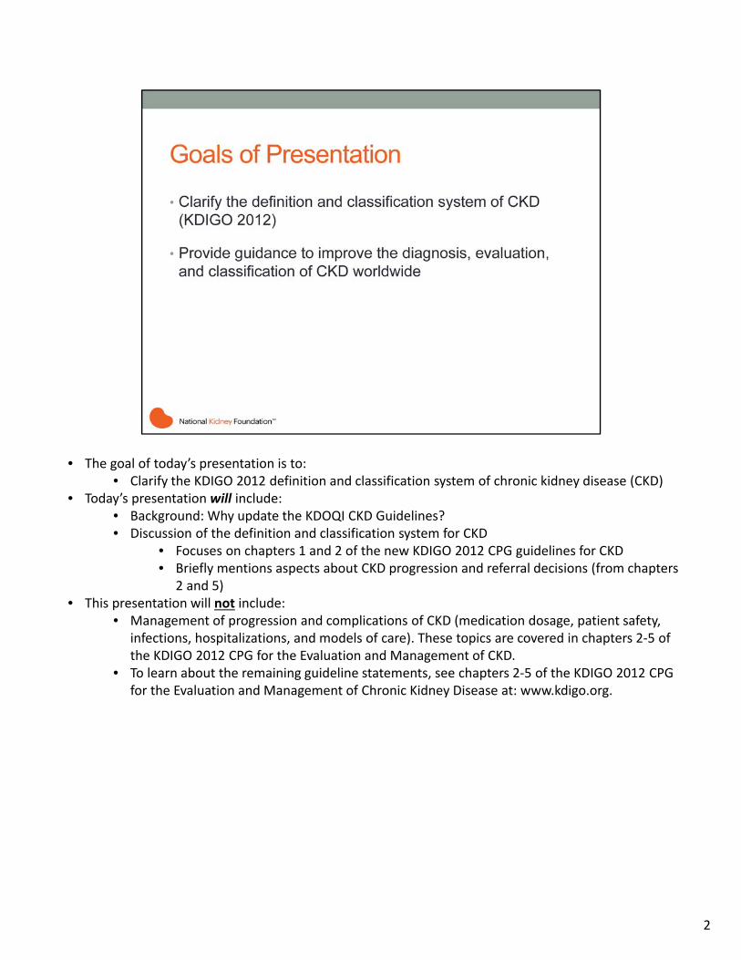
• The goal of today’s presentation is to:• Clarify the KDIGO 2012 definition and classification system of chronic kidney disease (CKD)
• Today’s presentation will include:• Background: Why update the KDOQI CKD Guidelines?• Discussion of the definition and classification system for CKD
• Focuses on chapters 1 and 2 of the new KDIGO 2012 CPG guidelines for CKD• Briefly mentions aspects about CKD progression and referral decisions (from chapters
2 and 5)• This presentation will not include:
• Management of progression and complications of CKD (medication dosage, patient safety, infections, hospitalizations, and models of care). These topics are covered in chapters 2‐5 of the KDIGO 2012 CPG for the Evaluation and Management of CKD.
• To learn about the remaining guideline statements, see chapters 2‐5 of the KDIGO 2012 CPG for the Evaluation and Management of Chronic Kidney Disease at: www.kdigo.org.
2

Sections:• Background
Slides 4 to 9• Definition of CKD
Slides 10 to 16• Classification of CKD
Slides 17 to 22• Evaluation
Slides 23 to 36• Risk stratification
Slides 37 to 44• Summary
Slides 45 to 49• Conclusion
Slides 50 to 53
3
4
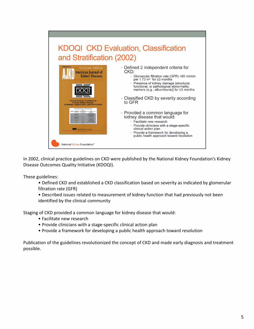
In 2002, clinical practice guidelines on CKD were published by the National Kidney Foundation’s Kidney Disease Outcomes Quality Initiative (KDOQI).
These guidelines:• Defined CKD and established a CKD classification based on severity as indicated by glomerular filtration rate (GFR)• Described issues related to measurement of kidney function that had previously not been identified by the clinical community
Staging of CKD provided a common language for kidney disease that would: • Facilitate new research• Provide clinicians with a stage‐specific clinical action plan• Provide a framework for developing a public health approach toward resolution
Publication of the guidelines revolutionized the concept of CKD and made early diagnosis and treatment possible.
5

In 2004, Kidney Disease: Improving Global Outcomes (KDIGO) endorsed the KDOQI CKD classification with minimal modifications, integrating kidney transplant recipients and clarifying stage 5 and dialysis patients.
In less than a decade, this staging framework has had enormous impact on clinical practice, research, and public health policy.
6

New information on albuminuria and GFR and their association(s) with mortality and other outcomes has emerged since the 2002 KDOQI CKD classification.
There has also been increasing recognition on limitations of the CKD definition and classification, leading to the debate which:
•Reflects changing knowledge•Provides opportunities for improvement
7

8
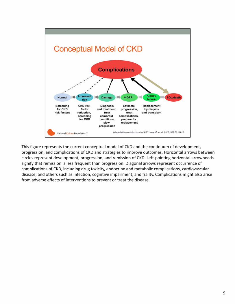
This figure represents the current conceptual model of CKD and the continuum of development, progression, and complications of CKD and strategies to improve outcomes. Horizontal arrows between circles represent development, progression, and remission of CKD. Left‐pointing horizontal arrowheads signify that remission is less frequent than progression. Diagonal arrows represent occurrence of complications of CKD, including drug toxicity, endocrine and metabolic complications, cardiovascular disease, and others such as infection, cognitive impairment, and frailty. Complications might also arise from adverse effects of interventions to prevent or treat the disease.
9
10

CKD is defined as abnormalities of kidney structure or function, present for >3 months, with implications for health.
How does this definition compare with the previous definition (2002 KDOQI)?• The definition of CKD remains intact. However, the classification and risk stratification now
includes “with implications for health.”• The addition of ‘with implications for health’ is intended to reflect the notion that a variety of
abnormalities of kidney structure or function may exist, but not all have implications for health of individuals, and therefore need to be contextualized.
11

• Kidney diseases may be acute or chronic. KDIGO explicitly but arbitrarily defines duration of >3 months (>90 days) as delineating “chronic” kidney disease.
• The rationale for defining chronicity is to differentiate CKD from acute kidney diseases (such as acuteglomerulonephritis), including acute kidney injury (AKI), which may require different interventions, and have different etiologies and outcomes. (See “KDIGO AKI Work Group. KDIGO clinical practice guideline for acute kidney injury. Kidney inter., Suppl. 2012; 2: 1‐138).
• KDIGO does not define acute kidney disease (AKD) because there does not appear be an evidence base for a precise definition.
• Most kidney diseases do not have symptoms or findings until later in their course and are detected only when they are chronic.
• Most causes of CKD are irreversible with a life‐long course, and treatment is aimed at slowing progression to kidney failure. However, chronicity is not synonymous with irreversibility.
12
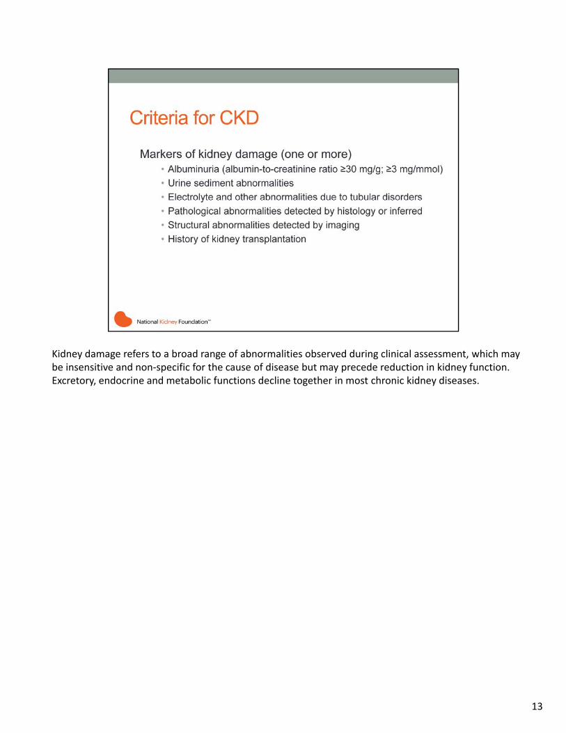
Kidney damage refers to a broad range of abnormalities observed during clinical assessment, which may be insensitive and non‐specific for the cause of disease but may precede reduction in kidney function. Excretory, endocrine and metabolic functions decline together in most chronic kidney diseases.
13
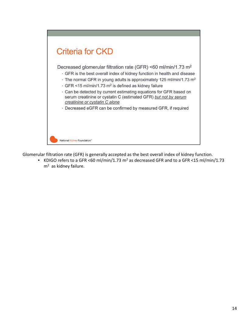
Glomerular filtration rate (GFR) is generally accepted as the best overall index of kidney function. • KDIGO refers to a GFR <60 ml/min/1.73 m2 as decreased GFR and to a GFR <15 ml/min/1.73
m2 as kidney failure.
14

• CKD is associated with a wide range of complications leading to adverse health outcomes. • For some complications, the causal pathway between kidney disease and adverse outcomes is well‐
known. For these complications, there are clinical practice guidelines for testing and treatment for modifiable factors to prevent adverse outcomes. Interested readers can refer to the KDIGO CKD‐MBD, Blood Pressure and Anemia guidelines for more details.
• Since 2002, a large number of epidemiologic studies have linked decreased GFR and albuminuria to the risk of adverse health outcomes not previously identified as CKD complications. The exploration of the mechanisms for the relationships between CKD and its complications is a rapidly growing area for basic and clinical research.
15

In general the definition of CKD in adults applies to children (birth‐18 years) with the following exceptions or allowances:• the criteria for duration >3 months does not apply to newborns or infants ≤3 months of age.• the criteria of a GFR <60 ml/min/1.73 m2 does not apply to children <2 years of age in whom an age
appropriate value should be applied.• a urinary total protein or albumin excretion rate above the normal value for age may be substituted
for albuminuria ≥30 mg/24 hours.• all electrolyte abnormalities are to be defined in light of age normative values.
16
17

• A CKD classification scheme encompassing cause and severity, as expressed by the level of GFR and the level of albuminuria, directly links to the risks of adverse outcomes including mortality and kidney outcomes.
• The inclusion of two additional domains represents a revision of the KDOQI CKD guidelines, which previously included staging only by level of GFR.
• Cause of disease is included because of its fundamental importance in predicting the outcome of CKD and choice of cause‐specific treatments.
• Albuminuria is included as an additional expression of severity of disease not only because it is a marker of the severity of injury but also because albuminuria itself is strongly associated with progression of kidney disease. Numerous studies have identified the adverse prognostic implication of albuminuria irrespective of level of kidney function.
• KDIGO proposes that this classification of CKD by Cause, GFR and Albuminuria, respectively be referred to as CGA staging. It can be used to inform the need for specialist referral, general medical management, and indications for investigation and therapeutic interventions. It will also be a tool for the study on the epidemiology, natural history, and prognosis of CKD.
Pediatric considerations:The principles inherent in this recommendation are fully applicable to children.
18

Assign cause of CKD based on presence or absence of systemic disease and the location within the kidney of observed or presumed pathologic‐anatomic findings.
• Cause is included so as to ensure that clinicians are alerted to the fact that CKD is not a diagnosis in and of itself, and that the assignment of cause is important for prognostication and treatment.
• There is wide geographic variation in the cause of kidney disease:• In developed countries, hypertension and diabetes are the most frequent causes of CKD,
especially in the elderly. In populations with a high prevalence of diabetes and hypertension, it can be difficult to distinguish CKD due to these disorders from CKD due to other disorders.
• In other countries, other causes of CKD may be as frequent as hypertension and diabetes (for example, glomerular disease in East Asia) or co‐exist with them.
• Specialized diagnostic testing, such as kidney biopsy or invasive imaging studies are performed only when it is essential to confirm some diagnoses, and the benefits justify the risks and cost.
• It is anticipated that cause of disease will not be known with certainty for many patients with CKD, but can be either inferred or not known.
Pediatric considerations:The principles inherent in this guideline are fully applicable to children.
19

Assign GFR categories• The purpose of assigning GFR categories is to ensure clarity in communication. The terms associated
with each of the GFR categories are descriptors which need to be taken in the context of the individual, and are all references to normal young adults.
• In the absence of evidence of kidney damage, neither GFR category G1 nor G2 (mildly decreased kidney function) fulfill the criteria for CKD.
• The associations of lower categories of GFR and risks of metabolic and endocrine complications formed the basis for the previous stratification into 5 stages. This current classification further acknowledges the importance of dividing stage 3 into categories G3a and G3b based on data supporting different outcomes and risk profiles.
• A number of other concurrent complications are associated with decreased categories of GFR including infection, impaired cognitive and physical function, and threats to patient safety.
Pediatric considerations:Criteria of GFR <60 ml/min/1.73 m2 does not apply to children <2 years of age in whom an age appropriate value should be applied.
20

Assign albuminuria categories• Albuminuria category is an important predictor of outcomes. The association of high levels of
proteinuria with signs and symptoms of nephrotic syndrome is well known. The detection and evaluation of lesser quantities have gained significance as multiple studies have demonstrated its diagnostic, pathogenic and prognostic importance.
• There is a continuous risk associated with albuminuria, but the use of a simple categorical approach was selected to simplify the concept for clinical practice. There is a graded increase in risk for higher albuminuria categories, at all GFR categories, without any clear threshold value. Even for subjects with GFR >60 ml/min/1.73 m2, the increased relative risk is statistically significant for urine ACR ≥30 mg/g (≥3 mg/mmol) for mortality and kidney outcomes.
• For simplicity, and to reflect the fact that it is an approximation, 3.4 mg/mmol as the current guideline threshold has been rounded to 3.0 mg/mmol.
Pediatric considerations:In children with CKD any expression of abnormal urinary protein excretion, irrespective of the marker:• must account for variation in that measurement as seen across age, sex, puberty and/or body size
(BMI). • should account for the possibility of tubular versus glomerular proteinuria dominance dependent on
the underlying disease.• may utilize proteinuria in place of albuminuria.
21
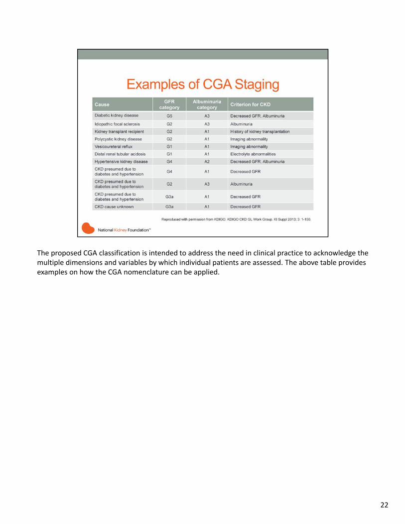
The proposed CGA classification is intended to address the need in clinical practice to acknowledge the multiple dimensions and variables by which individual patients are assessed. The above table provides examples on how the CGA nomenclature can be applied.
22

23

Chronicity• In people with GFR <60 ml/min/1.73 m2 or markers of kidney damage, review past history and previous
measurements to determine duration of kidney disease.• If duration is >3 months, CKD is confirmed. Follow recommendations for CKD.• If duration is ≤3 months or unclear, CKD is not confirmed. Patients may have CKD or acute kidney disease
(including AKI) or both, and tests should be repeated accordingly.
When evidence of CKD is first ascertained, proof of chronicity can be obtained or confirmed by:• review of past measurements of GFR;• review of past measurements of albuminuria or proteinuria and urine examinations;• imaging findings such as reduced kidney size and reduction in cortical thickness;• pathological findings such as fibrosis and atrophy;• medical history especially duration of disorders known to cause CKD;• repeat measurements within and beyond the 3 month point.
Pediatric considerations:In any child with GFR <60 (or more than 1 standard deviation (SD) below expected for their age and sex) or with markers of kidney damage, a complete review of their past history and previous measurement or estimate of renal function and full consideration of the clinical context (e.g., prenatal history, drug exposures of fetus or mother, genetic conditions, coincident organ abnormalities, physical examination, fetal and post‐natal laboratory measures including amniotic fluid, pre‐ and post‐natal imaging and pathologic diagnosis including those of the fetus and placenta) should be used to determine the cause(s) of kidney disease.
24

• It is essential to establish a cause for CKD. This will inform specific management and modify risk projections. • The diagnosis will be reached by standard clinical method (i.e., history examination) and special investigation,
based on knowledge of the common causes of CKD and their manifestations. Not all evaluations are required in all patients.
• For most patients the following evaluations are indicated:• Reagent strip urinalysis to detect hematuria or pyuria. If positive, use urine microscopy to detect RBC
casts or WBC casts.• Ultrasound to assess kidney structure (i.e., kidney shape, size, symmetry and evidence of obstruction)
as clinically indicated.• Serum and urine electrolytes to assess renal tubular disorders, as clinically indicated.
• Many individuals found to have CKD will not have a primary kidney disease but kidney damage caused by diabetes mellitus, vascular disease, and hypertension. The issue for the clinician will be to decide whether the presence of these is a sufficient explanation and if not, to investigate further.
Pediatric considerations:In any child with GFR <60 (or more than 1 SD below expected for their age and sex) or with markers of kidney damage, a complete review of their past history and previous measurement or estimate of renal function and full consideration of the clinical context (e.g., prenatal history, drug exposures of fetus or mother, genetic conditions, coincident organ abnormalities, physical examination, fetal and post‐fetal laboratory measures including amniotic fluid, pre‐ and post‐natal imaging and pathologic diagnosis including those of the fetus and placenta) should be used to determine the cause(s) of kidney disease.
25

For most clinical circumstances, estimating GFR from serum creatinine (SCr) is appropriate for diagnosis, staging, and tracking the progression of CKD. However, like all diagnostic tests, interpretation is influenced by varying test characteristics in selected clinical circumstances and the prior probability of disease. In particular, an isolated decreased eGFR in otherwise healthy individuals is more likely to be a false positive than in individuals with risk factors for kidney disease or markers of kidney damage. Confirmation of decreased eGFR by measurement of an alternative endogenous filtration marker (cystatin C) or a clearance measurement is warranted in specific circumstances when GFR estimates based on SCr are thought to be inaccurate and when decisions depend on more accurate knowledge of GFR, such as confirming a diagnosis of CKD, determining eligibility for kidney donation, or adjusting dosage of toxic drugs that are excreted by the kidneys.
Pediatric considerations:The use of SCr and recently derived pediatric specific GFR estimating equations, which incorporate a height term, are preferred over the use of SCr alone in the initial assessment of pediatric renal function(see KDIGO 2012 CKD Guideline, Reference Keys on p. ix)
26

Sources of error in GFR estimation from SCr concentration include non‐steady state conditions, non‐GFR determinants of SCr, measurement error at higher GFR, and interferences with the creatinine assays. GFR estimates are less precise at higher GFR levels than at lower levels. The clinician should remain aware of caveats for any estimating equation which may influence the accuracy in a given individual patient.
27

• KDIGO recommends measuring cystatin C in adults with eGFRcreat 45‐59 ml/min/1.73 m2, who do not have other markers of kidney damage if confirmation of CKD is required.
• If eGFRcys / eGFRcreat‐cys is also <60 ml/min/1.73 m2, the diagnosis of CKD is confirmed.• If eGFRcys / eGFRcreat‐cys is ≥60 ml/min/1.73 m2, the diagnosis of CKD is not confirmed.
• If cystatin C is measured, KDIGO suggests that health professionals:• Use a GFR estimating equation to derive GFR from serum cystatin C rather than relying on the serum
cystatin C concentration alone.• Understand clinical settings in which eGFRcys and eGFRcreat‐cys are less accurate
• Evaluation of eGFR with cystatin C requires several important considerations, including:• Clinicians may not want or need to confirm the diagnosis of CKD in patients with eGFRcreat 45‐59
ml/min/1.73 m2 without markers of kidney damage, either because the likelihood of CKD is high because of the presence of risk factors for CKD or presence of complications of CKD.
• Second, cystatin C is not universally available, so it may not be practical for a clinician to request a cystatin C blood test.
• Third, in certain clinical settings, the cost of measuring cystatin C may be prohibitive. For all these reasons, the guideline statement 1.4.3.5 is stated as a suggestion.
Pediatric considerations:This recommendation is fully applicable in pediatrics.
28

In clinical practice, there may be a requirement to measure GFR when the need for a ‘truer’ more precise value is identified (such as for organ donation or for dosing of toxic drugs). GFR is measured as the clearance of an exogenous filtration marker. The ‘‘gold standard’’ method is the urinary clearance of inulin during a continuous intravenous infusion. To simplify the procedure there are a number of alternative clearance methods and alternative filtration markers, with minor differences among them. For further details, see KDIGO 2012 CKD Guideline Table 18.
Pediatric considerations:This recommendation is fully applicable in pediatrics.
29
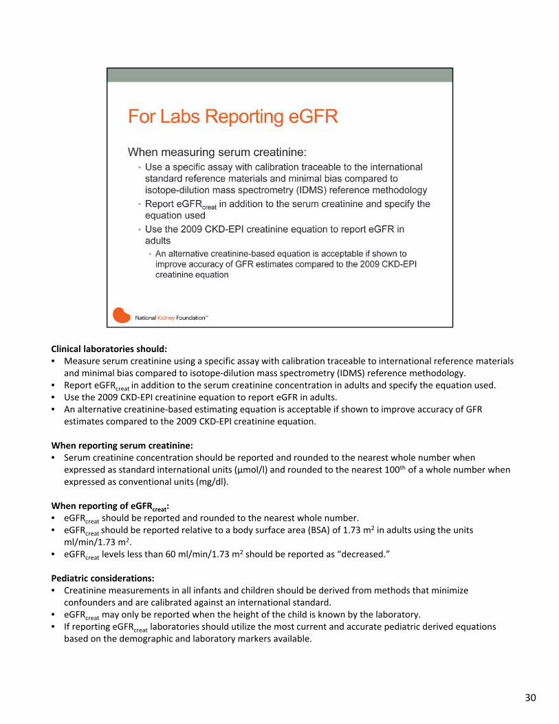
Clinical laboratories should:• Measure serum creatinine using a specific assay with calibration traceable to international reference materials
and minimal bias compared to isotope‐dilution mass spectrometry (IDMS) reference methodology. • Report eGFRcreat in addition to the serum creatinine concentration in adults and specify the equation used.• Use the 2009 CKD‐EPI creatinine equation to report eGFR in adults.• An alternative creatinine‐based estimating equation is acceptable if shown to improve accuracy of GFR
estimates compared to the 2009 CKD‐EPI creatinine equation.
When reporting serum creatinine:• Serum creatinine concentration should be reported and rounded to the nearest whole number when
expressed as standard international units (μmol/l) and rounded to the nearest 100th of a whole number when expressed as conventional units (mg/dl).
When reporting of eGFRcreat:• eGFRcreat should be reported and rounded to the nearest whole number.• eGFRcreat should be reported relative to a body surface area (BSA) of 1.73 m2 in adults using the units
ml/min/1.73 m2.• eGFRcreat levels less than 60 ml/min/1.73 m2 should be reported as “decreased.”
Pediatric considerations:• Creatinine measurements in all infants and children should be derived from methods that minimize
confounders and are calibrated against an international standard.• eGFRcreat may only be reported when the height of the child is known by the laboratory.• If reporting eGFRcreat laboratories should utilize the most current and accurate pediatric derived equations
based on the demographic and laboratory markers available.
30

When measuring cystatin C, clinical laboratories should:• Measure serum cystatin C using an assay with calibration traceable to the international standard reference
material.• Report eGFR from serum cystatin C in addition to the serum cystatin C concentration in adults and specify the
equation used whenever reporting eGFRcys and eGFRcreat‐cys.• Use 2012 CKD‐EPI cystatin C equation or 2012 CKD‐EPI creatinine‐cystatin C equation to report eGFRcys and
eGFRcreat‐cys, respectively.• An alternative cystatin C‐based GFR estimating equation is acceptable if shown to improve accuracy of GFR
estimates compared to the 2012 CKD‐EPI cystatin C and 2012 CKD‐EPI creatinine‐cystatin C equations.
When reporting serum cystatin C:• Serum cystatin C concentration should be reported and rounded to the nearest 100th of a whole number
when expressed as conventional units (mg/l).
When reporting of eGFRcys and eGFRcreat‐cys:• eGFRcys and eGFRcreat‐cys should be reported and rounded to the nearest whole number.• eGFRcys and eGFRcreat‐cys should be reported relative to a body surface area (BSA) of 1.73 m2 in adults using the
units ml/min/1.73 m2.• eGFRcys and eGFRcreat‐cys levels less than 60 ml/min/1.73 m2 should be reported as “decreased.”
Pediatric considerations:• Measure serum cystatin C using an immunonephelometrically determined method in which the assay is
calibrated and traceable to the international standard reference material.• Report eGFRcys in addition to the serum cystatin C concentration in children.• Report eGFRcys in children specifying the specific equation used.
31

For adults:Use the following measurements for initial testing of proteinuria (in descending order of preference, in all cases an early morning urine sample is preferred):
1. urine albumin‐to‐creatinine ratio (ACR);2. urine protein‐to‐creatinine ratio (PCR);3. reagent strip urinalysis for total protein with automated reading;4. reagent strip urinalysis for total protein with manual reading.
Urine albumin measurement provides a more specific and sensitive measure of changes in glomerular permeability than urinary total protein. There is substantial evidence linking increased albuminuria to outcomes of CKD and there is also evidence that urinary albumin is a more sensitive test to enable detection of glomerular pathology associated with some other systemic diseases including diabetes, hypertension and systemic sclerosis.
For children:Use the following measurements for initial testing of proteinuria in children (in descending order of preference):
1. urine PCR, early morning urine sample preferred;2. urine ACR, early morning urine sample preferred;3. reagent strip urinalysis for total protein with automated reading;4. reagent strip urinalysis for total protein with manual reading.
Currently urinary PCR should be favored over urine ACR in children. Unlike in adults where powerful evidence exists in support of the use of measures of albumin rather than total protein to predict adverse outcomes, this level of evidence is currently lacking in children.
32
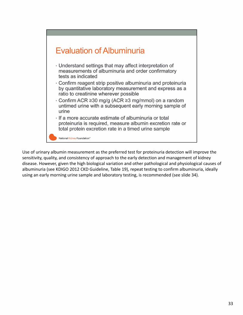
Use of urinary albumin measurement as the preferred test for proteinuria detection will improve the sensitivity, quality, and consistency of approach to the early detection and management of kidney disease. However, given the high biological variation and other pathological and physiological causes of albuminuria (see KDIGO 2012 CKD Guideline, Table 19), repeat testing to confirm albuminuria, ideally using an early morning urine sample and laboratory testing, is recommended (see slide 34).
33

Commonly used reagent strip devices measuring total protein are insufficiently sensitive for the reliable detection of proteinuria, do not adjust for urinary concentration, and are only semi‐quantitative. Furthermore, there is no standardization between manufacturers. The use of such strips should be discouraged in favor of quantitative laboratory measurements of albuminuria or proteinuria. When used, reagent strip results should be confirmed by laboratory testing. The algorithm above is a suggested protocol for further investigation of an individual demonstrating a positive reagent strip test for albuminuria/proteinuria or quantitative albuminuria/proteinuria test. • Reagent strip device results should be confirmed using laboratory testing of the ACR on at least two further
occasions. Patients with two or more positive (≥30 mg/g or ≥3 mg/mmol) tests on early morning samples 1‐2 weeks apart should be diagnosed as having persistent albuminuria. The possibility of postural proteinuria should be excluded by the examination of an EMU. PCR measurement can be substituted for the ACR but is insensitive in the detection of moderately increased albuminuria/proteinuria. Approximate PCR equivalent to an ACR of 30 mg/mmol is 50 mg/mmol.
• Do consider other causes of increased ACR (e.g., menstrual contamination, uncontrolled hypertension, symptomatic urinary tract infection, heart failure, other transitory illnesses, and strenuous exercise), especially in the case of type 1 diabetes present for less than 5 years. The presence of hematuria may indicate non‐diabetic renal disease.
Abbreviations: ACR, albumin‐to‐creatinine ratio; C&S, culture and sensitivity; CKD, chronic kidney disease; EMU, early morning urine; MSU, mid‐stream urine; PCR, protein‐to‐creatinine ratio.
34

There have been concerns that replacing urinary total protein measurement with albumin measurement may cause nonalbuminuric (effectively tubular and overproduction) proteinuria to be missed. However, total protein assays will also be poor at detecting tubular proteinuria. When investigating patients for tubular proteinuria, it is advisable to use assays targeted at specific tubular proteins.
Pediatric considerations:This statement is fully applicable to pediatrics.
35

• Clinical laboratories should report ACR and PCR in untimed urine samples in addition to albumin concentration or proteinuria concentrations rather than the concentrations alone.
• Do not use “Normoalbuminuria,” “Microalbuminuria” or “Macroalbuminuria” in defining albuminuria because they are antiquated non‐descriptive definitions. The proposed albuminuria categories A1‐A3 are a more clinically meaningful way to express information about categories within the continuum of albumin excretion.
• Use the current terminology for albuminura:• Category A1: Normal to mildly increased• Category A2: Moderately increased• Category A3: Severely increased
Pediatrics considerations:This statement is fully applicable to pediatrics.
36

37

• For all CKD complications, prognosis will vary depending on: 1) cause; 2) GFR; 3) degree of albuminuria; and 4) other comorbid conditions.
• Risk for kidney disease endpoints, such as kidney failure and AKI, is predominately driven by an individual patient’s clinical diagnosis, GFR, and the degree of albuminuria or other markers of kidney damage and injury.
• For cardiovascular disease, risk will be determined by history of cardiovascular disease and traditional and non‐traditional cardiovascular disease risk factors.
• For other conditions, the risk will be determined by risk factors specific for those conditions. • For all conditions, the cause of CKD, GFR category and albuminuria category will still have important
influence as “risk multipliers,” but will have smaller overall influence on disease prediction than risk factors specific for the condition. All these conditions have an impact on life expectancy and quality of life and contribute substantially to predicting the prognosis of CKD.
Pediatric considerations:The rationale and principles behind this statement would apply to pediatrics though the data are not available.
38

• For all CKD complications, prognosis will vary depending on: • Cause of CKD; • GFR category; • Albuminuria category; • Other risk factors and comorbid conditions.
• The risk associations of GFR and albuminuria categories appear to be largely independent of one another. Therefore, neither the category of GFR nor the category of albuminuria alone can fully capture the prognosis for a patient with CKD.
• All of the parameters above have an impact on life expectancy and quality of life and contribute substantially to predicting the prognosis of CKD.
Key to color grid:Colors indicate groups of patients at higher risk of major outcomes:• Green represents low risk. If the patient does not have other markers of kidney disease, then CKD is
not diagnosed or confirmed.• Compared with the Green box (eGFR>60 ml/min/1.73 m2 and ACR<30 mg/g [<3 mg/mmol]):
• Yellow = is one step away from normal down or across. It represents moderately increased risk.
• Orange = is two steps away from normal: down two, across two, or down one/across one. It represents high risk.
• Red = is three steps away from normal. It represents very high risk.
39

Extensive work by the CKD Prognosis Consortium has defined the relative risks across GFR and albuminuria stages for several important outcomes, including all‐cause mortality, cardiovascular disease, and kidney failure. • Levels of risk can be identified and grouped into categories, but they may differ somewhat for each
outcome. • Additional research is needed to map these GFR and albuminuria categories and cause of kidney
disease to other important outcomes of CKD.
40

This slide summarizes the pooled relative risks of varying levels of eGFR and albuminuria in the general population cohorts, expressed as continuous variables, for all five outcomes.
Mortality is reported for general population cohorts assessing albuminuria as urine ACR. Kidney outcomes are reported for general population cohorts assessing albuminuria as either urine ACR or dipstick (reagent strip). Estimated glomerular filtration rate (eGFR) is expressed as a continuous variable.
The three lines represent urine ACR of <30 mg/g or reagent strip negative and trace (blue), urine ACR 30–299 mg/g or reagent strip 1+ positive (green), and urine ACR ≥300 mg/g or reagent strip ≥2+ positive (red) [<3, 3‐29, ≥30 mg/mmol, respectively]. All results are adjusted for covariates and compared with reference point of eGFR of 95 ml/min per 1.73 m2 and ACR of <30 mg/g or reagent strip negative (diamond).
Each point represents the pooled relative risk from a meta‐analysis. Solid circles indicate statistical significance compared with the reference point (P <0.05); triangles indicate non‐significance. Red arrows indicate eGFR of 60 ml/min per 1.73 m2, threshold value of eGFR for the current definition of chronic kidney disease (CKD). HR, hazards ratio; OR, odds ratio.
41

This slide summarizes the pooled relative risks of varying levels of eGFR and albuminuria in the general population cohorts, expressed as categorical variables, for all five outcomes.
Mortality is reported for general population cohorts assessing albuminuria as urine ACR. Kidney outcomes are reported for general population cohorts assessing albuminuria as either urine ACR or reagent strip. Estimated glomerular filtration rate (eGFR) and albuminuria are expressed as categorical variables. All results are adjusted for covariates and compared with the reference cell (Ref).
Each cell represents a pooled relative risk from a meta‐analysis; bold numbers indicate statistical significance at P <0.05. Incidence rates per 1000 person‐years for the reference cells are 7.0 for all‐cause mortality, 4.5 for cardiovascular disease mortality, 0.04 for kidney failure, 0.98 for acute kidney injury (AKI), and 2.02 for kidney disease progression. Absolute risk can be computed by multiplying the relative risks in each cell by the incidence rate in the reference cell.
Colors reflect the ranking of adjusted relative risk. The point estimates for each cell were ranked from 1 to 28 (the lowest RR having rank number 1, and the highest number 28). The categories with rank numbers 1–8 are green, rank numbers 9–14 are yellow, the rank numbers 15–21 are orange, and the rank numbers 22–28 are colored red. (For the outcome of CKD progression, two cells with RR <1.0 are also green, leaving fewer cells as yellow, orange and red.)
42

General parameters to monitoring people with CKD:• Assess GFR and albuminuria at least annually in people with CKD.• Assess GFR and albuminuria more often for individuals at higher risk of progression, and/or where
measurement will impact therapeutic decisions.• More frequent measures of eGFR and albuminuria should be considered in patients with a lower GFR and
greater albuminuria as these people are more likely to progress. Frequency of measurement should also be individualized based on the patient history and underlying cause of kidney disease.
Recognize that:• Regular monitoring of stable patients may include more frequent monitoring than annually, but will be
dictated by underlying cause, history, and estimates of GFR and ACR values obtained previously.• Small fluctuations in GFR are common and are not necessarily indicative of progression.• CKD progression is defined based on one of more of the following:
• Decline in GFR category (≥90, 60–89, 45–59, 30–44, 15–29, <15 ml/min/1.73 m2). A certain drop in eGFR is defined as a drop in GFR category accompanied by a 25% or greater drop in eGFR from baseline.
• Rapid progression is defined as a sustained decline in eGFR of more than 5 ml/min/1.73 m2/yr.• The confidence in assessing progression is increased with increasing number of serum creatinine
measurements and duration of follow‐up.
Note: These are general parameters only based on expert opinion and must take into account underlying comorbid conditions and disease state, as well as the likelihood of impacting a change in management for any individual patient. Not all individuals with CKD require close surveillance and monitoring; clinical context remains an important modifier for all recommendations.
43

In general, refer to specialist kidney care services when:• GFR <30 ml/min/1.73 m2*;• There is a consistent finding of significant albuminuria: ACR ≥300 mg/g [≥30 mg/mmol] or AER ≥300
mg/24 hours, approximately equivalent to PCR ≥500 mg/g [≥50 mg/mmol] or PER ≥500 mg/24 hours)
Caveats: *If this is a stable isolated finding, formal referral may not be necessary and advice from a specialist services may be all that is required to facilitate best care for the patients. This will be health‐care system dependent.
The grid above can serve as a guide if an individual’s kidney function is relatively stable (rate of decline in GFR <5 ml/min/1.73 m2/year) but for certain people, such as those with diabetes, transition to a severe reduction in GFR and kidney failure may progress rapidly. In such individuals early nephrology referral is the watchword.
Other referral circumstances to consider: • AKI or abrupt sustained fall in GFR;• Progression of CKD;• Urinary red cell casts, RBC >20 per high power field sustained and not readily explained;• CKD and hypertension refractory to treatment with 4 or more antihypertensive agents;• Persistent abnormalities of serum potassium;• Recurrent or extensive nephrolithiasis;• Hereditary kidney disease;• People with progressive CKD in whom the risk of kidney failure within 1 year is 10–20% or higher,
should be referred for planning renal replacement therapy. The actual amount of time required at a minimum is at least 1 year to ensure appropriate education, understanding and referrals to other practitioners (e.g., vascular surgeons, transplant teams, etc.)
44

45

• The definition of CKD remains intact. However, the classification and risk stratification now includes “with implications for health.”
• The addition of ‘with implications for health’ is intended to reflect the notion that a variety of abnormalities of kidney structure or function may exist, but not all have implications for health of individuals, and therefore need to be contextualized.
• CKD is classified by: ‐ Cause‐ GFR category‐ Albuminuria category
• Collectively referred to as “CGA Staging”• GFR level equivalent to previous CKD Stage 3 is now subdivided into 2 GFR categories: 3a and 3b; this
division acknowledges the data supporting different outcomes and risk profiles for these two GFR categories.
• GFR (G1‐G5) and albuminuria (A1‐A3) are grouped as categories (as opposed to stages).
46

Albuminuria categories have been added because of the graded increase in risk for mortality, progression of CKD, and ESRD at higher levels of albuminuria, independent of eGFR, without an apparent threshold value. The use of a simple categorical approach was selected to simplify the concept for clinical practice.
47

In KDIGO 2012:• Risk relationship between GFR and albuminuria is defined for:
• Overall mortality• CVD • Kidney failure• AKI• CKD progression
• The new guideline elaborates on the determination of eGFR using creatinine, cystatin C or both with updated equations (in adults and pediatrics)
• For initial assessment, KDIGO recommends use of the 2009 CKD‐EPI creatinine equation;• KDIGO recommends additional tests in specific circumstances when eGFR based on serum
creatinine is less accurate • Cystatin C‐based equations• Clearance measurement
Confirmation of decreased eGFR by measurement of an alternative endogenous filtration marker (cystatin C) or a clearance measurement is warranted in specific circumstances when GFR estimates based on serum creatinine are thought to be inaccurate or when decisions depend on more accurate knowledge of GFR, such as confirming a diagnosis of CKD, determining eligibility for kidney donation, or adjusting dosage of toxic drugs that are excreted by the kidneys. See also Recommendation 1.4.3.8 of the KDIGO 2012 CKD Guideline.
48

There is considerable controversy as to what constitutes normal progression of CKD. The potential for biological and analytical variation associated with use of serum creatinine measurements should be taken into account as they represent reversible fluctuations in GFR and are not necessarily indicative of progression. Further, it is important to recognize that the degree of precision with which progression is able to be estimated is highly dependent upon two factors: the number of serum creatinine measurements used to define progression and the duration of follow‐up.
The importance of determining the rate of decline in kidney function over time is to identify individuals who are progressing at a more rapid rate than anticipated, which is associated with increased morbidity and mortality. Individuals who are ‘‘rapid progressors’’ should be targeted to slow their progression and associated adverse outcomes. A progressive decline in kidney function is influenced by baseline GFR category and albuminuria category.
Given the recognized limitations in defining rapid progression, the Work Group aimed to provide options for the determination of progression based on their clinical utility and ease of use. A criterion requiring both a change in GFR category (e.g., change from G2 to G3a) and percent change would ensure that small changes GFR from 61 to 59 ml/min/1.73 m2 for example, which represents a change in category but a minimal change in GFR, would not be misinterpreted to represent progression. A change of <25% in a pair of GFR estimates may reflect physiologic variation rather than true progression.
Management of progression and complications of CKD is addressed in further detail in KDIGO 2012 CKD Guideline Chapters 3 and 4.
49

50

51

52
53
54
55

56