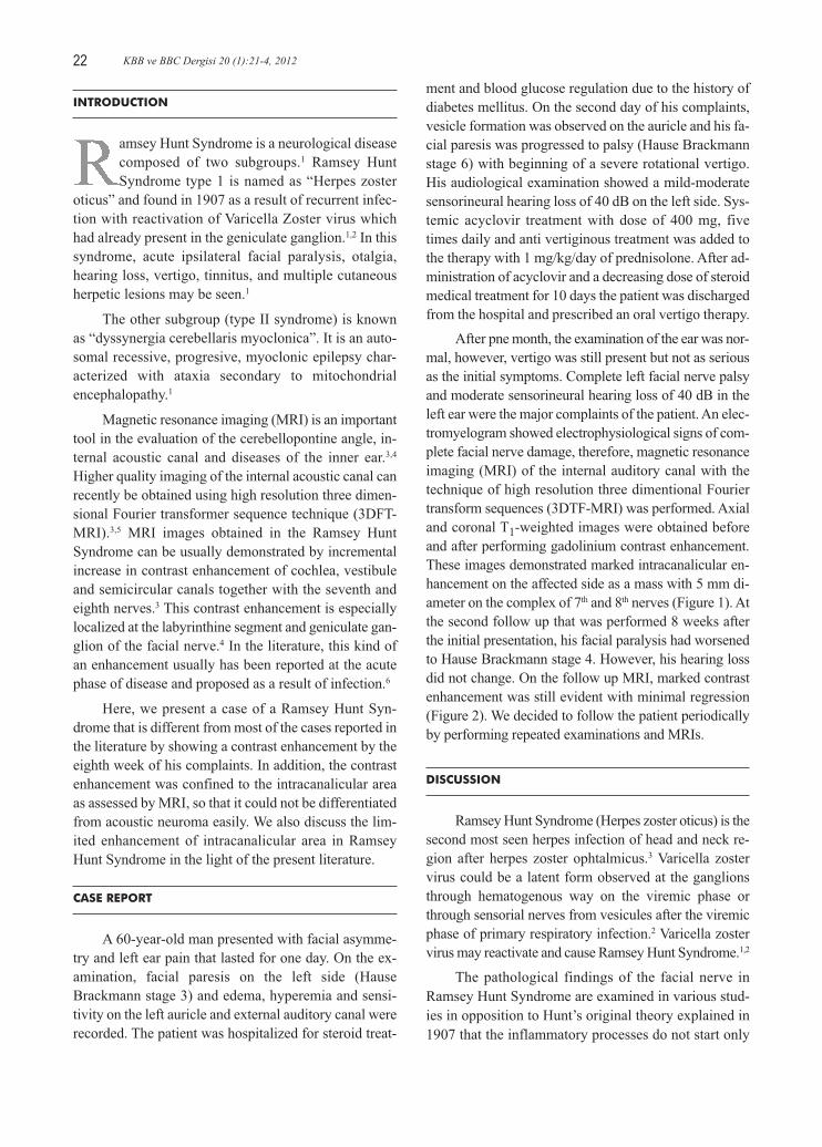
KBB ve BBC Dergisi 20 (1):21-4, 2012 Intracanalicular...
4
≈ Correspondence Fulya ÖZER, MD Baskent University Adana Teaching and Medical Research Center, Department of Otorhinolaryngology Head and Neck Surgery, Adana/TURKEY E-mail: [email protected] Turkiye Klinikleri J Int Med Sci 2008, 4 21 Intracanalicular Contrast Enhancement on Magnetic Resonance Imaging in Ramsey Hunt Syndrome Mimicking Acoustic Neuroma: Case Report Manyetik Rezonans Görüntülemede İntrakanaliküler Kontrast Tutulumu ile Akustik Nörinomu Taklit Eden Ramsey Hunt Sendromu: OIgu Sunumu *Fulya ÖZER, MD, *Cem ÖZER, MD, **Özlem ALKAN, MD, *Haluk YAVUZ, MD, *Levent N. ÖZLÜOĞLU, MD * Başkent University Medical Faculty, Department of Otorhinolaryngology Head and Neck Surgery, ** Başkent University Medical Faculty, Department of Radiology, Ankara ABSTRACT Magnetic resonance imaging (MRI) is a gold standard method for the diagnosis of lesions that are located in the internal acoustic canal. A 60- year-old man with vesicular lesions on his left auricle, erupted after the facial paresis was presented. He also had vertigo and hearing loss and his complaints did not re- cover after one month. MRI was obtained and an abnormal contrast enhancement was found at the intracanalicular area, similar to the view of an acoustic neuroma. During follow-up, this image of contrast enhancement was still evident in the MRI with minimal reduction. Abnormal contrast enhancement of the labyrinthine segment and other cranial nerves localized on the intratemporal area could be seen frequently in the Ramsey Hunt Syndrome. However, a con- trast enhancement confined to the intracanalicular area and persistence of findings more than eight weeks is not frequent. Ramsey Hunt Syndrome may mimic acoustic neuroma with its late clinical and MRI findings. At the differential diagnosis, repeated clinical and radiological evaluation of the patient is crucial. Keywords Herpes zoster oticus; neuroma, acoustic; magnetic resonance imaging ÖZET Magnetik rezonans görüntüleme (MRG) internal akustik kanal yerleşimli lezyonlarda altın standarddır. Altmış yaşında erkek hasta fasial parezi ile baş- vurmuş, izleminde aurikulada veziküler lezyonlar çıkması üzerine tedavi altına alınmıştır. Bir ay sonra halen fasial parezinin devam etmesi ve beraberinde vertigo ve aynı tarafta işitme kaybı olması üzerine çekilen MRG görüntülerinde intrakanaliküler anormal kontrast tutulumu tespit edilmiş; bu görünüm akus- tik nörinomdan ayırt edilememiştir. İkinci ayda yapılan kontrol MRG’ da aynı kontrast tutulum devam etmiş ancak minimal gerileme tespit edilmiştir. Hasta uzun süreli takiplerinde fasial fonksiyonlarını yeniden kazanmıştır. Ramsey Hunt Sendromunda intratemporal bölgede labirint ve diğer kranyal si- nirlerin anormal kontrast tutulumu sık görülen bir bulgudur. Ancak sadece kanal içinde görülen ve sekiz haftadan daha uzun süren anormal kontrast tutu- lum sık görülen bir durum değildir. Ramsey Hunt Sendromu, geç dönem klinik ve MRG bulguları ile akustik nörinomu taklit edebilir. Hastanın tanısının serolojik olarak teyit edilmesi önemli olmakla beraber tanı koyarken en önemli nokta tekrarlanan çekimlerde ve takiplerde yapılan değerlendirmedir. Anahtar Sözcükler Herpes zoster otikus; nöroma, akustik; manyetik rezonans görüntüleme This study was presented in 31 th National Congress of Otolaryngology Head and Neck Surgery, October 28-31, 2009, Antalya, Turkey Çalıșmanın Dergiye Ulaștığı Tarih: 30.10.2010 Çalıșmanın Basıma Kabul Edildiği Tarih: 21.06.2011 KBB ve BBC Dergisi 20 (1):21-4, 2012
Transcript of KBB ve BBC Dergisi 20 (1):21-4, 2012 Intracanalicular...

≈≈Correspondence
Fulya ÖZER, MDBaskent University Adana Teaching and Medical Research Center,
Department of Otorhinolaryngology Head and Neck Surgery,Adana/TURKEY
E-mail: [email protected]
Turkiye Klinikleri J Int Med Sci 2008, 4 21
Intracanalicular Contrast Enhancement on Magnetic Resonance Imaging in Ramsey Hunt Syndrome
Mimicking Acoustic Neuroma: Case ReportManyetik Rezonans Görüntülemede
İntrakanaliküler Kontrast Tutulumu ile Akustik Nörinomu Taklit Eden Ramsey Hunt Sendromu: OIgu Sunumu
*Fulya ÖZER, MD, *Cem ÖZER, MD, **Özlem ALKAN, MD, *Haluk YAVUZ, MD, *Levent N. ÖZLÜOĞLU, MD* Başkent University Medical Faculty, Department of Otorhinolaryngology Head and Neck Surgery,
** Başkent University Medical Faculty, Department of Radiology, Ankara
ABSTRACT
Magnetic resonance imaging (MRI) is a gold standard method for the diagnosis of lesions that are located in the internal acoustic canal. A 60- year-old manwith vesicular lesions on his left auricle, erupted after the facial paresis was presented. He also had vertigo and hearing loss and his complaints did not re-cover after one month. MRI was obtained and an abnormal contrast enhancement was found at the intracanalicular area, similar to the view of an acousticneuroma. During follow-up, this image of contrast enhancement was still evident in the MRI with minimal reduction. Abnormal contrast enhancement of thelabyrinthine segment and other cranial nerves localized on the intratemporal area could be seen frequently in the Ramsey Hunt Syndrome. However, a con-trast enhancement confined to the intracanalicular area and persistence of findings more than eight weeks is not frequent. Ramsey Hunt Syndrome may mimicacoustic neuroma with its late clinical and MRI findings. At the differential diagnosis, repeated clinical and radiological evaluation of the patient is crucial.
KeywordsHerpes zoster oticus; neuroma, acoustic; magnetic resonance imaging
ÖZET
Magnetik rezonans görüntüleme (MRG) internal akustik kanal yerleşimli lezyonlarda altın standarddır. Altmış yaşında erkek hasta fasial parezi ile baş-vurmuş, izleminde aurikulada veziküler lezyonlar çıkması üzerine tedavi altına alınmıştır. Bir ay sonra halen fasial parezinin devam etmesi ve beraberindevertigo ve aynı tarafta işitme kaybı olması üzerine çekilen MRG görüntülerinde intrakanaliküler anormal kontrast tutulumu tespit edilmiş; bu görünüm akus-tik nörinomdan ayırt edilememiştir. İkinci ayda yapılan kontrol MRG’ da aynı kontrast tutulum devam etmiş ancak minimal gerileme tespit edilmiştir.Hasta uzun süreli takiplerinde fasial fonksiyonlarını yeniden kazanmıştır. Ramsey Hunt Sendromunda intratemporal bölgede labirint ve diğer kranyal si-nirlerin anormal kontrast tutulumu sık görülen bir bulgudur. Ancak sadece kanal içinde görülen ve sekiz haftadan daha uzun süren anormal kontrast tutu-lum sık görülen bir durum değildir. Ramsey Hunt Sendromu, geç dönem klinik ve MRG bulguları ile akustik nörinomu taklit edebilir. Hastanın tanısınınserolojik olarak teyit edilmesi önemli olmakla beraber tanı koyarken en önemli nokta tekrarlanan çekimlerde ve takiplerde yapılan değerlendirmedir.
Anahtar SözcüklerHerpes zoster otikus; nöroma, akustik; manyetik rezonans görüntüleme
This study was presented in 31th National Congress of Otolaryngology Head and Neck Surgery, October 28-31, 2009, Antalya, Turkey
Çalıșmanın Dergiye Ulaștığı Tarih: 30.10.2010 Çalıșmanın Basıma Kabul Edildiği Tarih: 21.06.2011
KBB ve BBC Dergisi 20 (1):21-4, 2012
INTRODUCTION
amsey Hunt Syndrome is a neurological diseasecomposed of two subgroups.1 Ramsey HuntSyndrome type 1 is named as “Herpes zoster
oticus” and found in 1907 as a result of recurrent infec-tion with reactivation of Varicella Zoster virus whichhad already present in the geniculate ganglion.1,2 In thissyndrome, acute ipsilateral facial paralysis, otalgia,hearing loss, vertigo, tinnitus, and multiple cutaneousherpetic lesions may be seen.1
The other subgroup (type II syndrome) is knownas “dyssynergia cerebellaris myoclonica”. It is an auto-somal recessive, progresive, myoclonic epilepsy char-acterized with ataxia secondary to mitochondrialencephalopathy.1
Magnetic resonance imaging (MRI) is an importanttool in the evaluation of the cerebellopontine angle, in-ternal acoustic canal and diseases of the inner ear.3,4
Higher quality imaging of the internal acoustic canal canrecently be obtained using high resolution three dimen-sional Fourier transformer sequence technique (3DFT-MRI).3,5 MRI images obtained in the Ramsey HuntSyndrome can be usually demonstrated by incrementalincrease in contrast enhancement of cochlea, vestibuleand semicircular canals together with the seventh andeighth nerves.3 This contrast enhancement is especiallylocalized at the labyrinthine segment and geniculate gan-glion of the facial nerve.4 In the literature, this kind ofan enhancement usually has been reported at the acutephase of disease and proposed as a result of infection.6
Here, we present a case of a Ramsey Hunt Syn-drome that is different from most of the cases reported inthe literature by showing a contrast enhancement by theeighth week of his complaints. In addition, the contrastenhancement was confined to the intracanalicular areaas assessed by MRI, so that it could not be differentiatedfrom acoustic neuroma easily. We also discuss the lim-ited enhancement of intracanalicular area in RamseyHunt Syndrome in the light of the present literature.
CASE REPORT
A 60-year-old man presented with facial asymme-try and left ear pain that lasted for one day. On the ex-amination, facial paresis on the left side (HauseBrackmann stage 3) and edema, hyperemia and sensi-tivity on the left auricle and external auditory canal wererecorded. The patient was hospitalized for steroid treat-
ment and blood glucose regulation due to the history ofdiabetes mellitus. On the second day of his complaints,vesicle formation was observed on the auricle and his fa-cial paresis was progressed to palsy (Hause Brackmannstage 6) with beginning of a severe rotational vertigo.His audiological examination showed a mild-moderatesensorineural hearing loss of 40 dB on the left side. Sys-temic acyclovir treatment with dose of 400 mg, fivetimes daily and anti vertiginous treatment was added tothe therapy with 1 mg/kg/day of prednisolone. After ad-ministration of acyclovir and a decreasing dose of steroidmedical treatment for 10 days the patient was dischargedfrom the hospital and prescribed an oral vertigo therapy.
After pne month, the examination of the ear was nor-mal, however, vertigo was still present but not as seriousas the initial symptoms. Complete left facial nerve palsyand moderate sensorineural hearing loss of 40 dB in theleft ear were the major complaints of the patient. An elec-tromyelogram showed electrophysiological signs of com-plete facial nerve damage, therefore, magnetic resonanceimaging (MRI) of the internal auditory canal with thetechnique of high resolution three dimentional Fouriertransform sequences (3DTF-MRI) was performed. Axialand coronal T1-weighted images were obtained beforeand after performing gadolinium contrast enhancement.These images demonstrated marked intracanalicular en-hancement on the affected side as a mass with 5 mm di-ameter on the complex of 7th and 8th nerves (Figure 1). Atthe second follow up that was performed 8 weeks afterthe initial presentation, his facial paralysis had worsenedto Hause Brackmann stage 4. However, his hearing lossdid not change. On the follow up MRI, marked contrastenhancement was still evident with minimal regression(Figure 2). We decided to follow the patient periodicallyby performing repeated examinations and MRIs.
DISCUSSION
Ramsey Hunt Syndrome (Herpes zoster oticus) is thesecond most seen herpes infection of head and neck re-gion after herpes zoster ophtalmicus.3 Varicella zostervirus could be a latent form observed at the ganglionsthrough hematogenous way on the viremic phase orthrough sensorial nerves from vesicules after the viremicphase of primary respiratory infection.2 Varicella zostervirus may reactivate and cause Ramsey Hunt Syndrome.1,2
The pathological findings of the facial nerve inRamsey Hunt Syndrome are examined in various stud-ies in opposition to Hunt’s original theory explained in1907 that the inflammatory processes do not start only
KBB ve BBC Dergisi 20 (1):21-4, 201222

at geniculate ganglion or not always comprise only thisarea.7 In this disease, when facial paralysis accompanythe audiological and/or vestibular findings it is knownthat inflammatory process is localized at nerves insidethe internal acoustic canal.8
With the improvement in the techniques on MRIespecially the use of 3DFT-MRI,3 evaluation of the in-ternal acoustic canal and structures of inner ear becameeasier and prevalence of the disease increased. By thisway, the contrast enhancement on MRI has been shownnot only in acoustic neuroma but also in non-neoplasticdiseases, inflammatory neuropathies and post-traumaticfacial paralysis. MRI became an important technique forthe diagnosis of sensorineural hearing loss with non-neoplastic causes and/or facial paralysis.4
Gadolinium diethylenetriamine penthaasetic acid(Gd-DTPA) is a paramagnetic contrast substance thatwas traditionally used in the imaging of cerebral areaand showed a marked enhancement on T1 weighed MRIin cases of destruction of the blood-brain barrier and/ orincreased vascular permeability because of imperme-able feature of Gd-DTPA normally.7,8 Enhancement ofcontrast substance on peripheral nerves is explained asa result of increase in vascular permeability and/or de-struction of blood-nerve barrier due to inflammation.8
There are many case reports and clinical researchesin the literature about the use of MRI with enhancementof Gd-DTPA in the diagnosis and prognosis of RamseyHunt Syndrome.6-9 Yanagida et al.8 examined MR im-ages of 14 patients with Ramsey Hunt Syndrome andfound an enhancement of contrast substance at the dis-tal part of the internal acoustic canal and the labyrinthinesegment in most of the cases. This enhancement was es-pecially evident in patients presented with more find-ings of the seventh cranial nerve. When the findings ofthe cochleovestibular nerve were added to symptoms ofthe patients, the contrast enhancement could be seen inthe canal or in the inner ear at the images of the patients.Tada et al.9 could not establish a relationship betweenfindings of MRI and clinical symptoms in patients ofRamsey Hunt Syndrome and proposed that MRI wasmore specific in the differential diagnosis between Ram-sey Hunt Syndrome and tumors.
In a recent study by Kim et al.,6 the results of de-compression surgery of 13 patients with Ramsey HuntSyndrome was reported. The findings of MRI and sur-gical results of patients were comparable and thelabyrinthine segment was the most frequently estab-lished area for the enhancement and pathology.
Unique intracanalicular enhancement on MRI isnot prevalent in the patients with Ramsey Hunt Syn-drome.10,11 Particularly, isolated intracanalicular en-hancement on MRI is rarely seen during non-infectiousperiod after the acute phase of infection.11 In this case,
Effects of Smoking and Body Mass Index on Hearing Thresholds in Workers...
Turkiye Klinikleri J Int Med Sci 2008, 4 23
Intracanalicular Contrast Enhancement on Magnetic Resonance Imaging in Ramsey Hunt Syndrome... 23
Figure 1. The con trast en han ce ment in si de the left in ter nal aco us tic ca nal isshown on T1 axi al scan.
Figure 2. This con trast en han ce ment is smal ler mi ni mally shown on T1 axi -al scans.

although the correct diagnosis and treatment were ap-plied in the event that herpes vesicules were exist at thepresent, MRI scans could be evaluated easily as acousticneuroma. The contrast enhancement on MRI in that pa-tient persisted for a long time which may cause confu-sion about the diagnosis because of the presence of thecontrast substance only in the intracanalicular compart-ment. Downie et al.10 published a case of Ramsey HuntSyndrome with contrast enhancement on MRI with a sixmonths follow-up time. However, this contrast en-hancement was found to be located in cochlear andvestibular area by MRI. Goldsmith et al.11 had reporteda case of unique intracanalicular enhancement on MRIin Ramsey Hunt Syndrome which was not easily differ-entiated from acoustic neuroma resembling our case.
MRI was obtained to identify the continuedsymptoms of hearing loss, vertigo and facial paralysisalthough the vesicules on the ear of the patient wereimproved. We thought that MRI may show the pro-gression of neuritis of the cochleovestibular nerve. Ini-tially, we were a little bit confused about the diagnosisbecause of persistence of contrast enhancement onlyin the intracanalicular compartment mimicking acousticneuroma. After the second MRI, the diagnosis of in-flammatory neuritis was certain due to minimal de-creasing in the contrast enhancement. However, this wasagain a rare condition that the contrast enhancement onMRI in RHS or in any inflammatory neuritis was local-ized only in the intracanalicular area and showing pro-gression more than two months.
In our case, use of different techniques of MRI to-gether with repeated MRI scans was effective in the di-agnosis and improved our clinical findings. Theevaluation of internal acoustic canal and the cerebello-pontine angle was achieved accurately in a short dura-tion. In addition, the reconstruction of scans could bedone easily so that the inner ear and the cochleovestibu-lar and the facial nerve have been shown simply withthe technique of 3DFT-MRI instead of other conven-tional spin-echo MRI techniques.3,5
When the intracanalicular enhancement was presentat the scans of the patients with facial paralysis with sen-sorineural hearing loss, one of the first possible diagnoseswould be acoustic neuroma. However, patient historyshould be reviewed for varicella zoster infection too. In suchcases, mucocutaneous vesicules may not be present and /orcochleovestibular findings may be more evident than facialparalysis.2 The combination of antiviral and steroid man-agement is suggested to be started in these patients.1,2
Although isolated intracanalicular enhancement onMRI is thought to be an acoustic neuroma, even if MRIfindings were not evident at the acute phase, patientshould also be investigated for Ramsey Hunt Syndromeand other inflammatory neuropathies. It should be keptin mind that a small intracanalicular acoustic neuromamay be confused with a self inflicted disease of RamseyHunt Syndrome. Therefore, the most important point inthe diagnosis of this disease is the repeated MRI scansand the evaluation of clinical symptoms of the patientduring follow-up.
KBB ve BBC Dergisi 20 (1):21-4, 201224
1. Gupta J, Hutchins T, Palacios E. Ramsay Hunt syndrome,type I. Ear Nose Throat J 2007;86(3):138, 140.
2. Avcı S, Kansu L, Akkuzu B, Özgirgin N, Özlüoğlu L. A caseof herpetic facial paralysis in which cochleovestibular symp-toms outweigh facial nevre symptoms. The Turkish ENTJournal (article in Turkish) 2008; 18(1): 40-3.
3. Canellas AR, Sanches Torres C, Isern G,Capellades F, FierroEC, Planas G. Ramsey-Hunt Syndrome and High-Resolu-tion 3DFT MRI. J Comput Assist Tomogr 1993; 17(3): 495-7.
4. Kinoshita T, Ishii K, Okitsu T, Okudera T, Ogawa T. Facialnerve palsy: Evaluation by contrast-enhanced MR Imaging.Clin Radiol 2001; 56(11): 926-32.
5. Casselman JW, Kuhweide R, Deimling M, Ampe W, DehaeneI, Meeus L. Constructive interference in steady state-3D DFTMR imaging of the inner ear and cerebellopontine angle. AmJ Neuroradiol 1993; 14(1): 47-57.
6. Kim J, Chung MS, Moon IS, Lee HK, Lee WS. Correlation
between enhanced MRI and surgical findings in herpeticzoster oticus. Acta Otolaryngol 2009; 129(8): 900-5.
7. Kuo MJ, Grago PC, Proops DW, Chavda SV. Early diagnosisand treatment of Ramsey Hunt syndrome: the role of magneticresonance imaging. J Laryngol Otol 1995; 109(8): 777-80.
8. Yanagida M, Ushiro K, Toshio Y, Kumazawa T, Katoh T. Enhanced MRI in patients with Ramsey- Hunt’s Syndrome.Acta Otolaryngol 1993; 113 (suppl. 500): 58-61.
9. Tada Y, Aoyagi M, Hitoshi T, Inamura H, Saito O, Maeyama H,Kohsyu H, Koike Y. Gd-DTPA Enhanced MRI in Ramsey HuntSyndrome. Acta Otolaryngol 1994; 114(Suppl 511): 170-4.
10. Downie AC, Howlett DC, Koefman RJ, Banerjee AK, Tonge KA. Case report: prolenged contrast enhancementof the inner ear on magnetic resonance imaging in RamseyHunt syndrome. Br J Radiol 1994; 67(800): 819-21.
11. Goldsmith P, Zammit-Maempel I, Meikle D. Ramsey Huntsyndrome mimicking acoustic neuroma on MRI. J LaryngolOtol 1995; 109(10): 1013-5.
REFERENCES