Katrin König*, Uwe Kobold, Gerhard Fink, Andreas ...
9
DOI 10.1515/cclm-2013-0142 Clin Chem Lab Med 2013; 51(9): 1761–1769 Katrin König*, Uwe Kobold, Gerhard Fink, Andreas Leinenbach, Thomas Dülffer, Roland Thiele, Johannes Zander and Michael Vogeser Quantification of vancomycin in human serum by LC-MS/MS Abstract Background: The aim of our work was to develop and validate a reliable LC-MS/MS-based measurement proce- dure for the quantification of vancomycin in serum, to be applied in the context of efforts to standardize and harmo- nize therapeutic drug monitoring of this compound using routine assays. Methods: Sample preparation was based on protein pre- cipitation followed by ultrafiltration. In order to minimize differential modulation of ionization by matrix constitu- ents extended chromatographic separation was applied leading to a retention time of 9.8 min for the analyte. Measurement was done by HPLC-ESI-MS/MS. For internal standardization the derivative vancomycin-glycin (ISTD) prepared by chemical synthesis was used, HPLC condi- tions ensured coelution of ISTD with the analyte. Results: In a bi-center validation total CVs of < 4% were observed for quality control material ranging from 5.3 mg/L to 79.4 mg/L; accuracy was ± 4%. No relevant ion suppression was observed. Comparative measurement of aliquots from 70 samples at the two validation sites dem- onstrated close agreement. Conclusions: Employing a closely related homologue molecule for internal standardization and the use of MS/ MS following highly efficient sample pre-fractionation by HPLC, the method described here can be considered to offer the highest level of analytical reliability realized so far for the quantification of vancomycin in human serum. Thus, the method is suitable to be used in a comprehen- sive reference measurement system for vancomycin. Keywords: LC-MS/MS; liquid chromatography; mass spec- trometry; serum; vancomycin. *Corresponding author: Katrin König, Institute of Laboratory Medicine, Hospital of the University of Munich, Marchioninistrasse 15, 81375 Munich, Germany, Phone: +49 89 70953221, E-mail: [email protected] Uwe Kobold, Gerhard Fink, Andreas Leinenbach, Thomas Dülffer and Roland Thiele: Roche Diagnostics GmbH, Penzberg, Germany Michael Vogeser and Johannes Zander: Institute of Laboratory Medicine, Hospital of the University of Munich, Munich, Germany Introduction The glycopeptide compound vancomycin is one of the most widely used antimicrobial agents for the treatment of serious gram-positive infections including methicillin- resistant Staphylococcus aureus (MRSA) [1]. Vancomycin is also among the highest volume target analytes in thera- peutic drug monitoring (TDM) for many years now [1–3]. TDM of vancomycin mainly aims to balance therapeutic efficacy against the risk of nephrotoxicity. It has been shown in several studies – applying various analytical methods – that high vancomycin trough levels are asso- ciated with the incidence and extend of nephrotoxicity [4, 5]. However, low trough levels of vancomycin may lead to increased occurrence of resistant strains of S. aureus and failure of treatment in complicated infections. Based on these issues the American Society of Health-System Pharmacists, the Infectious Diseases Society of America, and the Society of Infectious Diseases Pharmacists have published expert panel recommendations for vancomycin TDM, recommending trough serum concentrations of van- comycin of 15–20 mg/L in complicated infections [1]. These recommendations, however, do not take into consideration that routine serum vancomycin quanti- fication by commercial immunoassays is still lacking between-method standardization and substantial method bias can be found [6]. In the proficiency testing scheme of the German Association of Clinical Chemistry and Labo- ratory Medicine [DGKL; Referenzinstitut für Bioanalytik (RfB), Bonn, Germany] at present 14 tests are monitored. The bias between the lowest reading vancomycin test and the highest reading test is continuously found in the range of 40% in this external quality assessment program. Considering the clear-cut vancomycin target concentra- tion ranges, it is likely that different clinical dosing deci- sions are made today in a substantial number of patients depending on the assay which is used in an individual institution. Notably, these consensus target concentra- tions ranges cannot be traced back conclusively to a defined analytical method. From these considerations it has to be concluded that improved harmonization and standardization of serum Bereitgestellt von | Bayerische Staatsbibliothek Angemeldet Heruntergeladen am | 05.11.15 11:17
Transcript of Katrin König*, Uwe Kobold, Gerhard Fink, Andreas ...

DOI 10.1515/cclm-2013-0142 Clin Chem Lab Med 2013; 51(9): 1761–1769
Katrin König*, Uwe Kobold, Gerhard Fink, Andreas Leinenbach, Thomas Dülffer, Roland Thiele, Johannes Zander and Michael Vogeser
Quantification of vancomycin in human serum by LC-MS/MS
Abstract
Background: The aim of our work was to develop and validate a reliable LC-MS/MS-based measurement proce-dure for the quantification of vancomycin in serum, to be applied in the context of efforts to standardize and harmo-nize therapeutic drug monitoring of this compound using routine assays.Methods: Sample preparation was based on protein pre-cipitation followed by ultrafiltration. In order to minimize differential modulation of ionization by matrix constitu-ents extended chromatographic separation was applied leading to a retention time of 9.8 min for the analyte. Measurement was done by HPLC-ESI-MS/MS. For internal standardization the derivative vancomycin-glycin (ISTD) prepared by chemical synthesis was used, HPLC condi-tions ensured coelution of ISTD with the analyte.Results: In a bi-center validation total CVs of < 4% were observed for quality control material ranging from 5.3 mg/L to 79.4 mg/L; accuracy was ± 4%. No relevant ion suppression was observed. Comparative measurement of aliquots from 70 samples at the two validation sites dem-onstrated close agreement.Conclusions: Employing a closely related homologue molecule for internal standardization and the use of MS/MS following highly efficient sample pre-fractionation by HPLC, the method described here can be considered to offer the highest level of analytical reliability realized so far for the quantification of vancomycin in human serum. Thus, the method is suitable to be used in a comprehen-sive reference measurement system for vancomycin.
Keywords: LC-MS/MS; liquid chromatography; mass spec-trometry; serum; vancomycin.
*Corresponding author: Katrin König, Institute of Laboratory Medicine, Hospital of the University of Munich, Marchioninistrasse 15, 81375 Munich, Germany, Phone: +49 89 70953221, E-mail: [email protected] Kobold, Gerhard Fink, Andreas Leinenbach, Thomas Dülffer and Roland Thiele: Roche Diagnostics GmbH, Penzberg, GermanyMichael Vogeser and Johannes Zander: Institute of Laboratory Medicine, Hospital of the University of Munich, Munich, Germany
Introduction
The glycopeptide compound vancomycin is one of the most widely used antimicrobial agents for the treatment of serious gram-positive infections including methicillin-resistant Staphylococcus aureus (MRSA) [1]. Vancomycin is also among the highest volume target analytes in thera-peutic drug monitoring (TDM) for many years now [1–3]. TDM of vancomycin mainly aims to balance therapeutic efficacy against the risk of nephrotoxicity. It has been shown in several studies – applying various analytical methods – that high vancomycin trough levels are asso-ciated with the incidence and extend of nephrotoxicity [4, 5]. However, low trough levels of vancomycin may lead to increased occurrence of resistant strains of S. aureus and failure of treatment in complicated infections. Based on these issues the American Society of Health-System Pharmacists, the Infectious Diseases Society of America, and the Society of Infectious Diseases Pharmacists have published expert panel recommendations for vancomycin TDM, recommending trough serum concentrations of van-comycin of 15–20 mg/L in complicated infections [1].
These recommendations, however, do not take into consideration that routine serum vancomycin quanti-fication by commercial immunoassays is still lacking between-method standardization and substantial method bias can be found [6]. In the proficiency testing scheme of the German Association of Clinical Chemistry and Labo-ratory Medicine [DGKL; Referenzinstitut für Bioanalytik (RfB), Bonn, Germany] at present 14 tests are monitored. The bias between the lowest reading vancomycin test and the highest reading test is continuously found in the range of 40% in this external quality assessment program. Considering the clear-cut vancomycin target concentra-tion ranges, it is likely that different clinical dosing deci-sions are made today in a substantial number of patients depending on the assay which is used in an individual institution. Notably, these consensus target concentra-tions ranges cannot be traced back conclusively to a defined analytical method.
From these considerations it has to be concluded that improved harmonization and standardization of serum
Bereitgestellt von | Bayerische StaatsbibliothekAngemeldet
Heruntergeladen am | 05.11.15 11:17
1762 König et al.: Quantification of vancomycin in human serum by LC-MS/MS
vancomycin measurement is warranted. A future compre-hensive reference measurement system for vancomycin measurement has to include, on the one hand, reliable reference materials, but on the other hand, a robust and reliable method for the specification of working calibration materials, and proficiency testing materials, as well as for the evaluation of routine immunometric tests referring to large reference serum panels. The aim of our work was to develop and to validate such a candidate reference method.
Due to the rather high molecular weight and the limited thermal stability of vancomycin, LC-MS/MS was the most promising technology for this aim. Indeed, several LC-MS/MS methods for the quantification of serum vancomycin concentrations have been described previously [7–9]. These methods, however, rely on inter-nal standard compounds with are structurally not related to the target analyze (teicoplanin, atenolol).
LC-MS/MS-based reference methods [10] usually involve stable isotope labeled compounds for internal standardization. Since vancomycin is a bio-product, pro-duction of a respective material is hardly possible. Conse-quently we designed a suited internal standard compound by stable chemical derivatization of vancomycin which showed almost identical behavior during sample prepa-ration and HPLC-MS/MS separation. The molecule was modified only marginal by introduction of a small func-tional group not changing the polarity of the molecule leading to a different molecular mass.
The intended use of this method protocol described herein is the specification of serum-based samples within the calibration range of the method in both an industrial and research setting. Consequently, high sample through-put did not have major priority in the development of this method, while robustness and reproducibility in differ-ent instrument settings was an essential goal. Thus, we decided to apply highly efficient chromatographic frac-tionation in order to minimize matrix effects, and to apply an innovative bi-centric validation protocol.
Materials and methods
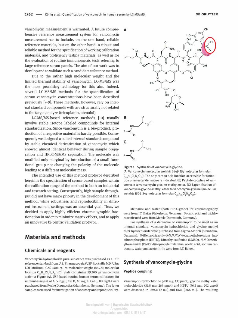
Chemicals and reagentsVancomycin-hydrochloride pure substance was purchased as a USP reference standard from U.S. Pharmacopeia (USP Rockville MD, USA; LOT M0H006; CAS 1404–93–9; molecular weight 1485.71; molecular formula C66H75Cl2N9O24.HCl; vials containing 99,300 µg vancomycin activity; Figure 1A). USP-based routine human serum calibrators for immunoassays (Cal A, 5 mg/L; Cal B, 40 mg/L; Cal C, 80 mg/L) were purchased from Roche Diagnostics (Mannheim, Germany). The latter samples were used for investigation of accuracy and reproducibility.
A
B
C
Figure 1 Synthesis of vancomycin-glycine. (A) Vancomycin (molecular weight: 1449.25; molecular formula: C66H75Cl2N9O24). The only carbon acid function accessible for forma-tion of an ester derivative is indicated. (B) Peptide coupling of van-comycin to vancomycin-glycine-methyl ester. (C) Saponi fication of vancomycin-glycine-methyl ester to vancomycin-glycine (molecular weight: 1506.34; molecular formula: C68H78Cl2N10O25).
Methanol and water (both HPLC-grade) for chromatography were from J.T. Baker (Griesheim, Germany). Formic acid and trichlo-roacetic acid were from Merck (Darmstadt, Germany).
For synthesis of a derivative of vancomycin to be used as an internal standard, vancomycin-hydrochloride and glycine methyl ester hydrochloride were purchased from Sigma-Aldrich (Steinheim, Germany). O-(Benzotriazol-1-yl)-N,N,N′,N′-tetramethyluronium hex-afluorophosphate (HBTU), Dimethyl sulfoxide (DMSO), N,N-Dimeth-ylformamide (DMF), diisopropylethylamine, acetic acid, sodium car-bonate, water and acetonitrile were from J.T. Baker.
Synthesis of vancomycin-glycine
Peptide coupling
Vancomycin-hydrochloride (200 mg; 135 µmol), glycine methyl ester hydrochloride (33.8 mg; 269 µmol) and HBTU (76.5 mg; 202 µmol) were dissolved in DMSO (2 mL) and DMF (0.66 mL). The resulting
Bereitgestellt von | Bayerische StaatsbibliothekAngemeldet
Heruntergeladen am | 05.11.15 11:17
König et al.: Quantification of vancomycin in human serum by LC-MS/MS 1763
solution was cooled to 0°C, and diisopropylethylamine (117 µL; 673 µmol) was added. After stirring at room temperature for 2.5 h, the mixture was diluted by acetic acid (0.5 mL), water (2 mL) and ace-tonitrile (0.5 mL). Using preparative reverse phase HPLC (C18; Vydac 218TP152050; 5 × 25 cm) the reaction product vancomycin-glycine-methyl ester (Vancomycin-Gly-OMe; Figure 1B) was purified and sub-sequently dried to a white powder (approx. 150 mg).
Saponification of vancomycin-glycine-methyl ester
Vancomycin-Gly-OMe (75 mg; 49.3 µmol) was added to sodium car-bonate buffer (2%; 5 mL; pH 10.0) at room temperature. The reac-tion mixture was stirred for 6 h and then acidified with acetic acid (0.5 mL). The main reaction product vancomycin-glycine (Figure 1C) was again purified by preparative reverse phase HPLC. After drying a white powder was obtained.
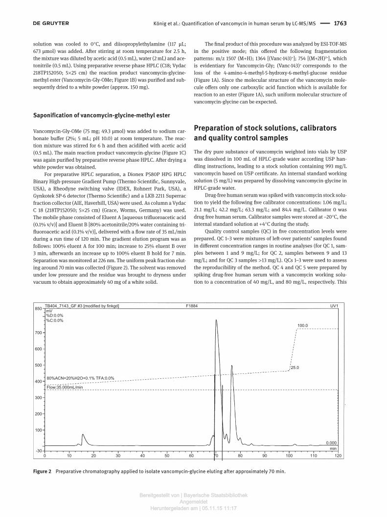
For preparative HPLC separation, a Dionex P580P HPG HPLC Binary High-pressure Gradient Pump (Thermo Scientific, Sunnyvale, USA), a Rheodyne switching valve (IDEX, Rohnert Park, USA), a Gynkotek SP-6 detector (Thermo Scientific) and a LKB 2211 Superrac fraction collector (AIE, Haverhill, USA) were used. As column a Vydac C 18 (218TP152050; 5 × 25 cm) (Grace, Worms, Germany) was used. The mobile phase consisted of Eluent A [aqueous trifluoroacetic acid (0.1% v/v)] and Eluent B [80% acetonitrile/20% water containing tri-fluoroacetic acid (0.1% v/v)], delivered with a flow rate of 35 mL/min during a run time of 120 min. The gradient elution program was as follows: 100% eluent A for 100 min; increase to 25% eluent B over 3 min, afterwards an increase up to 100% eluent B hold for 7 min. Separation was monitored at 226 nm. The uniform peak fraction elut-ing around 70 min was collected (Figure 2). The solvent was removed under low pressure and the residue was brought to dryness under vacuum to obtain approximately 40 mg of a white solid.
The final product of this procedure was analyzed by ESI-TOF-MS in the positive mode; this offered the following fragmentation patterns: m/z 1507 (M+H); 1364 [(Vanc-143)+]; 754 [(M+2H)2+], which is evidentiary for Vancomycin-Gly; (Vanc-143)+ corresponds to the loss of the 4-amino-4-methyl-5-hydroxy-6-methyl-glucose residue ( Figure 1A). Since the molecular structure of the vancomycin mole-cule offers only one carboxylic acid function which is available for reaction to an ester (Figure 1A), such uniform molecular structure of vancomycin-glycine can be expected.
Preparation of stock solutions, calibrators and quality control samplesThe dry pure substance of vancomycin weighted into vials by USP was dissolved in 100 mL of HPLC-grade water according USP han-dling instructions, leading to a stock solution containing 993 mg/L vancomycin based on USP certificate. An internal standard working solution (5 mg/L) was prepared by dissolving vancomycin-glycine in HPLC-grade water.
Drug-free human serum was spiked with vancomycin stock solu-tion to yield the following five calibrator concentrations: 1.06 mg/L; 21.1 mg/L; 42.2 mg/L; 63.3 mg/L; and 84.4 mg/L. Calibrator 0 was drug free human serum. Calibrator samples were stored at −20°C, the internal standard solution at +4°C during the study.
Quality control samples (QC) in five concentration levels were prepared. QC 1–3 were mixtures of left-over patients’ samples found in different concentration ranges in routine analyses (for QC 1, sam-ples between 1 and 9 mg/L; for QC 2, samples between 9 and 13 mg/L; and for QC 3 samples > 13 mg/L). QCs 1–3 were used to assess the reproducibility of the method. QC 4 and QC 5 were prepared by spiking drug-free human serum with a vancomycin working solu-tion to a concentration of 40 mg/L, and 80 mg/L, respectively. This
mV%D:0.0%%C:0.0%
700
850 TB404_7143_GF #3 [modified by finkgd] F1884
80%ACN+20%H2O+0.1% TFA:0.0%
Flow:35.000mL/min
UV1
100.0
25.0
600
500
400
300
200
100
-300 10 20 30 40 50 60 70 80 90 100 110 120
0.000min
Figure 2 Preparative chromatography applied to isolate vancomycin-glycine eluting after approximately 70 min.
Bereitgestellt von | Bayerische StaatsbibliothekAngemeldet
Heruntergeladen am | 05.11.15 11:17
1764 König et al.: Quantification of vancomycin in human serum by LC-MS/MS
vancomycin solution was prepared separately from the solution used for the preparation of calibrator samples. All QC samples were ali-quoted and stored at −20°C until analysis on either validation site.
High-performance liquid chromatography conditionsAt laboratory site 1 (Munich) a Waters Alliance 2795, at site 2 ( Penzberg) a Waters Acquity LC system was used. As analytical column a Fortis C8 (100 mm × 2.1 mm, 3 µm) (dichrom GmbH, Marl, Germany) was used. The column temperature was 40°C. The injection volume was 20 µL. The mobile phase consisted of two solvents; Eluent A: aqueous formic acid (0.1% v/v); Eluent B: methanol containing 0.1% formic acid (0.1% v/v). Run time was 21 min with a flow rate of 0.3 mL/min and an gradient elution program as follows: 100% A for 3 min; linear increase to 30% B over 7 min, hold for 2 min; afterwards a linear increase up to 80% B within 1 min, hold for 2 min; return to the initial condition within 1 min and re-equilibration for 5 min. Via a post-column switching valve, the HPLC eluate was directed into the mass spectrometer between 4.0 and 10.5 min after injection; during the residual run time the eluent was diverged into waste.
Mass spectrometric conditionsMass spectrometric analysis was performed using Waters Quattro Micro instruments in the positive ionization mode on both laboratory sites. The following settings were applied: Capillary voltage, 3.2 kV;
cone voltage, 20.0 V; collision energy, 12 eV; source temperature, 120°C; and desolvation temperature 480°C. The dwell time was 0.2 ms; inter-channel delay, 0.02 ms; and inter-scan delay 0.02 ms. Mass spec-trometric data were acquired from 4.5 to 12.0 min after injection.
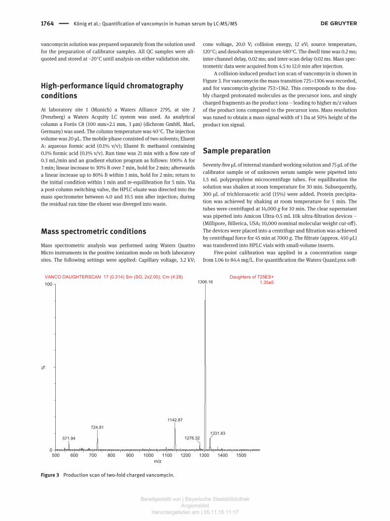
A collision-induced product ion scan of vancomycin is shown in Figure 3. For vancomycin the mass transition 725 > 1306 was recorded, and for vancomycin-glycine 753 > 1362. This corresponds to the dou-bly charged protonated molecules as the precursor ions, and singly charged fragments as the product ions – leading to higher m/z values of the product ions compared to the precursor ions. Mass resolution was tuned to obtain a mass signal width of 1 Da at 50% height of the product ion signal.
Sample preparationSeventy-five µL of internal standard working solution and 75 µL of the calibrator sample or of unknown serum sample were pipetted into 1.5 mL polypropylene microcentrifuge tubes. For equilibration the solution was shaken at room temperature for 30 min. Subsequently, 300 µL of trichloroacetic acid (15%) were added. Protein precipita-tion was achieved by shaking at room temperature for 5 min. The tubes were centrifuged at 14,000 g for 10 min. The clear supernatant was pipetted into Amicon Ultra-0.5 mL 10k ultra-filtration devices – (Millipore, Billerica, USA; 10,000 nominal molecular weight cut-off). The devices were placed into a centrifuge and filtration was achieved by centrifugal force for 45 min at 7000 g. The filtrate (approx. 450 µL) was transferred into HPLC vials with small-volume inserts.
Five-point calibration was applied in a concentration range from 1.06 to 84.4 mg/L. For quantification the Waters QuanLynx soft-
m/z500 600 700 800 900 1000 1100 1200 1300 1400 1500
%
0
100VANCO DAUGHTERSCAN 17 (0.314) Sm (SG, 2x2.00); Cm (4:28) Daughters of 725ES+
1.35e51306.16
1142.87
724.81
571.94 1276.321331.83
Figure 3 Production scan of two-fold charged vancomycin.
Bereitgestellt von | Bayerische StaatsbibliothekAngemeldet
Heruntergeladen am | 05.11.15 11:17
König et al.: Quantification of vancomycin in human serum by LC-MS/MS 1765
ware module was used with the following setting: Polynome type, linear; origin, include; weighting function, 1/x; axis transformation, none; smoothing method, mean; smoothing width, 1; smoothing it-erations, 1.
Method validationThe method was validated in a bi-centric protocol: Institute of Labo-ratory Medicine, Hospital of the University of Munich, Germany, site 1; and Roche Diagnostics, Penzberg, Germany, site 2.
Prior to each analytical run, a system suitability screening test was performed; for this the preparation of calibrator 1 (1 µg/mL) was injected. A signal-to-noise ratio of > 10:1 for the MRM-trace of vanco-mycin was defined as mandatory for a subsequent analytical run.
In order to test the specificity of the method, 10 leftover clinical serum samples from intensive care patients not treated with vanco-mycin were used. These samples were processed and analyzed with-out addition of the internal standard solution in order to verify the absence of peaks within the retention time window of the analyte or the internal standard, respectively.
In order to characterize the potential impact of residues of the sample matrix on the ionization of analyte and internal standard, two investigations were performed. In the post-column-infusion set-ting [11], a pure solution of vancomycin (1 mg/L) was infused with a syringe pump at constant rate using a T-piece into the column efflu-ent, in order to generate a MS/MS background signal. Upon injection of processed serum samples, potential modulation of the background signal was monitored. In a second experiment, extracted serum from three patients not treated with vancomycin was spiked with a solu-tion of vancomycin to a concentration of 21 mg/L each. For compari-son, a neat sample in water was spiked in the same way in triplicate. Both sets of samples were analyzed with the MS/MS method and peak areas were recorded in order to estimate potential effects of the serum-derived sample matrix on ionization of vancomycin, as matrix effect (%) according to Matuszewski et al. [12].
Accuracy of the method was tested by analyzing three external reference calibrator samples (USP Calibrators, Roche) and the qual-ity control samples QC 4 and QC 5 which were prepared on valida-tion site 2. Each sample was analyzed in triplicate on both validation sites.
Imprecision of the method was studied by analyzing aliquots of the QC samples 1–5 in two series of six-fold determination on both study sites, leading to a total of 24 results for each sample.
To roughly characterize a lower limit of detection and the per-formance of the method implementation in a concentration range be-low the lowest calibrator sample, a spiked sample with a vancomycin concentration of 0.1 µg/mL was injected in triplicate on both study sites and the signal-to-noise ratio was assessed.
In order to test the stability of processed samples, extracts of three patients’ samples were stored at +8°C and −20°C. These sam-ples were re-quantified in analytical series after 1 day, 2 days, and 1 week. These results were compared with the results found in the initial analytical run.
Left-over serum from 70 patients’ samples send to the Institute of Laboratory Medicine for clinically indicated vancomycin measure-ment were used to study the agreement between analyses performed with the described LC-MS/MS method on both validation sites. The samples were recruited consecutively without any selection, thus re-flecting the typical distribution of concentrations found in a tertiary
care hospital. After anonymization, two aliquots of these samples were prepared and stored at −20°C until analysis on either study site within 4 weeks. This procedure was approved by the Institutional Review Board.
ResultsA representative LC-MS/MS chromatogram is shown in Figure 4. Analyte and the internal standard compound vancomycin-glycine co-eluted as requested. Fifty MS/MS-data points were acquired over both chromato-graphic peaks. Minor signals from isobaric compounds were observed eluting slightly beforehand and follow-ing the peaks. Their pattern was found constant in both calibrator samples and patients’ samples on both study sites, and peak integration was performed consistently. The pattern did not change with the storage time of samples.
In all analytical series the regression coefficient r2 was ≥ 0.99 for all calibration runs over the concentration range from 1.06 to 84.41 mg/L. The slope of the calibration line was 0.25 ( ± 5%).
The signal-to-noise ratio observed in the system suit-ability screening test (injection of the lowest calibrator sample) was > 400:1 in all analytical series on study site 1, and > 100:1 on site 2.
The handling of the method was found convenient, including the ultrafiltration step. The entire instrumental setting was robust throughout the study period on both sites, in particular regarding HPLC back-pressure and ion-ization yield.
The analysis of 10 serum samples from patients which were not treated with vancomycin proved the specificity of the method; no peak signals in retention time windows of vancomycin or vancomycin-glycin were observed.
Figure 5 displays the results of the post-column infu-sion experiment which was performed to characterize the impact of serum-derived sample materials on the ioniza-tion yield of vancomycin. The figure shows an overlay of the signal pattern generated by injection of extract from a vancomycin-free sample, and from a patient’s sample. During the time period in which the chromatographic eluate was transferred to the MS/MS system a con-stant signal of the continuously infused analyte can be observed. There was no obvious drop or increase in ion yield at the retention time of vancomycin. In the spiking experiment, a matrix effect of –9% according to Matusze-wski et al. [12] was observed.
Table 1 displays the analytical accuracy realized for the analysis of standard and quality control materials;
Bereitgestellt von | Bayerische StaatsbibliothekAngemeldet
Heruntergeladen am | 05.11.15 11:17

1766 König et al.: Quantification of vancomycin in human serum by LC-MS/MS
for each single determination a bias of ≤ ± 4% was observed for the five materials investigated on both study sites which is within the ± 3 SD range of the method.
The data of the imprecision study are given in Table 2. Intra-assay imprecision observed for the five samples ranged from 1.1% to 3.9% CV. The total CV observed for aliquots of these samples analyzed on both study sites (4 series, 24 determinations) ranged from 1.9% to 3.8%.
In a sample spiked to a vancomycin concentration of 0.1 mg/L, a mean signal-to-noise ratio from three injec-tions of 52:1 (site 1) and 14:1 (site 2) was observed.
Stability of processed samples (autosampler on board stability) is shown in Table 3; sample extracts were found to be stable for at least 1 week at +8°C with bias of ≤ ± 3% compared to initial measurement.
For 70 unselected left-over serum samples which were analyzed to study the between-laboratory agree-ment of the method, mean concentrations of 9.4 mg/L and 9.9 mg/L were observed on site 1 and site 2, respectively, with median concentrations of 8.8 mg/L and 9.6 mg/L (Figure 6). Coefficient of variation observed for the inter-nal standard peak area was 14% on site 1 and 10% on site 2. Pearson’s r was 0.99. Linear regression demonstrated the following equation:
Figure 4 Representative MS/MS chromatogram of vancomycin (calibrator sample 1, with vancomycin-glycin as the internal standard).
Bereitgestellt von | Bayerische StaatsbibliothekAngemeldet
Heruntergeladen am | 05.11.15 11:17

König et al.: Quantification of vancomycin in human serum by LC-MS/MS 1767
site 2 = 1.04 * site 1+0.03 (95% confidence interval for slope: 1.00–1.09; 95% confidence interval for intercept: −0.42 mg/L to 0.49 mg/L)
DiscussionWe here report a HPLC-MS/MS method for the quantifica-tion of the antibiotic compound vancomycin in serum, which is intended to be used in the context of standardiza-tion and harmonization of routine assays for this analyte. An essential novelty of the method protocol is the fact that an internal standard compound was prepared as a homo-logue by derivatization of vancomycin to vancomycin-gly-cin. Compared to previously reported LC-MS/MS methods
for vancomycin quantification [7–9], the molecular struc-ture of this internal standard compound is much closer related to that of the target analyte and ensures identi-cal chromatographic behavior in the chosen setup. In general, matrix effect on the ionization of analytes is best compensated for by the use of stable isotope labeled inter-nal standard compounds; however, since vancomycin is a biological product such labeled compounds are not avail-able. We conclude that the method for the quantification of vancomycin described here has the highest metrologi-cal level realized so far, suggesting this protocol as a can-didate reference method.
A bi-centric method validation characterized the method as specific, accurate and precise. The between-laboratory agreement was found acceptable with < 4% bias for the analysis of a large series of authentic clinical
Time6.00 7.00 8.00 9.00 10.00 11.00 12.00 13.00 14.00
%
0
100MRM of 2 Channels ES+TIC5.00e4
13.3812.538.85
7.8010.999.96
9.53 10.2710.80 11.6211.32 12.2212.94 13.81 14.7714.1214.23
Figure 5 Post-column infusion experiment to assess matrix effects on the ionization of vancomycin. Overlay of a run with extract from drug-free serum and of a clinical sample.
Table 1 Analytical accuracy observed for external calibration material (Cal A-C) and quality control material manufactured on study site 2 (both materials based on USP vancomycin standard).
Target concentration Cal A, 5.0 mg/L Cal B, 40.0 mg/L Cal C, 80.0 mg/L QC 4, 39.7 mg/L QC 5, 79.4 mg/L
Penzberg (n = 3) Observed mean conc. 5.1 40.4 79.6 40.1 78.0 Recovery % 104/100/102 101/100/102 101/99/99 100/101/99 96/98/99Großhadern (n = 3) Observed mean conc. 5.1 40.6 80.4 40.4 81.8 Recovery % 104/100/104 103/102/100 103/99/100 99/102/103 103/101/103
Bereitgestellt von | Bayerische StaatsbibliothekAngemeldet
Heruntergeladen am | 05.11.15 11:17

1768 König et al.: Quantification of vancomycin in human serum by LC-MS/MS
sample materials on two study sites. Thus, the perfor-mance of this method – which is based on a homologue for internal standardization – is almost equal to that of typical isotope dilution LC-MS/MS methods. Control of matrix effects was verified by post-column-infusion and spiking experiments.
Minor isobaric interference was observed in the MS/MS chromatograms of analyte and internal standard (Figure 4); the nature of the respective compounds has not been disclosed. Efficient chromatographic separation and consistent peak integration is important for optimum reproducibility of analytical series.
The method described here was not optimized regard-ing a routine use in the clinical laboratory. Extended chromatography was applied in order to minimize matrix effects on the ionization of the analyte and the internal
5
15
00 5 10 15
Site 1, munich20 25 30
10
25
20
Site
2, p
enzb
erg
30
Figure 6 Between-laboratory comparison of serum vancomycin measurement by LC-MS/MS (n = 70; vancomycin in mg/L).
Table 2 Imprecision (coefficient of variation) observed for quality control materials.
QC 1, 5.3 mg/L
QC 2, 12.4 mg/L
QC 3, 13.9 mg/L
QC 4, 39.7 mg/L
QC 5, 79.4 mg/L
PenzbergIntra-assay (n = 6)Day ACV % 3.5 1.4 2.6 1.3 1.4Intra-assay (n = 6)Day BCV % 3.9 1.5 1.1 1.6 2.7
GroßhadernIntra-assay (n = 6)Day ACV % 1.5 1.5 1.3 2.1 0.8Intra-assay (n = 6)Day BCV % 1.4 1.1 3.1 2.0 1.6
Pooled data from both study sitesInter-assay (n = 24)CV % 2.8 2.2 3.8 1.9 2.2
Table 3 Stability of sample extracts (autosampler stability).
Recovery in % Storage
Serum 1 Serum 2 Serum 3
+8°C −20°C +8°C −20°C +8°C −20°C
Penzberg1 day 102 102 104 101 102 1012 days 104 109 103 104 104 1041 week 99 99 98 97 100 98
Großhadern1 day 96 99 99 95 100 1012 days 98 97 101 97 99 1011 week 100 96 103 96 101 101
standard, thereby optimizing the analytical reliability. This, however, resulted in a retention time of 9.8 min and a total run-time above 20 min. The sample preparation protocol is straightforward and based on protein precipi-tation. Subsequent ultrafiltration was performed in order to maximize the stability and robustness of the method.
To date no generally accepted validation protocol for quantitative bio-medical analytical methods using LC-MS/MS is available. The widely used FDA protocol [13] has important limitations and is mainly designed for the
Bereitgestellt von | Bayerische StaatsbibliothekAngemeldet
Heruntergeladen am | 05.11.15 11:17

König et al.: Quantification of vancomycin in human serum by LC-MS/MS 1769
validation of pharmacokinetic batch studies in individ-ual laboratories. However, a prerequisite for a reference method is robustness over time and space. Our approach of a bi-centric validation can give an impression on the general robustness of the method realized in different instrument installations and laboratories. We suggest such an assessment of between-instrument or between-laboratory agreement for validation protocols applied for clinical mass spectrometry methods, just as it is standard in the validation of commercial immunoassays.
We defined a signal-to-noise ratio for the lowest con-centration calibrator sample which is required as the acceptance criterion in a system suitability test. The con-centration range below the lowest calibrator sample was not addressed systematically in the validation study, since this would be outside the intended use of the method.
Besides a robust analytical method, the purity of standard compounds used is crucial for the implementa-tion of a reference measurement system for vancomycin in the future. Detailed characterization of the primary vanco-mycin standard has not been addressed in this work. All concentrations are based on the USP Pharmacopeia cer-tificates delivered with the purchased material.
In summary, we were able to describe a robust and reliable LC-MS/MS-based measurement procedure for the quantification of vancomycin in human serum which may be used in the context of a reference measurement system. Such a system may contribute to increase the added clini-cal value of routine vancomycin therapeutic drug moni-toring which is so far limited by rather poor agreement between routine methods.
Acknowledgments: We thank the Referenzinstitut für Bio-analytik (RfB) for the permission to report the results of a vancomycin proficiency testing program.
Conflict of interest statement
Authors’ conflict of interest disclosure: The authors stated that there are no conflicts of interest regarding the publication of this article.Research funding: None declared.Employment or leadership: None declared.Honorarium: None declared.
Received February 22, 2013; accepted March 21, 2013; previously published online April 11, 2013
References1. Rybak M, Lomaestro B, Rotschafer JC, Moellering R Jr., Craig W,
Billeter M, et al. Therapeutic monitoring of vancomycin in adult patients: a consensus review of the American Society of Health-System Pharmacists, the Infectious Diseases Society of America, and the Society of Infectious Diseases Pharmacists. Am J Health-Syst Pharm 2009;66:82–98.
2. Roberts JA, Norris R, Paterson DL, Martin JH. Therapeutic drug monitoring of antimicrobials. Br J Clin Pharmacol 2012;73: 27–36.
3. Moise-Broder PA, Forrest A, Birmingham MC, Schentag JJ. Pharmacodynamics of vancomycin and other antimicrobials in patients with Staphylococcus aureus lower respiratory tract infections. Clin Pharmacokinet 2004;43:925–42.
4. Cano EL, Haque NZ, Welch VL, Cely CM, Peyrani P, Scerpella EG, et al. Incidence of nephrotoxicity and association with vancomycin use in intensive care unit patients with pneumonia: retrospective analysis of the IMPACT-HAP Database. Clin Ther 2012;34:149–57.
5. Wong-Beringer A, Joo J, Tse E, Beringer P. Vancomycin-associated nephrotoxicity: a critical appraisal of risk with high-dose therapy. Int J Antimicrob Agents 2011;37:95–101.
6. Wilson JF, Davis AC, Tobin CM. Evaluation of commercial assays for vancomycin and aminoglycosides in serum: a comparison of accuracy and precision based on external quality assessment. J Antimicrob Chemother 2003;52:78–82.
7. Cass RT, Villa JS, Karr DE, Schmidt DE Jr. Rapid bioanalysis of vancomycin in serum and urine by high-performance liquid
chromatography tandem mass spectrometry using on-line sample extraction and parallel analytical columns. Rapid Commun Mass Spectrom 2001;15:406–12.
8. Zhang T, Watson DG, Azike C, Tettey JN, Stearns AT, Binning AR, et al. Determination of vancomycin in serum by liquid chromatography-high resolution full scan mass spectrometry. J Chromatogr B 2007;857:352–6.
9. Shibata N, Ishida M, Prasad YV, Gao W, Yoshikawa Y, Takada K. Highly sensitive quantification of vancomycin in plasma samples using liquid chromatography-tandem mass spectrometry and oral bioavailability in rats. J Chromatogr B 2003;789:211–8.
10. Stepman HC, Vanderroost A, Van Uytfanghe K, Thienpont LM. Candidate reference measurement procedures for serum 25-hydroxyvitamin D3 and 25-hydroxyvitamin D2 by using isotope-dilution liquid chromatography-tandem mass spectrometry. Clin Chem 2011;57:441–8.
11. Annesley TM. Ion suppression in mass spectrometry. Clin Chem 2003;49:1041–4.
12. Matuszewski BK, Constanzer ML, Chavez-Eng CM. Strategies for the assessment of matrix effect in quantitative bioanalytical methods based on HPLC-MS/MS. Anal Chem 2003;75:3019–30.
13. Guidance for Industry-Bioanalytical Method Validation. U.S. Department of Health and Human Services-Food and Drug Administration; 2001. Available from: http://www.fda.gov/downloads/Drugs/…/Guidances/ucm070107.pdf. Accessed on February, 2012.
Bereitgestellt von | Bayerische StaatsbibliothekAngemeldet
Heruntergeladen am | 05.11.15 11:17