Kadriye Ciftci, Ph.D. and Anshul Gupte, Ph.D. · 333 13 Gene Therapy: An Overview of the Current...
46
333 13 Gene Therapy: An Overview of the Current Viral and Nonviral Vectors Kadriye Ciftci, Ph.D. and Anshul Gupte, Ph.D. CONTENTS Introduction ............................................................................................................ 333 Gene Delivery Systems ......................................................................................... 337 Viral Vectors .................................................................................................. 337 Retroviruses ........................................................................................... 337 Adenoviruses.......................................................................................... 339 Adeno-Associated Viruses ..................................................................... 342 Other Viruses ......................................................................................... 343 Nonviral Vectors ............................................................................................ 345 Direct Injection of Naked DNA ............................................................ 348 Liposomes .............................................................................................. 349 Polymers ................................................................................................ 353 Receptor-Mediated Gene Delivery ........................................................ 358 Hybrid Vectors (Viral and Nonviral Components) ............................... 359 Nuclear Localization Signal Peptides and Membrane-Modifying Agents .................................................... 360 Membrane-Modifying Agents ............................................................... 361 Electroporation............................................................................................... 362 References .............................................................................................................. 365 INTRODUCTION Gene therapy holds great promise for the treatment of many diseases (e.g., cancer, AIDS, cystic fibrosis, adenosine deaminase deficiency, cardiovascular diseases, Gaucher disease, α 1-antitrypsin deficiency, rheumatoid arthritis, and several others) (1,2). Advances in genomics and molecular biology have revealed that almost all diseases have a genetic component. In some cases, such as cystic fibrosis or hemophilia, © 2006 by Taylor & Francis Group, LLC
Transcript of Kadriye Ciftci, Ph.D. and Anshul Gupte, Ph.D. · 333 13 Gene Therapy: An Overview of the Current...

333
13
Gene Therapy: An Overview of theCurrent Viral andNonviral Vectors
Kadriye Ciftci, Ph.D. and Anshul Gupte, Ph.D.
CONTENTS
Introduction............................................................................................................333Gene Delivery Systems .........................................................................................337
Viral Vectors ..................................................................................................337Retroviruses ...........................................................................................337Adenoviruses..........................................................................................339Adeno-Associated Viruses .....................................................................342Other Viruses .........................................................................................343
Nonviral Vectors ............................................................................................345Direct Injection of Naked DNA ............................................................348Liposomes ..............................................................................................349Polymers ................................................................................................353Receptor-Mediated Gene Delivery ........................................................358Hybrid Vectors (Viral and Nonviral Components) ...............................359Nuclear Localization Signal Peptides
and Membrane-Modifying Agents ....................................................360Membrane-Modifying Agents ...............................................................361
Electroporation...............................................................................................362References..............................................................................................................365
INTRODUCTION
Gene therapy holds great promise for the treatment of many diseases (e.g., cancer,AIDS, cystic fibrosis, adenosine deaminase deficiency, cardiovascular diseases, Gaucherdisease,
α
1-antitrypsin deficiency, rheumatoid arthritis, and several others) (1,2).Advances in genomics and molecular biology have revealed that almost all diseaseshave a genetic component. In some cases, such as cystic fibrosis or hemophilia,
PH1873_C013.fm Page 333 Thursday, June 30, 2005 10:38 AM
© 2006 by Taylor & Francis Group, LLC

334
Pharmaceutical Biotechnology
mutations in a single gene can result in the disease (3–5). In other cases, such ashypertension or high cholesterol, certain genetic variations may interact with envi-ronmental stimuli to cause the disease (6–8) or pathological conditions associatedwith aging frequently result in the loss of gene activity in specific types of cells.
The main targets of gene therapy are to repair or replace mutated genes, regulategene expression and signal transduction, manipulate the immune system or targetmalignant and other cells for destruction (1). There are several factors involved ineffective gene transfer to somatic cells in patients: (a) the type of vehicle used forgene delivery that will determine efficacy of delivery; (b) interaction of gene vehicle;(c) targeting to the specific area; (d) entrance to the target cell; (e) release from thecytoplasmic compartment, transport to the nucleus; (f) type and potency of regulatoryelements; (g) expression (transcription) of the transgene and translation into protein.
Compared to conventional small molecule drug therapies with a transient effecton their molecular targets, gene therapy usually requires an efficient transfer by deliverysystem to target cells resulting in a permanent change to the genetic constitution. Theapplication of gene delivery technology to a growing roster of clinical indications ispredicated on significant advances in both genomics and gene delivery systems.
There is a wide variety of vectors used to deliver DNA or oligonucleotides
intomammalian cells, either
in vitro
or
in vivo
. The most common
vector systems arebased on viral [retroviruses (9, 10), adeno-associated
virus (AAV) (11), adenovirus(12, 13), herpes simplex virus
(HSV) (14)] and nonviral [cationic liposomes (15, 16),polymers and receptor-mediated
polylysine-DNA] complexes (17). Other viral vec-tors that are currently under development are
based on lentiviruses (18), humancytomegalovirus (CMV) (19),
Epstein-Barr virus (EBV) (20), poxviruses (21), neg-ative-strand
RNA viruses (influenza virus), alphaviruses and herpesvirus
saimiri (22).Also a hybrid adenoviral/retroviral vector has successfully
been used for
in vivo
genetransduction (23). A simplified schematic representation of basic human gene therapymethods is described in Figure 13.1.
The choice of the appropriate delivery system for successful gene therapyrequires understanding of the drawbacks and advantages of each delivery system(Table 13.1 for comparison of viral vectors and Table 13.2 for comparison of nonviralmethods for gene therapy), such as limitations in the total length of the DNA thatcan be introduced, including the plasmid size and control elements. Understandingof the pathophysiology of the disease and the cell targets (IV, IP, intratumoral, SCinjection) is required. The type of control elements required for the tissue-specificexpression of the construct, the presence of viral or other origins of replication aswell as of the cDNA encoding the viral replication initiator protein for an episomalreplication of the transgene and sequences that prompt integration is also importantfor successful gene transfer. While no single vector developed to date is optimal forall clinical indications, the growing number of viral and synthetic vectors will enablegene therapy to be used in treating a wide variety of significant diseases.
Current gene therapy programs apply gene delivery technology across a broaderspectrum of disease conditions (2). Since 1989, when the first human gene therapystudy was performed, enormous research efforts have followed (3). Although mucheffort has been directed in the last decade
toward improvement of protocols in humangene therapy, the therapeutic
applications of gene transfer technology still remain
PH1873_C013.fm Page 334 Thursday, June 30, 2005 10:38 AM
© 2006 by Taylor & Francis Group, LLC

Gene Therapy: An Overview of the Current Viral and Nonviral Vectors
335
mostly
theoretical. The weakest point of gene therapy development programs
is vectordesign, followed by gene regulation and
avoidance of immune responses. Basicresearch is cautiously progressing
to address these pressing issues. The characteristicsof the most developed gene delivery systems are discussed in the following section.
FIGURE 13.1
Schematic representation of human gene therapy.
TABLE 13.1Comparison of Different Viral Vectors for Gene Therapy
Vector Advantages Disadvantages
Retrovirus Integration into host DNA All viral genes removed Relatively safe
Insertional mutagenesis Requires cell division Relatively low titer
Adenovirus Higher titer Efficient in nondividing cells
Toxicity Immunological response
Adeno-associated
virus All viral genes removed Limited size of foreign DNA Labor-intensive productionStatus of genome not fully elucidated
Lentivirus Provide long-term and stable geneexpression
Infect nondividing cells
Similar retrovirus
Delivery vectors A : Viral B : Synthetic
�erapeutic gene
�erapeutic gene
Electroporation
Patient
PH1873_C013.fm Page 335 Thursday, June 30, 2005 10:38 AM
© 2006 by Taylor & Francis Group, LLC

336
Pharmaceutical B
iotechnology
TABLE 13.2Methods of Nonviral Gene Transfer
MethodSize of DNA Target Cells
TransfectionEfficiency Transfection Cellular Toxicity Gene Expression Preparation Application
Naked DNA No limit Especiallymyocytes
10–30% of cellsat injection site
Extra chromosomal Lymphocytic infiltration
Until death of cell Easy and cheap
In vivo
Microinjection No limit Mitosis/resting Stable <0.1–1% Integration possible 30% survival 200–400 injections/hr
In vitro
Electroporation 150 Kb Mitosis/resting Stable <0.1–1% 1–2 copies 20–60% survival Easy
In vitro
Particlebombardment
10,000copies
Mitosis/resting Stable <0.1–1%,transient <20%
Persistent and integration?
85–95% survival 2–12 month Easy
In vitro
and
in vivo
Lipofection No limit Mitosis/resting Stable <0.1–1%,transient 80%
Integration possible Membranetoxic
Easy
In vitro
and
in vivo
Ligand mediated >48 Kb Mitosis/resting Up to 50% Extra chromosomal High High, Transient Labor intensive
In vitro
and
in vivo
Calcium phosphateprecipitation
No limit Mitosis/resting Stable <0.1% Often multiplecopies
High High, Transient Labor intensive and time consuming
In vitro
and
in vivo
PH1873_C
013.fm Page 336 T
hursday, June 30, 2005 10:38 AM
© 2006 by Taylor & Francis Group, LLC

Gene Therapy: An Overview of the Current Viral and Nonviral Vectors
337
GENE DELIVERY SYSTEMS
V
IRAL
V
ECTORS
Viral vectors are the first used vectors for gene therapy research. It has been knownthat many viruses have the capability of efficiently transferring their nucleic acidgenomes to mammalian cells in order to initiate their first step in life cycle. Viralvectors take advantage of the ability of the virus to enter cells and deliver geneticmaterial to the nucleus. Most viral vectors are engineered in such a way that theycan enter the cells but they do not have the ability to replicate in the cell. Tosuccessfully develop viral vectors, the important consideration includes introducingtherapeutic genes into their genomes while concurrently removing the native viralgenes that code for harmful viral proteins. To develop viral vectors first viral DNAis removed and is replaced with a therapeutic gene and the recombinant virus is thusproduced and functions purely as a delivery system for the therapeutic genes to thenucleus of the target cell without causing cellular damage or subsequent virus prop-agation (24). Depending on the therapeutic aim of a particular gene therapy, transientor permanent expression may be desirable. There are several different classes of viralvectors including retrovirus, adenovirus, adeno-associated virus, lentiviruses, herpessimplex, and alpha(
α
)-viruses used for gene therapy. The characteristics
and applica-tions of these vectors are discussed below.
Retroviruses
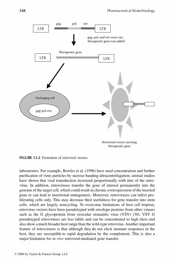
The retroviruses are enveloped viruses, roughly spherical, about 120 nm in diameter.They contain a diploid RNA genome of 7 to 11 kb that is converted into a DNAintermediate by the reverse transcriptase upon entry into the cytoplasm of a cell (25,26). The DNA is then transported to the nucleus, where it integrates randomly intothe genome (25). Retroviruses can only accommodate less than 9 kb of foreigngenetic information. The use of retroviruses for gene transfer requires a two-com-ponent approach as described in Figure 13.2. The first involves the replacement ofthe genetic material encoding the gag, pol, and env proteins with the DNA to betransferred. This DNA is expressed under the control of the promoter elements inthe 5LTR. The second component involves the introduction of this DNA into aretroviral packaging cell line to produce virus able to infect the appropriate hostspecies. This cell line contains a replication-defective helper retrovirus that willprovide the gag, pol, and env proteins and an encapsidation signal for efficient viralpackaging (27, 28).
Before the
in vivo
gene therapy with retroviruses becomes a successful realitya number of problems must be overcome. The major limitation of retroviruses hasbeen poor gene expression
in vivo
, which has been overcome through the use oftissue-specific promoters. Use of internal ribosome entry sites from picornavirusesin retroviral vectors has provided stable expression of multiple gene enhancers.Another drawback of retroviruses for their exploitation in gene therapy has been thelow viral titers obtained, too low to achieve therapeutic levels of gene expression;methods for the efficient concentration from large volumes of supernatant andpurification of amphotropic retrovirus particles have been developed in several
PH1873_C013.fm Page 337 Thursday, June 30, 2005 10:38 AM
© 2006 by Taylor & Francis Group, LLC
338
Pharmaceutical Biotechnology
laboratories. For example, Bowles et al. (1996) have used concentration and furtherpurification of virus particles by sucrose banding ultracentrifugation; animal studieshave shown that viral transduction increased proportionally with titer of the retro-virus. In addition, retroviruses transfer the gene of interest permanently into thegenome of the target cell, which could result in chronic overexpression of the insertedgene or can lead to insertional mutagenesis. Moreover, retroviruses can infect pro-liferating cells only. This may decrease their usefulness for gene transfer into stemcells, which are largely noncycling. To overcome limitations of host cell tropism,retrovirus vectors have been pseudotyped with envelope proteins from other virusessuch as the G glycoprotein from vesicular stomatitis virus (VSV) (30). VSV Gpseudotyped retroviruses are less labile and can be concentrated to high titers andalso show a much broader host range than the wild-type retrovirus. Another importantfeature of retroviruses is that although they
do not elicit immune responses in thehost, they are susceptible
to rapid degradation by the complement. This is also amajor
limitation for
in vivo
retroviral-mediated gene transfer.
FIGURE 13.2
Formation of retroviral vectors.
LTR LTR
LTR LTR
gag-pol-env
gag pol env
�erapeutic gene
Packaging cell
Retroviral vector carryingtherapeutic gene
gag, pol, and env were cut;therapeutic gene was added
PH1873_C013.fm Page 338 Thursday, June 30, 2005 10:38 AM
© 2006 by Taylor & Francis Group, LLC
Gene Therapy: An Overview of the Current Viral and Nonviral Vectors
339
The most common retroviral vector is based on the amphotropic
Moloney murineleukemia virus (MLV) (31). This system is particularly suitable
for efficient
in vitro
cell transduction: the amphotropic MLV has
a broad cell tropism, it can be producedat relatively high
titers (10
6
–10
7
iu/mL), and allows for long-term transgene expres-sion
because of the viral integration in the host chromosomal DNA.Retrovirus vectors were subjected to the first clinical trial on human gene therapy
to correct adenosine deaminase (ADA) deficiency (32). White-blood cells isolatedfrom patients were infected
ex vivo
with an MLV-based vector expressing ADA anda neomycin marker gene. After selection with G418, neomycin-resistant cells wereisolated and reintroduced into patients. The treatment improved the physical condi-tion of the patients and the ADA-containing provirus was stable in the blood forseveral years.
Investigators have been considering the engineering
of chimeric retroviruses withspecific cell tropism. This would
greatly facilitate the in vivo application of retroviralvectors
in clinical trials. In this respect, there have been many attempts
to alter thecell tropism of ecotropic retroviruses, which do
not infect human cells. This approachconsists of placing foreign
genes (CD4), single-chain antibodies, the polypeptideerythropoietin, short peptides binding to several integrins, and human heregulin(33–35). The retroviral systems used
in these studies were: avian leukosis virus,ecotropic
MLV and spleen necrosis virus. The foreign genes
used in the early studiesto generate hybrid envelopes were:
In some cases, there has been a partial successin
redirecting the cell tropism of ecotropic retroviruses (36), but the transductionefficiency is far from being optimal for
in vivo
applications. Retrovirus vectors have demonstrated some promising results in cancer therapy
and bone marrow transplantation. The introduction of retrovirus particles expressingHSV-TK and administration of GCV suggested that the treatment of graft-versus-host disease was efficient (37). The demonstration of the full correction of theSCID-X1 phenotype in infants is a further indication of the efficacy of retrovirusvectors (38).
Adenoviruses
Adenoviruses are nonenveloped DNA viruses with 80 to 110 nm diameter icosahe-dral protein shell containing double-strand DNA genome of 36 kb that encodes fourearly proteins (E1 to E4) and five late proteins (L1 to L5) (39). In order to enter thehost cell the adenovirus first attaches with a high affinity to a cell surface receptor,whose nature still remains elusive, using the head domains of the protruding viralfibers; the fibronectin-binding integrin on the cell surface then associates with thepenton base protein on the adenovirus triggering endocytosis of the virus particlevia coated pits and coated vesicles (40, 41). The third step in adenovirus entry intothe host cell includes penetration of the adenoviral particles by acid-catalyzed ruptureof the endosomal membrane involving the penton protein and the integrins andallowing escape to the cytoplasmic compartment; a decrease in endosome pH duringinternalization expose hydrophobic domains of these adenoviral capsid proteins,which permits these proteins to insert into the vesicle membrane in a fashion thatultimately disrupts its integrity (42). At the final step the adenoviral particle is
PH1873_C013.fm Page 339 Thursday, June 30, 2005 10:38 AM
© 2006 by Taylor & Francis Group, LLC

340
Pharmaceutical Biotechnology
attached to the cytoplasmic side of pore complexes and the DNA is released to theinterior of pore annuli entering the nucleoplasm.
Because replication is controlled by E1, it is usually deleted in adenoviralvectors used for gene therapy and replaced by the gene to be transferred, as shownin Figure 13.3. The resultant recombinant adenovirus is replication incompetent.This recombinant adenoviral DNA is then transferred into a complementing cellline containing E1 sequences in its genome (but lacking other sequences requiredfor replication) to generate viral particles that are infectious but replicationdefective (43).
Adenoviruses have certain advantages over retroviruses for gene therapy. Theycan be produced in high titer (>10
13
viral particles per milliliter) and can transfergenes efficiently into both replicating and nonreplicating cells (44). Adenovirusespossess a linear double-stranded genome which can be manipulated to accommodateup to 7.5 kb of DNA. Although early versions of adenoviruses showed toxic sideeffects and strong immune responses, newer second- and third-generation vectorswith many of the viral genes deleted, have demonstrated significant improvements(45). As the transferred genetic material is located episomally, the risks of perma-nently altering the genetic material of the cell and of insertional mutagenesis are
FIGURE 13.3
Gene delivery with adenoviral vectors.
E1
�erapeutic gene
Adenoviral vector with E1
E1 deleted, therapeutic gene added
Replication of virusin helper cell line
Viral particles
Infection of thetarget cell Expression of the
therapeutic protein
PH1873_C013.fm Page 340 Thursday, June 30, 2005 10:38 AM
© 2006 by Taylor & Francis Group, LLC

Gene Therapy: An Overview of the Current Viral and Nonviral Vectors
341
avoided (46). For safety, replication-deficient, infectious adenoviruses are being usedin somatic gene transfer; for example, deletion in a portion of the E3 region of thevirus permits encapsidation whereas deletion of a portion of the E1A codingsequence impairs viral replication (47, 48).
A disadvantage of adenoviral vectors is that the viral proteins are immunogenicand can induce nonspecific inflammation and specific cellular responses (43). Also,episomes tend to be lost from infected cells within 2 to 4 weeks, so repeatedadministration may be necessary (46). The efficacy of adenovirus delivery might beseverely hampered because most people have been exposed to natural adenovirusesinfections, even when using replication-deficient vectors. In a novel approach, thesurfaces of viral particles were coated with a multivalent copolymer based on poly-[
N
-(2-hydroxypropyl) methacrylamide] (pHPMA) (49). To improve targeting, fibro-blast growth factor (FGF) and vascular endothelial growth factor (VEGF) wereincorporated in the polymer, which resulted in targeting of bFGF receptor-positiveA549 cells. Targeting of endothelial human umbilical endothelial cells with polymer-VEGF coated adenovirus was also highly efficient (49). The
in vivo
targeting ofpolymer-bFGF ADLacZ virus in nude mice bearing intraperitoneal xenografts ofhuman SUIT2 cells was highly efficient.
In addition, the polymer-coated adenovirus particles were able to shield againstantibody recognition. In another approach, the PEGylation of the adenovirus capsidprotein prolonged transgene expression after systemic delivery of E1-deleted aden-ovirus, and allowed partial readministration with native virus (50). Adenovirus hasbeen explored as vector for the treatment of cystic fibrosis (51), for Duchennemuscular dystrophy (52), to deliver tumor suppressor genes for cancer treatment(53), for gene transfer to the brain (54) and for melanoma specific vector (55).
Recombinant adenovirus vectors have been used for variety of applicationsincluding, the transfer of factor IX gene in hemophilia B to dogs via vein injection(56) and in mice for the transfer of genes into neurons and glia in the brain for thetransfer of the gene of ornithine transcarmylase in deficient mouse and humanhepatocytes (57) for the transfer of the very low density lipoprotein receptor genefor treatment of familial hypercholesterolaemia in the mouse model (58) for thetransfer of low density lipoprotein receptor gene in normal mice and for the
ex vivo
transduction of T cells from ADA-deficient patients (59). The adenovirus major latepromoter was linked to a human a1-antitrypsin gene for its transfer to lung epitheliaof cotton rat respiratory pathway as a model for the treatment of a1-antitrypsindeficiency;
in vitro
and
in vivo
infections have shown production and secretion ofa1-antitrypsin by the lung cells (60).
To overcome one of the major limitations of the clinical utility of adenoviruseswhich is the low efficiency of gene transfer achieved in vivo, Arcasoy et al. (1997)found that the presence of the polycations polybrene, protamine, DEAE-dextran,and poly-L-lysine significantly increased the transfection efficiency in cell cultureusing the lacZ gene; because the polyanion heparin did not significantly alter genetransfer efficiency, but completely abrogated the effects of polycations it supportsthe idea that the negative charges presented by membrane glycoproteins reduce theefficiency of adenovirus-mediated gene transfer, an obstacle that can be overcomeby polycations.
PH1873_C013.fm Page 341 Thursday, June 30, 2005 10:38 AM
© 2006 by Taylor & Francis Group, LLC

342
Pharmaceutical Biotechnology
Adeno-Associated Viruses
Adeno-associated viruses (AAV) are parvoviruses that are not pathogenic in humans.They are extremely small, nonenveloped icosahedral virus of 18 to 26 nm in diametercarrying a single-stranded ~5 kb DNA genome with short, inverted terminal repeatsthat are required for genome replication and packaging. Unlike adenoviruses, AAVmay integrate into the host genome and do so at preferred locations, in particular, atone site on chromosome 19 (43). AAV has established its position as one of the mostpopular gene delivery systems. This is mainly because of the long-term and efficienttransgene expression in various cell types in many tissues such as liver, muscle, retinaand the central nervous system (62). Recombinant AAV vectors used for gene transfercontain 145 bp terminal repeat sequence and a polyadenylation site. They have hadmost of the viral genome deleted and replaced with DNA encoding the therapeuticgene. Since a few viral proteins are expressed, these viruses induce less of an immuneresponse than for adenoviruses. Like adenoviruses, AAV vectors do not require cellreplication for integration but high AAV titers are often difficult to obtain becausethe production of infectious AAV requires the use of an adenovirus, in which casecontamination of the AAV with adenovirus is a concern (43).
There are some disadvantages associated with the application of AAV. Genetransfer with AAV vectors has been shown to be low. Difficulties in generatingrecombinant virus on a large scale sufficient for preclinical and clinical trials and inobtaining high-titer virus stocks after the initial transfection into producer cells is alimiting factor for the widespread usage of AAV vectors. AAV vector particles in celllysates could be concentrated by sulfonated cellulose column chromatography to atiter higher than 10
8
cfu/mL or 5
×
10
10
particles/mL (63). A method for transfectingcells at extremely high efficiency with a rAAV vector and complementation plasmidwhile simultaneously infecting those cells with replication competent adenovirususing adenovirus-polylysine-DNA complexes has been developed (64).
After infection of cell cultures with recombinant AAV there is a decline in thepercentage of cells expressing the transferred gene with time in culture. This declinewas associated with ongoing losses of vector genomes (65). The packaging capacityis relatively restricted and the large-scale production inefficient. In addition, the pre-existing immunity to human AAV vectors is comparable to adenovirus and theintegration into the host genome is random, which can lead to unexpected activationor inhibition of endogenous gene expression.
Different AAV serotypes have shown remarkably different expression patternsbecause of differences in cell entry and intracellular activities (66, 67). Applicationof the dimerizer-inducible transcriptional regulatory system for AAV has allowedpharmacological regulation of heterologous gene expression
in vivo
(68). AAV normally contains a single-stranded copy of its genome. Transduction with
AAV can be enhanced in the presence of adenovirus gene products through theformation of double-stranded, nonintegrated AAV genomes. AAV has been reportedto have advantages over other viruses for gene transfer to hematopoietic stem cellsdue to their high titers and relative lack of dependence on cell cycle for target cellintegration. A robust CMV/LacZ reporter gene expression in primary humanCD34
+
CD2
-
progenitor cells induced to undergo T-cell differentiation was obtained
PH1873_C013.fm Page 342 Thursday, June 30, 2005 10:38 AM
© 2006 by Taylor & Francis Group, LLC

Gene Therapy: An Overview of the Current Viral and Nonviral Vectors
343
without toxicity or alteration in the pattern of T-cell differentiation. Seventy percentto 80% of the cells isolated from either adult bone marrow or umbilical cord bloodwere efficiently transduced with AAV, however, the expression was transient withoutintegration; this limits the potential use of AAV in gene therapy strategies for diseasessuch as AIDS (69).
Gene transduction by AAV vectors in cell culture can be stimulated over 100-fold by treatment of the target cells with agents that affect DNA metabolism, suchas irradiation or topoisomerase inhibitors (70), great improvements in transductionefficiency can also be achieved
in vivo
: previous g-irradiation increased the trans-duction rate in mouse liver by up to 900-fold, and the topoisomerase inhibitoretoposide increased transduction by about 20-fold after direct liver injection or aftersystemic delivery via tail vein injection; up to 3% of hepatocytes could be transducedafter a single systemic vector injection (71). This is a significant advantage comparedto stealth liposomes which, although concentrating in the liver, spleen and tumors,can transduce Kupffer cells but not hepatocytes after systemic delivery.
In another study, AAV-mediated delivery of the lacZ gene by direct injection tobrain tumors which were induced from human glioma cells in nude mice showedthat 30% to 40% of the cells along the needle track expressed b-galactosidase;subsequent delivery of the HSV-tk/IL-2 genes to these tumors with AAV and admin-istration of GCV to the animals for 6 days resulted in a 35-fold reduction in themean volume of tumors compared with controls by a significant contribution fromthe bystander effect (72).
Other Viruses
Lentiviruses
Although lentiviruses belong to retroviral class, gene therapy vectors derived fromlentiviruses offer many potentially unique advantages over more conventional ret-roviral gene delivery systems. Many of the lentivirus vectors used in gene therapyare based on the human immunodeficiency virus (HIV) (73). An advantage of HIVvectors has been the broad range of tissues and cell types they can transduce, aproperty granted because lentiviral vectors are pseudotyped with vesicular stomatitisvirus G glycoprotein. Human lentiviral (HIV)-based vectors can transduce nondi-viding cells
in vitro
and deliver genes
in vivo
; expression of transgenes in the brainhas been detected for more than 6 months. HIV vectors have been also used tointroduce genes directly into liver and muscle; 3% to 4% of the total liver tissuewas transduced by a single injection of 1-3
×
10
7
infectious units (IU) of recombinantHIV with no inflammation or recruitment of lymphocytes at the site of injection.Whereas expression of green fluorescent protein (GFP), used as a surrogate fortherapeutic protein, was observed for more than 22 weeks in the liver and for morethan 8 weeks in the muscle using lentiviral vectors, little or no GFP could be detectedin liver or muscle transduced with the Moloney murine leukemia virus (Mo-MLV),a prototypic retroviral vector (74).
The development of a stable noninfectious HIV-1 packaging cell line capableof generating high-titer HIV-1 vectors is another important step towards use of HIVvectors in gene therapy (75). HIV-mediated gene transfer was used to transfer the
PH1873_C013.fm Page 343 Thursday, June 30, 2005 10:38 AM
© 2006 by Taylor & Francis Group, LLC

344
Pharmaceutical Biotechnology
GFP gene under control of CMV to retinal cells by injection into the subretinalspace of eyes in rats; the GFP gene was efficiently expressed in both photoreceptorcells and retinal pigment epithelium; predominant expression in photoreceptor cellswas achieved using the rhodopsin promoter. The transduction efficiency was highand photoreceptor cells in
>
80% of the area of whole retina were expressing GFP(76). Intron-containing constructs have been successfully introduced into recentversions of lentivirus vectors (77).
Recently, a series of lentivirus vectors were developed for transduction of hepa-tocytes
in vivo
(78). Various promoters, such as the human CMV, the human phos-phoglycerate kinase (PGK) and the mouse albumin promoter, were introduced intothe HIV-1–based vector. These vectors showed enhanced nuclear translocation inhepatocytes and improved transgene expression. Interestingly, targeted expressionto the liver could be accomplished by the use of the albumin promoter. Therapeuticlevels of human factor IX were achieved after a single injection.
However, the use of lentiviral-based vectors in the clinic raises specific safetyand ethical issues. Concerns include the possible generation of replication competentlentiviruses during vector production, mobilization of the vector by endogenousretroviruses in the genomes of patients, insertional mutagenesis leading to cancer,germline alteration resulting in transgenerational effects and dissemination of newviruses from gene therapy patients (79). One approach to address safety issues hasbeen to develop lentivirus vectors incapable of replication in human cells. Genetransfer into hematopoietic stem cells using lentiviral vector (80) have been developedthat are able to deliver and express genes in nondividing cells
in vitro
and
in vivo.
Herpes Simplex Viruses
Herpes simplex virus (HSV-1) has a capacity of inserting up to 30 kb of exogenousDNA, which is a clear advantage over the adenovirus (up to 7.5 kb of exogenousDNA). High-titer viral stocks can be prepared from HSV-1. HSV-1 also displays awide range of host cells and can infect nonreplicating cells such as neuron cellsin which the vectors can be maintained indefinitely in a latent state. However,infection with HSV-1 is cytotoxic to cells because of residual viral proteins producedby the virus. Strategies to circumvent this drawback led to the development of viralvectors with a very large capacity for insertion (almost as large as the size of thevirus), which depend on defective helper virus for replication and packaging intoinfectious virions (see below). A mini viral vector can combine the advantage ofcloning the gene in bacterial plasmids, the high efficiency of virus-mediated genetransfer, and the possibility to transfer large genomic DNA fragments including farupstream, downstream and intronic regulatory elements.
Two types of viral vectors have been used for gene transfer to cancer cells:replication-incompetent vectors expressing a gene product that leads to the destruc-tion of the tumor or replication-competent vectors that are inherently cytotoxic tothe tumor cells. In order to combine the two modes of action Miyatake et al. useda defective HSV vector that consisted of a defective particle, containing tandemrepeats of the HSV-tk gene, and a replication-competent, non-neurovirulent HSVmutant as a helper virus. When glioma GL261 cells were infected with the tk-defective vector/helper virus the HSV-TK activity was significantly higher than thatin helper virus-infected cells which contained a single copy of HSV-tk; subcutaneous
PH1873_C013.fm Page 344 Thursday, June 30, 2005 10:38 AM
© 2006 by Taylor & Francis Group, LLC

Gene Therapy: An Overview of the Current Viral and Nonviral Vectors 345
injection of these cells to C57BL/6 mice inducing gliomas led to a significantdecrease in tumor size after GCV treatment.
Epstein Barr VirusesEpstein Barr virus (EBV) is an episomaly-replicating virus in synchrony with thecell cycle. EBV infects human cells causing mononucleosis; the presence of theunique latent origin of replication (oriP) in EBV allows for episomal replication ofthe virus in human cells without entering the lytic cycle. The presence of oriP andof the replication initiator protein EBNA1 cDNA on a vector allows episomalreplication in human cells; in addition, plasmids containing only oriP can replicateepisomally into cell lines expressing EBNA-1 (82). Infection of tumor-derived fibro-blast and epithelial cell lines in culture and local injection of human liver tumors innude mice was used to demonstrate 95% to 99% efficiency of infection and transferof the reporter b-galactosidase gene.
NONVIRAL VECTORS
An alternative to the use of viral vectors for gene delivery is to deliver geneticmaterial in the form of bacterial plasmid DNA. In the simplest form, naked plasmidDNA can be injected into skeletal muscle leading to transfection of muscle fibersclose to the site of delivery (83). Though the transfection efficiency by nonviralvectors is relatively lower than that by viral vectors, synthetic nonviral vectors aredesigned to overcome many of the problems associated with viral vectors, such asrisk of generating the infectious form or inducing tumorigenic mutations, risk ofimmune reaction, limitation to the size of genes incorporated, and difficulty for theproduction to scale up (84, 85).
The advantages of nonviral carriers over their viral counterparts are: (1) theyare easy to prepare and to scale-up; (2) they are generally safer in vivo; (3) they donot elicit a specific immune response and can therefore be administered repeatedly;(4) nonviral vectors allow for the delivery of large DNA fragments and are alsoparticularly suitable to deliver oligonucleotides to mammalian cells, which is anexcellent feature for the application of antisense strategies to downregulate theexpression of certain genes; and (5) they are better for delivering cytokine genesbecause they are less immunogenic than viral vectors (84, 86, 87).
Nonviral vector systems are usually either composed of a plasmid based expressioncassette alone (“naked” DNA), or are prepared with a synthetic amphipathic DNA-complexing agent (84, 88). Gene delivery systems based on nonviral vectors mainlycomprise cationic liposomes, DNA-polymer–protein complexes, and mechanic admin-istration of naked DNA. An idealized/optimized multifunctional nonviral gene deliverysystem is depicted in Figure 13.4.
Several major barriers need to be overcome for the development of nonviral genedelivery systems into true therapeutic products for use in humans. These barriers fallinto three classes: manufacturing, formulation, and stability (extracellular barriersand intracellular barriers) (85). Cationic lipids and cationic polymers self-assemblewith DNA to form small particles that are suitable for cellular uptake. At the thera-peutic doses positively charged particles readily aggregate as their concentrationincreases, and are quickly precipitated above their critical flocculation concentration.
PH1873_C013.fm Page 345 Thursday, June 30, 2005 10:38 AM
© 2006 by Taylor & Francis Group, LLC

346 Pharmaceutical Biotechnology
To circumvent this problem, hydrophilic polymers like polyethylene glycol (PEG)have been used to create PEGylated particles to provide steric stabilization. Theability to prepare well-defined particles and uniform morphology at high concentra-tion is essential to the development of a pharmaceutical product. In addition storagestability is important for all gene delivery systems. Lyophilization is a feasible methodof preparing nonviral gene delivery systems for storage. Lyophilization of lipoplexes(89) and polyplexes (90, 91) in the presence of lyoprotectants, such as trehalose andsugars, appears to provide for long-term storage. In addition to formulation andstability issues, the ability to scale-up of the nonviral vectors needs to be addressed.
The nonviral gene delivery systems must show low toxicity, escape the immunesystem, minimize interactions with plasma proteins, extracellular matrices, andnontargeted cell surfaces and not aggregate. Efforts to prepare nonviral gene deliverysystems that have ideal characteristics are ongoing. One limitation of nonviral genedelivery systems is their toxicity. Recent studies are involved in preparing carriersthat have lower toxicity. For example, recent evidence shows that low molecularweight preparations of polycations such as chitosan (92), polyethyleneimine (PEI)(93) and β-cyclodextrins-containing polymers (94) are significantly less toxic thanhigh molecular weight polycation both in cultured cells and in animals. Additionally,the molecular architecture of the nonviral delivery system can modulate the toxicity,and these data suggest that the toxicity should be controllable.
The stabilization of nonviral gene delivery particles is necessary to extendedcirculation time that is required to target particular cell. Strong positive charge onthe particles facilitates nonspecific interactions to the extracellular matrix, cell sur-faces, and plasma proteins (all negatively charged), whereas strong negative chargecan cause scavenging by phagocytosis via the macrophage polyanion receptor. Stericstabilization can be achieved by using hydrophilic polymer on the surface of
FIGURE 13.4 An optimized/ideal multifunctional nonviral gene delivery system.
Condensed plasmids
Targeting ligand
PH-sensitive fusogenic peptide
Envelope
Nuclear localization signal
PH1873_C013.fm Page 346 Thursday, June 30, 2005 10:38 AM
© 2006 by Taylor & Francis Group, LLC

Gene Therapy: An Overview of the Current Viral and Nonviral Vectors 347
lipoplexes or polyplexes, thereby decreasing interactions. For example, the PEGy-lation of polyplexes (95), covalent grafting of PEG (96) or HPMA (poly [N-(2-hydroxy-propyl) methacrylamide]) (97), after particle formation can all increasestability against aggregation and reduce nonself-interactions. Steric stabilization ofthese systems can be accomplished without alteration of polyplex morphology or,obviously, disruption of the polyplex. For further stabilization, the polymer strandsare crosslinked in preformed polyplexes (95). These crosslinked particles do notshow sufficient gene expression. Stabilization of lipoplexes and polyplexes shouldtarget to specific cell by using surface receptors and ligand-containing nonviral genedelivery systems. These ligands can be small molecules (e.g., folate, galactose, etc.)or peptides and proteins (e.g., transferrin and antibodies). Numerous systems havebeen investigated. For example, transferrin is a common ligand used to target tumorcells (96) and galactose-containing ligands have been used to target hepatocytes (95).
In most cases nonviral vectors enter cells either by charge-mediated interactionswith proteoglycans on cell membranes or by receptor-mediated endocytosis byligand–receptor binding interactions. Both methods result in uptake into vesicularcompartments that ultimately deliver their contents to lysosomes and escape fromthe lysosomes, trafficking to the nucleus, nuclear entry and vector dissociation thatare required for gene delivery. Dissociation of the lipid or polycation from the DNAmay occur upon vesicle escape or any time thereafter. Currently, the optimal releaserate remains unknown (98). The proposed mechanism for the transfer oflipoplexes/genosomes to the nucleus is schematically shown in Figure 13.5. Othermethods of assisting plasmid transport to the nucleus include the use of nuclearlocalization signal peptides. Nuclear localizing signals (NLS) are present on his-tones, transcription factors, nuclear enzymes, and a number of other nuclear proteins;nascent chains of DNA-binding polypeptides could bind to the supercoiled plasmidin the cytoplasm mediating its translocation to the nucleus. Recent evidence alsosuggests that particular targeting ligands (e.g., fibroblast growth factor) (99) can
FIGURE 13.5 Proposed mechanism for transfer of lipoplexes/genosomes to the nucleus.
Endocytosis
Adsorption
Fusion
Degradation in lysosome
Lipid exchange
PH1873_C013.fm Page 347 Thursday, June 30, 2005 10:38 AM
© 2006 by Taylor & Francis Group, LLC

348 Pharmaceutical Biotechnology
influence the trafficking of polyplex-mediated delivery to cell nuclei. Much morework on intracellular trafficking and nuclear entry need to be done to obtain efficientnonviral gene delivery in vivo.
Direct Injection of Naked DNA
The simplest nonviral gene transfer system in use for gene therapy is the injectionof naked plasmid DNA (pDNA) into local tissues or the systemic circulation (88,100). Naked DNA systems are composed of a bacterial plasmid that contains thecDNA of a reporter or therapeutic gene under the transcriptional control of variousregulatory elements (101, 102). In recent years, work in several laboratories hasshown that naked plasmid DNA (pDNA) can be delivered efficiently to cells in vivoeither via electroporation, or by intravascular delivery, and has great prospects forbasic research and gene therapy (101). Efficient transfection levels have also beenobtained on direct application of naked DNA to the liver (103, 104), solid tumours(105), the epidermis (106), and hair follicles (106).
One of the obstacles with these systems is, in systemic circulation, that nakedDNA is degraded rapidly by nucleases and cleared by the mononuclear-phagocytesystem. However, naked DNA injection (into mice) can induce efficient gene transferin internal organs (101). Since physical pressure (e.g., hydrodynamic or hydrostatic)is probably the major driving force in delivering DNA into cells, it might be possibleto treat human cancer patients using simple intramuscular injections of naked pDNAexpressing a cytokine gene, delivered on an infrequent basis (107, 108).
The major problem associated with plasmid based gene delivery to skeletalmuscle is the relatively low efficiency of transfection. Recent developments havedemonstrated improved delivery associated with the application of an electrical fieldto the muscle after injection of the plasmid DNA (109). It is clear that the applicationof naked DNA close to the site of pathology and away from degradative elementssuch as plasma is thus a viable strategy for gene delivery. However, this method isineffective if DNA dosing to anatomically inaccessible sites (e.g., solid tumors inorgans) is desired.
It has been reported that the plasmid vector is unable to translocate to the nucleusunless complexed in the cytoplasm with nuclear proteins possessing NLS. NLS areshort karyophilic peptides on proteins that bind to specific transporter molecules inthe cytoplasm, mediating their passage through the pore complexes to the nucleus.Examples of these peptides will be given later in this section. DNA can also bepresented to cells in culture as a complex with polycations such as polylysine, orbasic proteins such as protamine, total histones or specific histone fractions (110),cationized albumin, and others. These molecules increase the transfection effi-ciency. In addition histone H1 is identified as transfection-enhancing protein in cellculture (111).
Thee future prospects for naked DNA gene therapy include clinical trials forgenetic diseases (e.g., Duchenne muscular dystrophy, ischemia, hemophilia), whichwould be initiated in the next few years, and tail vein injections in rodents, whichwill become a widely used technique for rapidly testing expression vectors/genetherapy approaches (101).
PH1873_C013.fm Page 348 Thursday, June 30, 2005 10:38 AM
© 2006 by Taylor & Francis Group, LLC

Gene Therapy: An Overview of the Current Viral and Nonviral Vectors 349
Liposomes
Liposomes are well characterized and widely used as gene delivery systems becauseof their potential advantages such as the transient expression of delivered genes andavoidance of chromosomal integration (112–117). Among others, cationic lipids andliposomes are the most extensively investigated nonviral vectors (118–120). Cationicliposomes react spontaneously with the negatively charged DNA molecules (self-assembling system), forming complexes with DNA molecules participating in thereaction (121). These complexes have become a popular method for deliveringtherapeutic genes and are being tested in preclinical and clinical trials. Cationiclipoplexes are easy to produce, they are made up of nontoxic and nonimmunogenicprecursors, and they have the potential to deliver large polynucleotides into somaticcells. In addition, these reagents are easily manipulated in the laboratory to incor-porate novel biological functions, or to produce new formulations that can bescreened for in vivo gene transfer activity (122–126). The cationic lipid componentis amphipathic and can vary in its chemical structure. An example of cationic lipidsare dioctadecylamidoglicylspermin (DOGS or “transfectam”) (127) or “lipofectin”(128, 129). One of the first cationic lipids used, dioleoxypropyl trimethylammoniumchloride (DOTMA) (129) whose polar head group has the quaternary amine moiety.Thus, vesicles formed by DOTMA carry a positive charge on their surface. Eachcationic lipid can have single or multiple cationic charges and the overall positivecharge must be preserved. In other words cationic lipids may have monocationichead groups, such as DOTMA, dimyristooxypropyl dimethyl hydroxyethyl ammo-nium bromide (DMRI) (130), dioleoyloxy-3-(trimethylammonio)propane (DOTAP)(130, 131), DC-Chol or polycationic head groups (DOSPA and DOGS) (130, 131).It is shown that two processes are involved in the complex formation. A fast exo-thermic process is attributed to the electrostatic binding of DNA to the liposomesurface (132). A subsequent slower endothermic reaction is likely to be caused bythe fusion of the two components and their rearrangement into a new structure.During this process, the homogenous and physically stable suspensions are oftenformed (133).
Successful gene delivery by use of cationic liposomes requires the followingconditions (134): (1) condensation of DNA into the complex and its protection fromdegradation by intracellular nucleases; (2) adhesion of DNA–lipid complex ontothe cellular surface; (3) complex internalization; (4) fusion of an internalized DNA–cationic liposome complex with the endosome membrane; (5) escape of DNA fromthe endosome; (6) entry of DNA into the nucleus followed by gene expression.
Entry of the DNA liposomes into the cell may occur by endocytosis with subse-quent destruction of an endosome within the cell (135, 136) and direct fusion withcellular membrane (129). It was shown (137) that the major parts of the complexesare internalized by endocytosis and only 2% of cells are transfected through directcomplex-membrane fusion. The positive charge on the particle surface ensures theirbinding to the negatively charged cellular membranes. Adhesion of the complex,containing the positively charged cationic liposomes onto the negatively charged outermembrane of the cell occurs through electrostatic interactions. Removal of the neg-atively charged glycoprotein from the cellular membrane diminishes the transfection
PH1873_C013.fm Page 349 Thursday, June 30, 2005 10:38 AM
© 2006 by Taylor & Francis Group, LLC

350 Pharmaceutical Biotechnology
efficiency, whereas the treatment of cells with poly-L-lysine (PLL) prior to transfec-tion enhances it. PLL probably helps the formation of a protective layer, consistingof positively charged polypeptide residues on the negatively charged cellular surface,or to the enhanced adhesion of the complex (136). To facilitate endocytosis, theincorporation of proteins such as anti–MNS-antibodies, transferrin and Senday virusinto liposomes may be accomplished, which will allow for plasmid DNA penetrationfrom the endosome into the cytoplasm, thereby avoiding degradation. In addition,incorporation of only small amounts of anionic lipid into liposomes leads to DNAassociation with the inner surface of the liposomal membrane, which protects DNAfrom enzymatic degradation (135, 136).
It was believed that the main factors affecting transfection efficiency were thestructure of the cationic lipid, the type of helper lipid used and their susceptibilityto disruption by serum proteins. For gene transfer in vivo, apart from DOTMA-basedliposomes, other complexes (in equimolar ratios) are also used––such as dioctade-cylamidoglicylspermidin (DLS)/DOPE (137), DOPE/DOTMA (1:1), DOPE/DOTAP(1:1) (138, 139), dimethyloctadecylammonium bromide (DDAB), and DOTAP withcholesterol (1:1) (mol/mol) (139).
Cationic liposomes are usually formed from a variety of cationic lipids that areincorporated with a neutral lipid such as DOPE (dioleoylphosphatidyl-ethanolamine)(excluding DOGS-based liposomes which cannot be used this way) to facilitatemembrane fusion. In many cases, the equimolar mixture of a cationic lipid andDOPE ensures the optimally efficient transfection (130). However, it has been shownin some studies that inclusion of DOPE into the complex essentially lowers thetransfection efficiency (131–139). The commercially available liposomes such asLipofectamine™ and Lipofectin® (Invitrogen Corporation, Carlsbad, CA) have beendeveloped as DNA delivery vehicles using DOTAP (N-1(-(2,3-dioleoyloxy)propyl)-N,N,N-trimethylammoniumethyl sulphate) with DOPE, and DOTAP with DOTMA(N-(1-(2,3-dioleoyloxy)propyl)-N,N,N-trimethylammonium chloride), respectively(140–142). Usage of neutral lipids such as cholesterol and its derivatives allows oneto attain higher transfection levels in vivo (131, 133, 143). Reliably higher expressionin many organs was observed upon application of cholesterol-containing liposomes(143) as compared to other liposomes. DOPE-containing liposomes, as well asvarious galactosylated cholesterol derivatives, exhibit low toxicity and high trans-fection efficiency in human hepatoma cells, Hep G2. The efficiency enhancementis caused apparently by affinity to the asyaloglycoprotein receptor, specific forparenchymal liver cells (144). Taking into account an important role of cholesterolin efficient gene transfer, a series of oligocations containing one, two, or threecholesteryl moieties (143), provide a possibility to vary hydrophobicity/hydrophi-licity ratio inside the same group of transfection agents, which could be importantto provide targeted delivery of therapeutic genes. They represent a new group oftransfection mediators with high transfection efficiency (145). In a recent studyliposomes formed by O,O-ditetradecanoyl-N-(-trimethylammonioacetyl) diethano-lamine chloride (DC-6-14) and DOPE or cholesterol as helper lipid, exhibit hightransfection efficiency with regard to disseminated peritoneal tumour cells. They aremore efficient than commercial cationic liposomes such as “lipofectin,” “lipofectACE,”and “lipofectamine” (146).
PH1873_C013.fm Page 350 Thursday, June 30, 2005 10:38 AM
© 2006 by Taylor & Francis Group, LLC

Gene Therapy: An Overview of the Current Viral and Nonviral Vectors 351
The major disadvantage of cationic liposomes as a gene delivery system is theirmuch lower transfection efficiency compared to viral vector systems. The limitationsof cationic liposome usage for transfection purposes (as a gene therapy tool) areclosely connected to a short lifetime of the complexes, as well as to their inactivationby serum proteins and toxicity of cationic lipids in high concentrations. Anotherdrawback of cationic lipid-mediated gene transfer is the lipoplexes colloidal insta-bility, which leads to the formation of large aggregates. This creates difficulties forin vivo studies and clinical trials, which require homogenous lipoplexes with sizecompatible with in vivo delivery. It has been found that addition of high sucrose andtrehalose concentrations in freeze dried products of DOTAP/DNA lipoplex-enhancedtransfection efficiency and physical characteristics of the complex. Although manycell culture studies have been documented, systemic delivery of genes with cationiclipids in vivo has been very limited (135–140). After systemic application, lipoplexeswere rapidly cleared from the bloodstream due to aggregation of the complexes andthe highest expression levels are observed in first-pass organs, particularly the lungs.Therefore, all clinical protocols use subcutaneous, intradermal, intratumoral andintracranial injection as well as intranasal, intrapleural, or aerosol administration butnot IV delivery because of the toxicity of the cationic lipids and DOPE (147).Liposomes formulated from DOPE and cationic lipids based on diacyltrimethylam-monium propane (dioleoyl-, dimyristoyl-, dipalmitoyl-, disteroyl-trimethylammo-nium propane or DOTAP, DMTAP, DPTAP, DSTAP, respectively) or DDAB werehighly toxic when incubated in vitro with phagocytic cells (macrophages and U937cells), but not toward nonphagocytic T lymphocytes; the rank order of toxicity wasDOPE/DDAB > DOPE/DOTAP > DOPE/DMTAP > DOPE/DPTAP >DOPE/DSTAP (145–148).
Another factor to be considered before IV injections are undertaken is that nega-tively charged serum proteins can interact and cause inactivation of cationic liposomes.Condensing agents used for plasmid delivery including polylysine, transferrin-polyl-ysine, a fifth-generation poly(amidoamine) (PAMAM) dendrimer, poly(ethylene-imine), and several cationic lipids (DOTAP, DC-Chol/DOPE, DOGS/DOPE, andDOTMA/DOPE) were found to activate the complement system to varying extents.Strong complement activation was seen with long-chain polylysines, the dendrimer,poly(ethyleneimine), and DOGS; complement activation was considerably reduced bymodifying the surface of preformed DNA complexes with polyethyleneglycol (148).
The interaction of plasmid DNA with protamine sulfate followed by the additionof DOTAP cationic liposomes offered a better protection of plasmid DNA againstenzymatic digestion and gave consistently higher gene expression in mice via tailvein injection compared with DOTAP/DNA complexes, gene expression wasdetected in the lung as early as 1 hour after injection, peaked at 6 hour and declinedthereafter. Intraportal injection of protamine/DOTAP/DNA led to about a 100-folddecrease in gene expression in the lung as compared with IV injection; endothelialcells were the primary locus of lacZ transgene expression (147). Protamine sulfateenhanced plasmid delivery into several different types of cells in vitro using themonovalent cationic liposomal formulations (DC-Chol and lipofectin); this effectwas less pronounced with the multivalent cationic liposome formulation, lipo-fectamine (149).
PH1873_C013.fm Page 351 Thursday, June 30, 2005 10:38 AM
© 2006 by Taylor & Francis Group, LLC

352 Pharmaceutical Biotechnology
Spermine has been found to enhance the transfection efficiency of DNA-cationicliposome complexes in cell culture and in animal studies; this biogenic polyamineat high concentrations caused liposome fusion most likely promoted by the simul-taneous interaction of one molecule of spermine (four positively charged aminogroups) with the polar head groups of two or more molecules of lipids. At lowconcentrations (0.03–0.1 mM) it promoted anchorage of the liposome–DNA com-plex to the surface of cells and enhanced significantly transfection efficiency.
A number of factors for DOTAP-cholesterol/DNA complex preparation includ-ing the DNA/liposome ratio, mild sonication, heating, and extrusion were found tobe crucial for improved systemic delivery; maximal gene expression was obtainedwhen a homogeneous population of DNA/liposome complexes (200–450 nm) wasused. Cryoelectron microscopy showed that the DNA was condensed on the interiorof liposomes between two lipid bilayers in these formulations, a factor that wasthought to be responsible for the high transfection efficiency in vivo and for thebroad tissue distribution (150).
Steric stabilization of liposomes generally increases biocompatibility, reducesimmune response, increases in vivo stability and delays clearance by the retic-ulo–endothelial system. However, toxicity remains a major barrier to the use ofcationic lipids in clinical trials (135). Cationic lipids increase the transfection effi-ciency by destabilizing the biological membranes including plasma, endosomal, andlysosomal membranes; indeed, incubation of isolated lysosomes with low concen-trations of DOTAP caused a striking increase in free activity of b-galactosidase, andeven a release of the enzyme into the medium demonstrating that lysosomal mem-brane is deeply destabilized by the lipid. The mechanism of destabilization wasthought to involve an interaction between cationic liposomes and anionic lipids ofthe lysosomal membrane, allowing a fusion between the lipid bilayers; the processwas less pronounced at pH 5.0 than at pH 7.4 and anionic amphipathic lipids wereable to prevent partially this membrane destabilization (151). In contrast to DOTAPand DMRIE (100% charged at pH 7.4), DC-Chol was only about 50% charged asmonitored by a pH-sensitive fluorophore; this difference decreases the charge on theexternal surfaces of the liposomes and promotes dissociation of DC-Chol from theplasmid DNA and an increase in release of the DNA-lipid complex into the cytosolfrom the endosomes (152). Encapsulation of oligonucleotides into liposomesincreased their therapeutic index, avoided degradation in human serum and reducedtoxicity to cells (153–155). In addition, conjugation to a fusogenic peptide enhancedthe biological activity of antisense oligonucleotides (156). It has been found thatcomplexes of oligonucleotides with DOTAP liposomes entered the cell using anendocytic pathway, oligonucleotides redistributed from cytoplasmic regions into thenucleus and the process was independent of acidification of the endosomal vesicles.The nuclear uptake of oligonucleotides depended on charge of the particle, andnegative charges on the cell membrane are required for efficient fusion with cationicliposome-oligonucleotide complexes to promote entry to the cell (157). Transfectionby the use of anionic or neutral liposomes is not very efficient. Anionic and neutralliposmes are transfection efficient when applied in vitro and much less efficient invivo. In recent studies, transfection was conducted based on the use of amphiphilicphospholipid vesicles in the presence of high concentration of bivalent metal ions
PH1873_C013.fm Page 352 Thursday, June 30, 2005 10:38 AM
© 2006 by Taylor & Francis Group, LLC

Gene Therapy: An Overview of the Current Viral and Nonviral Vectors 353
(Ca, Mg) (158). Phosphate groups of DNA within the neutral liposome are bridgedwith phosphoryl groups of phospholipids by divalent metal ions. Such complexesform a network consisting of nucleic acid and liposomes, bridged by metal (II) ions.The structure of the complexes was investigated by electron microscopic and theresults showed that DNA is enwrapped into cylindrical phospholipid bilayers (159–160), allowing it to retain stability in transfection conditions in vivo. Neutral phos-pholipids are not subject to the major drawback of cationic liposomes which formcomplex with the DNA too tightly and prevent DNA release and its nuclear uptake.The addition of anionic liposomes to the DNA–lipid complex is known to lead tothe rapid destabilization of a membrane and to DNA release (160).
Polymers
Polymer-based systems (e.g., lactic or glycolic acid, polyanhydride or polyethylenevinyl coacetate and collagen) provide several potential advantages for the therapeuticdelivery of DNA including: (a) DNA can be delivered to target tissues in sustained,controlled, predictable manner; (b) DNA is effectively protected before beingreleased; (c) site-specific delivery is possible with simple implantation or directinjection; and (d) repeated injection is not necessary. For polymer-based gene deliv-ery system, three different mechanisms are reported: (1) cationic polymers executetheir effect by both condensing large genes into smaller structures as well as maskingthe negative DNA charges to necessitate transfecting to most types of cells; (2)neutrally-charged polymers such as PVA (polyvinyl alcohol), which cannot condenseDNA, are considered to protect DNA from extracellular nuclease degradation there-fore to retain their integrity and biological function at the site of action; (3) polymericnanoparticles or microparticles based on PLGA (polylactic-coglycolic acid), gelatin,alginate, chitosan, etc., which absorb or encapsulate genes, are regarded as importantmatrices for sustained release of gene materials.
Cationic Polymer-Based Gene Delivery SystemsUnder a variety of conditions, plasmid DNA undergoes a dramatic compaction inthe presence of condensing agents such as multivalent cations and cationic polymers.Naked DNA coils, typically with a hydrodynamic size of hundreds of nanometers,after condensation it may become only tens of nanometer in size. Contrary to proteinswhich show a unique tertiary structure, DNA coils do not condense into uniquecompact structure. Cationic polymers execute their gene carrier function by theircondensation effect on gene materials and, furthermore, their protection effect onDNA from nuclease digestion. Currently, the most widely used cationic polymersin research include linear or branched PEI (poly (ethyleneimine) (161–165), poly-peptides such as PLL (poly-L-lysine) (166–169), PLA (poly-L-arginine) (170).
Poly (ethylenimine) (PEI) has been demonstrated as an efficient gene deliveryvehicle both in vitro and in vivo (161–163). Linear (22 kDa) and branched PEIformulations of varying molecular weights (0.6–800 kDa) have been reported. Whilepolyplexes from higher molecular weight branched PEIs (70–800 kDa) were foundto be more efficient in vitro but on intravenous administration the smaller and linearPEIs seem in general to be more efficient (171). However, questions as to the
PH1873_C013.fm Page 353 Thursday, June 30, 2005 10:38 AM
© 2006 by Taylor & Francis Group, LLC
354 Pharmaceutical Biotechnology
mechanism of PEI-mediated transfection remain largely unanswered. Recently, itwas discovered that PEI–DNA complexes enter cell nuclei intact during the trans-fection process. This finding brings to light the question of what effect the polyca-tionic polymer has on cells after nuclear entry. Polycations (such as PEI) act tospontaneously bind with and condense plasmid DNA in the test tube, so it is notunwarranted to predict that PEI in cell nuclei might also interact with host DNA(161–163). Such an alteration of the nuclear environment has the potential to alterhost transcriptional processes and thereby affect the well-being of the cell (ororganism) as a whole. The charge ratio is usually defined as the molar ratio ofpositive charges on the polymers to the negative charge of the DNA phosphate group,for PEI (where protonated amine groups depend on pH) the charge ratio could bedefined as the molar N/P ratio of PEI nitrogen to DNA phosphates (172). Poly-(ethylenimine) (PEI) is a highly branched cationic polymer with a ratio of 1:2:1 ofprimary:secondary:tertiary amines. The high-density amine groups give PEI severaladvantages over other cationic carriers, such as to help the formation of tighter andsmaller complexes with negative DNA through charge interactions to act as protonsponge to facilitate the release of DNA from the endosomes and to aid the deliveredplasmid to enter the nucleus (173). The polycation has terminal amines that areionizable at pH 6.9 and internal amines that are ionizable at pH 3.9 and because ofthis organization can generate a change in vesicle pH which leads to vesicle swellingand, eventually, release from endosome entrapment. PEI polyplexes have been usedto achieve gene expression in experimental animals by direct application to variousanatomical sites such as rat kidneys by intrarterial injection (174), mouse brains(175,176) and mouse tumors (177, 178) by direct injection and rabbit lungs byintratracheal administration (179, 180). Overall the gene expression seen with linearPEI is superior to that seen with cationic liposomes both on intravenous (181) andintratracheal administration.
PEI with molecular weight 25kDa has been successfully shown to mediateintracellular DNA delivery and transgene expression in terminally differentiatedneurons, when transgene expression was investigated in rat spinal cord throughrepeated intrathecal administration of plasmid DNA complexed with 25 kDa PEIinto the lumbar subarachnoid space, with a single injection, DNA/PEI complexescould provide transgene expression in the spinal cord 40-fold higher than nakedplasmid DNA (182). It has been shown that linear 22 kDa PEI gives better lungdelivery than branched 25 kDa PEI or liposomes but gene expression is mainlyrestricted to alveolar cells, such as pneumocytes (183).
Poly-L-Iysine (PLL) and poly-L-arginine (PLA) are synthetic poly-cations thatconsist of repeating lysine and arginine residues, respectively, thus possess a biode-gradable nature (166–169). The biodegradable nature is an important factor for invivo use. PLL has been extensively studied for gene and oligonucleotide deliverydue to several advantages: (a) easily available and cost effective; (b) presence ofside substituents for the ligand or drug incorporation. Encapsulation of DNA–PLLcomplex in controlled release drug delivery devices such as PLGA microspheres,nanoparticles have been reported earlier as an alternative for improving gene delivery.But due to drawbacks such as low plasmid DNA incorporation and formation of
PH1873_C013.fm Page 354 Thursday, June 30, 2005 10:38 AM
© 2006 by Taylor & Francis Group, LLC
Gene Therapy: An Overview of the Current Viral and Nonviral Vectors 355
acidic byproducts during the polymer degradation and release of DNA have ham-pered the development of these encouraging strategies (184). PLL or PLA can besynthesized in various sizes ranging from 15 to over 1000 residues in length. Thebinding of the DNA to PLL or PLA occurs between the positive charge of the aminogroups and the negative charge of the phosphate groups on the DNA. This contributesto complete charge neutralization on the DNA molecule. Although poly-L-lysinepolyplexes prevent the degradation of DNA by serum nucleases (185) in a similarmanner to liposomes (186, 187); on intravenous injection, these polyplexes, arebound by plasma proteins and rapidly cleared from the plasma (188), again likecationic liposomes (189). Polyplex opsonization by plasma proteins may be sup-pressed by coating the polyplexes with a hydrophilic polymer such as hydroxypropylmethacrylic acid, and the cellular uptake of the polyplexes may once again bepromoted by the conjugation of targeting ligands such as transferrin (190) or fibro-blast growth factor (191) to the surface of the coated polyplexes. In fact PLL hasvery low transfection ability when applied alone without any modification, one ofthe most common modification is coating with PEG which not only increases thehalf-life of these vectors but also the transfection efficiency.
In addition the efficiency of synthetic vectors like PLL can be improved usingartificial nucleic-acid carriers incorporating functional elements that mimic viruses.For example, the adenovirus hexon protein enhances the nuclear delivery and increasesthe transgene expression of polyethyleneimine–pDNA vectors (192). Furthermore,artificial viral envelopes that mimic the lipid composition of retroviruses can be usedto encapsulate condensed pDNA. Nonviral vectors have also been designed to mimicthe receptor-mediated cell entry of adenoviruses; early attempts to deliver genes bynonviral receptor-mediated endocytosis had failed to mediate gene transfer effec-tively, mainly owing to a lack of cellular endosome escape. The inclusion of viralfusogenic peptides or endosomolytic agents in nonviral vectors has enhanced thegene delivery capacity of these systems. The adenovirus penton protein has alsobeen used in this way in vitro. In addition, formulations that combine the merits ofviral and nonviral systems have been developed, such as a virus–cationic-lipo-some–DNA complex (193), “haemagglutinating virus of Japan” liposomes (194),and cationic-lipid–DNA mixed with the G glycoprotein from the “vesicular stoma-titis virus” envelope (195).
It has been shown that poly-(2-(dimethylamino) ethyl methacrylate)(pDMAEMA) as well as other copolymers are able to introduce DNA into cells(196). Freeze drying was shown to be an excellent method to preserve the size andtransfection potential of pDMAEMA/plasmid complexes (polyplexes), even afteraging at 40˚C (197). In another study, it was demonstrated that the DNA topologyaffected pDMAEMA-mediated transfection: the circular forms of DNA (supercoiledand open circular) had a higher transfection activity than the linear forms (198).Dextran, a branched polymer consisting of repeating units of glucose, is a commonlyused biopolymer in drug delivery systems to enhance the circulation time of drug.Its branched structure might not only provide better shielding effect than those linearpolymers, such as PEG, to minimize the charge interactions with serum proteins butalso allow the conjugation of multiple ligands on each dextran molecule to increase
PH1873_C013.fm Page 355 Thursday, June 30, 2005 10:38 AM
© 2006 by Taylor & Francis Group, LLC

356 Pharmaceutical Biotechnology
the valance of the modified vector. The conjugation of dextran onto PEI has beenshown to improve the stability of the DNA–polymer complex in the presence ofserum (199). Nevertheless it is unclear how dextran molecular weight and the degreeof dextran grafting affect the stability of DNA–polymer complexes.
Poly-(3-hydroxybutanoic acid) (PHB), belongs to the large family of poly-(hydroxyalkanoates) (PHAs), high molecular weight natural polymers produced byvarious microorganisms and stored in cell cytoplasm (200). Low molecular weightPHB, also present in bacteria and are primarily involved in transport of ions andDNA across inner bacterial membrane (201). PHB could be developed as a valuablebiocompatible material with possible applications in gene delivery after cytotoxic,safety, and efficacy evaluations.
Another cationic polymer, poly-{-(4-aminobutyl)-L-glycolic) acid} (PLAGA)has been shown to condense DNA efficiently and also to be less cytotoxic than PLL.PLAGA is biodegradable, not toxic to the cells, and enhances transfection in culturedcells (202).
Neutrally Charged Polymer-Based Gene Delivery SystemIntrinsic drawbacks with cationic carriers, such as solubility, cytotoxicity, and lowtransfection efficiency, have limited their use in vivo. These vectors sometimes attractserum proteins and blood cells when entering the circulation, resulting in dynamicchanges in their physicochemical properties. Therefore recent studies have focusedon the development of neutrally charged gene delivery systems. In this category,PVP (polyvinylpyrolidone) and PVA (polyvinyl alcohol) are the representativereagents. When used for gene delivery, their basic function is considered to be aprotective effect for the DNA from degradation by nuclease. Because of their neu-trally charged structure, the transfection efficiency of this group of polymers isalways relatively low. But they could be very useful and an important compositionfor gene delivery when combined with other cationic polymers.
These stabilizers are added to the formulation in order to stabilize the emulsionformed during particle preparation. These stabilizers, however, can also influencethe properties of the particles formed. The type and concentration of the stabilizerselected may affect the particle size. Being present at the boundary layer betweenthe water phase and the organic phase during particle formation, the stabilizer canalso be incorporated on the particle surface, modifying particle properties such asparticle zeta potential and mucoadhesion (203). Other polymers have also beenevaluated as stabilizers in earlier studies such as cellulosic derivatives methylcellu-lose (MC), hydroxyethylcellulose (HEC), hydroxypropylcellulose (HPC), andhydroxypropylmethylcellulose (HPMC), as well as gelatin type A and B, carbomerand poloxamer (203).
Polymeric Nanoparticles or Microparticles-BasedGene Delivery SystemParticulate systems (e.g., collagen, lactic or glycolic acids, polyanhydride or poly-ethylene vinyl coacetate) have several potential advantages for the therapeutic deliv-ery of DNA (or of drugs), such as DNA encapsulation within the polymer can protectDNA degradation until release; injection or implantation of the polymer into the
PH1873_C013.fm Page 356 Thursday, June 30, 2005 10:38 AM
© 2006 by Taylor & Francis Group, LLC
Gene Therapy: An Overview of the Current Viral and Nonviral Vectors 357
body can be used to target a particular cell type or organ; drug release from thepolymer and into the tissue can be designed to occur rapidly (a bolus delivery) orover an extended period of time, thus, the delivery system can be tailored to aparticular application. The choice of polymer and its physical form determine theDNA release characteristics.
Control over DNA delivery can be achieved by the formation of both syntheticand natural polymers in a variety of dosage forms such as matrix, reservoir,nano/microparticles.
Nanoparticles based on their size can be endocytosed by the cells, which wouldeffectively increase the cellular uptake of the entrapped DNA. The DNA entrappedin the nanoparticles would be released slowly with the hydrolysis of the polymermatrix; thus achieving a sustained gene expression in the target tissue. PLGA (D,L-lactide co-glycolide) is a biocompatible and biodegradable polymer frequentlyemployed to formulate nanoparticles/microspheres for gene therapy systems,although other polymers such as chitosan, gelatin and other biodegradable polymershave also been investigated for nonviral gene therapy. Chitosan is a biodegradablepolysaccharide, which has an established toxicity profile. Due to its good biocom-patibility and toxicity profile, it has been widely used in pharmaceutical researchand in industry as a carrier for drug delivery and as biomedical material for artificialskin and wound healing bandage applications (204). Chitosan has also been shownto effectively bind DNA in saline and acetic acid solution and partially protect itfrom nuclease degradation (205).
Nanoparticles (NPs) formulated from the biodegradable PLGA is able to crossthe endosomal barrier and deliver the encapsulated therapeutic agents into the cyto-plasm. NPs are colloidal systems that typically range in diameter from 10 to 1000nm, with the therapeutic agent either entrapped into or adsorbed or chemicallycoupled onto the polymer matrix (206). The PLGA NP formulation with a therapeuticagent entrapped into the polymer matrix provides sustained drug release. The deg-radation products of PLGA are lactic and glycolic acids that are formed at a veryslow rate and are easily metabolized in the body via the Krebs cycle and areeliminated.
Nanoparticles formulated with PLGA have been shown to be rapidly uptakenby the endothelial cells, the uptake was shown to depend on the nanoparticle con-centration and the particles where mainly shown to localize in the cytoplasm (207).These nanoparticles were also shown to be biocompatible with the cells with noeffect on cell viability (207). This is important due to the fact that endothelium isan important target for gene therapy in a number of disorders including angiogenesis,atherosclerosis, tumor growth, myocardial infarction, limb and cardiac ischemia,restenosis (207).
Polymers like polyalkylcyanoacrylate (PACA), polybutylcyanoacrylate (PBCA),polyisohexylcyanoacrylate (PIHCA), and polyhexylcyanoacrylate (PHCA) have alsobeen used in nanoparticle formulations. Because of the negative surface potentialon the particles, a cationic polymer or cationic detergent was generally combinedwith the polymers to facilitate the binding of the DNA (208). Diethylaminoethyl(DEAE)-dextran is a polycationic derivative of dextran containing diethyl aminogroups coupled with glucose residues with ether linkage has also been used in
PH1873_C013.fm Page 357 Thursday, June 30, 2005 10:38 AM
© 2006 by Taylor & Francis Group, LLC
358 Pharmaceutical Biotechnology
conjunction with PHCA and other polymers in the formulation of positively chargednanoparticles (209).
Microspheres can deliver DNA in two ways: DNA can be physically entrappedin the microspheres, or DNA can be bound through electrostatic interaction with thepositive charged surface of the particles. Microparticles in particular have a numberof advantages over other delivery forms for gene delivery including, depending ontheir composition they can be delivered through different ways (oral, subcutaneous,intramuscular), they are suitable for scale up to industrial size production. Micro-spheres can be delivered in a minimally invasive manner (e.g., by direct injectionor by oral delivery), and matrices can be implanted at the appropriate site, forexample, for applications in tissue repair and wound healing. Targeting of genetransfer has also been achieved by modification of gene carriers using cell targetingligands, such as asialoglycoproteins for hepatocytes, transferrin for some cancercells, insulin, or galactose. In addition, a targeted folate-expressing, cationic-lipo-some–based transfection complex has been shown to specifically transfect folate-receptor–expressing cells and tumors, suggesting that this is a potential therapy forintraperitoneal cancers (210).
Dendrimers are synthetic hyperbranched polymers which are highly soluble inwater. Although they are less efficient than the viral vectors they can be used in genetherapy based on their safety and the lack of immunogenicity (211). The branchedstructure terminating in charged groups, results in the binding of DNA. Dendrimerswith positively charged terminal groups can bind DNA forming complexes calledas dendriplexes (211). The overall positive charge on the dendriplexes is thought toaffect its interaction with the cell surface, thus these groups can be modified toincorporate molecules for targeting (211).
Receptor-Mediated Gene Delivery
As an extension of lipofection or polyfection, targeting ligands have been incorpo-rated into DNA complexes to serve both aims of (a) targeting specific cell types and(b) enhancing intracellular delivery. The receptor-mediated gene delivery providesan opportunity to achieve cell specific delivery of DNA complexes. The struc-ture–function relationships for many receptor ligands are known, so it is possible toobtain receptor ligands with high binding affinities (1–10 nM). Most of the receptorligands are proteins and in general the receptor binding ligand domain of the proteinhas been identified by site specific mutagenesis. The mechanisms and routes ofinternalization of several receptor–ligand complexes in vivo have been partiallycharacterized, therefore, the biodistribution of the DNA complex can be predicted.
The asialoglycoprotein receptor has been widely used because it is a part of thesurveillance system in the circulation and it removes proteins as they age by thespontaneous loss of sialic acid (212, 213). This hepatic receptor has been widely used(214) to deliver reporter genes to the rat liver in vivo. In a study, a ligand for theasialoglycoprotein receptor (ASOR) was covalently linked to poly(L-lysine). Afterhepatic uptake of the complex of DNA and the ASOR–poly (L-lysine) conjugate, thereporter gene product is found in the liver (214). Other examples of receptor-mediated
PH1873_C013.fm Page 358 Thursday, June 30, 2005 10:38 AM
© 2006 by Taylor & Francis Group, LLC

Gene Therapy: An Overview of the Current Viral and Nonviral Vectors 359
gene delivery include the transferrin receptor (215), epidermal growth factor EGFreceptor (216), polymeric immunoglobulin receptor (217), CD3-T cell receptor(218), lectins (219, 220), folate receptor (221), malarial circumsporozoite proteinreceptor (222), integrins (223), the insulin receptor (224), and the thrombomodulinreceptor (225).
Use of fusogenic peptides from influenza virus hemagglutinin HA-2 enhancedgreatly the efficiency of transferrin-polylysine–DNA complex uptake by cells; inthis case the peptide was linked to polylysine and the complex was delivered by thetransferrin receptor-mediated endocytosis. Curiel (226) used the transferrin receptoron the surface of mammalian cells to deliver plasmid-polylysine-transferrin com-plexes to cells. These complexes are taken up by endosomes following receptorbinding, a method that suffers in that the endocytosed DNA is trapped in theintracellular vesicle and is later largely destroyed by lysosomes; use of the capacityof the adenoviruses to disrupt endosomes as part of their entry mechanism to thecells have augmented over 1000-fold the efficiency of gene transfer.
The obstacles of receptor-mediated gene delivery are the naturally occurringreceptor ligands (either proteins or complex carbohydrates), which are extremelydifficult to obtain in high purity and in sufficient quantity. These receptor ligandsare usually covalently crosslinked to poly-(L-lysine), thereby creating novel anti-genic epitopes (227). Because the conformation of the binding surface is formedby chance, binding of DNA to the conjugates is variable. The differences in poly-dispersity of the commercially available poly-(L-lysine) is one of the major causesof variability in the formulation of reproducible, stable formulations. The polydis-persity of commercial poly-(L-lysine) means that the individual molecular speciesof the polycation interact with DNA with individually distinct kinetics, for boththe electrostatic and the hydrophobic interactions. The extreme heterogeneitygreatly complicates both the kinetics of DNA–poly-(L-lysine) interaction and thethermodynamic stability of the final DNA complexes. Further, poly-(L-lysine) existsas a random coil at neutral pH, an -helix at alkaline pH and a mixture of confor-mations at pH 7.4 (228). In addition to the molecular heterogeneity of poly (L-lysines),they are toxic to living cells in nM concentrations, which limits their generalapplicability.
Hybrid Vectors (Viral and Nonviral Components)
As shown in Figure 13.6. hybrid vectors incorporate viral components in cationic-amphiphile based vectors. The efficiency of synthetic vectors can be improved usingartificial nucleic-acid carriers incorporating functional elements that mimic viruses.For example, the adenovirus hexon protein enhances the nuclear delivery andincreases the transgene expression of polyethyleneimine–pDNA complex (229).Nonviral vectors have also been designed to mimic the cell entry of adenoviruses;early attempts had failed to mediate gene transfer effectively, mainly owing to a lackof cellular endosome escape. The inclusion of viral fusogenic peptides or endoso-molytic agents in nonviral vectors has enhanced the gene delivery capacity of thesesystems. The adenovirus penton protein has also been used in this way in vitro.
PH1873_C013.fm Page 359 Thursday, June 30, 2005 10:38 AM
© 2006 by Taylor & Francis Group, LLC

360 Pharmaceutical Biotechnology
Nuclear Localization Signal Peptidesand Membrane-Modifying Agents
NLS PeptidesTranslocation of exogenous DNA through the nuclear membrane is a major concernof gene delivery technologies. Translocation of macromolecules across the nuclearmembrane is via pores that serve as size-exclusion barriers, with small moleculesentering the nucleus by passive diffusion while larger macromolecules only entervia highly regulated active processes. Selective nuclear import of macromoleculeslarger than 40 kDa is mediated by nuclear localizing signals (NLS). The NLS isrecognized by a heterodimeric protein complex of importin-α (also known as karyo-pherins-α, Kap-α) and importin-β (Kap-β). Importin-α then interacts directly withthe cargo NLS, whereas importin-β docks the complex to the nuclear pore complex(NPC) by specifically binding to a subset of hydrophobic phenylalanine–glycine-rich repeats (FG repeats, standard IUPAC single-letter amino acid code), repeatcontaining NPC proteins (nucleoporins, or nups) (230–231).
The ability of NLS–peptides to mediate delivery of biologically active pDNAhas been tested indirectly by monitoring the reporter gene activity following directintroduction into the cell via microinjection and/or transfection of the DNA–NLSconjugate into the cells. Ludtke et al. (1999) showed that microinjection of a con-struct consisting of a biotinylated 900-bp green fluorescent protein (GFP) expressionvector with streptavidin conjugated to a 39 amino acid peptide (H-CKKKSSSD-DEATADSQHSTPPKKKRKVEDPKDFPSELLS) containing a functional SV40large T antigen NLS resulted in a fourfold increase in GFP expression compared to
FIGURE 13.6 Viral–nonviral hybrid vectors. DNA is bound to a poly-L-lysine (PLL)–trans-ferrin conjugate (A) to form a PLL–transferrin–DNA complex (B). Transferrin binds tospecific receptors on the surface of some cancer cells, thereby targeting gene delivery to thesecells (C).
(a) DNA
Transferrin-PLL
(b)
(c)
PH1873_C013.fm Page 360 Thursday, June 30, 2005 10:38 AM
© 2006 by Taylor & Francis Group, LLC

Gene Therapy: An Overview of the Current Viral and Nonviral Vectors 361
that seen with constructs coupled to mutant NLS (232). In another study (233) NLSpeptide (PKKKRKVEDPYC) was conjugated to closed linear plasmid DNA. ThisDNA fragment contained the minimal immuno genetically defined gene expression(MIDGE) vector for hepatitis B surface antigen (HbsAg) required to stimulate anantibody response to HbsAg after gene gun immunization of BALB/c mice. Com-pared to the identical expression vector not conjugated to the NLS peptide, theHbsAg plasmid–NLS conjugate enhanced by 15-fold, both the priming of antibodyresponses to HbsAg after intramuscular injection and transfection efficiency in vitro.The enhanced immune response observed following treatment with import-compe-tent NLS conjugated plasmid was not, however, compared to a control import-deficient conjugated plasmid. There are a number of potential drawbacks in the use ofNLS peptides to enhance DNA nuclear import. Firstly, the approach involves the useof chemically synthesized proteinaceous material which triggers the host immune sur-veillance. Even humanized NLS peptides might not be able to escape this fate as mostcommon conjugation chemistries necessitate the addition of non-native cysteine resi-dues. Secondly, and even though some recent evidence has demonstrated the sequence-specific interaction of the “classic” SV40 TAg NLS with DNA/DNA complexes, muchstill remains to be learned.
Membrane-Modifying Agents
Gene transfer was found to be also strongly enhanced by the endosome-destabilizingactivity of replication-defective adenovirus particles or rhinovirus particles whichwere either added to the transfection medium (234) or directly linked to the DNAcomplex (235, 236). A variety of membrane-modifying agents including pH-specificfusogenic or lytic peptides, bacterial proteins, lipids, glycerol, or inactivated virusparticles have been evaluated for the enhancement of DNA–polycation complex-based gene transfer. The enhancement depends on the characteristics of both thecationic carrier for DNA and the membrane-modifying agent. Peptides derived fromviral sequences have been used, such as the N-terminus of influenza virus hemag-glutinin HA-2, the N-terminus of rhinovirus HRV2 VP-1 protein, and other syntheticor natural sequences such as the amphipathic peptides GALA, KALA, EGLA, JTS1,or gramicidin S.
Investigations with influenza HA2-derived sequences showed that the transfec-tion levels largely correlate with the capacity of peptides to disrupt liposomes ofnatural lipid composition or erythrocytes in a pH-specific manner (237). Few changesin the influenza peptide sequence (introduction of acidic residues; effect of residuestryptophane-14 and -21 and length of amphipathic sequence; effect of the conservedstretch of glycines) have a strong effect on membrane-disruption and polylysine-mediated gene transfer. The following observations further confirm the hypothesisthat the peptides enhance transfection activity by endosomal release: the block ofendosomal protonation by the specific inhibitor bafilomycin reduces gene transferactivity; influenza peptides can trigger the pH-specific endosomal release of fluo-rescent molecules into the cytoplasm of cultured cells.
PH1873_C013.fm Page 361 Thursday, June 30, 2005 10:38 AM
© 2006 by Taylor & Francis Group, LLC

362 Pharmaceutical Biotechnology
ELECTROPORATION
In recent years, the physical techniques of gene transfer have gained increasingimportance in gene therapy. As shown in Figure 13.7 electroporation designates theuse of short high-voltage pulses to overcome the barrier of the cell membrane (238).Compared with other methods, electroporation has numerous advantages. It is sim-ple, rapid, and relatively nontoxic to target cells (239). In general, electroporationcan deliver any charged molecule such as chemical compounds, peptides, or evenlarge proteins. Moreover, instruments for electroporation operation are commerciallyavailable and are easy to operate. Other advantages include the minimal need formanipulation of the transgene; the ability to transfect nondividing cells and thenonlimitation of transgene size (240). Despite these favorable features, limitationsof electroporation include low transfection efficiency compared to virus-mediatedgene transfer and the possibility of integration of the transgene. Over the pastdecades, the electroporation technology has been progressing in regards to equip-ments and sophistication of electroporation protocols.
Application of strong electric field pulses to cells and tissue is known to causesome type of structural rearrangement of the cell membrane. These rearrangementsconsist of temporary aqueous pathways with the electric field playing the dual roleof causing pore formation and providing a local driving force for ionic and moleculartransport through the pores.
Electroporation is a universal bilayer membrane phenomenon (241,242). Short(µs to ms) electric field pulses cause electroporation. For isolated cells, the necessarysingle electric field pulse amplitude is the range of 103–104 V/cm, with the valuedepending on cell size (243). A very general consideration is that the smaller thecell radius, the larger the electric field needed to achieve permeabilization. The extentof permeabilization can be controlled by pulse amplitude. For instance, the higherthe pulse amplitude, the greater the area through which diffusion can take place(244). The degree of permeabilization can be controlled by the pulse duration, the
FIGURE 13.7 Electroporation occurs when an applied external field exceeds the capacity ofthe cell membrane.
+ E
+ +
+ +
++
++
Electroporative deformation
Conical hydrophobic pore formation
Inverted hydrophilic pore
−
−
− − −−
− − −
PH1873_C013.fm Page 362 Thursday, June 30, 2005 10:38 AM
© 2006 by Taylor & Francis Group, LLC

Gene Therapy: An Overview of the Current Viral and Nonviral Vectors 363
longer the pulse the greater the perturbation of the membrane in a given area (245).Reversible electrical breakdown (REB) then occurs and is accompanied by greatlyenhanced transport of molecules across the membrane.
Because of the nature of electroporation, virtually any molecule can be intro-duced into cells. For transfer of DNA, the electroporation forces are important. Anelectrophoretic effect of the field causes the polyanion DNA to travel toward thepositive electrode. Fluorescence studies have shown that DNA enters the cell throughthe pole facing the negative electrode, where the membrane is more destabilized andwhere the field will drive the DNA towards the center of the cell (245). Membraneresealing occurs after pore formation. Whereas pore formation happens in the micro-second time frame, membrane resealing happens over a range of minutes withvariations depending on electrical parameters and temperature (246).
Commercial instruments for electro cell manipulation (ECM) have been avail-able for more than 20 years. For in vitro application as shown in Figure 13.8, ECMsystems consist of a generator providing the electric signals and a chamber in whichthe cells are subject to the electric fields created by the voltage pulse from thegenerator. A third optional component is a monitoring system, which measures theelectrical parameters as the pulse passes through the system. The instruments for invivo electroporation include a voltage generator, applicators that transform the volt-age into an efficacious electric field and electrodes. The generator provides a voltageoutput to the electrodes. This voltage between electrodes results in the generationof an electric field in the volume between the electrodes. The voltage needs to beselected so that in the volume between the electrodes the efficacious field strengthis achieved. New ECM systems are under development. For example, a square waveelectroporation system is designed for all mammalian in vitro and in vivo electropo-ration applications. The generator uses the new power platform technology and anall-new digital user interface. It has wide applications such as mammalian celltransfections/gene therapy, mammalian cell protein/drug electroincorporation, invivo applications, nuclear transfer, plant bacterial and yeast applications, and bac-terial and yeast electroporation.
FIGURE 13.8 An electrical circuit diagram for a simple electroporation device.
Cell suspension
Cuvette
Electrical circulate
PH1873_C013.fm Page 363 Thursday, June 30, 2005 10:38 AM
© 2006 by Taylor & Francis Group, LLC

364 Pharmaceutical Biotechnology
Electroporation had its inception in the 1960s with investigations into electri-cally induced breakdown of cell membranes (247). In 1980s electroporation wasdemonstrated to transfect intact cultured eukaryotic cells (248). Since then, in vitrotransfection of animal and plant cells has become a principal application of elec-troporation. The success of in vitro transfection has led to the development of invivo applications. Given the advantages of electroporation, it is a potential andpromising method in the clinic setting, e.g., production of missing proteins in variousdeficiency syndromes, production of cytokines in the treatment of malignant cancers,or production of antigens such as vaccines to enhance an antitumoral host immuneresponse.
Skeletal muscle is the most widely targeted tissue for in vivo electroporationgene delivery because it is easily accessible and able to produce secreted proteins.Muscle fibers have a long lifespan, which allows a long-term transgene expression.Thus, it is a good candidate as an endocrine tissue for expression of cytokines,growth factors or coagulation factors. An example is transfecting the tibial cranialmuscle in mice with the gene encoding secreted alkaline phosphatase (SEAP).Electroporation boosted expression levels from 10- to 100-fold higher than nakedDNA injection and produced long-term expression (249).
In vivo electroporation in tumor tissues is an exciting area of cancer genetherapy. Therapeutic genes including suicide genes, immune system stimulatingcytokines, antiangiogenesis, and tumor suppressors can be used. For example, suicidegene therapy using HSVtk/ganciclovir technology suppressed the growth andmetastasis of subcutaneously grafted mammary tumors in mice (250). Electropora-tion of DNA encoding cytokines such as IFN-, IL-12, and IL-18 is widely used.They have shown to reduce tumor growth and increase survival times in differenttumor models.
Pretreatment of the muscle with enzymes such as hyaluronidase enhances theelectroporation effect thus allowing a reduction in the field strength and consequentlyreducing the myofiber damage associated with electroporation (251). This raises theefficiency to the equivalent of the best that can be achieved with local delivery usingthe best viral vector.
Skin electroporation for increasing transdermal drug delivery have been explored.Drug delivery across skin offers advantages over conventional modes of administra-tion. It avoids gastrointestinal degradation and the hepatic first-pass effect and haspotential for controlled and sustained delivery. Using electroporation, transdermaldelivery might be applicable to a broad range of drugs. Flurbiprofen, a potent non-steroid anti-inflammatory drug, has been delivered transdermally by electroporation.Compared with iontophoresis of same amount of transported charges, flurbiprofentransport across skin by electroporation was much greater and more rapid (252).
Gene knockout is a commonly used method to study molecular functions ofgenes. Electroporation could be a powerful alternative for gene knockout. Recently,morpholino oligo, an efficient antisense DNA to inhibit the function of a correspond-ing gene, has been electroporated into developing tissues and check its effects onpattern formation (253). Electroporation is also increasingly being used for RNAtransfer. mRNA has already been shown to be delivered by elecroporation (254).
PH1873_C013.fm Page 364 Thursday, June 30, 2005 10:38 AM
© 2006 by Taylor & Francis Group, LLC

Gene Therapy: An Overview of the Current Viral and Nonviral Vectors 365
In general, electroporation is a very promising technique both in gene therapyand functional genomics research as a laboratory tool. The successful use of elec-troporation is rapidly becoming clinically feasible for a wide range of gene correc-tion, gene therapy and DNA vaccine applications. It may also useful for functionalgennomics study via direct somatic tissue transfection.
REFERENCES
1. Russell, S.J. (1997). Science, medicine, and the future: gene therapy. BMJ, 315,1289–1292.
2. Russell, S.J., Peng, K.W. (2003). Primer on medical genomics. Part X: Gene therapy.Mayo Clinic Proc., 78(11), 1370–1383.
3. Kasid, A., Morecki, S., Aebersold, P., et al. (1990). Human gene transfer: character-ization of human tumor-infiltrating lymphocytes as vehicles for retroviral–mediatedgene transfer in man. Proc. Natl. Acad. Sci. U.S.A., 87, 473–477.
4. Flotte, T.R., Afione, S.A., Solow, R., et al. (1993). Expression of the cystic fibrosistransmembrane conductance regulator from a novel adeno-associated virus promoter.J. Biol. Chem., 268(5), 3781–3790.
5. Walters, R.W., Yi, S.M., Keshavjee, S., et al. (2000). Binding of adeno-associatedvirus type 5 to 2, 3-linked sialic acid is required for gene transfer. Nat. Genet., 24(3),257–261.
6. Corvol, P. (1995). Liddle’s syndrome: heritable human hypertension caused by muta-tions in the Beta subunit of the epithelial sodium channel. J. Endocrinol. Invest.,18(7), 592–594.
7. Sellers, K.W., Katovich, M.J., Gelband, C.H., Raizada, M.K. (2001). Gene therapyto control hypertension: current studies and future perspectives. Am. J. Med. Sci.,322(1), 1–6.
8. Takada, D., Ezura, Y., Ono, S., et al. (2003). Apolipoprotein H variant modifies plasmatriglyceride phenotype in familial hypercholesterolemia: a molecular study in aneight-generation hyperlipidemic family. J. Atheroscler. Thromb., 10(2), 79–84.
9. Cournoyer, D., Caskey, C.T. (1993). Gene therapy of the immune system. Annu. Rev.Immunol., 11, 297–329.
10. Gilboa, E. (1990). Retroviral gene transfer. Applications to human gene therapy. Prog.Clin. Biol. Res., 352, 301–311.
11. Blacklow, N.R., Hoggan, M.D., Kapikian, A.Z., et al. (1968). Epidemiology of ade-novirus-associated virus infection in a nursery population. Am. J. Epidemiol., 88,368–378.
12. Stewart, P.L., Burnett, R.M., Cyrlaff, M., et al. (1991). Image reconstruction revealsthe complex molecular organization of adenovirus. Cell, 67, 145–154.
13. Yamada, M., Lewis, J.A., Grodzicker, T. (1985). Overproduction of the protein prod-uct of a nonselected foreign gene carried by an adenovirus vector. Proc. Natl. Acad.Sci. U.S.A., 82, 3597–3571.
14. Glorioso, J.C., De Luca, N.A., Fink, D.J. (1995). Development and application ofherpes simplex virus vectors for human gene therapy. Annu. Rev. Microbiol., 49,675–710.
15. Bennet, C.F., Chiang, M.Y., Chan, H., et al. (1992). Cationic lipids enhance cellularuptake and activity of phosphothioate antisense oligonucleotides. Mol. Pharmacol.,41, 1023–1033.
PH1873_C013.fm Page 365 Thursday, June 30, 2005 10:38 AM
© 2006 by Taylor & Francis Group, LLC

366 Pharmaceutical Biotechnology
16. Ropert, C., Malvy, C., Couvreur, P. (1993). Inhibition of the Friend retrovirus byantisense oligonucleotides encapsulated in liposomes: mechanism of action. Pharm.Res., 10, 1427–1433.
17. Ryser, H.P., Shen, W.C. (1978). Conjugation of methotrexate to poly(L–lysine)increases drug transport and overcomes drug resistance in cultured cells. Proc. Natl.Acad. Sci. U.S.A., 75, 3867–3870.
18. Bayard, B., Bisbal, C., Lebleu, B. (1986). Activation of ribonuclease L by (2–5)(A)4–poly(L–lisine) conjugates in intact cells. Biochemistry, 25, 3730–3736.
19. Mocarski, E.S., Kemble, G.W., Lyle, J.M., et al. (1996). A deletion mutant in thehuman cytomegalovirus gene encoding IE1491aa is replication defective due to a failurein autoregulation. Proc. Natl. Acad. Sci. U.S.A., 93, 11321–11326.
20. Robertson, E.S., Ooka, T., Kieff, E.D. (1996). Epstein-Barr virus vectors for genedelivery to B lymphocytes. Proc. Natl. Acad. Sci. U.S.A., 93, 11334–11340.
21. Moss, B. (1996). Genetically engineered poxviruses for recombinant gene expres-sion, vaccination, and safety. Proc. Natl. Acad. Sci. U.S.A., 93, 11341–11348.
22. Frolov, I., Hoffman, T.A., Pragai, B.M., et al. (1996). Alphavirus-based expressionvectors: strategies and applications. Proc. Natl. Acad. Sci. U.S.A., 93, 11371–11377.
23. Feng, M., Jackson, W.H., Goldman, C.K., et al. (1997). Stable in vivo gene transductionvia a novel adenoviral/retroviral chimeric vector. Nat. Biotech., 15, 866–870.
24. Dobbelstein, M. (2003). Viruses in therapy—royal road or dead end? Virus Res., 92,219–221.
25. Boris-Lawrie, K.A., Temin, H.M. (1993). Recent advances in retrovirus vector tech-nology. Curr. Opin. Genet. Dev., 3(1), 102–109.
26. Miller, A.D. (1992). Retroviral vectors. Curr. Top. Microbiol. Immunol., 158, 1–24.27. Buchschacher, G.L., Jr. (2001). Introduction to retroviruses and retroviral vectors.
Somat. Cell. Mol. Genet., 26, 1–11.28. Pear, W.S., Nolan, G.P., Scott, M.L., Baltimore, D. (1993). Production of high-titer
helper-free retroviruses by transient transfection. Proc. Natl. Acad. Sci. U.S.A., 90(18),8392–8396.
29. Bowles, N.E., Eisensmith, R.C., Mohuiddin, R., Pyron, M., Woo, S.L. (1996). Asimple and efficient method for the concentration and purification of recombinantretrovirus for increased hepatocyte transduction in vivo. Hum. Gene Ther., 7,1735–1742.
30. Burns, J.C. (1993). Vesicular stomatitis virus G glycoprotein pseudotyped retroviralvectors: concentration to very high titer and efficient gene transfer into mammalianand nonmammalian cells. Proc. Natl. Acad. Sci. U.S.A., 90, 8033–8037.
31. Shinnick, T.M., Lerner, R.A., Sutcliffe, J.G. (1981). Nucleotide sequence of Moloneymurine leukemia virus. Nature, 293, 543–548.
32. Blaese, R.M. (1995). T-lymphocyte–directed gene therapy for ADA-SCID: Initial trialresults after 4 years. Science, 270, 475–480.
33. Matano, T., Odawara, T., Iwamoto, A., et al. (1995). Targeted infection of aretrovirus bearing a CD4-Env chimera into human cells expressing human immu-nodeficiency virus type 1. J. Gen Virol, 76, 3165–3169.
34. Han, X., Kasahara, N., Kan, Y.W. (1995). Ligand-directed retroviral targeting ofhuman breast cancer cells. Proc. Natl. Acad. Sci. U.S.A., 92, 9747–9751.
35. Kasahara, K., Dozy, A.M., Kan, Y.W. (1994). Tissue-specific targeting of retroviralvectors through ligand-receptor interactions. Science, 266, 1373–1376.
36. Valsesia-Wittman, S., Morling, F.J., Nilson, B.H.K., et al. (1996). Improvement ofretroviral retargeting by using amino acid spacers between an additional binding
PH1873_C013.fm Page 366 Thursday, June 30, 2005 10:38 AM
© 2006 by Taylor & Francis Group, LLC
Gene Therapy: An Overview of the Current Viral and Nonviral Vectors 367
domain and the N terminus of Moloney murine leukemia virus SU. J. Virol., 70,2059–2064.
37. Bonini, C. (1997). HSV-TK gene transfer into donor lymphocytes for control ofallogeneic graft-versus-leukemia. Science, 276, 1719–1724.
38. Cavazzana-Calvo, M. (2000). Gene therapy of human severe combined immunedeficiency (SCID)-X1 disease. Science, 288, 669–672.
39. Tannock, L., Hill, R. (1998). The Basic Science of Oncology, 3rd ed. McGraw-HillHealth Professions Division, 420–442.
40. Svensson, V., Persson, R. (1984). Entry of adenovirus 2 into HeLa cells. J. Virol.,51(3), 687–694.
41. Greber, U.F., Webster, P., Weber, J., Helenius, A. (1996). The role of the adenovirusprotease on virus entry into cells. EMBO J, 15(8), 1766–1777.
42. Seth, P., Willingham, M.C., Pastan I. (1984). Adenovirus-dependent release of 51Crfrom KB cells at an acidic pH. J. Biochem., 259(23), 14350–14353.
43. Yang, Y., Nunes, F.A., Berencsi, K., Furth, E.E., Gonczol, E., Wilson, J.M. (1994).Cellular immunity to viral antigens limits E1-deleted adenoviruses for gene therapy.Proc. Natl. Acad. Sci. U.S.A., 91(10), 4407–4411.
44. Smith, T.A., Mehaffey, M.G., Kayda, D.B., et al. (1993). Adenovirus mediated expres-sion of therapeutic plasma levels of human factor IX in mice. Nat. Genet., 5(4),397–402.
45. Schiedner, G. (1998). Genomic DNA transfer with a high-capacity adenovirus vectorresults in improved in vivo gene expression and decreased toxicity. Nat. Genet., 18,180–183.
46. Kochanek, S., Schiedner, G., Volpers, C. (2001). High-capacity ‘gutless’ adenoviralvectors. Curr. Opin. Mol. Ther., 3, 454–463.
47. Gilardi, P., Courtney, M., Pavirani, A., Perricaudet, M. (1990). Expression of humanalpha 1-antitrypsin using a recombinant adenovirus vector. FEBS Lett., 267(1), 60–62.
48. Rosenfeld, M.A., Siegfried, W., Yoshimura, K., et al. (1991). Adenovirus-mediatedtransfer of a recombinant alpha 1-antitrypsin gene to the lung epithelium in vivo.Science, 252(5004), 431–434.
49. Fisher, K.D. (2001). Polymer-coated adenovirus permits efficient retargeting andevades neutralising antibodies. Gene Ther., 8, 341–348.
50. Croyle, M.A. (2002). PEGylation of E1–deleted Adenovirus vectors allows significantgene expression on readministration to liver. Hum. Gene Ther., 13, 1887–1900.
51. Perricone, M.A., Morris, J.E., Pavelka, K., et al. (2001). Aerosol and lobar adminis-tration of a recombinant adenovirus to individuals with cystic fibrosis. II. Transfectionefficiency in airway epithelium. Hum. Gene Ther., 12(11), 1383–1394.
52. Wakefield, P.M., Tinsley, J.M., Wood, M.J., Gilbert, R., Karpati, G., Davies, K.E.(2000). Prevention of the dystrophic phenotype in dystrophin/utrophin-deficient mus-cle following adenovirus-mediated transfer of a utrophin minigene. Gene Ther., 7(3),201–204.
53. Cerrato, J.A., Yung, W.K., Liu, T.J. (2001). Introduction of mutant p53 into a wild-type p53-expressing glioma cell line confers sensitivity to Ad-p53-induced apoptosis.Neuro-oncol., 3(2), 113–122.
54. Chen, S.H., Shine, H.D., Goodman, J.C., Grossman, R.G., Woo, S.L. (1994). Genetherapy for brain tumors: regression of experimental gliomas by adenovirus-mediatedgene transfer in vivo. Proc. Natl. Acad. Sci. U.S.A., 91(8), 3054–3057.
55. McCart, J.A., Wang, Z.H., Xu, H., et al. (2002). Development of a melanoma-specificadenovirus. Mol. Ther., 6(4), 471–480.
PH1873_C013.fm Page 367 Thursday, June 30, 2005 10:38 AM
© 2006 by Taylor & Francis Group, LLC
368 Pharmaceutical Biotechnology
56. Kay, M.A., Landen, C.N., Rothenberg, S.R., et al. (1994). In vivo hepatic genetherapy: complete albeit transient correction of factor IX deficiency in hemophiliaB dogs. Proc. Natl. Acad. Sci. U.S.A., 91, 2353–2357.
57. Morsy, M.A., Alford, E.L., Bett, A., Graham, F.L., Caskey, C.T. (1993). Efficientadenoviral-mediated ornithine transcarmylase expression in deficient mouse andhuman hepatocytes. J. Clin. Invest., 92, 1580–1586.
58. Kozarsky, K.F., Jooss, K., Donahee, M., Strauss, J.F., III, Wilson, J.M. (1996).Effective treatment of familial hypercholesterolaemia in the mouse model usingadenovirus-mediated transfer of the VLDL receptor gene. Nature Genet., 13, 54–62.
59. Bordignon, C., Notarangelo, L.D., Nobili, N., et al. (1995). Gene therapy in peripheralblood lymphocytes and bone marrow for ADA-immunodeficient patients. Science,270, 470–475.
60. Rosenfeld, M.A., Yoshimura, K., Trapnell, B.C., et al. (1992). In vivo transfer of thehuman cystic fibrosis transmembrane conductance regulator gene to the airway epi-thelium. Cell, 68(1), 143–155.
61. Arcasoy, S.M., Latoche, J.D., Gondor, M., Pitt, B.R., Pilewski, J.M. (1997). Polyca-tions increase the efficiency of adenovirus-mediated gene transfer to epithelial andendothelial cells in vitro. Gene Ther., 4, 32–38.
62. Rabinowtz, J.E., Samulski, J. (1998). Adeno-associated virus expression systemsfor gene transfer. Curr. Opin. Biotechnol., 9, 470–475.
63. Tamayose, K., Hirai, Y., Shimada, T. (1996). A new strategy for large-scale preparationof high-titer recombinant adeno-associated virus vectors by using packaging cell linesand sulfoNat.ed cellulose column chromatography. Hum. Gene Ther., 7, 507–513.
64. Mamounas, M., Leavitt, M., Yu, M., Wong-Staal, F. (1995). Increased titer of recom-binant AAV vectors by gene transfer with adenovirus coupled to DNA-polylysinecomplexes. Gene Ther., 2, 429–432.
65. Malik, P., McQuiston, S.A., Yu, X.J., et al. (1997). Recombinant adeno-associatedvirus Med.iates a high level of gene transfer but less efficient integration in the K562human hematopoietic cell line. J. Virol., 71, 1776–1783.
66. Davidson, B.L. (2000). Recombinant adeno–associated virus type 2, 4, and 5 vectors:transduction of variant cell types and regions in the mammalian central nervoussystem. Proc. Natl. Acad. Sci. U.S.A., 97, 3428–3432.
67. Gao, G.P. (2002). Novel adeno-associated viruses from rhesus monkeys as vectorsfor human gene therapy. Proc. Natl. Acad. Sci. U.S.A., 99, 11854–11859.
68. Auricchio, A., Behling, K.C., Maguire, A.M., et al. (2002). Pharmacological regula-tion of protein expression from adeno-associated viral vectors in the eye. Mol. Ther.,6, 238–242.
69. Gately, S., Twardowski, P., Stack, M.S., et al. (1997). The mechanism of cancer-mediated conversion of plasminogen to the angiogenesis inhibitor angiostatin. Proc.Natl. Acad. Sci. U.S.A., 94, 10868–10872.
70. Russell, S.J. (1996). Peptide-displaying phages for targeted gene delivery. NatureMed., 2, 276–277.
71. Koeberl, D.D., Alexander, I.E., Halbert, C.L., Russell, D.W., Miller, A.D. (1997).Persistent expression of human clotting factor IX from mouse liver after intravenousinjection of adeno-associated virus vectors. Proc. Natl. Acad. Sci. U.S.A., 94,1426–1431.
72. Okada, H., Miyamura, K., Itoh, T., et al. (1996). Gene therapy against an experimentalglioma using adeno-associated virus vectors. Gene Ther., 3, 957–964.
73. Vigna, E., Naldini, L. (2000). Lentiviral vectors: excellent tools for experimentalgene transfer and promising candidates for gene therapy. J. Gene Med., 2, 308–316.
PH1873_C013.fm Page 368 Thursday, June 30, 2005 10:38 AM
© 2006 by Taylor & Francis Group, LLC

Gene Therapy: An Overview of the Current Viral and Nonviral Vectors 369
74. Kafri, T., Blomer, U., Peterson, D.A., Gage, F.H., Verma, I.M. (1997). Sustainedexpression of genes delivered directly into liver and muscle by lentiviral vectors. Nat.Genet., 17, 314–317.
75. Caputo, A., Rossi, C., Bozzini, R., et al. (1997). Studies on the effect of the combinedexpression of anti-tat and anti-rev genes on HIV-1 replication. Gene Ther., 4, 288–295.
76. Miyoshi, H., Takahashi, M., Gage, F.H., Verma, I.M. (1997). Stable and efficient genetransfer into the retina using an HIV-based lentiviral vector. Proc. Natl. Acad. Sci.U.S.A., 94, 10319–10323.
77. Kay, M.A., (2001). Viral vectors for gene therapy: The art of turning infectious agentsinto vehicles for therapeutics. Nat. Med., 7, 33–40.
78. Follenzi, A. (2002). Efficient gene delivery and targeted expression to hepatocytes invivo by improved lentiviral vectors. Hum. Gene Ther., 13, 243–260.
79. Kafri, T. (2001). Lentivirus vectors: difficulties and hopes before clinical trials. Curr.Opin. Mol. Ther., 3, 316–326.
80. Logan, A.C., Lutzko, C., Kohn, D.B. (2002). Advances in lentiviral vector design forgene-modification of hematopoietic stem cells. Curr. Opin. Biotechnol., 13(5),429–436.
81. Miyatake, S. (1997). Herpes simplex virus as a vector for gene therapy. Uirusu, 47(2),239–246.
82. Tonini, T., Claudio, P.P., Giordano, A., Romano, G. (2004). Retroviral and lentiviralvector titration by the analysis of the activity of viral reverse transcriptase. MethodsMol. Biol., 285, 155–158.
83. Wolff, J.A., Malone, R.W., Williams, P. (1990). Direct gene transfer into mousemuscle in vivo. Science, 247, 1465–1468.
84. Schmidt-Wolf, G.D., Schmidt-Wolf, I.G. (2003). Non-viral and hybrid vectors inhuman gene therapy: an update. Trends Mol. Med., 9(2), 67–72.
85. Davis, M.E. (2002). Non-viral gene delivery systems. Curr. Opin. Biotechnol., 13(2),128–131.
86. Desnick, R.J., Schuchman, E.H. (1998). Gene therapy for genetic diseases. ActaPaediatr. Jpn., 40(3), 191–203.
87. Kumar, V.V., Singh, R.S., Chaudhuri, A. (2003). Cationic transfection lipids in genetherapy: successes, set-backs, challenges and promises. Curr. Med. Chem., 10(14),1297–1306.
88. Nishikawa, M., Hashida, M. (2002). Nonviral approaches satisfying various require-ments for effective in vivo gene therapy. Biol. Pharm. Bull., 25(3), 275–283.
89. Allison, S.D., Molina, M.C., Anchordoquy, T.J, (2000). Stabilization of lipid/DNAcomplexes during the freezing step of the lyophilization process: the particle isolationhypothesis. Biochim. Biophys. Acta, 1468, 127–138.
90. Kwok, K.Y., Adami, R.C., Hester, K.C., Park, Y., Thomas, S., Rice, K.G. (2000).Strategies for maintaining the particle size of peptide DNA condensates followingfreeze-drying. Int. J. Pharm., 203, 81–88.
91. Anchordoquy, T.J., Allison, S.D., Molina, M.C., Girouard, L.G., Carson, T.K. (2001).Physical stabilization of DNA-based therapeutics. Drug Discov. Today, 6, 463–470.
92. Richardson, S.C.W., Kolbe, H.V.J., Duncan, R. (1999). Potential of low molecularmass chitosan as a DNA delivery system: biocompatibility, body distribution andability to complex and protect DNA. Int J. Pharm., 178, 231–243.
93. Fischer, D., Bieber, T., Li, Y., Elsasser, H–P, Kissel, T. (1999). A novel non-viralvector for DNA delivery based on low molecular weight, branched polyethylenimine:effect of molecular weight on transfection efficiency and cytotoxicity. Pharm. Res.,16, 1273–12791.
PH1873_C013.fm Page 369 Thursday, June 30, 2005 10:38 AM
© 2006 by Taylor & Francis Group, LLC
370 Pharmaceutical Biotechnology
94. Hwang, S., Bellocq, N., Davis, M.E. (2001). Effects of structure of -cyclodextrin-containing polymers on gene delivery. Bioconjugate Chem., 12, 280–290.
95. Collard, W.T., Yang, Y., Kwok, K.Y., Park, Y., Rice, K.G. (2000). Biodistribution,metabolism and in vivo gene expression of low molecular weight glycopeptide poly-ethylene glycol peptide DNA co-condensates. J. Pharm. Sci., 89, 499–512.
96. Kircheis, R., Blessing, T., Brunner, S., Wightman, L., Wagner, E. (2001). Tumortargeting with surface-shielded ligand–polycation DNA complexes. J. ControlRelease, 72, 165–170.
97. Oupicky, D., Howard, K.A., Konak, C., Dash, P.R., Ulbrich, K., Seymour, L.W.(2000). Steric stabilization of poly-l-lysine/DNA complexes by the covalent attach-ment of semitelechelic poly [N-(2-hydroxypropyl)methacrylamide]. BioconjugateChem., 11, 492–501.
98. Schaffer, D.V., Fidelman, N.A., Dan, N., Lauffenburger, D.A. (2000). Vector unpakingas a potential barrier for receptor-mediated polyplex gene delivery. Biotechnol.Bioeng., 67, 598–606.
99. Fisher, K.D., Stallwood, Y., Green, N.K., Ulbrich, K., Mautner, V., Seymour, L.W.(2001). Polymer-coated adenovirus permits efficient retargeting and evades neutral-ising antibodies. Gene Ther., 8(5), 341–348.
100. Hagstrom, J.E. (2003). Plasmid-based gene delivery to target tissues in vivo: theintravascular approach. Curr. Opin. Mol. Ther., 5(4), 338–344.
101. Herweijer, H., Wolff, J.A. (2003). Progress and prospects: naked DNA gene transferand therapy. Gene Ther., 10(6), 453–458
102. Lu, Q.L., Bou-Gharios, G., Partridge, T.A. (2003). Non-viral gene delivery in skeletalmuscle: a protein factory. Gene Ther., 10(2), 131–142.
103. Hickman, M.A., Malone, R.W., Lehmann-Bruinsma, K., et al. (1994). Gene expres-sion following direct injection of DNA into liver. Hum. Gene Ther., 5, 1477–1483.
104. Zhang, G., Vargo, D., Budker, V., Armstrong, N., Knechtle, S., Wolff, J.A. (1997).Expression of naked plasmid DNA injected into the afferent and efferent vessels ofrodent and dog livers. Hum. Gene Ther., 8, 1763–1772.
105. Yang, J.P., Huang, L. (1996). Direct gene transfer to mouse melanoma by intratumorinjection of free DNA. Gene Ther., 3, 542–548.
106. Yu, W.H., Kashani-Sabet, M., Liggitt, D., Moore, D., Heath, T.D., Debs, R.J. (1999).Topical gene delivery to murine skin. J. Invest. Dermatol., 112, 370–375.
107. Kawase, A., Nomura, T., Yasuda, K., Kobayashi, N., Hashida, M., Takakura, Y. (2003).Disposition and gene expression characteristics in solid tumors and skeletal muscleafter direct injection of naked plasmid DNA in mice. J. Pharm. Sci., 92(6), 1295–1304.
108. Nomura, T., Yasuda, K., Yamada, T., et al. (1999). Gene expression and antitumoreffects following direct interferon (IFN)-gamma gene transfer with naked plasmidDNA and DC-chol liposome complexes in mice. Gene Ther., 6(1), 121–129.
109. Aihara, H., Miyazaki, J. (1998). Gene transfer into muscle by electroporation invivo. Nat. Biotechnol., 16, 867–870.
110. Fritz, J.D., Herweijer, H., Zhang, G., Wolff, J.A. (1996). Gene transfer into mamma-lian cells using histone-condensed plasmid DNA. Hum. Gene Ther., 7, 1395–1404.
111. Zaitsev, S.V., Haberland, A., Otto, A., Vorob’ev, V.I., Haller, H., Bottger, M. (1997).H1 and HMG17 extracted from calf thymus nuclei are efficient DNA carriers in genetransfer. Gene Ther., 4, 586–592.
112. Yoshida, J., Mizuno, M. (2003). Clinical gene therapy for brain tumors. Liposomaldelivery of anticancer molecule to glioma. J. Neurooncol., 65(3), 261–267.
113. Maurer, N., Fenske, D.B., Cullis, P.R. (2001). Developments in liposomal drugdelivery systems. Expert Opin. Biol. Ther., 1(6), 923–947.
PH1873_C013.fm Page 370 Thursday, June 30, 2005 10:38 AM
© 2006 by Taylor & Francis Group, LLC

Gene Therapy: An Overview of the Current Viral and Nonviral Vectors 371
114. Chonn, A., Cullis, P.R. (1995). Recent advances in liposomal drug-delivery systems.Curr. Opin. Biotechnol., 6(6), 698–708.
115. Cullis, P.R., Chonn, A. (1998). Recent advances in liposome technologies and theirapplications for systemic gene delivery. Adv. Drug Deliv. Rev., 30(1–3), 73–83.
116. Audouy, S.A., de LeiJ, L.F., Hoekstra, D., Molema, G. (2002). In vivo characteristicsof cationic liposomes as delivery vectors for gene therapy. Pharm. Res., 19(11),1599–1605.
117. Liu, F., Huang, L. (2002). Development of non-viral vectors for systemic genedelivery. Control Release, 78(1–3), 259–266.
118. Hirko, A., Tang, F., Hughes, J.A. (2003). Cationic lipid vectors for plasmid DNAdelivery. Curr Med. Chem., 10(14), 1185–1193.
119. Pedroso de Lima, M.C., Neves, S., Filipe, A., Duzgunes, N., Simoes, S. (2003).Cationic liposomes for gene delivery: from biophysics to biological applications.Curr. Med. Chem., 10(14), 1221–1231.
120. Miller, A.D. (2003). The problem with cationic liposome/micelle-based non-viralvector systems for gene therapy. Curr. Med. Chem., 10(14), 1195–1211.
121. Nicolau, C., Papahadjopulos, D. (1998). Gene therapy: liposomes and gene delivery—a perspective. In: Lasic, D. and Papahadjopoulos, D., eds., Medical Applications ofLiposomes. Elsevier, Amsterdam, 347–352.
122. Zhdanov, R.I., Podobed, O.V., Vlassov, V.V. (2002). Cationic lipid-DNA complexes-lipoplexes-for gene transfer and therapy. Bioelectrochemistry, 58(1), 53–64.
123. Almofti, M.R., Harashima, H., Shinohara, Y., Almofti, A., Baba, Y., Kiwada, H.(2003). Cationic liposome-mediated gene delivery: Biophysical study and mechanismof internalization. Arch. Biochem. Biophys., 410(2), 246–253.
124. Clark, P.R., Hersh, E.M. (1999). Cationic lipid-mediated gene transfer: current con-cepts. Curr. Opin. Mol. Ther., 1(2), 158–176.
125. El-Aneed, A. (2004). An overview of current delivery systems in cancer gene therapy.J. Control Release, 94(1), 1–14.
126. Dass, C.R., Burton, M.A. (1999). Lipoplexes and tumours. A review. J. Pharm.Pharmacol., 51(7), 755–770.
127. Berh, J.P., Demeneix, B., Loeffler, J.P., Mutul, J.P. (1989). Efficient gene transfer intomammalian primary endocrine cells with lipopolyamine coated DNA. Proc. Natl.Acad. Sci. U.S.A., 86, 6982–6986.
128. Felgner, P.L. (1995). The evolving role of liposomes in gene delivery. J. LiposomeRes., 5, 725–734,
129. Felgner, P.L., Gadek, T.R., Holm, M., et al. (1987). Lipofection: a highly efficient,lipid mediated DNA––transfection procedure. Proc. Natl. Acad. Sci. U.S.A., 84,7413–7416.
130. Felgner, J.H., Kumar, R., Sridhar, C.N., et al. (1994). Enhanced gene delivery andmechanism studies with a novel series of cationic lipid formulations. J. Biol. Chem.,269, 2550–2561.
131. Basic, D. (1997). Liposomes in Gene Delivery. CRC Press, Boca Raton, FL.132. Pector, V., Backmann, J., Maes, D., Vandenbranden, M., Ruysschaert, J.M. (2000).
Biophysical and structural properties of DNAdiC(14)-amidine complexes. J. Biol.Chem., 275, 29533–29538.
133. Hong, K., Zheng, W., Baker, A., Papahadjopoulos, D. (1997). Stabilisation of cationicliposome/DNA complexes by polyamines and polyethylenglycol-phospholipid con-jugates for efficient in vivo gene delivery. FEBS Lett., 414, 187–192.
134. Hui, S.W., Langner, M., Yzhao, Y.L., Ross, P., Hurley, E., Chan, K. (1996). The roleof helper lipids in cationic liposome-mediated gene transfer. Biophys. J., 71, 590–599.
PH1873_C013.fm Page 371 Thursday, June 30, 2005 10:38 AM
© 2006 by Taylor & Francis Group, LLC

372 Pharmaceutical Biotechnology
135. Zabner, J., Fasbender, A.J., Moninger, T., Poellinger, K.A., Wellsh, M.J. (1995).Cellular and molecular barriers to gene transfer by a cationic lipid. J. Biol. Chem.,270, 18997–19007.
136. Leventis, R., Silvius, J. (1990). Interactions mammalian cells with lipid dispersionscontaining novel metabolizable cationic amphiphiles. Biochim. Biophys. Acta, 1023,124–132.
137. Zhou, X., Huang, L. (1994). DNA transfection mediated by cationic liposomescontaining lipopolysine: characterization and mechanism of action. Biochim. Biophys.Acta, 1189, 195–203.
138. Zhu, N., Liggit, D., Lin, Y., Debs R. (1993) Systemic gene expression after intravenousDNA delivery in adult mice. Science, 261, 209–211.
139. Hong, K., Zheng, W., Baker, A., Papahadjopoulos, D. (1997). Stabilisation of cationicliposome/DNA complexes by polyamines and polyethylenglycol–phospholipid con-jugates for efficient in vivo gene delivery. FEBS Lett., 414, 187–192.
140. Ota, T., Maeda, M., Tatsuka, M. (2002). Cationic liposomes with plasmid DNAinfluence cancer metastatic capability. Anticancer Res., 22(6C), 4049–4052.
141. Ren, T., Song, Y.K., Zhang, G., Liu, D. (2000). Structural basis of DOTMA for itshigh intravenous transfection activity in mouse. Gene Ther., 7(9), 764–768.
142. Ciani, L., Ristori, S., Salvati, A., Calamai, L., Martini, G. (2004) DOTAP/DOPE andDC-Chol/DOPE lipoplexes for gene delivery: zeta potential measurements and elec-tron spin resonance spectra. Biochim. Biophys. Acta., 1664(1), 70–79.
143. Zhdanov, R.I., Kutsenko, N.G., Podobed, O.V., et al. (1998). Cholesteroyl polyeth-ylen/propylen/imines as mediators of transfection of eukaryotic cells for gene therapy.Dokl. Akad. Nauk., 361, 695–699.
144. Farhood, H., Serbina, N., Huang, L. (1995). The role of dioleoyl phosphatidyletha-nolamine in cationic liposome mediated gene transfer. Biochim. Biophys. Acta.,1235(2), 289–295.
145. Zhdanov, R.I., Sviridov, V., Podobed, O.V., et al. (1998). In vitro and in vivo functionalgene transfer with cholesteroyl polyethylen/propylen/imines. In: 6th Symposium onGene Therapy Towards to Gene Therapeutics, MDC, Berlin-Buch, May 4–6, 101,Abstract.
146. Kikuchi, A., Aoki, Y., Sugaya, S., et al. (1999). Development of novel cationicliposomes for efficient gene transfer into peritoneal disseminated tumor. Hum. GeneTher., 106, 947–955.
147. Plank, C., Mechtler, K., Szoka, F.C., Jr, Wagner, E. (1996). Activation of the com-plement system by synthetic DNA complexes: a potential barrier for intravenous genedelivery. Hum. Gene Ther., 7, 1437–1446.
148. Yang, J.P., Huang, L. (1997). Overcoming the inhibitory effect of serum on lipo-fection by increasing the charge ratio of cationic liposome to DNA. Gene Ther., 4,950–960.
149. Li, S., Huang, L. (1997). In vivo gene transfer via intravenous administration ofcationic lipid-protamine-DNA (LPD) complexes. Gene Ther., 4, 891–900.
150. Templeton, N.S., Lasic, D.D., Frederik, P.M., Strey, H.H., Roberts, D.D., Pavlakis,G.N. (1997). Improved DNA: Liposome complexes for increased systemic deliveryand gene expression. Nat. Biotechnol., 15, 647–652.
151. Wattiaux, R., Jadot, M., Warnier-Pirotte, M.T., Wattiaux-De Coninck, S. (1997).Cationic lipids destabilize lysosomal membrane in vitro. FEBS Lett., 417, 199–202.
152. Zuidam, N.J., Barenholz, Y. (1997). Electrostatic parameters of cationic liposomescommonly used for gene delivery as determined by 4-heptadecyl-7-hydroxycoumarin.Biochim. Biophys. Acta, 1329, 211–222.
PH1873_C013.fm Page 372 Thursday, June 30, 2005 10:38 AM
© 2006 by Taylor & Francis Group, LLC

Gene Therapy: An Overview of the Current Viral and Nonviral Vectors 373
153. Thierry, A.R., Dritschilo, A. (1992). Intracellular availability of unmodified, phos-phorothioated and liposomally encapsulated oligodeoxynucleotides for antisenseactivity. Nucleic Acids Res., 20, 5691–5698.
154. Capaccioli, S., Di Pasquale, G., Mini, E., Mazzei, T., Quattrone, A. (1993). Cationiclipids improve antisense oligonucleotide uptake and prevent degradation in culturedcells and in human serum. Biochem Biophys. Res. Commun., 197, 818–825.
155. Morishita, R., Gibbons, G.H., Ellison, K.E., et al. (1993). Single intraluminal deliveryof antisense cdc2 kinase and proliferating-cell nuclear antigen oligonucleotides resultsin chronic inhibition of neointimal hyperplasia. Proc. Natl. Acad. Sci. U.S.A., 90,8474–8478.
156. Bongartz, J.P., Aubertin, A.M., Milhaud, P.G., Lebleu, B. (1994). Improved biologicalactivity of antisense oligonucleotides conjugated to a fusogenic peptide. Nucleic AcidsRes., 22, 4681–4688.
157. Jaaskelainen, I., Monkkonen, J., Urtti, A. (1994). Oligonucleotide-cationic liposomeinteractions. A physicochemical study. Biochim. Biophys. Acta., 1195, 115–123.
158. Venanzi, M., Zhdanov, R.I., Petrelli, C., Moretti, P., Amici, A., Petrelli, F. (1993).Entrapment of supercoiled DNA into performed amphiphilic lipid vesicles. In:Gregoriadis, G. and Florence, A., eds., Int. Conference Liposomes, Nineties andBeyond, London, 122–123, Abstract.
159. Tarahovsky, R., Khusainova, A., Gorelov, K., Dawson, A.K., Ivanitsky, G. (1996).DNA initiates polymorphic structural transition in lectin. FEBS Lett., 390, 133–137.
160. Harvie, P., Wong, F.M., Bally, M.B. (1998). Characterization of lipid DNA interaction:I. Destabilization of bound lipids and DNA dissociation. Biophys. J., 75, 1040–1051.
161. Godbey, W.T., Wu, K.K., Mikos, A.G. (1999). Poly(ethylenimine) and its role in genedelivery. J. Control Release, 60(2–3), 149–160.
162. Lemkine, G.F., Demeneix, B.A. (2001). Polyethylenimines for in vivo gene delivery.Curr. Opin. Mol. Ther., 3(2), 178–182.
163. Trubetskoy, V.S., Wong, S.C., Subbotin, V., et al. (2003). Recharging cationic DNAcomplexes with highly charged polyanions for in vitro and in vivo gene delivery.Gene Ther. 10(3), 261–271.
164. Kircheis, R., Wightman, L., Wagner, E. (2001). Design and gene delivery activity ofmodified polyethylenimines. Adv. Drug Deliv. Rev., 53(3), 341–358.
165. Coll, J.L., Chollet, P., Brambilla, E., Desplanques, D., Behr, J.P., Favrot, M. (1999).In vivo delivery to tumors of DNA complexed with linear polyethylenimine. Hum.Gene Ther., 10(10), 1659–1666.
166. Aral, C., Akbuga, J. (2003). Preparation and in vitro transfection efficiency ofchitosan microspheres. containing plasmid DNA: poly (L-lysine) complexes. J.Pharm. Pharm. Sci., 6(3), 321–326.
167. Read, M.L., Etrych, T., Ulbrich, K., Seymour, L.W. (1999). Characterisation of thebinding interaction between poly(L-lysine) and DNA using the fluorescamine assayin the preparation of non-viral gene delivery vectors. FEBS Lett., 461(1–2), 96–100.
168. Xiang, J.J., Tang, J.Q., Zhu, S.G., et al. (2003). IONP-PLL: a novel non-viral vectorfor efficient gene delivery. J. Gene Med., 5(9), 803–817.
169. Jeon, E., Kim, H.D., Kim, J.S. (2003). Pluronic-grafted poly-(L)–lysine as a newsynthetic gene carrier. J. BioMed. Mater. Res., 66A(4), 854–859.
170 Luo, D., Woodrow-Mumford, K., Belcheva, N., Saltzman, W.M. (1999). ControlledDNA delivery systems. Pharm. Res., 16(8), 1300–1308.
171. Brownlie, A., Uchegbu, I.F., Schatzlein, A.G. (2004). PEI-based vesicle-polymerhybrid gene delivery system with improved biocompatibility. Int. J. Pharm., 274(1–2), 41–52.
PH1873_C013.fm Page 373 Thursday, June 30, 2005 10:38 AM
© 2006 by Taylor & Francis Group, LLC

374 Pharmaceutical Biotechnology
172. Ogris, M., Wagner, E. (2002). Tumor-targeted gene transfer with DNA polyplexes.Somat. Cell. Mol. Genet., 27(1–6), 85–95.
173. Tseng, W.C., Jong, C.M. (2003). Improved stability of polycationic vector bydextran-grafted branched polyethylenimine. Biomacromolecules, 4(5), 1277–1284.
174. Boletta, A., Benigni, A., Lutz, J., Remuzzi, G., Soria, M.R., Monaco, L. (1997).Nonviral gene delivery to the rat kidney with polyethylenimine. Hum. Gene Ther.,8, 1243–1251.
175. Boussif, A., Lezoualch, F., Zanta, M.A., et al. (1995). A versatile vector for gene andoligonucleotide transfer into cells in culture and in-vivo-polyethylenimine. Proc. Natl.Acad. Sci. U.S.A., 92, 7297–7301.
176. Lemkine, F., Goula, O., Becker, N., Paleari, N., Levi, G., Demeneix, B.A. (1999).Optimisation of polyethylenimine-based gene delivery to mouse brain. J. Drug Target,7, 305.
177. Coll, L., Chollet, P., Brambilla, E., Desplanques, D., Behr, J.P., Favrot, M. (1999).In vivo delivery to tumors of DNA complexed with linear polyethylenimine. Hum.Gene Ther., 10, 1659–1666.
178. Aoki, K., Furuhata, S., Hatanaka, K., et al. (2001). Polyethylenimine-mediated genetransfer into pancreatic tumor dissemination in the murine peritoneal cavity. GeneTher., 8, 508–514.
179. Ferrari, S., Moro, E., Pettenazzo, A., et al. (1994). ExGen 500 is an efficient vectorfor gene delivery to lung epithelial cells in vitro and in vivo. Gene Ther., 4, 1100–1106.
180. Ferrari, S., Pettenazzo, A., Garbati, N., Zacchello, F., Behr, J.P., Scarpa, M. (1999).Polyethylenimine shows properties of interest for cystic fibrosis gene therapy. Bio-chim. Biophys. Acta, 1447, 219–225.
181. Bragonzi, B., Boletta, A., Biffi, A., et al. (1999). Comparison between cationicpolymers and lipids in mediating systemic gene delivery to the lungs. Gene Ther.,6, 1995–2004.
182. Shi, L., Tang, G.P., Gao, S.J., et al. (2003). Repeated intrathecal administration ofplasmid DNA complexed with polyethylene glycol-grafted polyethylenimine led toprolonged transgene expression in the spinal cord. Gene Ther., 10(14), 1179–1188.
183. Wiseman, J.W., Goddard, C.A., McLelland, D., Colledge, W.H. (2003). A comparisonof linear and branched polyethylenimine (PEI) with DCChol/DOPE liposomes forgene delivery to epithelial cells in vitro and in vivo. Gene Ther., 10(19), 1654–1662.
184. Ozbas-Turan, S., Aral, C., Kabasakal, L., Keyer-Uysal, M., Akbuga, J. (2003). Co-encapsulation of two plasmids in chitosan microspheres as a non-viral gene deliveryvehicle. J. Pharm. Pharm. Sci., 6(1), 27–32.
185. Chiou, H.C., Tangco, M.V., Levine, S.M., et al. (1994). Enhanced resistance tonuclease degradation of nucleic acids complexed to asialoglycoprotein-polylysinecarriers. Nucl. Acids Res., 22, 5349–5446.
186. Houk, B.E., Hochhaus, G., Hughes, J.A. (1999). Kinetic modeling of plasmid DNAdegradation in rat plasma. AAPS PharmSci., 1, E9.
187. Monck, M.A., Mori, A., Lee, D., et al. (2000). Stabilized plasmid-lipid particles:Pharmacokinetics and plasmid delivery to distal tumors following intravenous injec-tion. J. Drug Target, 7, 439–452.
188. Dash, P.R., Read, M.L., Barrett, L.B., Wolfert, M.A., Seymour, L.W. (1999). Factorsaffecting blood clearance and in vivo distribution of polyelectrolyte complexes forgene delivery. Gene Ther., 6, 643–650.
189. Ishiwata, H., Suzuki, N., Ando, S., Kikuchi, H., Kitagawa, T. (2000). Character-istics and biodistribution of cationic liposomes and their DNA complexes. J. ControlRelease, 69, 139–148.
PH1873_C013.fm Page 374 Thursday, June 30, 2005 10:38 AM
© 2006 by Taylor & Francis Group, LLC

Gene Therapy: An Overview of the Current Viral and Nonviral Vectors 375
190. Dash, P.R., Read, M.L., Fisher, K.D., et al. (2000). Decreased binding to proteinsand cells of polymeric gene delivery vectors surface modified with a multivalenthydrophilic polymer and retargeting through attachment of transferrin. J. Biol. Chem.,275, 3793–3802.
191. Fisher, K.D., Ulbrich, K., Subr, V., et al. (2000). A versatile system for receptor-mediated gene delivery permits increased entry of DNA into target cells, enhanceddelivery to the nucleus and elevated rates of transgene expression. Gene Ther., 7,1337–1343.
192. Carlisle, R.C., Bettinger, T., Ogris, M., Hale, S., Mautner, V., Seymour, L.W. (2001).Adenovirus hexon protein enhances nuclear delivery and increases transgene expres-sion of polyethylenimine/plasmid DNA vectors. Mol. Ther., 4(5), 473–483.
193. Tagawa, T., Manvell, M., Brown, N., et al. (2002). Characterisation of LMD virus-like nanoparticles self-assembled from cationic liposomes, adenovirus core peptidemu and plasmid DNA. Gene Ther., 9(9), 564–576.
194. Kaneda, Y., Yamamoto, S., Hiraoka K. (2003). The hemagglutinating virus of Japan—liposome method for gene delivery. Methods Enzymol., 373, 482–493.
195. Hopkins, N. (1993). High titers of retrovirus (vesicular stomatitis virus) pseudotypes,at last. Proc Natl. Acad Sci. U.S.A., 90 (19), 8759–8760.
196. Cherng, J.Y., Van de Wetering, P., Talsma, H., Crommelin, D.J.A., Hennink, W.E.(1996). Effect of size and serum proteins on transfection efficiency of poly ((2-dime-thylamino)ethyl methacrylate)-plasmid nanoparticles. Pharm. Res., 13, 1038–1042.
197. Cherng, J.Y., Talsma, H., Crommelin, D.J.A., Hennink, W.E., (1999). Long termstability of poly((2-dimethylAm.ino)ethyl methacrylate)-based gene delivery sys-tems. Pharm. Res., 16, 1417–1423.
198. Cherng, J.Y., Schuurmans-Nieuwenbroek, N.M., Jiskoot, W., Talsma, H., et al. (1999).Effect of DNA topology on the transfection efficiency of poly((2-dimethy-lamino)ethyl methacrylate)-plasmid complexes. J. Control Release, 60, 343–353.
199. Erbacher, P., Bettinger, T., Belguise-Valladier, P., Zou, S., Coll, J.L., Behr, J.P., Remy,J.S. (1999). Transfection and physical properties of various saccharide, poly(ethyleneglycol), and antibody-derivatized polyethylenimines (PEI). J. Gene Med., 1 (3), 210–222.
200. Anderson, A.J., Dawes, E.A. (1990). Occurrence, metabolism, metabolic role, andindustrial uses of bacterial polyhydroxyalkanoates, Microbiol Rev., 54(4), 450–472.
201. Piddubnyak, V., Kurcok, P., Matuszowicz, A., et al. (2004). Oligo-3-hydroxybutyratesas potential carriers for drug delivery. Biomaterials, 25(22), 5271–5279.
202. Koh, J.J., Ko, K.S., Lee, M., Han, S., Park, J.S., Kim, S.W. (2000). Degradablepolymeric carrier for the delivery of IL-10 plasmid DNA to prevent autoimmuneinsulitis of NOD mice. Gene Ther., 7(24), 2099–2104.
203. Vandervoort, J., Ludwig, A. (2002). Biocompatible stabilizers in the preparation ofPLGA nanoparticles: a factorial design study. Int. J. Pharm., 238(1–2), 77–92.
204. Mao, H.Q., Roy, K., Troung-Le, V.L., et al. (2001). Chitosan-DNA nanoparticlesas gene carriers: synthesis, characterization and transfection efficiency. J. ControlRelease, 70(3), 399–421.
205. MacLaughlin, F.C., Mumper, R.J., Wang, J., et al. (1998). Chitosan and depolymer-ized chitosan oligomers as condensing carriers for in vivo plasmid delivery. J. ControlRelease, 56, (1–3), 259–272.
206. Panyam, J., Zhou, W.Z., Prabha, S., Sahoo, S.K., Labhasetwar, V. (2002). Rapid endo-lysosomal escape of poly(DL-lactide-co-glycolide) nanoparticles: implications fordrug and gene delivery. FASEB J, 16(10), 1217–1226.
207. Davda, J., Labhasetwar, V. (2002). Characterization of nanoparticle uptake by endot-helial cells. Int J. Pharm., 233(1–2), 51–59.
PH1873_C013.fm Page 375 Thursday, June 30, 2005 10:38 AM
© 2006 by Taylor & Francis Group, LLC

376 Pharmaceutical Biotechnology
208. Zimmer, A. (1999). Antisense oligonucleotide delivery with polyhexylcyanoacrylatenanoparticles as carriers. Methods, 18(3), 286–295, 322.
209. Zobel, H.P., Kreuter, J., Werner, D., Noe, C.R., Kumel, G., Zimmer, A. (1997).Cationic polyhexylcyanoacrylate nanoparticles as carriers for antisense oligonucle-otides. Antisense Nucleic Acid Drug Dev., 7(5), 483–493.
210. Reddy, J.A., Abburi, C., Hofland, H., et al. (2002). Folate-targeted, cationic liposome-mediated gene transfer into disseminated peritoneal tumors. Gene Ther., 9(22),1542–1550.
211. Manunta, M., Tan, P.H., Sagoo, P., Kashefi, K., George, A.J. (2004). Gene deliveryby dendrimers operates via a cholesterol dependent pathway. Nucleic Acids Res.,32(9), 2730–2739.
212. Schwartz, A.L. (1989). The hepatic asialoglycoprotein receptor. CRC Crit. Rev. Bio-chem., 19, 207–233.
213. Spiess, M. (1990). The asialoglycoprotein receptor: model for endocytic transportreceptors. Biochemistry, 29, 1009–1018.
214. Wu, G.Y., Wu, C.H. (1988). Receptor-mediated gene delivery and expression invivo. J. Biol. Chem., 263, 14621–14624.
215. Wu, C.H., Wilson, J.M., Wu, G.Y. (1989). Targeting genes: Delivery and persistentexpression of a foreign gene driven by mammalian regulatory elements in vivo. J.Biol. Chem., 264, 16985–16987.
216. Chen, J., Gamou, S., Takayanagi, A., Shimizu, N. (1994). A novel gene deliverysystem using EGF receptor-mediated endocytosis. FEBS Lett., 338, 167–169.
217. Ferkol, T., Perales, J.C., Eckman, E., Kaetzel, C.S., Hanson, R.W., Davis, P.B. (1995).Gene transfer into the airway epithelium of animals by targeting the polymericimmunoglobulin receptor. J. Clin. Invest., 95, 493–502.
218. Buschle, M., Cotton, M., Kirlappos, H., et al. (1995) Receptor-mediated genetransfer into human T lymphocytes via binding of DNA/CD3 antibody particles tothe CD3 T cell receptor complex. Hum. Gene Ther., 6, 753–761.
219. Batra, R.K., Wang-Johanning, F., Wagner, E., Garver, R.I., Jr., Curiel, D.T. (1994).Receptor-mediated gene delivery employing lectin-binding specificity. Gene Ther., 1,255–260.
220. Yin, W., Cheng, P.W. (1994). Lectin conjugate-directed gene transfer to airwayepithelial cells. Biochem. Biophys. Res. Commun., 205, 826–833.
221. Gottschalk, S., Cristiano, R.J., Smith, L.C., Woo, S.L. (1994). Folate receptor medi-ated DNA delivery into tumor cells: potosomal disruption results in enhanced geneexpression. Gene Ther., 1, 185–191.
222. Ding, Z.M., Cristiano, R.J., Roth, J.A., Takacs, B., Kuo, M.T. (1995). Malarialcircumsporozoite protein is a novel gene delivery vehicle to primary hepatocytecultures and cultured cells. J. Biol. Chem., 270, 3667–3676.
223. Hart, S.L., Harbottle, R.P., Cooper, R., Miller, A., Williamson, R., Coutelle, C. (1995).Gene delivery and expression mediated by an integrin-binding peptide. Gene Ther.,2, 552–554.
224. Huckett, B., Ariatti, M., Hawtrey, A.O. (1990). Evidence for targeted gene transferby receptor-mediated endocytosis. Stable expression following insulin-directed entryof NEO into HepG2 cells. Biochem. Pharm., 40, 253–263.
225. Ross, G.F., Morris, R.E., Ciraolo, G., et al. (1995). Surfactant protein A-polylysineconjugates for delivery of DNA to airway cells in culture. Hum. Gene Ther., 6, 31–40.
226. Curiel, D.T. (1994). High-efficiency gene transfer employing adenovirus-polylysine-DNA complexes. Nat. Immun., 13, 141–164.
PH1873_C013.fm Page 376 Thursday, June 30, 2005 10:38 AM
© 2006 by Taylor & Francis Group, LLC

Gene Therapy: An Overview of the Current Viral and Nonviral Vectors 377
227. Thurnher, M., Wagner, E., Clausen, H., et al. (1994). Carbohydrate receptor-mediatedgene transfer to human T leukaemic cells. Glycobiology, 4, 429–435.
228. Shibata, A., Yamamoto, M., Yamashita, T., Chiou, J.S., Kamaya, H., Ueda, I. (1992).Biphasic effects of alcohols on the phase transition of poly(α-lysine) between α-helixand β-sheet conformations. Biochemistry, 31, 5728–5733.
229. Carlisle, R.C. (2001). Adenovirus hexon protein enhances nuclear delivery andincreases transgene expression of polyethylenimine/plasmid DNA vectors. Mol. Ther.4, 473–483.
230. Iovine, M.K., Watkins, J.L., Wente, S.R. (1995). The GLFG repetitive region of thenucleoporin Nup116p interacts with Kap95p, an essential yeast nuclear import factor.J. Cell Biol., 131, 1699–1713.
231. Rexach, M., Blobel, G. (1995). Protein import into nuclei: association and disso-ciation reactions involving transport substrate, transport factors, and nucleoporins.Cell, 83, 683–692.
232. Ludtke, J.J., Zhang, G., Sebestyen, M.G., Wolff, J.A. (1999). A nuclear localizationsignal can enhance both the nuclear transport and expression of 1 kb DNA. J. CellSci., 112, 2033–2041.
233. Schirmbeck, R., Konig-Merediz, S.A., Riedl, P., et al. (2001). Priming of immuneresponses to hepatitis B surface antigen with minimal DNA expression constructsmodified with a nuclear localization signal peptide. J. Mol. Med., 79, 343–350.
234. Zauner, W., Blaas, D., Küchler, E., Wagner, E. (1995). Rhinovirus Med.iated endo-somal release of transfection complexes. J. Virol., 69, 1085–1092.
235. Wu, G.Y., Zhan, P., Sze, L.L., Rosenberg, A.R., Wu, C.H. (1994). Incorporation ofadenovirus into a ligand-based DNA carrier system results in retention of originalreceptor specificity and enhances targeted gene expression. J. Biol. Chem., 269,11542–11546.
236. Curiel, D.T., Wagner, E., Cotton, M., et al. (1992). High-efficiency gene transfer byadenovirus coupled to DNA-polylysine complexes. Hum. Gene Ther., 3, 147–154.
237. Plank, C., Oberhauser, B., Mechtler, K., Koch, C., Wagner, E. (1994). The influenceof endosome-disruptive peptides on gene transfer using synthetic virus-like genetransfer systems. J. Biol. Chem., 269, 12918–12924.
238. Gel, J. (2003). Electroporation: theory and methods, perspectives for drug delivery,gene therapy and research. Acta Physiol. Scand., 177, 437–447.
239. Chang, D.C., Chassy, B.M., Saunders, J.A., Sowers, A.E. (1992). Guide to Electropo-ration and Electrofusion. Academic Press, San Diego, CA.
240. Keating, A., Toneguzzo, F. (1988). Gene transfer by electroporation. Exp. Hematol.Today, 71–74.
241. Tsong, T.Y. (1991). Electroporation of cell membrane. Biophys. J., 60, 297–306. 242. Weaver, J.C. (1993). Electroporation: a general phenomenon for manipulating cells
and tissue. J. Cell. Biochem., 51, 426–435.243. Nickoloff, J.A. (Ed.) (1995). Methods in Molecular Biology, Vol. 48: Animal Cell
Electroporation and Electrofusion Protocols. Human Press, pp. 3–38. 244. Garbriel, B., Teissie, J. (1997). Direct observation in the millisecond time range of
fluorescent molecule asymmetrical interaction with the electropermeabilized cellmembrane. Biophys. J., 73, 2630–2637.
245. Golzio, M., Teissie, J., Rols, M.P. (2002). Direct visualization at the single cell levelof electrically mediated gene delivery. Proc. Natl. Acad. Sci. U.S.A., 99, 1292–1297.
246. Rols, M.P., Teissie, J. (1990). Electropermeabilization of mammalian cells. Quin-titative analysis of the phenomenon. Biophys. J., 58, 1089–1098.
PH1873_C013.fm Page 377 Thursday, June 30, 2005 10:38 AM
© 2006 by Taylor & Francis Group, LLC

378 Pharmaceutical Biotechnology
247. Sale, A.J., Hamilton, W.A. (1967). Effects of high electric fields on micro-organ-isms. 1. Killing of bacteria and yeasts. Biochim. Biophys. Acta, 163, 37–43.
248. Neumann, E., Schaefer-Ridder, M., Wang, Y., Hofshneider, P.H. (1982). Gene transferinto mouse lyoma cells by electroporation in high electric fields. EMBO J.,1, 841–845.
249. Bettan, M., Emmanuel, F., Darteil, R., et al. (2000). High-level protein secretioninto blood circulation after electric pulse-mediated gene transfer into skeletal muscle.Mol. Ther., 2, 204–210.
250. Shibata, M.A., Morimoto, J., Otsuki, Y. (2002). Suppression of murine mammarycarcinoma growth and metastasis by HSVtk/GCV gene therapy using in vivo elec-troporation. Cancer Gene Ther., 9, 16–27.
251. McMahon, J.M., Signori, E., Wells, K.E., Fazio, V.M., Wells, D.J. (2001). Optimisationof electrotransfer of plasmid into skeletal muscle by pretreatment with hyaluronidase-increased expression with reduced muscle damage. Gene Ther., 8, 1264–1270.
252. Perez de la Cruz, M., Eeckhoudt, S., Verbeeck, R., Preat, V. (1997). Transdermaldelivery of flurbiprofen in the rat by iontophoresis and electroporation. Pharm. Res.,11, 309.
253. Tucker, R.P. (2001). Abnormal neural crest cell migration after the in vivo knockdownof tenascin-C expression with morpholono antisense oligonucleotides. Dev. Dyn.,222, 115–119.
254. Van Tendeloo, V.F., Ponsaerts, P., Lardon, F., et al. (2001). Highly efficient genedelivery by mRNA electroporation in human hematopoietic cells: superiority tolipofection and passive pulsing of mRNA and to electroporation of plasmid cDNAfor tumor antigen loading of dendritic cells. Blood, 98, 49–56.
PH1873_C013.fm Page 378 Thursday, June 30, 2005 10:38 AM
© 2006 by Taylor & Francis Group, LLC