Jurnal Pbt Multiple Pregnancy
19
MULTIPLE PREGNANCY Introduction The natural incidence of twinning has a large geographical variation,ranging from 54/100 in the UK to 4/1000 in japan.this difference is almost entirely due to variations in the rate of non-identical twins remains remarkably constant at around 3/100.in defeloped countries,the actual incidence of twin pregnancies is significantly greater than the natural incidence,due to in vitro fertilization and ovulation induction techniques.around 25% of twin pregnancies ,50-60% of triplet pregnancies,and 75% of quadruplet pregnancies are aresult of assisted reproduction techniques. Overall,the perinatal mortality in twin pregnancies is four to five times higher than for singleton pregnancies,largely because of preterm delivery,fetal growth restriction,twin-twin transfusion syndrome (TTTS),and a slightly increased incidence of congenital malformations.perinatal mortality rates rise exponentially with fetal number in higher-order pregnancies.the outcome of any multiple pregnancy is also significantly affected by its chorionicity (wheter each fetus has its own or shares a placenta) ( fig.36.1).
-
Upload
mimanasution -
Category
Documents
-
view
227 -
download
0
description
jurnal multiple pregnancy
Transcript of Jurnal Pbt Multiple Pregnancy
MULTIPLE PREGNANCY
Introduction
The natural incidence of twinning has a large geographical variation,ranging from 54/100
in the UK to 4/1000 in japan.this difference is almost entirely due to variations in the rate of non-
identical twins remains remarkably constant at around 3/100.in defeloped countries,the actual
incidence of twin pregnancies is significantly greater than the natural incidence,due to in vitro
fertilization and ovulation induction techniques.around 25% of twin pregnancies ,50-60% of
triplet pregnancies,and 75% of quadruplet pregnancies are aresult of assisted reproduction
techniques.
Overall,the perinatal mortality in twin pregnancies is four to five times higher than for
singleton pregnancies,largely because of preterm delivery,fetal growth restriction,twin-twin
transfusion syndrome (TTTS),and a slightly increased incidence of congenital
malformations.perinatal mortality rates rise exponentially with fetal number in higher-order
pregnancies.the outcome of any multiple pregnancy is also significantly affected by its
chorionicity (wheter each fetus has its own or shares a placenta) ( fig.36.1).
The nature of twinning and chorionicity
‘Zygosity’ refers to whether the twins have come from the same ovum or from different
ova-in other words whether they are identical or non-identical.’chorionecity’ refers to the
number of placentae (fig.36.2).
Dizygotic twinning (non-identical)
Dizigotic twins account for approximately 70% of twins.this process accurs when two
ova are fertilized and implant separately into the deciduas.each developing embryo will form its
own outer chorion (chorionic membrane and placenta) and its own inner amniotic
membrane.dizygotic twin pregnancies are described as dichorionic and diamniotic.

Monozygotic twinning (identical)
Monozygotic twins (30% of twins) are derived from the spilitting of a single ebryo,and
the exact configuration of placentation depends on the age of the embryo when the split occurs
(fig.32.2).A split that occurs at or before the eight-cell stage (3 days post-fertilization) will occur
before the outer chorion has differentiated and will therefore give rise to two separate embryos
that will each proceed to form their own chorion.These twin pregnancies,like dizygotic
twins,will therefore be diamniotic and dichirionic.Embryosplitting at the blastocyst stage (4-8
days post-fertilization) will accur after the chorion has started to differentiate and therefore the
fetuses will share an outer chorion (placenta and outer chorionic membrane).this is the more
common form of monozygotic twinning.division of the ebryo at between 8 and 14 days will
result in the inner amniotic cavity and membrane being shared (monochorionic monoamniotic
twins).Splitting beyond 14 days following fertilization is extremely rare,giving rise to conoined
twins (fig.36.3;table 36.1).
In monochorionic twins the shared placental mass inevitably contains a number of
vascular anastomoses between the two fetal-placental circulations.The number and nature of
these vascular connections places monochorionic twins at risk of specific complications and an
increased perinatal loss and morbidity rate.
Chorionicity determination is therefore essential to allow risk stratification (table
36.2),and has key implacations for prenatal diagnosis and antenatal monitoring.it is most easily
determined in the first or early second trimester by ultrasound:
Widely separated first-trimester sacs or separate placentae are dichorionic.
Those with a ‘lambda’ or ‘twin-peak’ sign at the membrane insertion are dichorionic
(fig.36.4A).
Those with a ‘T’ sign at the membrane insertion are monochorionic (fig.36.4B).
Different-sex fetuses are always dichorionic (and dizygous).
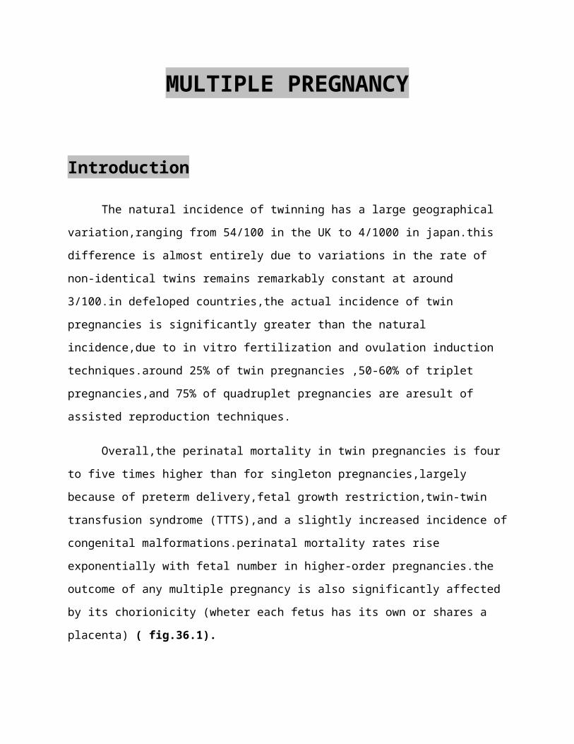
Table 36.1

Chorionicity in monozygous twins
Timming of
embryonic separation
after fertilization
Number of chorions Number of amniotic
sacs
Percentage of
monozygous twins
<4 days Dichorionic Diamniotic 30%
4-8 days Monochorionic Diamniotic 66%
8-4 days Monochorionic monoamniotic 3%
>14 days Monochorionic
(conjoined)
Monoamniotic <1%
Fig 36.1 Trichorionic placenta
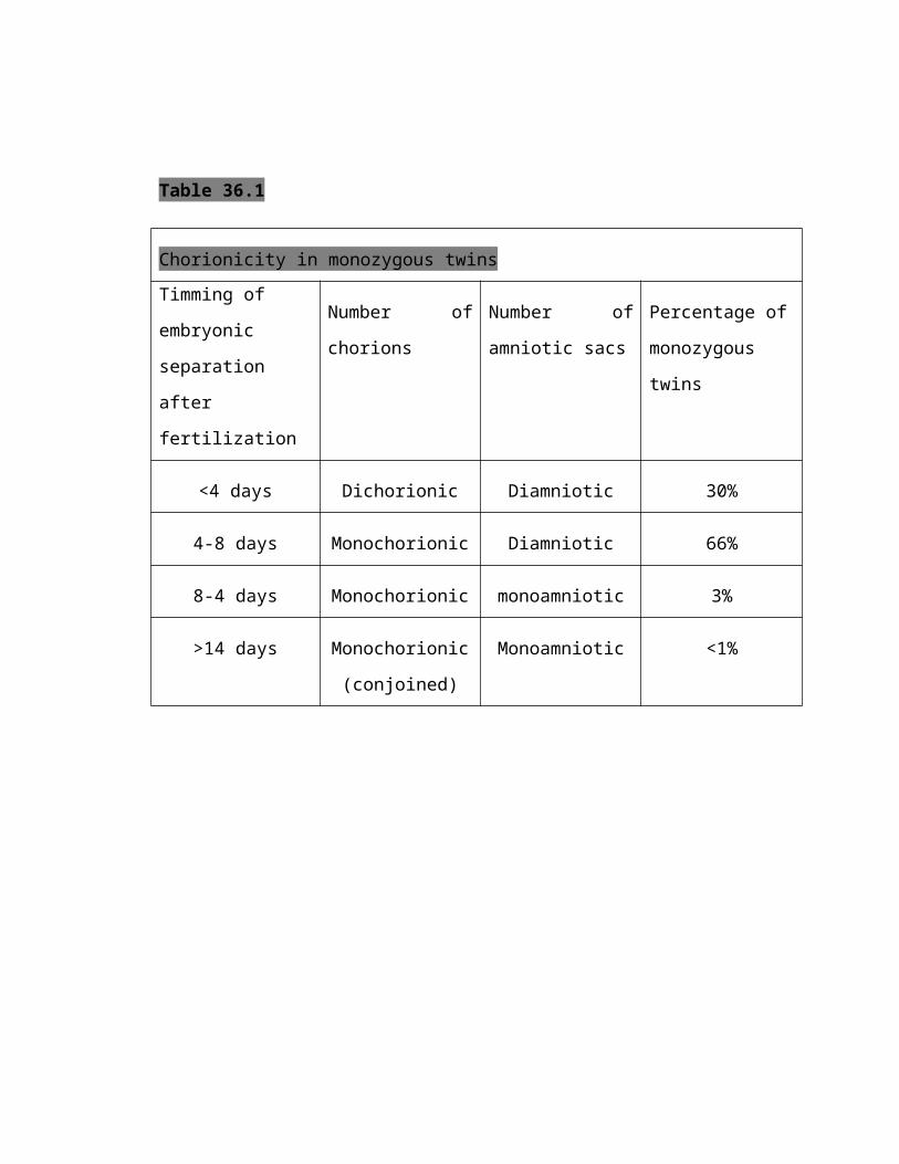
Fig.36.2 diagram of chorionicity. Monozygotic pregnancies may form any of the following combinations
depending on the gestation of embryo division : (A) dichorionic diamniotic; (B) monochorionic diamniotic; (C)
monochorionic monoamniotic; (D) conjoined twins.
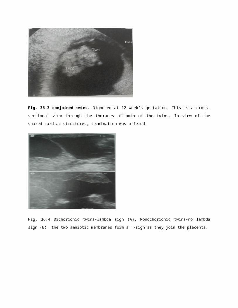
Fig. 36.3 conjoined twins. Dignosed at 12 week’s gestation. This is a cross-sectional view through the thoraces of
both of the twins. In view of the shared cardiac structures, termination was offered.
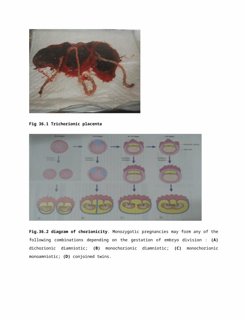
Fig. 36.4 Dichorionic twins-lambda sign (A), Monochorionic twins-no lambda sign (B). the two amniotic
membranes form a T-sign’as they join the placenta.
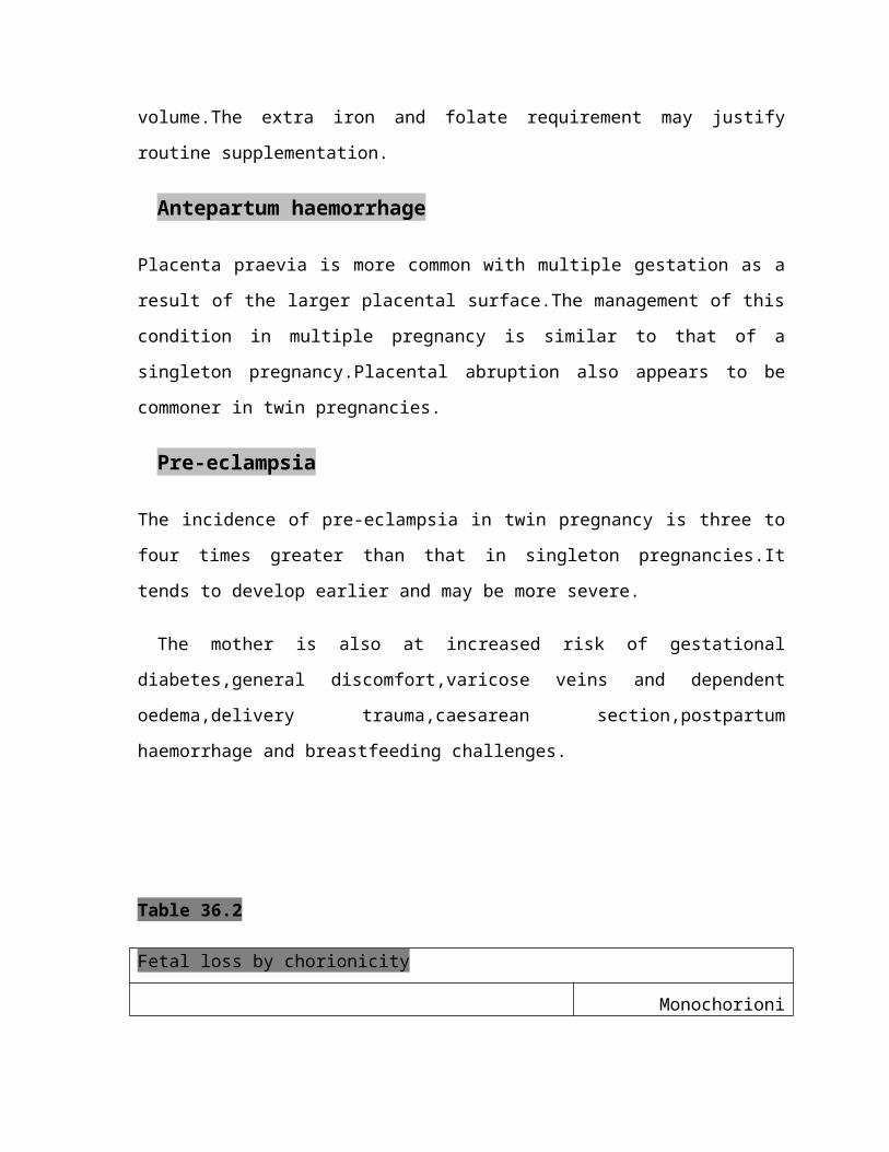
Fig. 36.5 Monochorionic twins demonstrating twin twin transfusion syndrome (oligohydramnion-
polyhtdramnios sequence)
Maternal complications
The incidence of all maternal complications is increased in multiple pregnancy.
Hyperemesis
The increased placental mass,and therefore increased maternal circulating human chorionic
gonadotrophin (hCG) concentration,is associated with an increased and earlier incidence of
hyperemesis.
Anaemia
There is a slight increase in the incidence of anaemia associated with multiple
pregnancy,which is not completely explained by a haemodilutional effect of the increased
plasma volume.The extra iron and folate requirement may justify routine supplementation.
Antepartum haemorrhage
Placenta praevia is more common with multiple gestation as a result of the larger placental
surface.The management of this condition in multiple pregnancy is similar to that of a
singleton pregnancy.Placental abruption also appears to be commoner in twin pregnancies.
Pre-eclampsia
The incidence of pre-eclampsia in twin pregnancy is three to four times greater than that in
singleton pregnancies.It tends to develop earlier and may be more severe.
The mother is also at increased risk of gestational diabetes,general discomfort,varicose
veins and dependent oedema,delivery trauma,caesarean section,postpartum haemorrhage
and breastfeeding challenges.
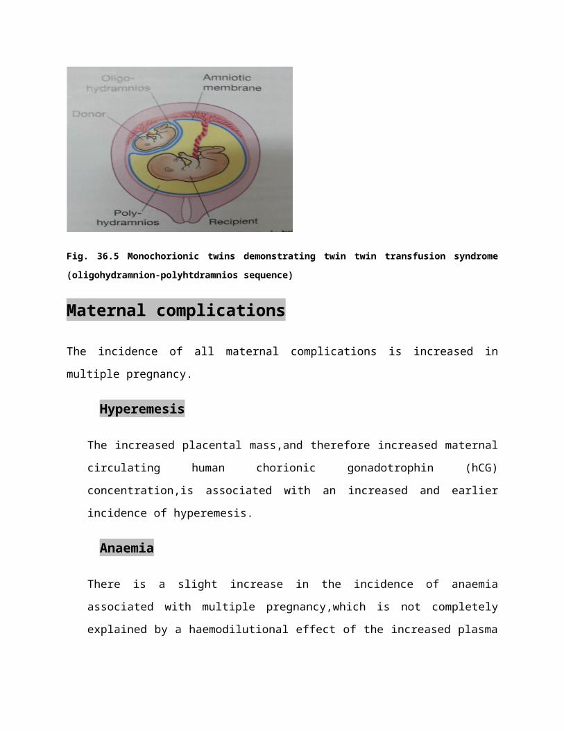
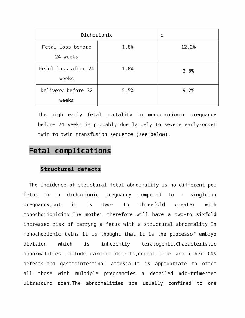
Table 36.2
Fetal loss by chorionicity
Dichorionic Monochorionic
Fetal loss before 24 weeks 1.8% 12.2%
Fetol loss after 24 weeks 1.6% 2.8%
Delivery before 32 weeks 5.5% 9.2%
The high early fetal mortality in monochorionic pregnancy before 24 weeks is probably due
largely to severe early-onset twin to twin transfusion sequence (see below).
Fetal complications
Structural defects
The incidence of structural fetal abnormality is no different per fetus in a dichorionic pregnancy
compered to a singleton pregnancy,but it is two- to threefold greater with monochorionicity.The
mother therefore will have a two-to sixfold increased risk of carryng a fetus with a structural
abnormality.In monochorionic twins it is thought that it is the processof embryo division which is
inherently teratogenic.Characteristic abnormalities include cardiac defects,neural tube and other
CNS defects,and gastrointestinal atresia.It is appropriate to offer all those with multiple pregnancies
a detailed mid-trimester ultrasound scan.The abnormalities are usually confined to one
twin(i.e.nonconcordent);for example,if there is a neural tube defect in one twin,the other twin is
normal in 85-90% of cases.Selective termination with intracardiac KCL is possible in dichorionic
pregnancies only,and is most safely carried out before 16-20 weeks.The procedure,however,carries
a 5% risk of miscarriage of both twins.In monochorionic twins,specialized cord occlusion
techniques may be considered,but carry an increased risk of loss to the other twin due to the
increased invasiveness of this procedure.
Chromosomal abnormalities
These are usually discordant in dizygotic twin and almost always concordant in monozygotic
twins.The maternal agerelated risk for carrying a fetus with down syndrome is there fore
approximately doubled in dichorionic twin pregnancies.Maternal serum screening for trisomy 21
performs poorly in twin pregnancy.Nuchal translucency measurement (without biochemistry) is a
more useful screening test.If invasive diagnostic testing is indicated,amniocentesis of each amniotic
sac is required in dichorionic pregnancies,and care must be taken to document which sample has
come from which sac.
Chorionic villous sampling is not usually appropriate for twin pregnancies as it difficult to be
sure that both placentae have been sampled, particularly if they are lying close to gether.
Premature birth
Twins typically deliver by 37-38 weeks’ gestation. Twins account for 25% of all premature
births despite accounting for only 2% of births per year. Preterm delivery is higher in
monochorionic compared to dichorionic twins (table 36.2). Increased uterine distension, early
myometrial contractility and TTTS may be causative factors in premature labour in multiple
pregnancy. At present there is no known effective treatment to prevent premature labour.
Women should be advised to present early with any symptoms of suspected preterm labour so
that corticosteroids can be administered to accelerate fetal lung maturation.
Intrauterine growth restriction
Twins typically reflect singleton size charts until 28-30 weeks’ gestation and then growth slows.
Approximately 30% of twins are small for gestational age by singleton standards and a
significant in the growth of one twin compared to the other is seen in 12% of pregnancies.
Placental dysfunction underlies intrauterine growth restriction in twin pregnancies as in singleton
pregnancies. Abdominal palpation is not reliable to monitor fetal growth in multiple pregnancy.
Serial ultrasound be performed to measure fetal abdominal circumferences. If diagnosed,
intrauterine growth restriction requires increased surveillance of fetal well-being with umbilical
artery Doppler and cardiotocography (CTG) monitoring, so that delivery can be optimally timed.
Monochorionic twins are at increased risk of intrauterine growth restriction, and require a lower
threshold for delivery owing to the adverse consequences of a single intrauterine death in these
twins.
Twins with one fetal death
First-trimester intrauterine death in a twin has not been shown to have adverse consequences for
the survivor. This probably also holds true for the early second trimester, but loss in the late
second or third trimester commonly precipitates labour such that 90% will have delivered both
twins within 3 weeks of the loss. Prognosis for a surviving dichorionic fetus is then influenced
primarily by its gestation. When a monochorionic twin dies in utero, however, there are
additional risks of death (approximately 20%) or cerebral damage (approximately 25%) in the
co-twin as a result of the shared fetal-placental circulations. As these are probably related to
acute hypotension in the co-twin at the time of the other’s death, early delivery of the surviving
twin is unlikely to improve its outcome and may compound morbidity if performed at a
premature gestation.
Antenatal problems specific to monochorionic twin
pregnancies
Twin-twin transfusion syndrome (TTTS)
This complicates 10-15% monochorionic multiple pregnancies and accounts for around 15% of
perinatal mortality in twins. In this condition there is a net blood flow from one twin to the other
through arterial to venous anastomoses in the shared placenta. The circulation of the recipient
becomes hyperdynamic, with the risk of high-output cardiac failure and polyhydramnios.
Conversely, the donor develops oliguria and oligohydramnios and often suffers growth
restriction (Figs.36.5, 36.6). The ultrasound finding of the oligohydramnios/polyhydramnios
sequence is the key to establishing an antenatal diagnosis.
Without treatment TTTS is associated with a greater than 80% pregnancy loss rate. Two
interventions have proven useful: serial amniodrainage and laser ablation of the causative
placental vascular anastomoses. Evidence has emerged that laser ablation is the most effective
intervention (70% survival vs 50% survival with amnioreduction). Laser therapy is also
associated with a lower rate of significant neurological morbidity in surviving twins compared to
amnioreduction (5% vs 15%).
Fig.36.6 twin-twin transfusion sequence. These monochorionic twins were born at 37 weeks’ gestation. Although
their weight were almost identical, there was significant oligohydramnions around the recipient. (A) Pre-transfusion.
(B) Post-transfusion. (with permission).
Monoamniotic twins
Twins who occupy a single amniotic sac, are at risk of cord entanglement in utero. Frequent
CTG monitoring is required once they reach viability. Delivery is indicated if cord compression
is subsequently diagnosed. Delivery is otherwise electively planned for 32 weeks’ gestation.
Delivey should be performed by caesarean section, as the risk of a cord accident is particularly
high during labour.
Twin reversed arterial perfusion sequence
If the heart of one monochorionic twin stops, it may continue to be partially perfused by the
surviving twin if large fetal arterial-to-arterial anastomoses axist in the shared placenta. The dead
twin undergoes atrophy of its upper body and heart due to the especially poor oxygenation of
these tissues, and becomes what has been described as an’acardiac monster’. The condition is
very rare and there is a high incidence of mortality in the donor twin due to intrauterine cardiac
failure and prematurity. Cord ligation has been used with success in isolated cases.
Management of pregnancy
Initial visit
As many as 10% of twin pregnancies diagnosed in the first trimester will proceed only as
singleton pregnancies and
Subsequent visits
These are ideally performed at a dedicated twins clinic and timed to coincide with the ultrasound
assessments ,the schedule of which will depend on chorionicity.
Monochorionic twins:
Every 2 weeks from 16 to 24 weeks to survey for TTTS.
Detailed structural survey at 18 weeks’ gestation.
Detailed fetal cardiac scan at 20-22 weeks’ gestation.
Every 2 weeks from 24 weeks for fetal growth assessment.
Dichorionic twins:
Detailed structural survey at 18 weeks’ gestation.
Every 2-4 weeks from 24 weeks for fetal growth assessment.
The mother should be monitored for complications such as pre-eclampsia and
anaemia.Discussions abouth the risk and management of premature delivery and fetal growth
problems are useful at 22-24 weeks’ gestation.In uncomplicated pregnancies,discussion around
mode of delivery and management of twin labour is usuful at 32 weeks’ gestation,when fetal
presentations are unlikely to change.Tailored parencraft advice or classes is worthwhile.
Management of twin delivery
Presentations at term are typically:
Cephalic/cephalic (40%)
Cephalic/breech (40%)
Breech/cephalic (10%)
Other,e.g. transverse (10%).
It is common practice to induce labour at 38-40 weeks in those who are suitable for
vaginal delivery,and to carry out a caesarean section at 38 weeks in those who are not.In general
with twins,providing the presentation of the first twin is cephalic,the balance of current evidence
would suggest that vaginal birth is appropriate.Significant growth discordance may be a reason
to consider caesarean section.If the labour is preterm (<34 weeks),many clinicians would also
consider delivery by caesarean section.
The first stage of labour is managed as for singleton pregnancies and care should be taken
to ensure that both twins are being monitored with CTG,rather than one twin twice.This is best is
best achieved by monitoring twin I with a fetal scalp electrode and twin II abdominally.
An experienced obstetrician,an anaesthetist,two paediatricians and two midwives should
be present for delivery,and,if not already required,a syntocinon infusion should be ready in case
uterine activity decreases after delivery of the first twin.After delivery of the first twin,it is often
helpful to have someone ‘stabilize’ the lie of second twin to longitudinal by abdominal palpation
while a vaginal examination is performed to assess the station of the presenting part.A
portable,ultrasound machine is helpful to confirm the lie and presentation of the second twin.The
membranes of twin II should not be broken until the presenting part has descended into the
prlvis.If twin II lies transversely after the delivery of twin I,external chapalic or breech version is
appropriate.If the lies is still transverse (particularly likely if the back is towards the fundus),the
choice is between breech extraction (gentle continuous traction on one or both feet through intact
membranes) and caesarean section.The CTG of twin II should be carefully monitored throughout
and delivery expedited if suspected fetal distress is observed.
A maternal epidural is useful in the management of twin labour owing to the increased
risks of obstetric intervention,particularly assisted delivery of twin il.
Triplets and higher multiples
In these cases,the perinatal mortality is high,mostly because of the high risk of premature
labour,and it may be appropriate to discuss reducing the number of fetuses to twins at 12-14
weeks’ gestation.With quadruplets or higher-order pregnancies,there is likely to be a greater
chance of at least one or two survivors if fetal reduction is carried out,despite the miscarriage
risk associated with the procedure itself.For triplets,the situation is less clear.The emotional and
ethical problems associated with these decisions are considerable.
Triplets and higher-order multiple pregnancies require intensive antenatal care.These
pregnancies are best delivered by caesarean section due to the inability to effectively monitor all
fetuses in labour and the higher risk of fetal malpresentation.
Key points
It is essential to establish chorionicity early to help advise about prenatal diagnosis and
stratify subsequent care.Prenatal screening should only be under taken after careful
discussion of its implications.
Monochorionic pregnancies are at further increased risk of preterm delivery and
intrauterine growth restriction.They also have specific additional risks such as twin-twin
transfusion syndrome and loss of co-twin complications.