JPET #155770 1 Extended methamphetamine self-administration in ...
36
JPET #155770 1 Extended methamphetamine self-administration in rats results in a selective reduction of dopamine transporter levels in the prefrontal cortex and dorsal striatum not accompanied by marked monoaminergic depletion Marek Schwendt, Angelica Rocha, Ronald E. See, Alejandra M. Pacchioni, Jacqueline F. McGinty, and Peter W. Kalivas Department of Neurosciences, Medical University of South Carolina, Charleston, South Carolina JPET Fast Forward. Published on July 31, 2009 as DOI:10.1124/jpet.109.155770 Copyright 2009 by the American Society for Pharmacology and Experimental Therapeutics. This article has not been copyedited and formatted. The final version may differ from this version. JPET Fast Forward. Published on July 31, 2009 as DOI: 10.1124/jpet.109.155770 at ASPET Journals on February 13, 2018 jpet.aspetjournals.org Downloaded from
-
Upload
phungquynh -
Category
Documents
-
view
215 -
download
0
Transcript of JPET #155770 1 Extended methamphetamine self-administration in ...

JPET #155770
1
Extended methamphetamine self-administration in rats results in a selective reduction of
dopamine transporter levels in the prefrontal cortex and dorsal striatum not accompanied
by marked monoaminergic depletion
Marek Schwendt, Angelica Rocha, Ronald E. See, Alejandra M. Pacchioni, Jacqueline F.
McGinty, and Peter W. Kalivas
Department of Neurosciences, Medical University of South Carolina, Charleston, South Carolina
JPET Fast Forward. Published on July 31, 2009 as DOI:10.1124/jpet.109.155770
Copyright 2009 by the American Society for Pharmacology and Experimental Therapeutics.
This article has not been copyedited and formatted. The final version may differ from this version.JPET Fast Forward. Published on July 31, 2009 as DOI: 10.1124/jpet.109.155770
at ASPE
T Journals on February 13, 2018
jpet.aspetjournals.orgD
ownloaded from

JPET #155770
2
Running Title: Methamphetamine alters dopamine transporters
Corresponding author:
Marek Schwendt, Ph.D.
Department of Neurosciences
Medical University of South Carolina
173 Ashley Ave, BSB 403
Charleston, SC 29425
Tel: 843-792-3689
Fax: 843-792-4423
E-mail: [email protected]
Number of text pages: 26
Number of tables: 1
Number of figures: 5
Number of references: 40
Number of words in abstract: 246
Number of words in introduction: 677
Number of words in discussion: 1583
Abbreviations: DAT, dopamine transporter; dSTR, dorsal striatum; GFAP, Glial fibrillary
acidic protein; Iba-1, ionized calcium-binding adaptor molecule 1; Meth, methamphetamine;
NAc, nucleus accumbens; NET, norepinephrine transporter; PBS, phosphate-buffered saline;
PFC, prefrontal cortex; SERT, serotonin transporter; TH, tyrosine hydroxylase.
Section Assignment: Neuropharmacology
This article has not been copyedited and formatted. The final version may differ from this version.JPET Fast Forward. Published on July 31, 2009 as DOI: 10.1124/jpet.109.155770
at ASPE
T Journals on February 13, 2018
jpet.aspetjournals.orgD
ownloaded from
JPET #155770
3
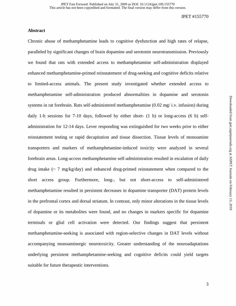
Abstract
Chronic abuse of methamphetamine leads to cognitive dysfunction and high rates of relapse,
paralleled by significant changes of brain dopamine and serotonin neurotransmission. Previously
we found that rats with extended access to methamphetamine self-administration displayed
enhanced methamphetamine-primed reinstatement of drug-seeking and cognitive deficits relative
to limited-access animals. The present study investigated whether extended access to
methamphetamine self-administration produced abnormalities in dopamine and serotonin
systems in rat forebrain. Rats self-administered methamphetamine (0.02 mg/ i.v. infusion) during
daily 1-h sessions for 7-10 days, followed by either short- (1 h) or long-access (6 h) self-
administration for 12-14 days. Lever responding was extinguished for two weeks prior to either
reinstatement testing or rapid decapitation and tissue dissection. Tissue levels of monoamine
transporters and markers of methamphetamine-induced toxicity were analyzed in several
forebrain areas. Long-access methamphetamine self-administration resulted in escalation of daily
drug intake (~ 7 mg/kg/day) and enhanced drug-primed reinstatement when compared to the
short access group. Furthermore, long-, but not short-access to self-administered
methamphetamine resulted in persistent decreases in dopamine transporter (DAT) protein levels
in the prefrontal cortex and dorsal striatum. In contrast, only minor alterations in the tissue levels
of dopamine or its metabolites were found, and no changes in markers specific for dopamine
terminals or glial cell activation were detected. Our findings suggest that persistent
methamphetamine-seeking is associated with region-selective changes in DAT levels without
accompanying monoaminergic neurotoxicity. Greater understanding of the neuroadaptations
underlying persistent methamphetamine-seeking and cognitive deficits could yield targets
suitable for future therapeutic interventions.
This article has not been copyedited and formatted. The final version may differ from this version.JPET Fast Forward. Published on July 31, 2009 as DOI: 10.1124/jpet.109.155770
at ASPE
T Journals on February 13, 2018
jpet.aspetjournals.orgD
ownloaded from
JPET #155770
4
Introduction
Methamphetamine (Meth) abuse in humans can quickly develop into a chronic relapsing
disorder, accompanied by a wide range of neuropsychological deficits. For example, Meth
addicts display impairments in memory functions, cognitive and psychomotor performance, as
well as increased impulsivity and aggressive behavior (reviewed by Nordahl et al., 2003; Scott et
al., 2007). Human brain imaging studies provide evidence that these neuropsychological deficits
are paralleled by significant changes in brain dopaminergic and serotonergic neurotransmitter
systems, as well as altered general metabolic activity in basal ganglia and frontal cortices
(reviewed by Chang et al., 2007). In particular, chronic Meth abuse reduces the density of
dopamine transporters (DAT) in the striatum and (to a lesser extent) in the frontal cortex of
abstinent Meth users (Volkow et al., 2001; Sekine et al., 2003; McCann et al., 2008). Similarly, a
decreased number of serotonin transporters (SERT) was detected across several brain regions in
abstinent Meth addicts (Sekine et al., 2006).
Most frequently, animal models of repeated Meth exposure have utilized short-term
noncontingent, multiple high-dose (‘binge’) regimens producing Meth-induced neurotoxicity,
specifically damaging dopamine and serotonin terminals in the brain. As a consequence,
persistent reductions in striatal dopamine content, DAT density, and activity of the dopamine-
synthesizing enzyme, tyrosine hydroxylase, have been described in brains of rodents and non-
human primates (reviewed by Volz et al., 2007). It has been repeatedly observed that Meth-
induced insult to dopamine terminals was more severe in the dorsal striatum (dSTR) in
comparison to nucleus accumbens (NAc) and prefrontal cortex (PFC), likely reflecting the
different densities of DAT in these regions (Prudencio et al., 2002; Volz et al., 2007). It is well
established that DAT plays a major role in cellular mechanisms of Meth-induced neurotoxicity.
This article has not been copyedited and formatted. The final version may differ from this version.JPET Fast Forward. Published on July 31, 2009 as DOI: 10.1124/jpet.109.155770
at ASPE
T Journals on February 13, 2018
jpet.aspetjournals.orgD
ownloaded from

JPET #155770
5
Thus, mice with a genetic deletion of DAT are significantly less vulnerable to the neurotoxic
effects of Meth (Fumagalli et al., 1998; Numachi et al., 2007). Disruption of dopamine
homeostasis by high Meth concentrations (in the striatum) leads to increased oxidative stress,
activation of apoptotic cascades and inflammatory mediators, as well as the activation of
microglia and astrocytes (Thomas et al., 2004; Quinton and Yamamoto, 2006). Even though the
mechanism of Meth-induced serotonergic toxicity is less understood, it is believed to involve
similar mechanisms to that of dopamine (Numachi et al., 2007). Except for the striatum, the
effects of Meth on serotonin neurons are distributed throughout many other brain structures,
including the hippocampus, septum and frontal cortex (Haughey et al., 2000; Armstrong and
Noguchi, 2004).
Binge models have been useful for the study of Meth-induced neurotoxicity and
recapitulate some changes seen in human Meth addicts (e.g., loss in striatal DAT density).
However, these models have failed to adequately address the motivational and cognitive facets of
Meth addiction in humans. Meth self-administration in rodents constitutes an addiction model
with greater face validity than noncontingent drug administration models. Similar to human
Meth abusers, rats with extended access to intravenous Meth demonstrate escalation of drug
intake (Kitamura et al., 2006) and enhanced drug-seeking and increased cognitive deficits
(Dalley et al., 2007; Rogers et al., 2008) relative to animals trained in shorter Meth access
protocols that do not elicit escalated drug intake. In contrast to drugs such as cocaine, there is
little information available on the neurobiological consequences of chronic Meth self-
administration. Previous studies have shown that limited Meth self-administration exposure
produces only transient changes in mesostriatal dopamine function present after short (1 day),
but not extended (30 days) withdrawal (Stefanski et al., 2002; Shepard et al., 2006).
This article has not been copyedited and formatted. The final version may differ from this version.JPET Fast Forward. Published on July 31, 2009 as DOI: 10.1124/jpet.109.155770
at ASPE
T Journals on February 13, 2018
jpet.aspetjournals.orgD
ownloaded from

JPET #155770
6
Since there has been no previous assessment of the integrity of monoaminergic function
after prolonged Meth self-administration, we utilized short and long-access Meth self-
administration treatment protocols in rats to study possible enduring abnormalities in dopamine
and serotonin systems in striatal and cortical brain regions. Akin to exacerbating cognitive
deficits, we hypothesized that long-access animals may show larger abnormalities in dopamine
and serotonin transmission. Towards this aim, we analyzed whether enhanced drug-seeking
behavior (Meth-induced reinstatement), would correspond with adaptations in monoaminergic
function (tissue levels of dopamine, serotonin, and levels of monoamine transporters), and with
changes in markers of Meth-induced toxicity in the forebrain.
This article has not been copyedited and formatted. The final version may differ from this version.JPET Fast Forward. Published on July 31, 2009 as DOI: 10.1124/jpet.109.155770
at ASPE
T Journals on February 13, 2018
jpet.aspetjournals.orgD
ownloaded from

JPET #155770
7
Methods
Subjects and surgery
Male Long-Evans rats (Charles-River; 275–300 g) were individually housed in a
temperature- and humidity-controlled vivarium on a 12-h reversed light–dark cycle (lights off 6
A.M. to 6 P.M.). Animals were provided water and standard rat chow (Harlan, Indianapolis, IN,
USA) ad libitum throughout the study, except during the first 2-3 days of Meth self-
administration, during which animals were maintained on 15-20 g of standard rat chow per day
to facilitate the acquisition of lever responding (Bongiovanni and See, 2008). Procedures were
conducted in accordance with the Guide for the Care and Use of Laboratory Rats of the National
Research Council and approved by the IACUC of the Medical University of South Carolina.
Rats were anesthetized using a mixture of ketamine hydrochloride and xylazine (66 and 1.33
mg/kg, respectively, i.p.), followed by equithesin (0.5 ml/kg with a solution of 9.72 mg/ml
pentobarbital sodium, 42.5 mg/ml chloral hydrate, and 21.3 mg/ml magnesium sulfate
heptahydrate dissolved in a 44% propylene glycol, 10% ethanol solution, i.p.) and chronic
indwelling catheters were implanted into the right jugular vein using previously described
methods (Bongiovanni and See, 2008). Catheter patency was maintained by flushing with 0.1 ml
of 10 U/ml heparinized saline immediately prior to self-administration sessions with a 0.1 ml
antibiotic solution of cefazolin (10 mg/ml; dissolved in 70 U/ml heparinized saline) and 0.1 ml
100 U/ml heparinized saline regimen following each session. Stylets were inserted into the
catheters when the rats were not connected to infusion pumps. To verify catheter patency, rats
occasionally received a 0.12 ml i.v. infusion of methohexital sodium (10 mg/ml; dissolved in
0.9% physiological saline), a short-acting barbiturate that produces a rapid loss of muscle tone
when administered.
This article has not been copyedited and formatted. The final version may differ from this version.JPET Fast Forward. Published on July 31, 2009 as DOI: 10.1124/jpet.109.155770
at ASPE
T Journals on February 13, 2018
jpet.aspetjournals.orgD
ownloaded from
JPET #155770
8
Methamphetamine self-administration
After a 5-day recovery period from surgery, rats were randomly assigned to Meth or yoked-
saline groups. Testing was conducted in self-administration chambers (30x20x20 cm, Med
Associates, St. Albans, VT, USA) linked to a computerized data collection program (MED PC).
Each chamber was contained within a sound-attenuating cubicle and equipped with two
retractable levers, two stimulus lights, a speaker for tone delivery, and a house light. The house
light always signaled the initiation of a session. Rats self-administered Meth (methamphetamine
hydrochloride; Sigma-Aldrich Co., St. Louis, MO, USA) according to a fixed ratio 1 (FR1)
schedule of reinforcement during daily sessions. Lever presses on the active lever resulted in a
2-s activation of the infusion pump (0.02 mg/50 μl bolus infusion) and a 5-s presentation of a
stimulus complex, consisting of activation of the white stimulus light above the active lever and
the tone generator (78 dB, 4.5 kHz), followed by a 20-s time-out period to prevent overdose.
Responses during the time-out or on the inactive lever were recorded, but resulted in no
programmed consequences. Yoked saline controls received 50-μl infusions of 0.9% sterile saline
whenever the matched self-administering subject received a Meth infusion. Daily 1-h sessions
continued for 7-10 days (“Acquisition”), after which subjects were assigned to stay on short-
access (1-h) or proceed with long-access (6-h) self-administration for 12 (Experiments 1 and 2)
or 12-14 (Experiment 3) days (“Maintenance”). Rats were distributed to access conditions such
that no a priori differences existed in prior drug self-administration for short- vs. long-access
groups.
Extinction, reinstatement testing, and termination of experiments
After the last day of self-administration, rats experienced daily 1-h extinction sessions.
Responses on either the active or inactive lever were recorded, but resulted in no programmed
This article has not been copyedited and formatted. The final version may differ from this version.JPET Fast Forward. Published on July 31, 2009 as DOI: 10.1124/jpet.109.155770
at ASPE
T Journals on February 13, 2018
jpet.aspetjournals.orgD
ownloaded from
JPET #155770
9
consequences. In experiment 1, subjects continued under extinction conditions until they reached
a criterion of a minimum of 10 days and <25 lever presses per session for two consecutive days.
Five reinstatement tests were then conducted with a minimum of 2 days of extinction trials
between each test. During the conditioned-cued reinstatement test, rats were placed into the
chambers for 1-h, and each active lever press resulted in a 5-s tone + light cue presentation in the
absence of drug reinforcement. For the drug-primed reinstatement test, a non-contingent dose of
Meth (0, 0.3, 1.0 or 3.0 mg/kg, i.p.) was administered 20 min prior to the 1-h session, and lever
responses had no programmed consequences. Doses of Meth were administered in random order
separated by a minimum of 2 extinction trials. Conditioned-cued reinstatement was always
conducted first, followed by drug-primed reinstatement, in order not to affect conditioned-cued
reinstatement with prior non-contingent Meth exposure. Multiple reinstatement trials of this type
have been successfully utilized by our laboratory (Kippin et al., 2006) and others (Shaham et al.,
2000).
In experiments 2 and 3, animals underwent the same acquisition, maintenance, and
extinction protocol as in experiment 1, but did not experience the series of reinstatement tests. At
the end of extinction, rats were anesthetized with equithesin, euthanized, the brains removed, and
tissues were collected for the analysis of tissue dopamine, serotonin, and their metabolites for
experiment 2 and dissected for immunoblotting analysis in experiment 3.
Tissue levels of dopamine, serotonin and metabolites measured by HPLC
Animals were decapitated after achieving extinction criterion (see above), the brain rapidly
removed and the NAc core, NAc shell, and dSTR were hand dissected on ice. Similarly, the
medial PFC was dissected and then bisected into approximately equal dorsal and ventral halves.
The dissected tissue was frozen on dry ice for later monoamine measurements using high-
This article has not been copyedited and formatted. The final version may differ from this version.JPET Fast Forward. Published on July 31, 2009 as DOI: 10.1124/jpet.109.155770
at ASPE
T Journals on February 13, 2018
jpet.aspetjournals.orgD
ownloaded from

JPET #155770
10
pressure liquid chromatography and electrochemical detection (HPLC-EC). In order to extract
monoamines, tissue samples were placed in 300 μl of mobile phase containing isoproterenol as
an internal standard (0.05 μM for PFC and 1 μM for NAc shell and core, dSTR), sonicated and
centrifuged (2 min at 13,000 rpm). The protein content in the resulting pellet was measured using
the Bradford assay (Pierce, Rockford, IL, USA). Monoamines were separated on a reversed-
phase HR-80 column (3 μm x 80 x 3.2 mm; ESA), and dopamine, 3,4-dihydroxyphenylacetic
acid (DOPAC), homovanillic acid (HVA), and serotonin, were quantified using an ESA
(Chelmsford, MA, USA) model 540 autosampler connected to an HPLC system with
electrochemical detection (mobile phase: 0.1 M trichloroacetic acid, 0.01 M sodium acetate, 0.1
mM EDTA and 16% MeOH; pH=4.1). The samples were reduced-oxidized using coulometric
detection (Coulochem II; ESA, Chelmsford, MA, USA). Three electrodes were used: a guard
cell (+ 400 mV), a reduction analytical electrode (-150 mV), and an oxidation analytical
electrode (+325 mV). A chart recorder recorded peaks and peak heights were measured. These
values were normalized by comparison with an external standard curve for each analyte
quantified. The data were expressed as picomoles per milligram of protein.
Immunoblotting
Tissues of interest were hand-dissected from 2 mm thick coronal slabs on the day of the
experiment, quickly frozen on dry ice and stored at −80 °C until processed. Samples were then
solubilized in 1% SDS/PBS buffer containing protease and phosphatase inhibitors: “Complete
Mini” protease inhibitor (Roche Diagnostics Corporation, Indianapolis, IN, USA), “Halt”
phosphatase inhibitor (Pierce, Rockford, IL, USA). Protein concentration in the samples was
measured by BCA assay (Pierce, Rockford, IL, USA) and diluted with loading buffer containing
This article has not been copyedited and formatted. The final version may differ from this version.JPET Fast Forward. Published on July 31, 2009 as DOI: 10.1124/jpet.109.155770
at ASPE
T Journals on February 13, 2018
jpet.aspetjournals.orgD
ownloaded from

JPET #155770
11
Inclusion Body Solubilization Buffer (G-Biosciences, Maryland Heights, MO, USA) and a
reducing agent. Samples were incubated at 45°C to prevent aggregation of hydrophobic
transporter proteins. Subsequently, equal amounts of total protein were resolved using SDS-
PAGE and transferred to a PVDF membrane (BioRad, Hercules, CA, USA). The membrane was
blocked with 5% milk/TBST and probed with antibodies against the following proteins: DAT,
GFAP, NET, SERT (Santa Cruz Biotechnology, Santa Cruz, CA, USA), TH (Pel-Freeze,
Trenton, NJ, USA) and Iba-1 (Wako Chemicals, Osaka, Japan). After the incubation with HRP-
conjugated secondary antibodies at 1:5000 (Jackson Immuno Research, West Grove, PA, USA),
immunoreactive bands on the membranes were detected by ECL+ chemiluminescence reagents
on Hyperfilm ECL (Amersham Biosciences, Piscataway, NJ, USA). Integrated density of the
bands was measured with Gel-Pro 3.1 software (Media Cybernetics, Silver Spring, MD, USA).
Equal loading and transfer of proteins were confirmed by immunolabeling of the same
membranes with anti-calnexin antibody (Stressgene, Victoria, BC, Canada).
Statistical Analysis
The evaluation of escalation was made using a paired Student’s t-test to compare the
average mg/kg of Meth infused over the first three days after switching to long-access Meth with
the average of the last three days of self-administration training. The reinstatement of drug-
seeking data were evaluated using a two-way ANOVA with repeated measures over trial to
evaluate the effect of cue- and Meth-induced reinstatement on lever pressing between short- and
long-access subjects, followed by a Dunnett’s test for post hoc comparisons to extinction levels
of lever pressing. The tissue levels of monoamines and the ratio of DOPAC to dopamine were
statistically compared using a one-way ANOVA to compare between yoked-saline controls and
short- and long-access animals, followed by a Dunnett’s post hoc comparison to control.
This article has not been copyedited and formatted. The final version may differ from this version.JPET Fast Forward. Published on July 31, 2009 as DOI: 10.1124/jpet.109.155770
at ASPE
T Journals on February 13, 2018
jpet.aspetjournals.orgD
ownloaded from
JPET #155770
12
Immunoblotting data, represented by band density values, were normalized for the density of
calnexin immunoreactivity within the same sample and analyzed by a one-way ANOVA
followed by a Dunnett’s post hoc test to determine differences between treatment groups and the
control (yoked saline). Protein data were then expressed as the percentage of the values from
saline-treated rats within the same time point or treatment. Sigma Stat (SPSS, Chicago, IL, USA)
software was used for statistical analysis.
This article has not been copyedited and formatted. The final version may differ from this version.JPET Fast Forward. Published on July 31, 2009 as DOI: 10.1124/jpet.109.155770
at ASPE
T Journals on February 13, 2018
jpet.aspetjournals.orgD
ownloaded from

JPET #155770
13
Results
Experiment 1: Long-access to Meth augments drug-, but not cue-induced reinstatement of Meth-
seeking.
Lever responding and Meth intake are shown in Figure 1. In general, there was a large
distinction between active and inactive lever responding throughout the self-administration
phase, demonstrating that animals distinguished between the active lever delivering Meth and the
inactive lever with no consequences (Figure 1 A and B). The transient increase in inactive lever
responding in long access, as compared to short access animals likely reflects initial extended
access to both levers, rather than a loss of lever discrimination. As seen in Figure 1C and 1D,
Meth intake (mg/kg/day) in the long access animals significantly increased (escalated) when
comparing the average intake over the first 3 days of long access drug intake with the average of
the last 3 days (two-tailed paired Student’s t-test, t(30) = 3.65, p = 0.001). In contrast, no increase
in drug intake was apparent over the time course of daily short access to Meth. Removal of Meth
reinforcement produced rapid extinction of lever pressing in all animals.
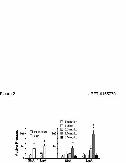
Figure 2 (left panel) shows that both access conditions elicited robust conditioned-cued
reinstatement as compared to extinction baseline, but reinstatement did not significantly differ
between animals with different access histories (two-way ANOVA, repeated measures over trial
revealed no effect of long- vs. short-access, significant effect of cue vs. extinction, F(1,44) =
15.97, p< 0.001, but no interaction between access group and reinstatement). In addition, both
access groups also showed significant Meth-primed reinstatement, with a biphasic dose response
curve (Figure 2; right panel). However, in contrast to conditioned-cued reinstatement, the long
access animals showed heightened drug-primed reinstatement. Thus, in long-access subjects, a
lower dose of Meth (0.3 mg/kg) significantly increased Meth-seeking, and the maximum
This article has not been copyedited and formatted. The final version may differ from this version.JPET Fast Forward. Published on July 31, 2009 as DOI: 10.1124/jpet.109.155770
at ASPE
T Journals on February 13, 2018
jpet.aspetjournals.orgD
ownloaded from

JPET #155770
14
response to the 1.0 mg/kg dose was significantly greater in long access animals (two-way
ANOVA with repeated measures over Meth reinstatement trial revealed a significant effect of
access group F(1,3) = 7.54, p = 0.007, reinstatement trial F(3,3) = 17.01, p< 0.001, and an
interaction between group and trial F(3,88) = 4.32, p = 0.007). Neither group showed significant
increases at the highest dose (3.0 mg/kg), which likely produced competing behaviors that could
have hindered drug-seeking, such as stereotypy. Even though stereotypy was not measured in the
current study, it has been shown that 2-4 mg/kg doses of Meth consistently induced stereotypic
behaviors in rats with a history of Meth treatment (Szumlinski et al 2000).
Experiment 2: Meth self-administration did not alter dopamine levels.
In the next study, changes in forebrain dopamine and serotonin levels were investigated in a
separate group of animals that underwent Meth self-administration and extinction protocol
identical to the previous experiment. For the two week Meth maintenance period in experiment
2, animals in the long-access group had a total Meth intake of 6.62 ± 1.17 mg/kg (n = 9; mean ±
sem over the last three days of self-administration), while short access animals had a total Meth
intake of 1.45 ± 0.23 mg/kg (n = 8). Figure 3 and table 1 reveal that in none of the brain regions
examined, including the dorsal and ventral PFC, shell and core of the NAc, or the dSTR, did
either short- or long-access to Meth self-administration significantly alter the tissue levels of
dopamine. As seen in Figure 3, the only significant effects were found in short-access animals,
whereby short-, but not long-access, subjects showed a reduction in DOPAC (F(2,24) = 3.86, p =
0.037) in the vPFC and an increase in short-access Meth animals in the DOPAC:dopamine ratio
in the NAc core (F(2,24) = 3.31, p = 0.054). No other effects on dopamine metabolites or
serotonin levels were detected (see Table 1).
This article has not been copyedited and formatted. The final version may differ from this version.JPET Fast Forward. Published on July 31, 2009 as DOI: 10.1124/jpet.109.155770
at ASPE
T Journals on February 13, 2018
jpet.aspetjournals.orgD
ownloaded from
JPET #155770
15
Experiment 3: Extended Meth self-administration reduced the level of DAT in the PFC and
dSTR.
In addition to tissue levels of dopamine and serotonin, the effects of Meth self-
administration on the levels of monoamine transporters were investigated in a separate group of
animals. For the two week Meth maintenance period in experiment 3, animals in the long-access
group had a total daily Meth intake of 6.69 ± 0.77 mg/kg (n = 6; mean ± sem over the last three
days of self-administration), while short access animals had a total daily Meth intake of 1.20 ±
0.12 mg/kg (n = 5). As depicted in Figure 4, long- but not short-access Meth administration
significantly reduced DAT protein levels in both the PFC (F(2,15) = 5.53, p = 0.043) and dSTR
(F(2,15) = 5.25, p = 0.021). There was no change of DAT protein detected in the NAc in any
group of rats. In contrast, no changes of total protein levels of norepinephrine or serotonin
transporters were detected in surveyed brain areas (Figure 5A and B). Further investigation
revealed that Meth did not produce persistent access-dependent changes in a number of protein
markers typically associated with Meth-induced neurotoxicity (Figure 5A and B). Thus, there
was no change in tyrosine hydroxylase (TH, dopamine synthetic enzyme), glial fibrillary acidic
protein (GFAP, marker of astrocyte activation and proliferation) and ionized calcium-binding
adaptor molecule 1 (Iba-1, marker of activated microglia).
This article has not been copyedited and formatted. The final version may differ from this version.JPET Fast Forward. Published on July 31, 2009 as DOI: 10.1124/jpet.109.155770
at ASPE
T Journals on February 13, 2018
jpet.aspetjournals.orgD
ownloaded from

JPET #155770
16
Discussion
The key finding of the current study is that extended, but not limited, daily self-administered
Meth results in persistent decreases in the levels of DAT protein in the PFC and the dSTR.
Importantly, these decreases are not likely to be a direct consequence of lasting Meth-induced
neurotoxicity, as we found only negligible alterations in dopamine metabolism in the PFC and
NAc in short access animals, and no changes in markers specific for dopamine terminals or glial
cell activation in either group. The downregulation of DAT activity may relate to lasting
functional consequences of prolonged Meth intake, including motivated drug-seeking behavior,
as seen by heightened Meth-primed reinstatement in animals with a history of long access Meth
intake.
Animal models of limited-to-extended daily access to Meth self-administration (as
applied in the current study) mimic the course of Meth administration in humans much more
closely than studies with experimenter-administered Meth. In support of this, an escalation of
Meth intake was observed in rats switched to long-access (6-h/day), but not in rats maintained on
limited access (1-h/day), which is in agreement with previous studies (Kitamura et al., 2006;
Rogers et al., 2008). As a result, we observed robust Meth self-administration (70-110 mg/kg
over the two weeks) in the long-access rats. This pattern arguably resembles a transition from
controlled (limited) to uncontrolled (binge and run) Meth use typical for human addicts (Cho and
Melega, 2002). In comparison, typical “binge” experiments employ acute i.p. injections in a
single day (ranging from one to four single doses of 1 to 10 mg/kg) that produce cumulative
totals ranging from 4 to 40 mg/kg (Quinton and Yamamoto, 2006; Volz et al., 2007).
Further, self-administration models of Meth addiction (in contrast to noncontingent binge
models) can also readily address questions of motivated drug-seeking under relapse conditions.
This article has not been copyedited and formatted. The final version may differ from this version.JPET Fast Forward. Published on July 31, 2009 as DOI: 10.1124/jpet.109.155770
at ASPE
T Journals on February 13, 2018
jpet.aspetjournals.orgD
ownloaded from

JPET #155770
17
Significantly, chronic Meth use in humans is typically associated with high rates of relapse. As
previously reported (Rogers et al., 2008), long-access Meth animals displayed a significant
enhancement of Meth-primed reinstatement, a finding similar to that seen after chronic cocaine
self-administration (Mantsch et al., 2004; Kippin et al., 2006). In the current study, reinstatement
tests with a range of priming doses (0.3 - 3.0 mg/kg) were performed following 10 days of
extinction in order to assess potential long-term changes in sensitivity to drug effects. Long-
access animals readily reinstated to a low priming dose of Meth (0.3 mg/kg) that failed to elicit
reinstatement in animals with a history of short access. While more doses would need to be
tested in order to make a definitive statement about any potential shifts in the dose-response
curve, the pattern of behavior in the present study suggests a leftward shift in long-access
animals that is indicative of increased sensitivity to a drug-prime. As opposed to Meth-primed
reinstatement where differences between short- and long-access animals were evident, there was
no difference in cue-reactivity between groups. The lack of difference in cue-reactivity has been
previously reported (Rogers et al., 2008) and may reflect the extensive overtraining with the cues
that all animals experienced, regardless of daily access duration. Additional forms of conditioned
cued reinstatement (e.g., contextual cues) or changes in cue parameters are warranted in future
studies in order to reveal possible access dependent differences in relapse.
Both animal and human data have previously shown that chronic Meth results in long-term
changes in the function of dopaminergic and serotonergic systems in the brain (as reviewed by
Chang et al., 2007; Volz et al., 2007). However, multiple interpretations exist regarding the
nature of these changes (compensatory adaptations vs. neuronal damage), as well as the degree to
which they exhibit recovery over the prolonged drug-free period (Volkow et al., 2001; Wang et
al., 2004; McCann et al., 2008). In the current study, dopamine and serotonin levels were
This article has not been copyedited and formatted. The final version may differ from this version.JPET Fast Forward. Published on July 31, 2009 as DOI: 10.1124/jpet.109.155770
at ASPE
T Journals on February 13, 2018
jpet.aspetjournals.orgD
ownloaded from

JPET #155770
18
assessed after prolonged withdrawal from Meth self-administration. Although animals with a
history of extended Meth access display altered motivational and cognitive performance well
after cessation of Meth (current results, Rogers et al., 2008), no corresponding changes in
dopamine or serotonin levels were detected in the forebrain areas examined. Interestingly, across
measures, the only changes suggestive of altered monoamine activity were changes in dopamine
metabolism in the ventral PFC and NAc core of rats with short (but not long) access to Meth.
Changes in DOPAC or the DOPAC:dopamine ratio have been used as estimates of dopamine
transmission, with lower relative production of DOPAC indicating reduced transmission. The
significance of the apparent reduction in dopamine transmission in the short access only group is
not clear. Since activity in the ventral PFC has been strongly linked to extinction learning and
relapse (Peters et al., 2008), differences in drug-seeking for groups with varied drug access
histories may be related to cortical dopamine transmission.
While long access Meth failed to produce lasting changes in monoamine levels, extended
Meth resulted in a significant downregulation of DAT levels in the PFC and dSTR as measured
two weeks after the end of self-administration. This finding is congruent with clinical reports that
have found long-term declines in the levels of DAT in the PFC and dSTR of abstinent Meth
users (Sekine et al., 2001; Sekine et al., 2003; McCann et al., 2008). Previous studies in animals
after Meth self-administration have found only transient changes in DAT and other dopaminergic
terminal markers (Stefanski et al., 2002; Shepard et al., 2006). However, these studies only
utilized short access Meth experience (Stefanski et al., 2002) and shorter total duration of self-
administration (Shepard et al., 2006). The current data suggest that more extensive Meth
exposure, akin to that seen in chronic human Meth addicts, is necessary for the persistent DAT
decrease to occur.
This article has not been copyedited and formatted. The final version may differ from this version.JPET Fast Forward. Published on July 31, 2009 as DOI: 10.1124/jpet.109.155770
at ASPE
T Journals on February 13, 2018
jpet.aspetjournals.orgD
ownloaded from

JPET #155770
19
High, neurotoxic doses of Meth (up to 40 mg/kg/daily) delivered in noncontingent binge-
type regimens also result in prolonged decreases in striatal DAT levels (Volz et al., 2007).
Decreased DAT due to Meth-induced neurotoxicity is typically accompanied by changes in other
markers of dopamine terminal damage, such as depletion of striatal dopamine, decrease in
tyrosine hydroxylase, activation of glia, and oxidative stress (Thomas et al., 2004; Quinton and
Yamamoto, 2006). Given that the maximum daily drug intake of rats with extended access to i.v.
Meth was within the range of 6-7 mg/kg, DAT decrease was not likely due to Meth-induced
dopamine terminal toxicity, particularly as no changes in a number of dopamine terminal
markers were detected following chronic Meth self-administration. In some previous studies,
repeated administration of below-toxic doses of Meth led to decreased DAT levels in the dSTR,
but in the absence of significant depletion of striatal dopamine levels (O'Neil et al., 2006;
Bjorklund et al., 2008). Therefore, we suggest that decreased DAT levels in the PFC and dSTR
represent a neuroadaptation resulting from chronic Meth effects directly on DAT regulation
rather than from Meth-induced dopaminergic toxicity. In this regard, it is intriguing that
postmortem studies in human Meth addicts have found reduced levels of dopamine terminal
markers in the striatum (likely associated with an acute dopamine depletion caused by Meth
overdose), but no signs of dopamine terminal degeneration (Wilson et al., 1996; Moszczynska et
al., 2004). However, the possibility that the current Meth paradigm did produce some
monoaminergic toxicity, which recovered (except for DAT) by the time of tissue analysis, cannot
be fully excluded. Therefore, our future studies will evaluate possible signs of Meth-induced
cellular toxicity after shorter withdrawal periods (e.g., 24 h).
Significantly, DAT changes occurred in the PFC and dSTR, as these brain structures are
part of the circuitry activated during the reinstatement of extinguished cocaine-seeking
This article has not been copyedited and formatted. The final version may differ from this version.JPET Fast Forward. Published on July 31, 2009 as DOI: 10.1124/jpet.109.155770
at ASPE
T Journals on February 13, 2018
jpet.aspetjournals.orgD
ownloaded from

JPET #155770
20
(McFarland and Kalivas, 2001; Fuchs et al., 2006). Therefore, vulnerability to reinstate
previously extinguished drug-seeking may be associated with decreased DAT levels in the PFC
and dSTR as observed in the current study. Decreases in DAT have also been observed in the
dSTR of depressed patients displaying a lack of ‘normal’ reward-motivated behavior or
anhedonia (Sarchiapone et al., 2006). Interestingly, anhedonia is also one of the symptoms
typically associated with withdrawal from chronic Meth use (McGregor et al. 2005). Further, it is
important to note that long-, but not short-access animals, showed a decrease in DAT levels in
the PFC and dSTR and only long-access animals exhibited drug-induced behavioral
dysregulations, such as escalation and greater reinstatement of Meth-seeking. However, whether
and how decreased DAT levels affect extracellular dopamine levels and reinstatement of Meth-
seeking is not clear. Therefore, future studies will investigate additional changes in cortical and
striatal dopamine function (dopamine levels, function of DAT, and vesicular monoamine
transporters) in the context of the cognitive and motivational deficits that result from extended
Meth self-administration. Alternatively, other factors need to be considered when characterizing
neuroadaptations underlying post-Meth behavioral deficits. First, extinction learning itself could
play a role in neuroadaptations involved in drug-seeking (Sutton et. al. 2003). Thus, comparison
of animals with a history of extinction vs. abstinence following Meth self-administration will be
critical. Second, other neurotransmitters not investigated in the current study are likely involved
in Meth-induced alterations. In particular, glutamate is involved in Meth-induced toxicity
(Quinton and Yamamoto, 2006), Meth reward and reinforcement (Kim and Jang, 1997; Gass et
al., 2009), and reinstatement of Meth-seeking (Gass et al., 2009).
Taken together, the present findings suggest that extended Meth self-administration
followed by extinction leads to increased Meth-primed reinstatement of drug-seeking and
This article has not been copyedited and formatted. The final version may differ from this version.JPET Fast Forward. Published on July 31, 2009 as DOI: 10.1124/jpet.109.155770
at ASPE
T Journals on February 13, 2018
jpet.aspetjournals.orgD
ownloaded from
JPET #155770
21
decreased DAT in the PFC and dSTR in the absence of persisting changes in dopamine,
serotonin, and dopamine metabolism in cortical and striatal subregions. By gaining a greater
understanding of these critical substrates in a relevant animal model of Meth addiction, potential
targets for therapeutic intervention may be identified and tested.
Acknowledgements: The authors thank Jason Rogers for technical assistance.
This article has not been copyedited and formatted. The final version may differ from this version.JPET Fast Forward. Published on July 31, 2009 as DOI: 10.1124/jpet.109.155770
at ASPE
T Journals on February 13, 2018
jpet.aspetjournals.orgD
ownloaded from

JPET #155770
22
References
Armstrong BD and Noguchi KK (2004) The neurotoxic effects of 3,4-
methylenedioxymethamphetamine (MDMA) and methamphetamine on serotonin,
dopamine, and GABA-ergic terminals: an in-vitro autoradiographic study in rats.
Neurotoxicology 25:905-914.
Bjorklund NL, Sorg BA and Schenk JO (2008) Neuronal dopamine transporter activity, density
and methamphetamine inhibition are differentially altered in the nucleus accumbens and
striatum with no changes in glycosylation in rats behaviorally sensitized to
methamphetamine. Synapse 62:736-745.
Bongiovanni M and See RE (2008) A comparison of the effects of different operant training
experiences and dietary restriction on the reinstatement of cocaine-seeking in rats.
Pharmacol Biochem Behav 89:227-233.
Chang L, Alicata D, Ernst T and Volkow N (2007) Structural and metabolic brain changes in the
striatum associated with methamphetamine abuse. Addiction 102 Suppl 1:16-32.
Cho AK and Melega WP (2002) Patterns of methamphetamine abuse and their consequences. J
Addict Dis 21:21-34.
Dalley JW, Laane K, Theobald DE, Pena Y, Bruce CC, Huszar AC, Wojcieszek M, Everitt BJ
and Robbins TW (2007) Enduring deficits in sustained visual attention during withdrawal
of intravenous methylenedioxymethamphetamine self-administration in rats: results from
a comparative study with d-amphetamine and methamphetamine.
Neuropsychopharmacology 32:1195-1206.
This article has not been copyedited and formatted. The final version may differ from this version.JPET Fast Forward. Published on July 31, 2009 as DOI: 10.1124/jpet.109.155770
at ASPE
T Journals on February 13, 2018
jpet.aspetjournals.orgD
ownloaded from

JPET #155770
23
Fuchs RA, Branham RK and See RE (2006) Different neural substrates mediate cocaine seeking
after abstinence versus extinction training: a critical role for the dorsolateral caudate-
putamen. J Neurosci 26:3584-3588.
Fumagalli F, Gainetdinov RR, Valenzano KJ and Caron MG (1998) Role of dopamine
transporter in methamphetamine-induced neurotoxicity: evidence from mice lacking the
transporter. J Neurosci 18:4861-4869.
Gass JT, Osborne MP, Watson NL, Brown JL and Olive MF (2009) mGluR5 antagonism
attenuates methamphetamine reinforcement and prevents reinstatement of
methamphetamine-seeking behavior in rats. Neuropsychopharmacology 34:820-833.
Haughey HM, Fleckenstein AE, Metzger RR and Hanson GR (2000) The effects of
methamphetamine on serotonin transporter activity: role of dopamine and hyperthermia.
J Neurochem 75:1608-1617.
Kim HS and Jang CG (1997) MK-801 inhibits methamphetamine-induced conditioned place
preference and behavioral sensitization to apomorphine in mice. Brain Res Bull 44:221-
227.
Kippin TE, Fuchs RA and See RE (2006) Contributions of prolonged contingent and
noncontingent cocaine exposure to enhanced reinstatement of cocaine seeking in rats.
Psychopharmacology (Berl) 187:60-67.
Kitamura O, Wee S, Specio SE, Koob GF and Pulvirenti L (2006) Escalation of
methamphetamine self-administration in rats: a dose-effect function.
Psychopharmacology (Berl) 186:48-53.
This article has not been copyedited and formatted. The final version may differ from this version.JPET Fast Forward. Published on July 31, 2009 as DOI: 10.1124/jpet.109.155770
at ASPE
T Journals on February 13, 2018
jpet.aspetjournals.orgD
ownloaded from

JPET #155770
24
Mantsch JR, Yuferov V, Mathieu-Kia AM, Ho A and Kreek MJ (2004) Effects of extended
access to high versus low cocaine doses on self-administration, cocaine-induced
reinstatement and brain mRNA levels in rats. Psychopharmacology (Berl) 175:26-36.
McCann UD, Kuwabara H, Kumar A, Palermo M, Abbey R, Brasic J, Ye W, Alexander M,
Dannals RF, Wong DF and Ricaurte GA (2008) Persistent cognitive and dopamine
transporter deficits in abstinent methamphetamine users. Synapse 62:91-100.
McFarland K and Kalivas PW (2001) The circuitry mediating cocaine-induced reinstatement of
drug-seeking behavior. J Neurosci 21:8655-8663.
McGregor C, Srisurapanont M, Jittiwutikarn J, Laobhripatr S, Wongtan T, White JM (2005) The
nature, time course and severity of methamphetamine withdrawal. Addiction 100:1320-
1329.
Moszczynska A, Fitzmaurice P, Ang L, Kalasinsky KS, Schmunk GA, Peretti FJ, Aiken SS,
Wickham DJ and Kish SJ (2004) Why is parkinsonism not a feature of human
methamphetamine users? Brain 127:363-370.
Nordahl TE, Salo R and Leamon M (2003) Neuropsychological effects of chronic
methamphetamine use on neurotransmitters and cognition: a review. J Neuropsychiatry
Clin Neurosci 15:317-325.
Numachi Y, Ohara A, Yamashita M, Fukushima S, Kobayashi H, Hata H, Watanabe H, Hall FS,
Lesch KP, Murphy DL, Uhl GR and Sora I (2007) Methamphetamine-induced
hyperthermia and lethal toxicity: role of the dopamine and serotonin transporters. Eur J
Pharmacol 572:120-128.
This article has not been copyedited and formatted. The final version may differ from this version.JPET Fast Forward. Published on July 31, 2009 as DOI: 10.1124/jpet.109.155770
at ASPE
T Journals on February 13, 2018
jpet.aspetjournals.orgD
ownloaded from

JPET #155770
25
O'Neil ML, Kuczenski R, Segal DS, Cho AK, Lacan G and Melega WP (2006) Escalating dose
pretreatment induces pharmacodynamic and not pharmacokinetic tolerance to a
subsequent high-dose methamphetamine binge. Synapse 60:465-473.
Peters J, LaLumiere RT and Kalivas PW (2008) Infralimbic prefrontal cortex is responsible for
inhibiting cocaine seeking in extinguished rats. J Neurosci 28:6046-6053.
Prudencio C, Abrantes B, Lopes I and Tavares MA (2002) Structural and functional cellular
alterations underlying the toxicity of methamphetamine in rat retina and prefrontal cortex.
Ann N Y Acad Sci 965:522-528.
Quinton MS and Yamamoto BK (2006) Causes and consequences of methamphetamine and
MDMA toxicity. Aaps J 8:E337-347.
Rogers JL, De Santis S and See RE (2008) Extended methamphetamine self-administration
enhances reinstatement of drug seeking and impairs novel object recognition in rats.
Psychopharmacology (Berl) 199:615-624.
Sarchiapone M, Carli V, Camardese G, Cuomo C, Di Giuda D, Calcagni ML, Focacci C, De
Risio S (2006) Dopamine transporter binding in depressed patients with anhedonia.
Psychiatry Res 147:243-248.
Scott JC, Woods SP, Matt GE, Meyer RA, Heaton RK, Atkinson JH and Grant I (2007)
Neurocognitive effects of methamphetamine: a critical review and meta-analysis.
Neuropsychol Rev 17:275-297.
Sekine Y, Iyo M, Ouchi Y, Matsunaga T, Tsukada H, Okada H, Yoshikawa E, Futatsubashi M,
Takei N and Mori N (2001) Methamphetamine-related psychiatric symptoms and reduced
brain dopamine transporters studied with PET. Am J Psychiatry 158:1206-1214.
This article has not been copyedited and formatted. The final version may differ from this version.JPET Fast Forward. Published on July 31, 2009 as DOI: 10.1124/jpet.109.155770
at ASPE
T Journals on February 13, 2018
jpet.aspetjournals.orgD
ownloaded from
JPET #155770
26
Sekine Y, Minabe Y, Ouchi Y, Takei N, Iyo M, Nakamura K, Suzuki K, Tsukada H, Okada H,
Yoshikawa E, Futatsubashi M and Mori N (2003) Association of dopamine transporter
loss in the orbitofrontal and dorsolateral prefrontal cortices with methamphetamine-
related psychiatric symptoms. Am J Psychiatry 160:1699-1701.
Sekine Y, Ouchi Y, Takei N, Yoshikawa E, Nakamura K, Futatsubashi M, Okada H, Minabe Y,
Suzuki K, Iwata Y, Tsuchiya KJ, Tsukada H, Iyo M and Mori N (2006) Brain serotonin
transporter density and aggression in abstinent methamphetamine abusers. Arch Gen
Psychiatry 63:90-100.
Shaham Y, Erb S and Stewart J (2000) Stress-induced relapse to heroin and cocaine seeking in
rats: a review. Brain Res Brain Res Rev 33:13-33.
Shepard JD, Chuang DT, Shaham Y and Morales M (2006) Effect of methamphetamine self-
administration on tyrosine hydroxylase and dopamine transporter levels in mesolimbic
and nigrostriatal dopamine pathways of the rat. Psychopharmacology (Berl) 185:505-
513.
Stefanski R, Lee SH, Yasar S, Cadet JL and Goldberg SR (2002) Lack of persistent changes in
the dopaminergic system of rats withdrawn from methamphetamine self-administration.
Eur J Pharmacol 439:59-68.
Sutton MA, Schmidt EF, Choi KH, Schad CA, Whisler K, Simmons D, Karanian DA,
Monteggia LM, Neve RL, Self DW (2003) Extinction-induced upregulation in AMPA
receptors reduces cocaine-seeking behaviour. Nature 421:70-75.
This article has not been copyedited and formatted. The final version may differ from this version.JPET Fast Forward. Published on July 31, 2009 as DOI: 10.1124/jpet.109.155770
at ASPE
T Journals on February 13, 2018
jpet.aspetjournals.orgD
ownloaded from

JPET #155770
27
Szumlinski KK, Balogun MY, Maisonneuve IM, Glick SD (2000) Interactions between iboga
agents and methamphetamine sensitization: studies of locomotion and stereotypy in rats.
Psychopharmacology (Berl) 151:234-241.
Thomas DM, Walker PD, Benjamins JA, Geddes TJ and Kuhn DM (2004) Methamphetamine
neurotoxicity in dopamine nerve endings of the striatum is associated with microglial
activation. J Pharmacol Exp Ther 311:1-7.
Volkow ND, Chang L, Wang GJ, Fowler JS, Franceschi D, Sedler M, Gatley SJ, Miller E,
Hitzemann R, Ding YS and Logan J (2001) Loss of dopamine transporters in
methamphetamine abusers recovers with protracted abstinence. J Neurosci 21:9414-9418.
Volz TJ, Fleckenstein AE and Hanson GR (2007) Methamphetamine-induced alterations in
monoamine transport: implications for neurotoxicity, neuroprotection and treatment.
Addiction 102 Suppl 1:44-48.
Wang GJ, Volkow ND, Chang L, Miller E, Sedler M, Hitzemann R, Zhu W, Logan J, Ma Y and
Fowler JS (2004) Partial recovery of brain metabolism in methamphetamine abusers after
protracted abstinence. Am J Psychiatry 161:242-248.
Wilson JM, Kalasinsky KS, Levey AI, Bergeron C, Reiber G, Anthony RM, Schmunk GA,
Shannak K, Haycock JW and Kish SJ (1996) Striatal dopamine nerve terminal markers in
human, chronic methamphetamine users. Nat Med 2:699-703.
This article has not been copyedited and formatted. The final version may differ from this version.JPET Fast Forward. Published on July 31, 2009 as DOI: 10.1124/jpet.109.155770
at ASPE
T Journals on February 13, 2018
jpet.aspetjournals.orgD
ownloaded from

JPET #155770
28
Footnotes
Authors Marek Schwendt, Angelica Rocha contributed equally to the research.
This work was supported by the Translational Research in Addiction Center at Medical
University of South Carolina [National Institutes of Health Grant P20 DA022658]; and the
Extramural Research Facilities Program of the National Center for Research Resources [Grant
CO6 RR01 5455].
This work has been previously presented at the 38th annual meeting of the Society for
Neuroscience, Washington, DC, Nov. 15 – 19, 2008, Program Numbers 358.13 and 455.16, 2008
Neuroscience Meeting Planner.
Reprint request should be addressed to:
Marek Schwendt, Ph.D.
Department of Neurosciences
Medical University of South Carolina
173 Ashley Ave, BSB 403
Charleston, SC 29425
E-mail: [email protected]
This article has not been copyedited and formatted. The final version may differ from this version.JPET Fast Forward. Published on July 31, 2009 as DOI: 10.1124/jpet.109.155770
at ASPE
T Journals on February 13, 2018
jpet.aspetjournals.orgD
ownloaded from

JPET #155770
29
Legends for Figures
Figure 1. Lever pressing and Meth intake for short- and long-access Meth self-administration
and extinction. A) Active lever presses delivering Meth. B) Inactive lever presses. C) Daily
intake of Meth. D) The development of escalating Meth intake in long-, but not short-access
subjects. Data are shown as mean ± sem, *p< 0.05 comparing the average of the last 3 days of
self-administration with the first 3 days after animals were placed on long-access. ShA, short
access Meth (n = 9); LgA - long-access Meth (n = 15).
Figure 2. Reinstatement of drug-seeking by conditioned cues or Meth priming after long- and
short-access Meth self-administration. The data are from the same animals shown in figure 1.
All animals experienced all reinstatement tests (i.e., cue and 3 different doses of Meth or saline).
*p< 0.05, comparing all trials to the average of the last three days of extinction trials. +p< 0.05
comparing short- and long-access data within each trial. ShA, short access Meth; LgA, long-
access Meth.
Figure 3. Levels of dopamine, DOPAC, HVA, and serotonin in the ventral PFC (vPFC) and
core of the accumbens (NAcore) in animals with a history of long- and short-access Meth self-
administration and extinction or yoked saline controls. Monoamine levels were examined one
day after the last extinction session. Data are shown as mean ± sem. n = 8-9 per group. *p< 0.05
compared to yoked saline controls. ShA, short access Meth; LgA, long-access Meth.
Figure 4. DAT protein levels in the prefrontal cortex (PFC), nucleus accumbens (NAc) and
dorsal-striatum (dSTR) in animals with a history of chronic Meth self-administration and
extinction. Representative immunoblots (top insets) depict immunoreactivity of DAT in total
cellular homogenates. DAT immunoreactivity was normalized to calnexin and expressed as
This article has not been copyedited and formatted. The final version may differ from this version.JPET Fast Forward. Published on July 31, 2009 as DOI: 10.1124/jpet.109.155770
at ASPE
T Journals on February 13, 2018
jpet.aspetjournals.orgD
ownloaded from

JPET #155770
30
percent of the yoked saline control ± sem (n = 5-6 per group). * p<0.05 yoked saline control vs.
long-access Meth. ShA, short access Meth; LgA, long-access Meth.
Figure 5. Protein levels of tyrosine hydroxylase, GFAP, Iba-1, NET, and SERT in the prefrontal
cortex (PFC), nucleus accumbens (NAc) and dorsal striatum (dSTR) in animals with a history of
chronic Meth self-administration and extinction. A) Representative immunoblots depicting
immunoreactivity of each protein in total cellular homogenates. B) Immunoreactivity of each
protein was normalized to calnexin and expressed as percent of the yoked saline control ± sem (n
= 5-6). GFAP, Glial fibrillary acidic protein; Iba-1, Ionized calcium binding adapter molecule 1;
LgA, long-access Meth; NET, norepinephrine transporter; Sal, yoked saline; SERT, serotonin
transporter; ShA, short access Meth; TH, Tyrosine hydroxylase.
This article has not been copyedited and formatted. The final version may differ from this version.JPET Fast Forward. Published on July 31, 2009 as DOI: 10.1124/jpet.109.155770
at ASPE
T Journals on February 13, 2018
jpet.aspetjournals.orgD
ownloaded from

JPET #155770
31
Table 1. Levels of biogenic amines were not altered by long- or short-access to Meth in the
shell of the accumbens (NAc shell), dorsal striatum (dSTR), or dorsal PFC (dPFC).
Brain Region Treatment Dopamine DOPAC HVA Serotonin DOPAC:DA
NAc shell Saline 628 ± 40 173 ± 11 49 ± 4 92 ± 14 0.28 ± 0.01
ShA 522 ± 26 173 ± 11 51 ± 4 80 ± 8 0.31 ± 0.02
LgA 638 ± 42 197 ± 12 65 ± 9 82 ± 8 0.33 ± 0.03
dSTR Saline 733 ± 164 182 ± 38 54 ± 7 16 ± 1 0.26 ± 0.04
ShA 682 ± 47 213 ± 22 60 ± 6 20 ± 1 0.31 ± 0.02
LgA 722 ± 63 257 ± 26 66 ± 16 18 ± 1 0.35 ± 0.01
dPFC Saline 4.1 ± 0.5 4.8 ± 0.4 3.8 ± 0.6 30.2 ± 3.0 1.23 ± 0.13
ShA 4.3 ± 0.5 5.0 ± 0.6 3.6 ± 0.3 32.6 ± 3.4 1.19 ± 0.10
LgA 4.6 ± 0.7 4.5 ± 0.5 3.5 ± 0.4 28.6 ± 3.0 1.04 ± 0.05
All data are shown as mean ± sem pmol/mg protein, n= 7-9 in each cell. ShA, short-access Meth;
LgA, long-access Meth.
This article has not been copyedited and formatted. The final version may differ from this version.JPET Fast Forward. Published on July 31, 2009 as DOI: 10.1124/jpet.109.155770
at ASPE
T Journals on February 13, 2018
jpet.aspetjournals.orgD
ownloaded from

This article has not been copyedited and formatted. The final version may differ from this version.JPET Fast Forward. Published on July 31, 2009 as DOI: 10.1124/jpet.109.155770
at ASPE
T Journals on February 13, 2018
jpet.aspetjournals.orgD
ownloaded from
This article has not been copyedited and formatted. The final version may differ from this version.JPET Fast Forward. Published on July 31, 2009 as DOI: 10.1124/jpet.109.155770
at ASPE
T Journals on February 13, 2018
jpet.aspetjournals.orgD
ownloaded from

This article has not been copyedited and formatted. The final version may differ from this version.JPET Fast Forward. Published on July 31, 2009 as DOI: 10.1124/jpet.109.155770
at ASPE
T Journals on February 13, 2018
jpet.aspetjournals.orgD
ownloaded from

This article has not been copyedited and formatted. The final version may differ from this version.JPET Fast Forward. Published on July 31, 2009 as DOI: 10.1124/jpet.109.155770
at ASPE
T Journals on February 13, 2018
jpet.aspetjournals.orgD
ownloaded from

This article has not been copyedited and formatted. The final version may differ from this version.JPET Fast Forward. Published on July 31, 2009 as DOI: 10.1124/jpet.109.155770
at ASPE
T Journals on February 13, 2018
jpet.aspetjournals.orgD
ownloaded from