JOURNAL OF Vol .268, 5, pp. 210ze-z1034,1~3 Q …Vol .268, No. Issue of October 5, pp....
7
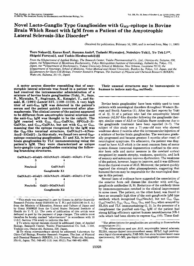
THE JOURNAL OF BIOLOGICAL CHEMISTRY Q 1993 by The American Society for Biochemistry and Molecular Biology, Inc. Vol .268, No. Issue of October 5, pp. 210ze-z1034,1~3 Printed in U.S.A. Novel Lacto-Ganglio Type Gangliosides with GM2-epitope in Bovine Brain Which React with IgM from a Patient of the Amyotrophic Lateral Sclerosis-like Disorder* (Received for publication, February 16, 1993, and in revised form, May 11, 1993) Toru NakaoSQ, Kazuo Konll, Susumu Andoll, Tadashi MiyatakeIJ, Nobuhiro YukiII , Yu-Teh Li**, Shigeki FuruyaSS, and Yoshio HirabayashiSSBQ From the $Department of Applied Biology, The Research Center, Taisho Pharmaceutical Co., Ltd., Ohmiya-shi, Saitama, 330, Japan, the llDepartment of Membrane Biochemistry, Tokyo Metropolitan Institute of Gerontology,Itabashi-ku, Tokyo, 173, Japan, the **Department of Biochemistry, Tulane University School of Medicine, New Orleans, Louisiana 70112, the ([Department of Neurology, School of Medicine, Tokyo Medical and Dental University, Bunkyo-ku, Tokyo, 113, Japan, and the $$Laboratory for Glyco-CellBiology, Frontier Research Program, The Institute of Physical and Chemical Research (RIKEN), Wako-shi, Saitarna, 351-01, Japan A motor neuron disorder resembling that of amy- otrophic lateral sclerosis was found in a patient who had receivedtheintramuscularadministrationofa mixture of bovine brain gangliosides (Yuki, N., Sato, S., Miyatake, T., Sugiyama, K., Katagiri, T., and Sa- saki, H. (1991) Lancet 337, 1109-1110). A very high titer of anti-GM2 IgM was detectedinthepatient’s serum and the patient quickly recovered after plas- mapheresis. The clinical course of the patient appeared to be different from amyotrophic lateral sclerosis and the anti-Gm2IgM was thought to be the culprit. The IgM reacted with GM~, GM~L-GBINAC, SPG(o2-3)- GalNAc, and GD1,-GalNAc, but not with GA2 or GD~, meaning that the epitope recognized by the IgM was the Gm-like terminal structure, GalNAcB1-4(Neu- Aca2-3)Gal/31-. In this study, we found two novel GM~- epitope containing gangliosides, X1 and X2, in bovine brain gangliosides by TLC immunostaining using the patient’s IgM. They were characterized as unique lacto-ganglio type gangliosides containing the follow- ing branching structures. GalNAc@1-4Gal@1-3GlcNAcj3l-3G~1-4Glc@1-l’Cer 3 4 NeuAca2 GalNAcBl Ganglioside X1 GalNAc@1-4Gal@l-3GlcNAc@1-3Gal@1-4Glc@l-1’Cer I I 3 4 NeuAcPa Gal@l-SGalNAc@l Ganglioside X2 I I Research Priority Areas 03255104 (to Y. H.) and 03255106 (to S. A.) * This study was supported in part by Grants-in-Aid for Scientific from the Ministry of Education, Science and Culture of Japan and by Grant NS09626 from the United States National Institute of Health (to Y.-T. L). The costs of publication of this article were defrayed in part by the payment of page charges. This article must therefore be hereby marked “advertisement” in accordance with 18 U.S.C. Section 1734 solely to indicate this fact. 8 To whom reprint requests should be addressed Dept. of Applied Biology, Research Center, Taisho Pharmaceutical Co. Ltd., 1-350, Yoshino-cyo, Omiya-shi, Saitama, 330, Japan. $8 To whom correspondence should be addressed Laboratory for Glyco-Cell Biology,Frontier Research Program, The Inst. of Physical and Chemical Research (RIKEN), 2-1, Hirosawa, Wako-shi, Saitama, 351-01,Japan. Tel.: 048-462-1111 (ext. 6511); Fax: 048-462-4690. Theirunusualstructuresmaybeimmunogenicto humans to induce anti-GM2 antibody. Bovine brain gangliosides‘ have been widely used to treat patients with neurological disorders throughout Western Eu- rope and South America (1). After the first reports by Yuki et al. (2, 3) of a patient who had the amyotrophic lateral sclerosis (ALS)’-like disorder following the ganglioside ther- apy, similar cases of ALS or Guillain-Barre syndrome due to the ganglioside treatment have been reported (4-7). The subject of thisstudy was a 71-year-old man who showed weakness about 2 months after the intramuscular injection of a mixture of bovine brain gangliosides. The weakness gradu- ally progressed and became greatest 5 months after onset of neurological symptoms. On admission, the patient was diag- nosed to have ALS which is the most common form of motor system disease (neuronal degeneration confined to the ante- rior horn cells and motor neurons in the cerebral cortex) recognized by muscle atrophy, weakness, spasticity, and lack of sensory and autonomic nervous dysfunction. The weakness of the patient, however, began to improve, and it was different from the clinical course of ALS. Moreover, the patient quickly regained the strength after plasmapheresis, suggesting that humoral factors may be responsible for the neurological dam- age in this patient. Several lines of evidence have suggested the association of theanterior horn cell disease-like disorder with anti-GMl ganglioside antibodies (8, 9). Reduction of the antibody titers by immunosuppression resulted in the clinical improvement in some cases. The patient, on the other hand, was found to have an unusually high titer of the anti-ganglioside IgM type antibody whichrecognized GdNeuAc), but not GAZ, GM~, GM2(NeuGc), GM~, GDl,, G D ~ ~ , GD~, and GTI~ when assayed by ELISA and TLC immunostaining (2,3). In thepresence of a complement the purified anti-GMz IgM of the patient had strong killing efficiency against human neuroblastoma NB-1 cells which had been shown to express GMz (10). These find- The abbreviations for gangliosides are according to Svennerholm nomenclature (31). The abbreviations used are: ALS, amyotrophic lateral sclerosis; ELISA, enzyme-linked immunosorbent assay; HPLC, high perform- ance liquid chromatography; FAB-MS, fast atom bombardment-mass spectrometry; GC-MS, gas-liquid chromatography-mass spectrome- try. 21028
Transcript of JOURNAL OF Vol .268, 5, pp. 210ze-z1034,1~3 Q …Vol .268, No. Issue of October 5, pp....
THE JOURNAL OF BIOLOGICAL CHEMISTRY Q 1993 by The American Society for Biochemistry and Molecular Biology, Inc. Vol .268, No. Issue of October 5, pp. 210ze-z1034,1~3
Printed in U.S.A.
Novel Lacto-Ganglio Type Gangliosides with GM2-epitope in Bovine Brain Which React with IgM from a Patient of the Amyotrophic Lateral Sclerosis-like Disorder*
(Received for publication, February 16, 1993, and in revised form, May 11, 1993)
Toru NakaoSQ, Kazuo Konll, Susumu Andoll, Tadashi MiyatakeIJ, Nobuhiro YukiII , Yu-Teh Li**, Shigeki FuruyaSS, and Yoshio HirabayashiSSBQ From the $Department of Applied Biology, The Research Center, Taisho Pharmaceutical Co., Ltd., Ohmiya-shi, Saitama, 330, Japan, the llDepartment of Membrane Biochemistry, Tokyo Metropolitan Institute of Gerontology, Itabashi-ku, Tokyo, 173, Japan, the **Department of Biochemistry, Tulane University School of Medicine, New Orleans, Louisiana 70112, the ([Department of Neurology, School of Medicine, Tokyo Medical and Dental University, Bunkyo-ku, Tokyo, 113, Japan, and the $$Laboratory for Glyco-Cell Biology, Frontier Research Program, The Institute of Physical and Chemical Research (RIKEN), Wako-shi, Saitarna, 351-01, Japan
A motor neuron disorder resembling that of amy- otrophic lateral sclerosis was found in a patient who had received the intramuscular administration of a mixture of bovine brain gangliosides (Yuki, N., Sato, S., Miyatake, T., Sugiyama, K., Katagiri, T., and Sa- saki, H. (1991) Lancet 337, 1109-1110). A very high titer of anti-GM2 IgM was detected in the patient’s serum and the patient quickly recovered after plas- mapheresis. The clinical course of the patient appeared to be different from amyotrophic lateral sclerosis and the anti-Gm2 IgM was thought to be the culprit. The IgM reacted with G M ~ , GM~L-GBINAC, SPG(o2-3)- GalNAc, and GD1,-GalNAc, but not with GA2 or G D ~ , meaning that the epitope recognized by the IgM was the Gm-like terminal structure, GalNAcB1-4(Neu- Aca2-3)Gal/31-. In this study, we found two novel G M ~ - epitope containing gangliosides, X1 and X2, in bovine brain gangliosides by TLC immunostaining using the patient’s IgM. They were characterized as unique lacto-ganglio type gangliosides containing the follow- ing branching structures.
GalNAc@1-4Gal@1-3GlcNAcj3l-3G~1-4Glc@1-l’Cer 3 4
NeuAca2 GalNAcBl Ganglioside X1
GalNAc@1-4Gal@l-3GlcNAc@1-3Gal@1-4Glc@l-1’Cer
I I
3 4
NeuAcPa Gal@l-SGalNAc@l Ganglioside X2
I I
Research Priority Areas 03255104 (to Y. H.) and 03255106 (to S. A.) * This study was supported in part by Grants-in-Aid for Scientific
from the Ministry of Education, Science and Culture of Japan and by Grant NS09626 from the United States National Institute of Health (to Y.-T. L). The costs of publication of this article were defrayed in part by the payment of page charges. This article must therefore be hereby marked “advertisement” in accordance with 18 U.S.C. Section 1734 solely to indicate this fact.
8 To whom reprint requests should be addressed Dept. of Applied Biology, Research Center, Taisho Pharmaceutical Co. Ltd., 1-350, Yoshino-cyo, Omiya-shi, Saitama, 330, Japan.
$ 8 To whom correspondence should be addressed Laboratory for Glyco-Cell Biology, Frontier Research Program, The Inst. of Physical and Chemical Research (RIKEN), 2-1, Hirosawa, Wako-shi, Saitama, 351-01, Japan. Tel.: 048-462-1111 (ext. 6511); Fax: 048-462-4690.
Their unusual structures may be immunogenic to humans to induce anti-GM2 antibody.
Bovine brain gangliosides‘ have been widely used to treat patients with neurological disorders throughout Western Eu- rope and South America (1). After the first reports by Yuki et al. ( 2 , 3) of a patient who had the amyotrophic lateral sclerosis (ALS)’-like disorder following the ganglioside ther- apy, similar cases of ALS or Guillain-Barre syndrome due to the ganglioside treatment have been reported (4-7). The subject of this study was a 71-year-old man who showed weakness about 2 months after the intramuscular injection of a mixture of bovine brain gangliosides. The weakness gradu- ally progressed and became greatest 5 months after onset of neurological symptoms. On admission, the patient was diag- nosed to have ALS which is the most common form of motor system disease (neuronal degeneration confined to the ante- rior horn cells and motor neurons in the cerebral cortex) recognized by muscle atrophy, weakness, spasticity, and lack of sensory and autonomic nervous dysfunction. The weakness of the patient, however, began to improve, and it was different from the clinical course of ALS. Moreover, the patient quickly regained the strength after plasmapheresis, suggesting that humoral factors may be responsible for the neurological dam- age in this patient.
Several lines of evidence have suggested the association of the anterior horn cell disease-like disorder with anti-GMl ganglioside antibodies (8, 9). Reduction of the antibody titers by immunosuppression resulted in the clinical improvement in some cases. The patient, on the other hand, was found to have an unusually high titer of the anti-ganglioside IgM type antibody which recognized GdNeuAc), but not GAZ, G M ~ , GM2(NeuGc), G M ~ , GDl,, G D ~ ~ , G D ~ , and GTI~ when assayed by ELISA and TLC immunostaining (2,3). In the presence of a complement the purified anti-GMz IgM of the patient had strong killing efficiency against human neuroblastoma NB-1 cells which had been shown to express GMz (10). These find-
The abbreviations for gangliosides are according to Svennerholm nomenclature (31).
The abbreviations used are: ALS, amyotrophic lateral sclerosis; ELISA, enzyme-linked immunosorbent assay; HPLC, high perform- ance liquid chromatography; FAB-MS, fast atom bombardment-mass spectrometry; GC-MS, gas-liquid chromatography-mass spectrome- try.
21028
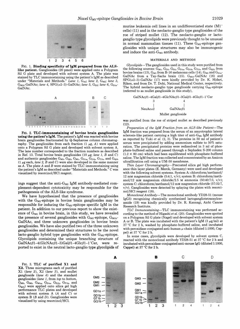
Novel GM2-epitope Gangliosides in Bovine Brain 21029
~. " - . - - - - - 1 2 3 4 5 6
FIG. 1. Binding specificity of IgM prepared from the ALS- like patient. Gangliosides (20 pmol) were applied onto a Polygram Si1 G plate and developed with solvent system A. The plate was stained by TLC immunostaining using the patient's IgM as described under "Materials and Methods." Lane 1, GA2; lane 2, G M ~ ; lane 3, GMlb-GalNAc; lane 4, SPG(a2-3)-GalNAc; lane 5, GDZ; lane 6, GD~.- GalNAc.
a
-x, * X?
. x3
,, : . . . $ , I , . . . . . : . . . ,.,,. ;::, , ,
FIG. 2. TLC-irnmunoetaining of bovine brain gangliosides using the patient's IgM. The patient's IgM was reacted with bovine brain gangliosides fractionated by Q-Sepharose column chromatog- raphy. The gangliosides from each fraction (1 pg, A ) were applied onto a Polygram Si1 G plate and developed with solvent system A. The lane number corresponded to the fraction numbers as described in Ref. 13. Total bovine brain gangliosides (10 pg; lane 1, B and C) and authentic gangliosides G M ~ , G M ~ , G M ~ , GD~,, GD1b, GT1b, and G ~ l b (1 pg each, lane 2, B and C) were also developed in the same manner as A. The plate A and B were stained by TLC immunostaining using the patient's IgM as described under "Materials and Methods." C was visualized by resorcinol/HCl reagent.
ings suggest that the a n t i - G ~ ~ IgM antibody-mediated com- plement-dependent cytotoxicity may be responsible for the pathogenesis of the ALS-like syndrome.
We have hypothesized that the presence of gangliosides with the G~z-epitope in bovine brain gangliosides may be responsible for inducing the G~z-epitope specific IgM in the patient. In addition to our previous report to show the exist- ence of GM2 in bovine brain, in this study, we have revealed the presence of several gangliosides with GM2-epitope, GMlb- GalNAc, and three unknown gangliosides in bovine brain gangliosides. We have also purified two of the three unknown gangliosides and determined their structures to be the novel lacto-ganglio hybrid type gangliosides with the GMz-epitope. Glycolipids containing the unique branching structure of GalNAcpl-4(GlcNAcpl-3)Gal~l-4Glc~l-l'Cer, were re- ported to exist in the neutral lacto-ganglio type glycolipids of
murine leukemia cell lines in an undifferentiated state (Ml- cells) (11) and in the neolacto-ganglio type gangliosides of the roe of striped mullet (12). The neolacto-ganglio or lacto- ganglio type glycolipids were previously thought to be unusual in normal mammalian tissues (11). These G ~ e p i t o p e gan- gliosides with unique structures may also be immunogenic and induce the anti-& antibody.
MATERIALS AND METHODS
Glycolipids-The gangliosides used in this study were purified from
bovine brains (13), G M ~ from B-16 melanoma cells (14), GM2 and GDW GalNAc from a Tay-Sachs brain (15). GMlb-GalNAc (16) and SPG(a2-3)-GalNAc (17) were kindly provided by Dr. K. Hidari, Riken, and from Dr. T. Dohi, National Medical Center, respectively. The hybrid neolacto-ganglio type ganglioside carrying G~~-epi tope (referred to as mullet ganglioside in this study),
the following sources: G M ~ , G D ~ , GD2, GD~., GDlb, GTlb, and Gglb from
GalNAc~1-4Gal~1-4GlcNAc~l-3Gal~l-4Glc~l-l'Cer 3 4
NeuAca2 GalNAcBl I I
Mullet ganglioside
was purified from the roe of striped mullet as described previously (12).
Preparation of the ZgM Fraction from an ALS-like Patient-The IgM fraction was prepared from the serum of an amyotrophic lateral sclerosis-like patient carrying a high titer of anti-% IgM antibody as reported by Yuki et al. (2, 3). The proteins in 50 ml of patient's serum were precipitated by adding ammonium sulfate to 50% satu- ration. The precipitated proteins were redissolved in 3 ml of phos- phate-buffered saline and passed through a Sephadex S-200 column (1.2 X 80 cm) which had been equilibrated with phosphate-buffered saline. The IgM fraction was collected and concentrated by an Amicon ultrafiltration cell using a UM-50 membrane.
Thin Layer Chromatography-Precoated silica gel high perform- ance thin layer plates (E. Merck, Germany) were used and developed with the following solvent systems. System A chloroform/methanol/ 12 mM magnesium chloride (5:41, v/v); system B: chloroform/meth- anol/l2 mM magnesium chloride/2.5 M ammonia (5040:7:3, v/v); system C: chloroform/methanol/12 mM magnesium chloride (57:32:7, v/v). Gangliosides were detected by splaying the plates with resorci- nol/HCl reagent (18).
Monoclonal Antibody-The monoclonal antibody YI328-51 (mouse IgG3) recognizing chemically synthesized lactogangliotetraosylcer- amide (19) was kindly provided by Dr. R. Kannagi, Aichi Cancer Research Institute.
TLC Zmmunostaining-TLC immunostaining was performed ac- cording to the method of Higashi et al. (20). Gangliosides were spotted on a Polygram Si1 G plate (Nagel) and developed with solvent system A or B. The plate was incubated with the patient's IgM (3 pg/ml) a t 37 "C for 2 h, washed by phosphate-buffered saline, and incubated with peroxidase-conjugated anti-human p-chain (diluted 1:1000, Cap- pel) a t 37 "C for 2 h.
In some cases, glycolipids were developed by solvent system C, reacted with the monoclonal antibody YI328-51 at 37 "C for 2 h and incubated with peroxidase-conjugated anti-mouse IgG (diluted 1:1000, Cappel) at 37 "C for 2 h.
A B C D
FIG. 3. TLC of purified X1 and X2. Three micrograms each of purified X1 ( l a n e 2), X2 ( l a n e 3), and mullet ganglioside ( l a n e 4 ) and the standard gangliosides ( l a n e 1 , from top to bottom,
GQIb) were applied onto silica gel high performance TLC plates and developed with solvent system A (A and C) or system B ( B and D). Gangliosides were visualized by using resorcinol/HCl.
G M ~ , G M ~ , GM., GDI,, Gib, Gib, and GM3 - 8
GM2 - GMl - GDla - GDlb - GQlb" GT1 b - "
1 2 3
GM3 - GM2 - GM1 - GDla- GDlbl GT1 b GQ 1 b'
1 2 3
1.. " c
" -
-3- r ..A .. .-
1 2 4 1 2 4

21030
A
Novel GM2-epitope Gangliosides in Bovine Brain X1 [M-H] ’ ;1950,1978
Ne:Ac 673)
R
I t 1 ”
R
n d
R
I : 1
R
a
HexNAc
NeuAc
(564 592
FIG. 4. Negative FAB-MS spectra of X1 and X2. Negative FAB-MS study of intact X1 ( A ) and X2 ( B ) were carried out as described under “Materials and Methods.”
TABLE I Partially methylated alditol acetates from X1 and X 2
2,3,6-Tri-O- 2,6-Di-0- 2,3,4,6-Tetra- Me-Glc Me-Gal 0-Me-Gal
4,6-Di-O- 3,6-Di-0- 3,4,6-Tri- 4,6-Di-0- Me-Glc- Me-Glc- NAcMe NAcMe
0-Me- GalNAcMe
Me-Gal- NAcMe
GMU + + + + Mullet ganglioside + + - - + + + x1 + + x2 + + + + + +
- - - - + - + -
-
ELISA-ELISA was performed by the method of Higashi et al. Isolation of GMz-epitope Gangliosides-Bovine brain gangliosides (5 (21). Gangliosides (50 pmol to 50 fmol) were reacted with the patient’s g) were separated by Q-Sepharose column chromatography using a IgM (5 #g/ml) at 37 “C for 1.5 h and followed by incubation with linear gradient system of chloroform/methanol/sodium acetate and peroxidase-conjugated anti-human p-chain (diluted 1:1000, Cappel) collected into 23 fractions (see Ref. 13 for details). Fraction 7 (4 mg), a t 37 “C for 2 h. containing several extremely minor gangliosides, was redissolved in

Novel GM2-epitope Gangliosides in Bovine Brain
V IV I11 I1 I
GalNAc - Gal - GlcNAc - Gal - Glc - Cer I I
NeuAc Gal - GalNAc VI 111‘
A
V
21031
4.5 4 . 0 3 .5 3 . 0
B
r I I I I I I I I I
4 . 5 4 . 0 3 . 5 3 . 5
FIG. 5. Proton NMR spectrum of X1 and X2. Proton NMR spectrum of X1 ( A ) and X2 ( B ) were obtained as described under “Materials and Methods.”

21032 Novel GM2-epitope Gangliosides in Bovine Brain
TABLE I1 Chemical shifts of the anomeric protons and their coupling constants
of X1 and X2 x1 X2
Chemical shift 5 1 . 2 5 1 . 2 shift
ppm H z ppm Hz I-Glc 4.15 7.3 4.16 8.0
4.25 7.7 4.26 7.8 111-GlcNAc" 4.64 7.7 4.68 8.0
111'-GalNAc" 4.62 6.2 4.65 IV-Gal
8.4 4.30 7.7 4.31 7.7
V-GalNAc 4.78 8.4 4.81 8.1 VI-Gal 4.30 7.0
11-Gal
or or or
a The exact assignment could not be achieved by one-dimensional MNR analysis.
1.0 I i
0.8 - 0.6 - 0.4
0.2 -
0' 1 - 1 - 1 - I
0 1 2 3 4 5 6 7
Ganglioside (50 x 2'" pmollwell)
FIG. 6. Reactivity of X1 and X2 with the patient's IgM in ELISA. Purified X1 (A), X2 (A), and G M 2 (0) were coated on a 96- well polystyrene plate (2-fold dilution from the top dose of 50 pmol/ well) and reacted with the patient's IgM (5 pg/ml) in ELISA. ELISA was performed as described under "Materials and Methods."
chloroform/methanol/water (60:25:1, v/v, system I) and injected onto an Aquasil SS-552N HPLC column (6 X 250 mm, Sensyu Chemicals, Tokyo) equilibrated with the solvent system I. The column was attached to a Jasco 880 HPLC system (Japan Spectroscopic Co. Ltd.). Gangliosides were eluted using a linear gradient program starting from the solvent system I and progressing in polarity until the solvent system of chloroform/methanol/water (60:304.5, v/v) was attained. The flow rate was 1 ml/min, and the elution was completed in 60 min. Each fraction (4 ml/tube) was evaporated to dryness under a stream of nitrogen. The residue was redissolved in 200 p1 of chloro- form/methanol (2:1, v/v), and 2-pl aliquots were analyzed for gan- gliosides by TLC.
Negative Zon Fast Atom Bombardment-Mass Spectrometry-Gan- gliosides were characterized by negative ion fast atom bombardment- mass spectrometry (FAB-MS). FAB-MS study was carried out with a JEOL JMS-DX304 mass spectrometer (JEOL Ltd., Tokyo) equipped with a JMA-DA 5000 data system (JEOL Ltd.). The sample (10-20 pg), dissolved in dimethyl sulfoxide, was mixed with 1 ml of triethanolamine as a matrix on a sample holder. Mass spectra were taken in accumulation mode at 3 kV of accelerating voltage (22).
N HCl and incubated at 100 "C for 1 h. The degradation was stopped Chemical Degradation-Gangliosides were dissolved in 300 pl of 1
by addition of 1 ml of 2.5 N ammonia. After lyophilization, the products were analyzed by TLC and TLC immunostaining using the monoclonal antibody YI328-51 to detect the lacto-ganglio branching structure GalNAc~1-4(GlcNAc~l-3)Gal-.
Enzymic Hydrolysis-X2 (10 pg) was hydrolyzed by P-galactosidase (bovine testis, Sigma, 10 milliunits/ml in 100 pl of 50 mM acetate
buffer, pH 5.5, containing 100 pg of sodium taurodeoxycholate) a t 37 "C for 2 h. The product was extracted from the reaction mixture using 1 ml of chloroform/methanol(2:1, v/v) after lyophilization and analyzed by TLC immunostaining using the patient's IgM.
Compositional Analysis-The sugar compositions of the ganglio- sides were analyzed by gas-liquid chromatography and mass spec- trometry (GC-MS) after methanolysis, N-acetylation, and trimethyl- silylation as described by Bhatti et al. (23). Fatty acid compositions were determined by gas-liquid chromatography. The system consisted of a Shimadzu GC-14A gas chromatograph (Kyoto) equipped with a capillary column of 3% OV-101 (0.2 mm X 25 m, Shimadzu) and Shimadzu LKB 9000B data system. The column temperature was increased from 160 to 240 "C at the rate of 4 "C/min.
Permethylution Analysis-Permethylation of gangliosides (100 pg) was performed according to the method of Hakomori (24). The acetates of partially methylated alditols were prepared as described previously (25, 26). An aliquot of the compounds were analyzed by GC-MS equipped with OV-101 column with a temperature program increasing from 160 to 240 "C at the rate of 4 "C/min. For analysis of permethylated sialic acid derivatives, the permethylated gangliosides were methanolyzed with 0.5 M HCl in methanol at 80 "C for 16 h and analyzed by GC-MS (27,28).
Proton Nuclear Magnetic Resonance-Proton nuclear magnetic resonance spectra were obtained by 400 MHz NMR spectrometer (model XL-400, Varian). Ganglioside (1 mg) was dissolved in 500 ml of dimethyl sulfoxide-Q/D20 (9:1, v/v) containing tetramethylsilane. The operation temperature was 40 "C.
RESULTS AND DISCUSSION
Reactivity of IgM of the ALS-like Patient with Ganglw- sides-Binding specificity of the ALS-like patient's IgM was examined by TLC immunostaining. The patient's IgM reacted with GMZ, GMlb-GalNAc, SPG(a2-3)-GalNAc, and GD~.- GalNAc (Fig. I), but not with GA2, GAI, GM3, GM1, G D ~ , G D ~ , GDl,, GTlb, and GBlb (data not shown), indicating the terminal structure of GalNAcpl-4(NeuAca2-3)Gal~l- (GM~- epitope) to be the epitope recognized by the IgM. The binding specificity was almost equal to that of IgM from a patient of the neuropathy associated with gammopathy (29). Bovine brain gangliosides which had been separated by Q-Sepharose column chromatography (13) were tested for reactivity with the IgM fraction (Fig. 2). Positive spots were observed only in monosialo-fractions (fractions 1-7). The positive spots corresponding to GMz and GMlt,-GalNAc were detected in fractions 1 and 4, respectively. Three other unidentified pos- itive spots containing GMz epitope, designated XI, X2, and X3, were detected in fractions 6 and 7 as shown in Fig. 2 A . GMz and GMlb-GalNAc were the major component of GMZ- epitope gangliosides in bovine brain (Fig. 2B). X1 and X2 could be detected as faint bands in 10 pg of total bovine brain gangliosides.
Isolation of Monosialogangliosides Carrying G,w Epitope- The gangliosides of fractions 7 (4 mg) were subjected to Aquasil SS-552N HPLC as described under "Materials and Methods." An aliquot (1/100) of each tube was analyzed by TLC. X1 (1.1 mg) and X2 (2.8 mg) were obtained from tubes 26-28 and tubes 34-38, respectively. The purity of X1 and X2 was examined by high performance TLC (Fig. 3). Both X1 and X2 gave single spots when using two different solvent systems and showed characteristically slow migration with the basic solvent system. The mobility of X1 was almost identical to that of mullet ganglioside in the neutral solvent system (Fig. 3C) but moved slower than mullet ganglioside in the basic solvent system (Fig. 30) .
Structural Characterization of X1 and X2"The sugar com- position of X1 and X2 was analyzed by GC-MS. X1 and X2 consisted of glucose, galactose, N-acetylglucosamine, N-ace- tylgalactosamine, and N-acetylneuraminic acid in molar ratio of 1:2:1:2:1 and 1:3:1:2:1, respectively. Fatty acid compositions of X1 and X2 were analyzed by GC. They resembled each

Novel GM2-epitope Gangliosides in Bovine Brain 21033
other in exhibiting C18:O as their major component. Negative FAB-MS spectra of intact X1 and X2 are shown
in Fig. 4. Molecular ion species, (M - H)-, of X1 were given by a pair of ions m/z 1950 and 1978 (Fig. 4A). These ions were consistent with the structures containing one N-acetyl- neuraminic acid, three hexoses, three N-acetylhexosamines, and a ceramide containing a C18:O fatty acid and a long-chain base of d l 8 1 or d20:l. Although molecular ion species of X2, (M - H)-, were detected by a pair of prominent ions at m/z 2112 and 2140 (Fig. 4B). These incremental increase in the quasi molecular ions as compared with those of X1 were equivalent to one additional hexose. FAB-MS also yielded the following fragment ions from both X1 and X2; m/z 726 and 754 for monohexosylceramide, m/z 888 and 916 for dihexo- sylceramide, and m/z 1294 and 1322 for the characteristic branching structure of HexNAc-(HexNAc-)Hex-Hex-Cer. The fragment ions specific for the G M ~ like terminal structure, HexNAc-(NeuAc-)Hex and HexNAc-(NeuAc-)Hex-HexNAc, were observed at m/z 673 and 876 in both X1 and X2, respectively. The ions at m/z 1484 and 1456 for Hex-HexNAc- (HexNAc-)Hex-Hex-Cer were detected only in X2.
XI, X2, GMla, and mullet ganglioside were permethylated and subjected to acetolysis followed by reduction and acety- lation. The products were analyzed by GC-MS using fragment ions, m/z 117 and 129 for alditol acetates and the ions m/z 116 and 159 for hexosaminitol acetates (Table I). The reten- tion times of the two partially methylated alditol acetates and a third methylated hexosaminitol acetate detected in X1 were identical to that derived from mullet ganglioside. They were identified as 2,3,6-tri-O-Me-1,4,5-tri-O-Ac-Glc-OH, 2,6-di-0- Me-l,3,4,5-tetra-O-Ac-Gal-OH, and 3,4,6-tri-O-Me-1,2,5-tri- 0-Ac-GalNAcMe-OH. The partially methylated hexosamini- to1 acetate, 3,6-di-0-Me-1,2,4,5-tetra-O-Ac-GlcNAcMe-OH, detected in mullet ganglioside was not observed in XI. XI, on the other hand, gave a peak of longer retention time than 3,6-di-0-Me-1,2,4,5-tetra-O-Ac-GlcNAcMe-OH. This peak was identified as 4,6-di-O-Me-l,2,3,5-O-Ac-HexNAcMe-OH by mass spectrometry (data not shown). Since the retention time of the latter peak was not identical to that of 4,6-di-0- Me-1,2,3,5-0-Ac-GalNAcMe-OH detected in GMla, but it gave the identical mass spectrum, suggests that the peak from X1 was 4,6-di-0-Me-1,2,3,5-tetra-O-Ac-GlcNAcMe-OH. This methylation study indicates that X1 is a lacto-ganglio gan- glioside, differing from neolacto-ganglio ganglioside isolated from mullet roe (12). Both X1 and mullet ganglioside pro- duced only 1,2,4,7,8,9-hexa-O-Me-NeuAcMe as the permeth- ylated N-acetylneuraminic acid derivative. All partially meth- ylated alditol acetates, hexosaminitol acetates, and the per- methylated N-acetylneuraminyl-methyl glycoside derived from X1 were also detected in X2. In addition, X2 gave 2,3,4,6- tetra-0-Me-5-0-Ac-Gal-OH and 4,6-di-O-Me-l,2,3,5-tetra-O- Ac-GalNAcMe-OH.
Mullet ganglioside, X1, and X2 were hydrolyzed in 1 N HC1 at 100 “C for 1 h. The partial degradation products were reacted with monoclonal antibody YI328-51 in the TLC im- munostaining system. The monoclonal antibody YI328-51 was raised against the chemically synthesized lactogangliote- traosylceramide by Kannagi et al. (19). The positive spots with same RF values were observed in the partial degradation products of mullet ganglioside, X1, and X2 (data not shown), indicating that GlcNAc and GalNAc of X1 and X2 were linked to the Gal residue of lactosylceramide through @I-3 and @I- 4 linkages, respectively.
X2 was hydrolyzed by @-galactosidase, and the products were analyzed by TLC immunostaining. X2 produced a GM2- epitope-containing ganglioside with the same RF value as X1
(data not shown), suggesting that the terminal Gal residue of X2 is attached to GalNAc in the branch of the lactogangliote- traosylceramide back bone.
The anomeric regions of the ’H NMR spectra of X1 and X2 showed six and seven resonances, respectively (Fig. 5). The assignment of each doublet is shown in Table 11. The chemical shifts and J values of H1 of I-Glc, 11-Gal, IV-Gal, V-GalNAc of X1 and X2, and VI-Gal of X2 are consistent with previously reported data (11, 12). The resonances for H1 of 111-GlcNAc and 111’-GalNAc were observed at 4.62 ppm (J1, = 6.2) and 4.64 ppm (J 1, = 7.7) in X1, and at 4.65 pprn (J1,2 = 8.4) and 4.68 ppm ( J I , z = 8.0) in X2, respectively. Based on the one-dimensional spectrum, it is not possible to assign the two HexNAc H1 peaks (4.62 and 4.64 ppm of XI, and 4.65 and 4.68 ppm of X2) to the H1 of 111-GlcNAc or 111’- GalNAc. However, the data indicate that the anomeric link- ages of both 111-GlcNAc and 111’-GalNAc are in a @-configu- ration. Whether the peaks are assigned to 111-GlcNAc or 111’- GalNAc, the chemical shifts of H1 of the branched GalNAcs of X1 and X2 are shifted to lower field than that of mullet roe ganglioside (12).
From the above experimental results, the structures of X1 and X2 were identified as follows.
GalNAcpl-4Gal~1-3GlcNAc~l-3Gal~l-4Glc~l-l’Cer 3 4
NeuAccu2 GalNAcpl Ganglioside X1
GalNAcpl-4Gal~l-3GlcNAc~l-3Gal~l-4Glc~l-l’Cer
I I
3 4
NeuAccuP Galpl-3GalNAcpl Ganglioside X2
It it worthy to note that X1 contains a type I chain where mullet roe ganglioside contains a type I1 chain.
Reactivity Determination of X 1 and X 2 with the Patient’s IgM in ELISA-Purified X1 and X2 were found to react with the patient’s IgM when examined by ELISA (Fig. 6). Both X1 and X2 exhibited nearly the same reactivity as GM2. This result strongly suggests that the patient’s IgM recognizes an epitope in the outer trisaccharide sequence GalNAcPl- 3(NeuAca2-3)Gallp- and that the inner core structure is not a significant factor for the binding with the antibody.
The glycolipids containing lactogangliotetraosylceramide have never been found in normal mammalian tissues. Such glycolipids were first purified from undifferentiated murine leukemia cells (Ml-) by Kannagi et al. (11). They described the structural uniqueness of the glycolipids and the possible involvement of the appearance of an aberrant glycosyltrans- ferase with loose specificity which is associated with oncogen- esis. This is the first report of the detection and isolation of lacto-ganglio type gangliosides X1 and X2 from normal mam- malian tissues. Many unknown minor components in brain gangliosides remain to be characterized. Some of them are novel hybrid types of ganglio and lacto series, and might have high immunogenic activity to induce anti-ganglioside anti- body, i.e. anti-neuron antibody as illustrated in this study. Although the antigens to induce anti-ganglioside antibodies or clinical significance of the antibodies have not been well established, there is evidence for anti-ganglioside antibody involvement in some forms of neuronal disorders (for a review see Ref. 30). Thus, extreme caution should be paid to the clinical use of gangliosides derived from natural sources.
I I

21034 Novel GM2-epitope Gangliosides in Bovine Brain
Acknowledgment-We are grateful to Dr. Yoshitaka Nagai, Tokyo Metropolitan Institute of Medicine, for helpful comments.
REFERENCES 1. Bradley, W. G. (1990) Muscle Nerve 13,833-842 2. Yuki, N., Sato, S., Miyatake, T., Sugiyama, K., Katagiri, T., and Sasaki,
3. Yuki, N., Sato, S., Miyatake, T., Sugiyama, K., Katagiri, T., and Sasaki,
4. Latov, N., Koski, C. L., and Walicke, P. A., (1991) Lancet 338,757 5. Schonhofer, P. S. (1992) N. Engl. J. Med. 326,493 6. Schonhofer, P. S. (1991) Lancet 338 ,757 7. Nobile-Orazio, E., Carpo, M., Meucci, N., Grassi, M. P., Capitani, E.,
Sciacco, M., Mangoni, A., and Scarlato, G. (1992) J. Neurol. Sci. 109 ,
8. Freddo, L., Yu, R. K., Latov, N., Donofrio, P. D., Hays, A. P., Greenberg, H. S., Albers, J. W., Allessi, A. G., and Keren, D. (1986) Neurology 3 6 ,
9. Pestronk, A,, Cornblath, D. R., Ilyas, A. A,, Baba, H., Quarles, R. H., Griffin, J. W., Alderson, K., and Adams, R. N. (1988) Ann. Neurol. 2 4 , 73-78
10. Yuki, N., Miyatake, T., Ichihashi, Y., Sato, S., and Katagiri, T. (19 92) Muscle Nerve 16,1371-1373
11. Kannagi, R., Levery, S. B., and Hakomori, S. (1984) J. Biol. Chem. 2 6 9 ,
H. (1991) Lancet 337,1109-1110
H. (1991) Lancet 338,314-315
200-206
454-458
RAdd-RdF(1 12. DeGasperi, R., Koerner, T. A. W., Quarles, R. H., Ilyas, A. A,, Ishikawa,
13. Hirabavashi. Y.. Nakao. T.. Matsumoto. M.. Obata. K.. and Ando. S. (1988) Y., Li, S.-C., and Li, Y.-T. (1987) J. Biol. Chem. 262,17149-17155
"" ""
J. Chromatog;. 445,377-384
14. Hiraha ashi, Y., Hamaoka, A., Matsumoto, M., Matsubara, T., Tagawa, M., kakahayashi, S., and Taniguchi, M. (1985) J. Biol. Chem. 2 6 0 ,
15. Svennerholm, L., Mansson, J.-E., and Li, Y.-T. (1973) J. Biol. Chem. 248 ,
16. Itoh, T., Li, Y.-T., Li, S.-C., and Yu, R. K. (1981) J. Biol. Chem. 266,165-
13328-13333
740-742 1 ca
17.
18. 19.
20.
21.
22.
23.
Dohi, T., Hanai, N., Yamaguchi, K., and Oshima, M. (1991) J. Biol. Chem. I".,
266.24038-24043 Svennerholm, L. (1957) Biochim. Biophys. Acta 24,604-611 Shigeta, K., Ito, Y., Ogawa, T., Kirihata, Y., Hakomori, S., and Kannagi,
Higashi, H., Fukui, Y., Ueda, S., Kato, S., Hirabayashi, Y., Matsumoto, M.,
Higashi, H., Ikuta, K., Ueda,,S., Kato, S., Hirabayashi, Y., Matsumoto, M.,
Hirabayashl, Y., F u J I ~ ~ , S. C., Kon, K., and Ando, S. (1991) J. Biol. Chem.
Bhatti. T.. Chambers. R. E.. and Alamu. J. R. (1970) Biochim. BwDhvs.
R. (1987) J. Biol. Chem. 262,135S-1362
and Naiki, M. (1984) J. Biochem. (Tokyo) 95,1517-1520
and Naik,i, M. (1984) J. Bmhem. (Tokyo) 9 6 , 785-794
266,10268-10274
Acta 222,339-347 ' . I . , I -
24. Hakomori S. (1964) J. Biochem. (Tokyo) 65,205-208 25. Sto_ffel,Wl, and Hanfland, P. (1973) Hoppe-Seyler's Z. Physwl. Chem. 364 ,
26. Taki, T., Hiraha ashi, Y., Ishikawa, H., Ando, S., Kon, K., Tanaka, Y., and
27. Levery, S. B., and Hakomori, S. (1987) Met&& Enzymol. 138,13-25 28. Nakao, T., Kon, K., Ando, S., and Hirabayashl, Y. (1991) Btochtm. Bmphys.
Acta 1086,305-309 29. Ilyas A. A. Li S.-C., Chou D. K. H., Li Y.-T. Jungalwara, F. B., Dalakas,
M.'C., aAd Buarles, R. H. (1988) J. Bhl. Cdm. 263,4369-4373 30. Quarles, R. H. (1989) Methods EnzymoL 179,291-299 31. Svennerholm, L. (1964) Lipids 6, 145-162
2,141
Matsumoto, d. (1986) J. Biol Chem. 261,3075-3078