Journal of the Taiwan Institute of Chemical...
13
Review Current developments of computer-aided drug design Hung-Jin Huang a,1 , Hsin Wei Yu a,1 , Chien-Yu Chen a , Chih-Ho Hsu a , Hsin-Yi Chen a , Kuei-Jen Lee b , Fuu-Jen Tsai b,c , Calvin Yu-Chian Chen a,b, * a Laboratory of Computational and Systems Biology, School of Chinese Medicine, China Medical University, Taichung 40402, Taiwan b Department of Bioinformatics, Asia University, Taichung 41354, Taiwan c Department of Medical Genetics, Pediatrics and Medical Research, China Medical University Hospital and College of Chinese Medicine, China Medical University, Taichung 40402, Taiwan Contents 1. Introduction ..................................................................................................... 624 2. Structure-based drug design ........................................................................................ 624 2.1. Protein structure determination................................................................................ 624 2.1.1. Homology modeling ................................................................................. 624 2.1.2. Folding recognition .................................................................................. 626 2.1.3. Ab initio protein modeling ............................................................................. 626 2.1.4. Hot spot prediction .................................................................................. 627 2.2. Docking ................................................................................................... 628 2.2.1. Autodock .......................................................................................... 628 2.2.2. CDOCKER .......................................................................................... 629 2.2.3. Flexible docking ..................................................................................... 629 2.2.4. LigandFit .......................................................................................... 629 2.2.5. Transmembrane protein modeling ...................................................................... 629 2.3. Binding free energy.......................................................................................... 629 2.4. Flexibility of protein–ligand complex ........................................................................... 630 2.5. De novo evolution ........................................................................................... 630 3. Ligand-based drug design .......................................................................................... 631 3.1. Quantitative structure–activity relationship (QSAR) ................................................................ 631 3.1.1. CoMFA .................................................................................................... 631 3.1.2. CoMSIA ................................................................................................... 632 4. Molecular dynamics simulations..................................................................................... 633 Journal of the Taiwan Institute of Chemical Engineers 41 (2010) 623–635 ARTICLE INFO Article history: Received 26 November 2009 Received in revised form 12 March 2010 Accepted 20 March 2010 Keywords: Molecular simulation Molecular dynamics (MD) Docking Computer-aided drug design (CADD) Quantitative structure–activity relationship (QSAR) ABSTRACT The continuous advancement in molecular biology and information technology aided the development of a rich molecular simulation repertoire that can be applied in system biology, proteomics, molecular biology, bioinformatics, and materials science. We attempt to introduce the latest developments in drug design based on computational techniques, including protein structure modeling, docking, binding site prediction, quantitative structure–activity relationship (QSAR), and molecular dynamics simulation. Furthermore, a brief discussion on current docking issues, including accuracy of protein structure and protein–ligand interaction, is also included. Weight equation and rules and a new concept on flexibility are also described here as possible solution for these issues. ß 2010 Taiwan Institute of Chemical Engineers. Published by Elsevier B.V. All rights reserved. * Corresponding author. Current address: Computational and Systems Biology, Massachusetts Institute of Technology, Cambridge, MA 02139, USA. Tel.: +1 617 353 7123. E-mail addresses: [email protected], [email protected] (C.-C. Chen). 1 These authors contributed equally. Contents lists available at ScienceDirect Journal of the Taiwan Institute of Chemical Engineers journal homepage: www.elsevier.com/locate/jtice 1876-1070/$ – see front matter ß 2010 Taiwan Institute of Chemical Engineers. Published by Elsevier B.V. All rights reserved. doi:10.1016/j.jtice.2010.03.017
Transcript of Journal of the Taiwan Institute of Chemical...

Journal of the Taiwan Institute of Chemical Engineers 41 (2010) 623–635
Review
Current developments of computer-aided drug design
Hung-Jin Huang a,1, Hsin Wei Yu a,1, Chien-Yu Chen a, Chih-Ho Hsu a, Hsin-Yi Chen a,Kuei-Jen Lee b, Fuu-Jen Tsai b,c, Calvin Yu-Chian Chen a,b,*a Laboratory of Computational and Systems Biology, School of Chinese Medicine, China Medical University, Taichung 40402, Taiwanb Department of Bioinformatics, Asia University, Taichung 41354, Taiwanc Department of Medical Genetics, Pediatrics and Medical Research, China Medical University Hospital and College of Chinese Medicine, China Medical University,
Taichung 40402, Taiwan
Contents
1. Introduction . . . . . . . . . . . . . . . . . . . . . . . . . . . . . . . . . . . . . . . . . . . . . . . . . . . . . . . . . . . . . . . . . . . . . . . . . . . . . . . . . . . . . . . . . . . . . . . . . . . . . 624
2. Structure-based drug design . . . . . . . . . . . . . . . . . . . . . . . . . . . . . . . . . . . . . . . . . . . . . . . . . . . . . . . . . . . . . . . . . . . . . . . . . . . . . . . . . . . . . . . . 624
2.1. Protein structure determination. . . . . . . . . . . . . . . . . . . . . . . . . . . . . . . . . . . . . . . . . . . . . . . . . . . . . . . . . . . . . . . . . . . . . . . . . . . . . . . . 624
2.1.1. Homology modeling . . . . . . . . . . . . . . . . . . . . . . . . . . . . . . . . . . . . . . . . . . . . . . . . . . . . . . . . . . . . . . . . . . . . . . . . . . . . . . . . . 624
2.1.2. Folding recognition . . . . . . . . . . . . . . . . . . . . . . . . . . . . . . . . . . . . . . . . . . . . . . . . . . . . . . . . . . . . . . . . . . . . . . . . . . . . . . . . . . 626
2.1.3. Ab initio protein modeling. . . . . . . . . . . . . . . . . . . . . . . . . . . . . . . . . . . . . . . . . . . . . . . . . . . . . . . . . . . . . . . . . . . . . . . . . . . . . 626
2.1.4. Hot spot prediction . . . . . . . . . . . . . . . . . . . . . . . . . . . . . . . . . . . . . . . . . . . . . . . . . . . . . . . . . . . . . . . . . . . . . . . . . . . . . . . . . . 627
2.2. Docking . . . . . . . . . . . . . . . . . . . . . . . . . . . . . . . . . . . . . . . . . . . . . . . . . . . . . . . . . . . . . . . . . . . . . . . . . . . . . . . . . . . . . . . . . . . . . . . . . . . 628
2.2.1. Autodock . . . . . . . . . . . . . . . . . . . . . . . . . . . . . . . . . . . . . . . . . . . . . . . . . . . . . . . . . . . . . . . . . . . . . . . . . . . . . . . . . . . . . . . . . . 628
2.2.2. CDOCKER . . . . . . . . . . . . . . . . . . . . . . . . . . . . . . . . . . . . . . . . . . . . . . . . . . . . . . . . . . . . . . . . . . . . . . . . . . . . . . . . . . . . . . . . . . 629
2.2.3. Flexible docking. . . . . . . . . . . . . . . . . . . . . . . . . . . . . . . . . . . . . . . . . . . . . . . . . . . . . . . . . . . . . . . . . . . . . . . . . . . . . . . . . . . . . 629
2.2.4. LigandFit . . . . . . . . . . . . . . . . . . . . . . . . . . . . . . . . . . . . . . . . . . . . . . . . . . . . . . . . . . . . . . . . . . . . . . . . . . . . . . . . . . . . . . . . . . 629
2.2.5. Transmembrane protein modeling . . . . . . . . . . . . . . . . . . . . . . . . . . . . . . . . . . . . . . . . . . . . . . . . . . . . . . . . . . . . . . . . . . . . . . 629
2.3. Binding free energy. . . . . . . . . . . . . . . . . . . . . . . . . . . . . . . . . . . . . . . . . . . . . . . . . . . . . . . . . . . . . . . . . . . . . . . . . . . . . . . . . . . . . . . . . . 629
2.4. Flexibility of protein–ligand complex . . . . . . . . . . . . . . . . . . . . . . . . . . . . . . . . . . . . . . . . . . . . . . . . . . . . . . . . . . . . . . . . . . . . . . . . . . . 630
2.5. De novo evolution . . . . . . . . . . . . . . . . . . . . . . . . . . . . . . . . . . . . . . . . . . . . . . . . . . . . . . . . . . . . . . . . . . . . . . . . . . . . . . . . . . . . . . . . . . . 630
3. Ligand-based drug design . . . . . . . . . . . . . . . . . . . . . . . . . . . . . . . . . . . . . . . . . . . . . . . . . . . . . . . . . . . . . . . . . . . . . . . . . . . . . . . . . . . . . . . . . . 631
3.1. Quantitative structure–activity relationship (QSAR) . . . . . . . . . . . . . . . . . . . . . . . . . . . . . . . . . . . . . . . . . . . . . . . . . . . . . . . . . . . . . . . . 631
3.1.1. CoMFA . . . . . . . . . . . . . . . . . . . . . . . . . . . . . . . . . . . . . . . . . . . . . . . . . . . . . . . . . . . . . . . . . . . . . . . . . . . . . . . . . . . . . . . . . . . . . . . . . . . . 631
3.1.2. CoMSIA . . . . . . . . . . . . . . . . . . . . . . . . . . . . . . . . . . . . . . . . . . . . . . . . . . . . . . . . . . . . . . . . . . . . . . . . . . . . . . . . . . . . . . . . . . . . . . . . . . . 632
4. Molecular dynamics simulations. . . . . . . . . . . . . . . . . . . . . . . . . . . . . . . . . . . . . . . . . . . . . . . . . . . . . . . . . . . . . . . . . . . . . . . . . . . . . . . . . . . . . 633
A R T I C L E I N F O
Article history:
Received 26 November 2009
Received in revised form 12 March 2010
Accepted 20 March 2010
Keywords:
Molecular simulation
Molecular dynamics (MD)
Docking
Computer-aided drug design (CADD)
Quantitative structure–activity relationship
(QSAR)
A B S T R A C T
The continuous advancement in molecular biology and information technology aided the development
of a rich molecular simulation repertoire that can be applied in system biology, proteomics, molecular
biology, bioinformatics, and materials science. We attempt to introduce the latest developments in drug
design based on computational techniques, including protein structure modeling, docking, binding site
prediction, quantitative structure–activity relationship (QSAR), and molecular dynamics simulation.
Furthermore, a brief discussion on current docking issues, including accuracy of protein structure and
protein–ligand interaction, is also included. Weight equation and rules and a new concept on flexibility
are also described here as possible solution for these issues.
� 2010 Taiwan Institute of Chemical Engineers. Published by Elsevier B.V. All rights reserved.
Contents lists available at ScienceDirect
Journal of the Taiwan Institute of Chemical Engineers
journal homepage: www.elsev ier .com/ locate / j t i ce
* Corresponding author. Current address: Computational and Systems Biology, Massachusetts Institute of Technology, Cambridge, MA 02139, USA. Tel.: +1 617 353 7123.
E-mail addresses: [email protected], [email protected] (C.-C. Chen).1 These authors contributed equally.
1876-1070/$ – see front matter � 2010 Taiwan Institute of Chemical Engineers. Published by Elsevier B.V. All rights reserved.
doi:10.1016/j.jtice.2010.03.017

H.-J. Huang et al. / Journal of the Taiwan Institute of Chemical Engineers 41 (2010) 623–635624
5. Sample course syllabuses . . . . . . . . . . . . . . . . . . . . . . . . . . . . . . . . . . . . . . . . . . . . . . . . . . . . . . . . . . . . . . . . . . . . . . . . . . . . . . . . . . . . . . . . . . 633
6. Conclusion . . . . . . . . . . . . . . . . . . . . . . . . . . . . . . . . . . . . . . . . . . . . . . . . . . . . . . . . . . . . . . . . . . . . . . . . . . . . . . . . . . . . . . . . . . . . . . . . . . . . . . 634
Acknowledgements . . . . . . . . . . . . . . . . . . . . . . . . . . . . . . . . . . . . . . . . . . . . . . . . . . . . . . . . . . . . . . . . . . . . . . . . . . . . . . . . . . . . . . . . . . . . . . . 634
References . . . . . . . . . . . . . . . . . . . . . . . . . . . . . . . . . . . . . . . . . . . . . . . . . . . . . . . . . . . . . . . . . . . . . . . . . . . . . . . . . . . . . . . . . . . . . . . . . . . . . . 634
1. Introduction
The research fields in chemical engineering have always beenchanging and evolving, from the field of applied industrialchemistry at the beginning of the last century, through therevolutionary reformulation of unit operations, transport phe-nomena and engineering science in the 1960s, to the extensive useof computing technology and the incorporation of molecularbiology over the last two decades. This latter change is graduallybeing adopted by prestigious research institutes and universities,including the Department of Chemical and Biomolecular Engi-neering of Johns Hopkins University, which has shifted researchfocuses to biological related issues and revised the engineeringundergraduate and graduate teaching curricula to integratebiomolecular modeling into process dynamics and control.Indeed, the integration of computational force and molecularbiology, such as to simulate the behavior of molecules, isbecoming a mainstream in the chemical engineering researchand has received much attention from the pharmaceuticalindustries.
Molecular simulations are an interdisciplinary science withdifferent applications in different research area. In polymerscience, much use of molecular simulation has been put instudying fluxional behavior. As for monitoring heat transfer insemi-conductor, simulation is used for studying thermal profile. Ininformatics, the emphasis is on developing more powerful, fastsimulation models that can accurately predict or describe scientificphenomenon. As for uses in drug design and bioinformatics, thereare two different research focuses concurrent in the scientific field.The first is to design new mathematic algorithm for more realisticcalculation. The second is to apply current algorithms on molecularbiology research, such as simulating protein–ligand or protein–protein interactions, and use the result for further biomolecularexperiments.
For a medicinal product to reach patients, commonly, morethan 8 years of time and millions of dollars in investment arerequired to finish the long tedious drug development process.Furthermore, only a handful can finish the clinical trial and pass thestrict inspection of drug regulatory agency, despite that thousandsof new therapeutic candidates are being discovered in laboratoriesevery year. However, the recent advances in technologies, namelyautomated platform, computational chemistry and computer-aided drug design (CADD), are now offering a fast track to somelimiting factors of therapeutic discovery as well. Computer-aideddrug design (CADD), that offers an in silico alternative to medicinalchemistry techniques for studying the structure and predicting thebiological activity of drug candidates, has the advantages of bothspeed and low cost and is becoming an indispensable program ofmajor pharmaceutical companies.
There are two major application areas of CADD, namelystructure-based drug design and ligand-based drug design.Structure-based drug design relies on three-dimensional knowl-edge of the receptor structure and its active sites to investigateinteraction, binding energy and steric relationship between ligandand receptor. Ligand-based designing approach, on the other hand,relies on knowledge of ligands that interact with target of interest.This technique employs statistical methods to link structuralfeatures to biological activities and attempts to identify specificstructural features of a ligand required for interacting with its
target. Both structure-based and ligand-based techniques can beapplied in the initial drug discovery process and aid the discoveryof a lead compound which serves as the starting basis for furthermodification to improve pharmacokinetics, solubility, selectivity,potency or stability. Two of the great advantages of CADD lie in theability of fast screening a large molecule databank and theaccelerated time steps of identifying notable medicinal chemistryfeatures. These characteristics are extremely beneficial in design-ing multi-target medicinal products.
As a short review to introduce the basic of molecular simulationin drug design, we arbitrarily categorized CADD into three majorsections: (a) structure-based drug design, (b) ligand-based drugdesign, and (c) molecular dynamics. A summary of CADD processflowchart is shown in Fig. 1. In addition, to the brief overview, wewill address on issues encountered by biochemists on usingdocking programs, including the low hit rate of docking programs(Kontoyianni et al., 2005; Warren et al., 2006) and the lowreplicability of predicted protein–ligand interaction (Baxter et al.,1998). For these issues, we will attempt to improve the dockingresults by introducing a parameter accounting for the flexibility ofprotein and ligand. Overall, we hope the readers can gain muchinsight into CADD by our concise introductions on major- and sub-topics, practical examples of relevant application cases, andsample curricula of CADD courses.
2. Structure-based drug design
To design a medicinal product for treating a disease or relieve asymptom, a clear understanding of the disease pathway andrelevant processes is crucial for selecting a therapeutic target.Thus, in the past we have employed programs such as GeneGo andKEGG to build pathway maps of hypoxia-inducible factor in braininjury, shown in Fig. 2, for identifying critical signal or transcrip-tion pathways, specific protein–protein interactions and relation-ships between upstream and downstream proteins. Thisbackground knowledge has proven helpful to us for selectingkey therapeutic target.
2.1. Protein structure determination
For structure-based drug design, a priority before investigatingreceptor–ligand relationship is to obtain the target structure. Thereare two major methods for protein structure determination byphysical measures, X-ray diffraction and NMR (Marti-Renom et al.,2000). The solved protein structures can be readily found at ProteinData Bank (www.rcsb.org/); however, for proteins that have notbeen solved or are difficult to isolate, modeling approach can beused.
2.1.1. Homology modeling
Homology modeling is a fast method to obtain proteinstructures that can not only be used in studying rational drugdesign but also for protein–protein interaction and site-directedmutagenesis (Josa et al., 2008; Mohan et al., 2009; Sujatha et al.,2009). Proteins lacking structural information could be con-structed if they have over 30% sequence identify with their relatedhomologous proteins (templates) (Marti-Renom et al., 2000). Thismodeling strategy has been widely applied in many researches andin our past studies as well (Chen, 2008a,b,c; Chen, 2009a,b,c,d;

[(Fig._1)TD$FIG]
Fig. 1. An example of a computer-aided drug design flowchart.
H.-J. Huang et al. / Journal of the Taiwan Institute of Chemical Engineers 41 (2010) 623–635 625
Chen, 2010a,b; Chen and Chen, 2007; Chen et al., 2008a,b; Chen et al.,2009, 2010; Ding et al., 2008; Lin et al., 2009; Sheu et al., 2009).
For our studies on H1N1 influenza virus, we have used homologymodeling approach to construct hemagglutinin and neuraminidasestructures from newly identified viral protein sequences and solvedprotein structures. A past sequence alignment result of the H1 andN1 sequences to templates, shown in Fig. 3, shows that both H1 andN1 have sequence identity and similar over 75%, which gives us highconfidence in using the templates for modeling.
The modeled structures can be further modified in modelrefinement to be consistent with the experiment data in covalentbonds, geometry, and energy configuration. Force fields, such asCHARMM, AMBER, CVFF, CFF91, and GROMOS can also be appliedto molecules for calculating energy minimization, which uses thefunction (Payne et al., 1992) shown below:
Etotal ¼ Estretching þ Ebending þ Edihedral þ Eout-of-plane þ Ecrossterms
þ EVdW þ Ecoulombic (1)
To ensure the rationality of the modeled structures, checks onstereochemistry, energy profile, residue environment, and struc-ture similarity are often needed. Stereochemistry considers thebond angles and lengths, the dihedral angles of major chains, andthe non-covalent bonds of amino acid residues within a protein.Two examples of our stereochemistry check are shown in Fig. 4. Forour modeled hemagglutinin, the Ramachandran graph shows that94.4% of H1 residues are in the allowed region while only 2.5% arein the disallowed region. Similarly, for modeled N1 structure, 91.4%of residues are found in the allowed region and only 2.5% ofresidues are in the disallowed region.
Energy profile is based on Profile-3D that analyzes thecompatibility of amino acid sequences with three-dimensionalenvironment (Al-Lazikani et al., 2001). The Profile-3D graphs of H1and N1 model are shown in Fig. 5. There are several factors that caninfluence the verify score. In conditions where the hydrophobicresidues are folded on protein surface or the polar residues arefolded into protein core, decreases in verify scores are likely to beseen. For regions that have verify scores above zero are considered

[(Fig._2)TD$FIG]
Fig. 2. The signal pathway of HIF protein.
H.-J. Huang et al. / Journal of the Taiwan Institute of Chemical Engineers 41 (2010) 623–635626
to have stable folding and more energetic favor three-dimensionalstructure.
2.1.2. Folding recognition
Also known as ‘‘threading,’’ folding recognition was brought upin 1991 by Bowie and colleagues whom employed this method todescribe the environment of residues interactions. Foldingrecognition calculates the probabilities of the 3D structures couldform by given protein sequences (Mishra, 2009). Both the
environment of residues interactions and the protein surface areaare considered in the threading protocol. Structure with thehighest probability is recommended to construct the proteinmodel.
2.1.3. Ab initio protein modeling
The ab initio method is based on physical principles, residueinteraction center and lattice representation of a protein to buildthe target (Adrian-Scotto and Vasilescu, 2008; Deepa and

[(Fig._3)TD$FIG]
Fig. 3. The sequence alignment of (a) H1 (75.4% sequence identity and 79.8% sequence similarity) and (b) N1 (83.6% sequence identity and 92.1% sequence similarity).
H.-J. Huang et al. / Journal of the Taiwan Institute of Chemical Engineers 41 (2010) 623–635 627
Kolandaivel, 2008). This method is extremely useful when theother protocols fail to predict an unknown protein structure(Huang et al., 1998). However, the identity and accuracy given byab initio modeling could be lower than other approaches. Proteinfolding is not only a physical action, but also involves manybiochemical actions originated from inherent residues interaction(Sippl, 1993). Based on this concept, ab initio method hypothesizesthat: when a protein folds, it would tend to achieve the mostenergetically favorable state (Luthy et al., 1992).
2.1.4. Hot spot prediction
Another important issue in structure-based drug design is todetermine the ligand active site. While the active site may bedetermined via ligand location in the crystal lattice after X-raycrystallography, this method is not possible for proteins that cannotbe crystallized. Several binding site determination methods have
been invented to address this issue and FTMAP (Brenke et al., 2009) isone of the recently developed methods being investigated in our lab.
The primary strategy of FTMAP utilizes small molecularfragments as a probe for exploring protein surface. Spots wheremolecular fragments clustered are predicted to be the favorabledruggable sites. Significant hydrogen bonds and non-boundedinteractions can also be explored between the probes and protein.In addition, the structure of the molecular probes can be thestarting basis for designing new medicinal products.
The reliability of FTMAP has been confirmed in the past bycomparing the predicted with the experiment results of Allen et al.
(1996) and Mattos et al. (2006). Consistencies in binding sitelocation and protein–probe interactions have been observed in thecomparisons (Brenke et al., 2009). Landon et al. (2009) have alsoconducted FTMAP researches with molecular experiments ver-ifications as well.

[(Fig._4)TD$FIG]
Fig. 4. The Ramachandran plot of (a) H1 and (b) N1.
[(Fig._5)TD$FIG]
Fig. 5. The verify score of (a) H1 and (b) N1.
[(Fig._6)TD$FIG]
Fig. 6. PDE-5 hot spots predicted by FTMP. Small molecular fragments used include
acetaldehyde, ethanal, ethane, acetamide, acetonitrile, acetone, methylamine,
benzene, dimethyl ether, urea, N,N-dimethylformamide, ethanol, benzaldehyde,
phenol, isopropanol.
H.-J. Huang et al. / Journal of the Taiwan Institute of Chemical Engineers 41 (2010) 623–635628
We have used FTMAP in our researches on protein phosphodi-esterase-5 (PDE-5) to determine potential hot spots. Potentialdruggable sites predicted by FTMAP are shown in Fig. 6, andhistograms of calculated protein–probe interactions are shown inFig. 7.
2.2. Docking
2.2.1. Autodock
The software AutoDock, developed by Olsen’s laboratory in theScripps Research Institute, is a program for docking small flexibleligands into a rigid 3D structure (Goodsell and Olson, 1990). A set ofgrid is used to describe the 3D structure, based on the AMBER forcefield, and generated with AutoGrid to calculate van der Waals andcoulombic interactions. In version 1.0 and 2.0, the geneticalgorithm and simulated annealing were utilized for searching
the best binding model, but version 3.0 incorporated LamarckianGenetic Algorithm (LGA) in the search, and the efficiency wasgreatly enhanced than the previous versions. In the currentversion, the linear regression analysis is used to obtain a free-energy scoring function, based on the AMBER force field, and alarger set of diverse receptor–ligand complexes is kept constantwhile the side-chains in the 3D structure are flexible. Theapplications of AutoDock are immense, including but not limitedto computer-aided structure-based drug design, X-ray crystallog-raphy analysis, high throughput virtual screening, combinatoriallibrary design, and protein–protein interaction study (Rajakrish-nan et al., 2008).
[(Fig._7)TD$FIG]
Fig. 7. Summary of H-bond interaction and non-bonded interaction found between PDE-5 protein residues and small molecular probes.
Fig. 8. A modeled GABA receptor with membrane force field.
H.-J. Huang et al. / Journal of the Taiwan Institute of Chemical Engineers 41 (2010) 623–635 629
2.2.2. CDOCKER
The CDOCKER protocol is a CHARMM-based docking algorithm(Wu et al., 2003) and retains all the advantages of full ligandflexibility. Ligand conformations are generated from the initialligand structure by high temperature molecular dynamics (MD)simulation. The random conformations are refined based on grid-based simulated annealing and full molecular mechanics minimi-zation. In the docking procedure, CDOCKER uses a sphere to definean active site, so the knowledge of the binding site is not required.
2.2.3. Flexible docking
The flexible docking protocol retains receptor flexibility duringdocking of flexible ligands (Koska et al., 2008). The target receptorside-chain conformations are calculated in the first step and aregenerated by the ChiFlex algorithm. The ChiFlex algorithm createsvarious protein conformations with different side-chain orienta-tions. The second step is providing low energy conformations ofligands for the docking process. The LibDock program is used forthis docking process, which indicates the binding site where ligandpolar and non-polar groups may be bound to the favorablepositions of protein. The next step is to remove similar ligandposes. The refinement is performed in the final steps; the side-chains are refined using the ChiRotor algorithm and the CDOCKERfor structure simulated annealing and energy minimization of eachligand pose. Overall, flexible docking can optimize the flexibility ofthe side-chains (Braun et al., 2008). However, it generally requiresextensive computing resources and generates more data thangeneral rigid docking protocol.
2.2.4. LigandFit
LigandFit is a grid-based method for calculating receptor–ligand interaction energies, which is crucial in initial ligand shapematch to the receptor binding site (Akten et al., 2009; Ramalhoet al., 2009; Venkatachalam et al., 2003). The LigandFit protocolcontains three essential steps for docking ligands to the specifiedsite: definition of the active site, analysis of ligand conformations,docking of ligands to a selected site, and scoring of the predictedposes. The first step is to determine the active site of a protein withknown 3D structure. As afore mentioned this can be achieved bylocating the ligand within the protein structure. If ligands are notavailable in the active site, flood-filling algorithm of LigandFit canbe used to determine possible cavity region on protein surface. Inthe second step, LigandFit utilizes Monte Carlo (MC) method togenerate the ligand conformations. As soon as one conformation isgenerated, it will be employed to dock with the receptor. The thirdstep is the estimation of binding affinity (score) by grid-based
energy calculation of the energy between the ligand and thereceptor. The ligands will have to be docked into the receptorbefore calculating the docking scores. The docking scores can becalculated according to the following scoring functions: DockScore, LigScore1, LigScore2, PLP1, PLP2, Jain, PMF, PMF04, Ludienergy estimate 1, Ludi energy estimate 2, and Ludi energyestimate 3.
2.2.5. Transmembrane protein modeling
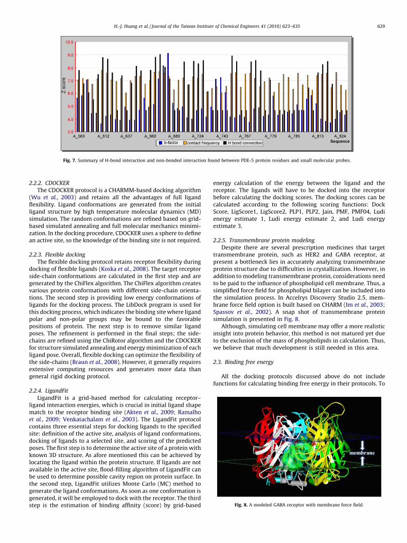
Despite there are several prescription medicines that targettransmembrane protein, such as HER2 and GABA receptor, atpresent a bottleneck lies in accurately analyzing transmembraneprotein structure due to difficulties in crystallization. However, inaddition to modeling transmembrane protein, considerations needto be paid to the influence of phospholipid cell membrane. Thus, asimplified force field for phospholipid bilayer can be included intothe simulation process. In Accelrys Discovery Studio 2.5, mem-brane force field option is built based on CHARM (Im et al., 2003;Spassov et al., 2002). A snap shot of transmembrane proteinsimulation is presented in Fig. 8.
Although, simulating cell membrane may offer a more realisticinsight into protein behavior, this method is not matured yet dueto the exclusion of the mass of phospholipids in calculation. Thus,we believe that much development is still needed in this area.
2.3. Binding free energy
All the docking protocols discussed above do not includefunctions for calculating binding free energy in their protocols. To[(Fig._8)TD$FIG]

H.-J. Huang et al. / Journal of the Taiwan Institute of Chemical Engineers 41 (2010) 623–635630
calculate the binding free energy, information on the energy statusof the protein–ligand complex, free ligands and unbound proteinmust be pre-determined. The energy is calculated using theformula (Kollman et al., 2000):
energy of binding ¼ energy of complex� energy of ligand
� energy of receptor: (2)
2.4. Flexibility of protein–ligand complex
Our research team has proposed a weight equation and rules(Chen, 2009d), attempting to improve the accuracy of theconsensus scoring. Although our results supported weight scoreover consensus score, there are still areas needed for furtherdevelopment. In here, we will propose a rough concept, based onthe flexible nature of protein and drug molecule. Currently, therotation and fluctuation of protein and drug molecules can besimulated by using molecular dynamics. However, moleculardynamics simulations require extensive computing unit and time.Hence, it is impractical to perform large scale screening of amolecule database with molecular dynamics. Therefore, ourcurrent experiments are limited to virtual screening of databaseand then to simulations of a few possible candidates.[(Fig._9)TD$FIG]
Fig. 9. The concept of D
Hereby, we propose a concept:
(1) The difference in result of flexible docking and LigandFit is dueto difference in flexibility of molecules, such that:
flexibility ¼ score of LigandFit� score of flexible docking
(2) The result of molecular simulation is related to flexibility, and apositive relationship can be obtained in flexibility vs. moleculardynamics.
Furthermore, we hope to introduce flexibility parameter intodocking algorithm to closely monitor real life situation. Theequation is shown below:
real docking score ¼ docking score� flexibility
2.5. De novo evolution
After docking program, we can modify ligands by two methods(shown in Fig. 9). The first method is based on active site features toidentify functional groups that can establish strong interactionswith the receptor. Then, the functional groups can be linked orattached to the original ligand scaffolds. The second method uses
e novo evolution.

[(Fig._10)TD$FIG]
Fig. 10. The core atom (blue) of the training set used for designing GABA receptor
inhibitor. (For interpretation of the references to color in this figure legend, the
reader is referred to the web version of the article.)
[(Fig._11)TD$FIG]
Fig. 11. CoMFA contour map, with steric favor region in green and disfavor region in
yellow. The electropositive contribution is in blue and electronegative region in red
(Chen, 2009a). (For interpretation of the references to color in this figure legend, the
reader is referred to the web version of the article.)
H.-J. Huang et al. / Journal of the Taiwan Institute of Chemical Engineers 41 (2010) 623–635 631
the original ligand scaffolds to develop derivatives that cancomplement the receptor.
3. Ligand-based drug design
When the target protein structure is unknown or cannot bepredicted by modeling techniques, the ligand-based drug design isthe alternative protocol. This method uses statistical approaches tocorrelate ligand activity to structural information (Singer andWilliam, 1967).
3.1. Quantitative structure–activity relationship (QSAR)
Quantitative structure–activity relationship is a widely usedtechnique in drug designing process. It employs statistics andanalytical tools to investigate the relationship between thestructures of ligands and their corresponding effects. Hence,mathematical models are built based on structural parameters todescribe this structure–activity relationship. Before, 2D-QSAR waswidely used to link structural property descriptors (such ashydrophobicity, steric, electrostatic and geometric effects) tomolecular biological activity; the results were often analyzed with[(Fig._12)TD$FIG]
Fig. 12. CoMSIA contour maps. (a) Steric region: favor (green) and disfavor (yellow). Elect
(white). (c) Hydrogen bond donor region: favor (cyan) and disfavor (purple). (d) Hydrogen
the references to color in this figure legend, the reader is referred to the web version o
multiple regression analysis. One of the most commonly used 2D-QSAR methods was proposed by Hansch (Clayton and Purcell,1969; Hansch, 1969). However, because 2D-QSAR cannot accu-rately describe the correlation between the 3D spatial arrange-ment of the physiochemical properties, and the biologicalactivities, recently 3D-QSAR approaches have been adapted.
In the past, we had used QSAR for drug design research,including GABA-A (Chen, 2009a) and mPGES-1 (Chen, 2009c).Using the case of GABA-A receptor as an example, the alignmentcore for the training set is shown in Fig. 10 and the contour mapsfrom CoMFA and CoMSIA are shown in Fig. 11 and Fig. 12separately.
In here, we will describe two frequently applied 3D-QSARmethodologies: comparative molecular field analysis (CoMFA) andcomparative molecular similarity indices analysis (CoMSIA).
3.1.1. CoMFA
Comparative molecular field analysis (CoMFA) is established onthe concept that the biological activity of a molecule is dependent
ropositive favored region (blue). (b) Hydrophobic region: favor (purple) and disfavor
bond acceptor: favor (green) and disfavor (red) (Chen, 2009a). (For interpretation of
f the article.)

[(Fig._13)TD$FIG]
Fig. 13. Standard dynamics simulation of mPGES-1. Snapshots taken at (a) initial conformation, (b) 300 ps, (c) 600 ps, (d) 900 ps, (e) 1200 ps, (f) 1500 ps, (g) 1800 ps, and (h)
2000 ps. The protein is quite dynamic and has loop movement and fluctuation in structures. However no large movement in protein backbone is observed in the simulation
time.
H.-J. Huang et al. / Journal of the Taiwan Institute of Chemical Engineers 41 (2010) 623–635632
of the surrounding molecular fields, such as steric and electrostaticfields. The steric and electrostatic fields were calculated by CoMFAusing Lennard–Jones potential, and coulombic potential, respec-tively. Although this method has been widely adopted, it hasseveral problems. Both potential functions changes dramaticallynear the van der Waals surface of the molecule and thus, cut-offvalues are often required. In addition, alignment of ligands must beconducted before energy calculation, but the orientation of thesuperimposed molecules is correlative to the calculation grid. It
could cause large changes in CoMFA results. Moreover, in order toexamine both fields in the same PLS analysis, a scaling factor needsto be added to the steric field (Cramer et al., 1989).
3.1.2. CoMSIA
Comparative molecular similarity index analysis (CoMSIA) is amethod developed recently as an extension of CoMFA. The CoMSIAmethod includes more additional field properties; these are: steric,electrostatic, hydrophobic, hydrogen bond donor and hydrogen

[(Fig._14)TD$FIG]
Fig. 14. Root mean square deviation (RMSD) of M2 influenza proton channel as a
function of the simulation time. The root mean square deviation (RMSD) of M2
shows that the protein gradually adapts and stabilizes at a configuration after
100 ns of simulation.
Table 2The lecture of structure bioinformatics.
Lesson Lecture description
1 Protein structure
2 Protein structure prediction
3 Homology modeling
4 Force fields
5 Folding recognition
6 Hot spot and binding site
7 Programming for binding affinity
8 Ab initio protein modeling
9 Perl programming
10 Protein–protein interaction
11 Modeling for lipid bilayer
12 Docking
13 Flexible docking
14 Binding free energy
15 3D-QSAR
16 NCI database
17 Traditional Chinese medicine database
18 Final examination
H.-J. Huang et al. / Journal of the Taiwan Institute of Chemical Engineers 41 (2010) 623–635 633
bond acceptor. CoMSIA is insensitive to the orientation of thealigned molecules and correlates to the grid by using Gaussianfunction. Furthermore, the improved function algorithm is leastinfluenced by the relative distance to the van der Waals surface.Overall, this model can offer a more accurate structural–activityrelationship than CoMFA (Klebe et al., 1994).
4. Molecular dynamics simulations
Molecular dynamics (MD) simulation is one of the importanttools in the theoretical study of biological molecules. Becausemolecular systems generally contain a large number of particles, itis impossible to analyze such complex systems. By using numericalmethods, molecular dynamics simulation can avoid such analyticintractability.
During simulation, atoms and molecules are allowed to interactfor a period of time. The motion for every atom is calculated andcan be played to examine the overall behavior (Mccammon et al.,1977). Overall, the background algorithm for a MD simulationincludes: (1) the determination of the initial positions andvelocities of every atom; (2) the calculation of forces applied onthe investigated atom using inter-atomic potentials; (3) theprogression of atomic positions and velocities through a short-time period. These new positions and velocities are then turnedinto new inputs to step 2, and when steps 2 and 3 are repeated,each repetition forms an additional time step.
Table 1The lecture of computer-aided drug design.
Lesson Lecture description
1 Introduction of modeling software
2 Protein structure prediction
3 Structure-based drug design
4 Docking
5 Virtual screening
6 Pharmacology and molecular simulations
7 Molecular dynamics
8 Protein folding prediction
9 Mid-term examination
10 Ligand-based drug design
11 CoMFA
12 CoMSIA
13 HypoGen
14 Scoring function
15 Chinese herb database and NCI database
16 Weight rules (Chen’s weight rules and equation) (I) [Chen, 2009d]
17 Weight rules (Chen’s weight rules and equation) (II) [Chen, 2009d]
18 Final examination
Molecular dynamics is now routinely employed to study thestructure, dynamics and thermodynamics of biological moleculesand their complexes. It provides detailed information on thefluctuations and conformational changes of proteins. Nucleic acidsstructural information can be investigated using this method aswell (Borkar et al., 2010; Roy and Thakur, 2010). In addition,solvent molecules can also be investigated on the impacts ofoverall protein structural changes.
In our studies, we are especially interested at studying theentering of drug molecule into target protein and the associatedprotein–ligand interaction. In the case of mPGES-1, we haveapplied molecular dynamics to study structural changes afterbinding of target to ligand (Fig. 13). An RMSD graph of M2 influenzaproton channel is also shown (Fig. 14) to illustrate the differencebetween the protein at a specific time and the initial reference.
5. Sample course syllabuses
An introductory syllabus on CADD, including structure-baseddrug design, ligand-based drug design and molecular dynamics, isshown in Table 1. The syllabus for structural bioinformatics isshown in Table 2, and this course emphasizes on physics andmolecular simulation algorithms. Moreover, syllabus of theprinciple and application of molecular simulation is shown inTable 3.
Table 3The lecture of molecular simulation.
Lesson Lecture description
1 An overview of molecular simulation
2 Monte Carlo methods
3 Free energy
4 Free-energy calculations
5 An overview of molecular dynamics
6 Force fields: AMBER, CHARMM, OPLS, GROMOS (I)
7 Application of force fields
8 Effects of solvents
9 Algorithms and computations
10 Choosing the time step
11 The Lennard–Jones potential
12 FENE potential
13 EAM potential
14 Potential for covalent carbon
15 Simulated annealing
16 Softwares (AMBER, CHARMM, VASP (DFT), XMD, CPMD)
17 Group presentation
18 Final examination

H.-J. Huang et al. / Journal of the Taiwan Institute of Chemical Engineers 41 (2010) 623–635634
6. Conclusion
From the aforementioned introduction, it is easy to see thatmolecular simulation has a vital role in drug design and CADD,whether it is in protein modeling, in docking or in moleculardynamics. In addition to these, we hope our flexibility concept cangreatly increase the hit rate and the accuracy of protein–ligandinteraction. This concept is different from our previous weightequation of which requires IC50 to obtain the parameter in thealgorithm. By introducing flexibility in docking protocol, we hopethat the simulation can be more close to real life events. With theadvancement in computing facilities and software algorithms,many simulation works that require supercomputer in the past canbe done in a workstation. By implementing molecular simulationinto biomolecular researches, not only the research steps can beaccelerated, but also the vast investment in money can be saved. Inthe future, molecular simulation and computer-aided drug designcan greatly influence the development of pharmaceutical industryand become a necessity before molecular experiments.
Acknowledgements
The research was supported by grants from the National ScienceCouncil of China (NSC 98-2221-E-039-007-) and China MedicalUniversity (CMU98-CT-15, CMU97-276) and Asia University(CMU98-ASIA-09). This study is also supported in part by TaiwanDepartment of Health Clinical Trial and Research Center ofExcellence (DOH99-TD-B-111-004) and Taiwan Department ofHealth Cancer Research Center of Excellence (DOH99-TD-C-111-005). We are grateful to the National Center of High-performanceComputing for computer time and facilities.
References
Adrian-Scotto, M. and D. Vasilescu, ‘‘Quantum Molecular Modeling of Glycyl-Adenylate,’’ J. Biomol. Struct. Dyn., 25, 697 (2008).
Akten, E. D., S. Cansu, and P. Doruker, ‘‘A Docking Study Using Atomistic ConformersGenerated Via Elastic Network Model for Cyclosporin A/Cyclophilin A Complex,’’ J.Biomol. Struct. Dyn., 27, 13 (2009).
Al-Lazikani, B., J. Jung, Z. Xiang, and B. Honig, ‘‘Protein Structure Prediction,’’ Curr. Opin.Chem. Biol., 5, 51 (2001).
Allen, K. N., C. R. Bellamacina, X. Ding, C. J. Jeffery, C. Mattos, G. A. Petsko, and D. Ringe,‘‘An Experimental Approach to Mapping the Binding Surfaces of CrystallineProteins,’’ J. Phys. Chem., 100, 2605 (1996).
Baxter, C. A., C. W. Murray, D. E. Clark, D. R. Westhead, and M. D. Eldridge, ‘‘FlexibleDocking Using Tabu Search and an Empirical Estimate of Binding Affinity,’’Proteins, 33, 367 (1998).
Borkar, A., I. Ghosh, and D. Bhattacharyya, ‘‘Structure and Dynamics of Double HelicalDNA in Torsion Angle Hyperspace: A Molecular Mechanics Approach,’’ J. Biomol.Struct. Dyn., 27, 695 (2010).
Braun, G. H., D. M. Jorge, H. P. Ramos, R. M. Alves, V. B. da Silva, S. Giuliatti, S. V. Sampaio,C. A. Taft, and C. H. Silva, ‘‘Molecular Dynamics, Flexible Docking, Virtual Screen-ing, Admet Predictions, and Molecular Interaction Field Studies to Design NovelPotential Mao-B Inhibitors,,’’ J. Biomol. Struct. Dyn., 25, 347 (2008).
Brenke, R., D. Kozakov, G. Y. Chuang, D. Beglov, D. Hall, M. R. Landon, C. Mattos, and S.Vajda, ‘‘Fragment-based Identification of Druggable ‘Hot Spots’ of Proteins UsingFourier Domain Correlation Techniques,’’ Bioinformatics, 25, 621 (2009).
Chen, C. Y. C., ‘‘Discovery of Novel Inhibitors for c-Met by Virtual Screening andPharmacophore Analysis,’’ J. Chin. Inst. Chem. Engrs., 39, 617 (2008a).
Chen, C. Y. C., ‘‘A Novel Perspective on Designing the Inhibitor of HER2 Receptor,’’ J.Chin. Inst. Chem. Engrs., 39, 291 (2008b).
Chen, C. Y. C., ‘‘Insights into the Suanzaoren Mechanism—From Constructing the 3DStructure of GABA-a Receptor to Its Binding Interaction Analysis,’’ J. Chin. Inst.Chem. Engrs., 39, 663 (2008c).
Chen, C. Y. C., ‘‘Chemoinformatics and Pharmacoinformatics Approach for Exploringthe GABA-A Agonist from Chinese Herb Suanzaoren,’’ J. Taiwan Inst. Chem. Engrs.,40, 36 (2009a).
Chen, C. Y. C., ‘‘De Novo Design of Novel Selective COX-2 Inhibitors: From VirtualScreening to Pharmacophore Analysis,’’ J. Taiwan Inst. Chem. Engrs., 40, 55 (2009b).
Chen, C. Y. C., ‘‘Pharmacoinformatics Approach for mPGES-1 in Anti-inflammation by3D-QSAR Pharmacophore Mapping,’’ J. Taiwan Inst. Chem. Engrs., 40, 155 (2009c).
Chen, C. Y. C., ‘‘Weighted Equation and Rules—A Novel Concept for Evaluating Protein–Ligand Interaction,’’ J. Biomol. Struct. Dyn., 27, 271 (2009d).
Chen, C. Y. C., ‘‘Virtual Screening and Drug Design for PDE-5 Receptor from TraditionalChinese Medicine Database,’’ J. Biomol. Struct. Dyn., 27, 627 (2010a).
Chen, C. Y. C., ‘‘Bioinformatics, Chemoinformatics, and Pharmainformatics Analysis ofHER2/HSP90 Dual-targeted Inhibitors,’’ J. Taiwan Inst. Chem. Engrs., 41, 143(2010b).
Chen, C. Y. C. and K. T. Chen, ‘‘Novel selective inhibitors of hydroxyxanthonederivatives for human cyclooxygenase-2,’’ Acta Pharmacol. Sin., 28, 2027 (2007).
Chen, C. Y. C., G. W. Chen, and Y. C. Chen, ‘‘Molecular Simulation: The Mechanism ofHER2/neu Degradation by Inhibiting HSP90,’’ J. Chin. Chem. Soc., 55, 297 (2008a).
Chen, C. Y. C., Y. F. Chen, C. H. Wu, and H. Y. Tsai, ‘‘What is the Effective Component inSuanzaoren Decoction for Curing Insomnia? Discovery by Virtual Screening andMolecular Dynamic Simulation. J. Biomol. Struct. Dyn., 26, 57 (2008b).
Chen, C. Y., Y. H. Cheng, D. T. Bau, H. J. Huang, F. J. Tsai, C. H. Tsai, and C. Y. C. Chen,‘‘Discovery of potent inhibitors for phosphodiesterase 5 by virtual screening andpharmacophore analysis,’’ Acta Pharmacol. Sin., 30, 1186 (2009).
Chen, C. Y., H. J. Huang, F. J. Tsai, and C. Y. C. Chem, ‘‘Drug Design for Influenza A VirusSubtype H1N1,’’ J. Taiwan Inst. Chem. Engrs., 41, 8 (2010).
Clayton, J. M. and W. P. Purcell, ‘‘Hansch and Free-Wilson Analyses of InhibitoryPotencies of Some 1-Decyl-3-carbamoylpiperidines against Butyrylcholinesteraseand Comparison of the Two Methods,’’ J. Med. Chem., 12, 1087 (1969).
Deepa, P. and P. Kolandaivel, ‘‘Studies on Tautomeric Forms of Guanine–Cytosine BasePairs of Nucleic Acids and Their Interactions with Water Molecules,’’ J. Biomol.Struct. Dyn., 25, 733 (2008).
Ding, Q., L. Huo, J. Y. Yang, W. Xia, Y. Wei, Y. Liao, C. J. Chang, Y. Yang, C. C. Lai, D. F. Lee, C.J. Yen, Y. J. Rita Chen, J. M. Hsu, H. P. Kuo, C. Y. Lin, F. J. Tsai, L. Y. Li, C. H. Tsai, and M.C. Hung, ‘‘Down-regulation of Myeloid Cell Leukemia-1 through Inhibiting Erk/Pin1 Pathway by Sorafenib Facilitates Chemosensitization in Breast Cancer,’’ CancerRes., 68, 6109 (2008).
Goodsell, D. S. and A. J. Olson, ‘‘Automated Docking of Substrates to Proteins bySimulated Annealing,’’ Proteins, 8, 195 (1990).
Hansch, C. A., ‘‘Quantitative Approach to Biochemical Structure–ActivityRelationships,’’ Acc. Chem. Res., 2, 232 (1969).
Huang, E. S., P. Koehl, M. Levitt, R. V. Pappu, and J. W. Ponder, ‘‘Accuracy of Side-chainPrediction upon Near-native Protein Backbones Generated by Ab Initio FoldingMethods,’’ Proteins, 33, 204 (1998).
Im, W., M. Lee, and C. Brooks, ‘‘Generalized Born Model with a Simple SmoothingFunction,’’ J. Comput. Chem., 24, 1691 (2003).
Josa, D., E. F. da Cunha, T. C. Ramalho, T. C. Souza, and M. S. Caetano, ‘‘HomologyModeling of Wild-type, D516v, and H526l Mycobacterium Tuberculosis RNAPolymerase and Their Molecular Docking Study with Inhibitors,’’ J. Biomol. Struct.Dyn., 25, 373 (2008).
Klebe, G., U. Abraham, and T. Mietzner, ‘‘Molecular Similarity Indices in a ComparativeAnalysis (CoMSIA) of Drug Molecules to Correlate and Predict Their BiologicalActivity,’’ J. Med. Chem., 37, 4130 (1994).
Kollman, P. A., I. Massova, C. Reyes, B. Kuhn, S. Huo, L. Chong, M. Lee, T. Lee, Y. Duan, W.Wang, O. Donini, P. Cieplak, J. Srinivasan, D. A. Case, and T. E. Cheatham,‘‘Calculating Structures and Free Energies of Complex Molecules: CombiningMolecular Mechanics and Continuum Models,’’ Acc. Chem. Res., 33, 889 (2000).
Kontoyianni, M., G. S. Sokol, and L. M. McClellan, ‘‘Evaluation of Library RankingEfficacy in Virtual Screening,’’ J. Comput. Chem., 26, 11 (2005).
Koska, J., V. Z. Spassov, A. J. Maynard, L. Yan, N. Austin, P. K. Flook, and C. M.Venkatachalam, ‘‘Fully Automated Molecular Mechanics Based Induced Fit Pro-tein–Ligand Docking Method,’’ J. Chem. Inf. Model., 48, 1965 (2008).
Landon, M. R., R. L. Lieberman, Q. Q. Hoang, S. Ju, J. M. Caaveiro, S. D. Orwig, D. Kozakov,R. Brenke, G. Y. Chuang, D. Beglov, S. Vajda, G. A. Petsko, and D. Ringe, ‘‘Detection ofLigand Binding Hot Spots on Protein Surfaces via Fragment-based Methods:Application to DJ-1 and Glucocerebrosidase,’’ J. Comput. Aided Mol. Des., 23, 491(2009).
Lin, Y. J., Y. C. Hou, C. H. Lin, Y. A. Hsu, J. C. Sheu, C. H. Lai, B. H. Chen, P. D. Lee, L. Wan, andF. J. Tsai, ‘‘Puerariae radix isoflavones and their metabolites inhibit growth andinduce apoptosis inbreast cancer cells,’’ Biochem. Biophys. Res. Commun., 378, 683(2009).
Luthy, R., J. U. Bowie, and D. Eisenberg, ‘‘Assessment of Protein Models with Three-dimensional Profiles,’’ Nature, 356, 83 (1992).
Marti-Renom, M. A., A. C. Stuart, A. Fiser, R. Sanchez, F. Melo, and A. Sali, ‘‘ComparativeProtein Structure Modeling of Genes and Genomes,’’ Annu. Rev. Biophys. Biomol.Struct., 29, 291 (2000).
Mattos, C., C. R. Bellamacina, E. Peisach, A. Pereira, D. Vitkup, G. A. Petsko, and D. Ringe,‘‘Multiple Solvent Crystal Structures: Probing Binding Sites, Plasticity andHydration,’’ J. Mol. Biol., 357, 1471 (2006).
Mccammon, J. A., B. R. Gelin, and M. Karplus, ‘‘Dynamics of Folded Proteins,’’ Nature,267, 585 (1977).
Mishra, S., ‘‘Function Prediction of Rv0079, a Hypothetical Mycobacterium Tubercu-losis Dosr Regulon Protein,’’ J. Biomol. Struct. Dyn., 27, 283 (2009).
Mohan, S. S., J. J. Perry, N. Poulose, B. G. Nair, and G. Anilkumar, ‘‘Homology Modeling ofGlut4, an Insulin Regulated Facilitated Glucose Transporter and Docking Studieswith ATP and Its Inhibitors,’’ J. Biomol. Struct. Dyn., 26, 455 (2009).
Payne, M. C., M. P. Teter, D. C. Allan, T. A. Arias, and J. D. Joannopoulos, ‘‘IterativeMinimization Techniques for Ab Initio Total-energy Calculations: Molecular Dy-namics and Conjugate Gradients,’’ Rev. Mod. Phys., 64, 1045 (1992).
Rajakrishnan, V., V. R. Manoj, and G. Subba Rao, ‘‘Computer-aided, Rational Design of aPotent and Selective Small Peptide Inhibitor of Cyclooxygenase 2 (Cox2),’’ J. Biomol.Struct. Dyn., 25, 535 (2008).
Ramalho, T. C., M. S. Caetano, E. F. da Cunha, T. C. Souza, and M. V. Rocha, ‘‘Constructionand Assessment of Reaction Models of Class I Epsp Synthase: Molecular Dockingand Density Functional Theoretical Calculations,’’ J. Biomol. Struct. Dyn., 27, 195(2009).

H.-J. Huang et al. / Journal of the Taiwan Institute of Chemical Engineers 41 (2010) 623–635 635
Roy, S. and A. R. Thakur, ‘‘20ns Molecular Dynamics Simulation of the AntennapediaHomeodomain–DNA Complex: Water Interaction and DNA Structure Analysis,’’ J.Biomol. Struct. Dyn., 27, 443 (2010).
Sheu, J. C., C. H. Hua, L. Wan, Y. J. Lin, H. C. Tseng, N. Jinawath, M. H. Tsai, N. W. Chang, C.F. Lin, C. C. Lin, L. J. Hsieh, T. L. Wang, I. M. Shih, and F. J. Tsai, ‘‘Functional genomicanalysis identified EGFR activation as the most common genetic event in oralsquamous cell carcinoma,’’ Cancer Res., 69, 2568 (2009).
Singer, J. A. and P. William, ‘‘Purcell Relationships among Current QuantitativeStructure–Activity Models,’’ J. Med. Chem., 10, 1000 (1967).
Sippl, M. J., ‘‘Recognition of Errors in Three-dimensional Structures of Proteins,’’Proteins, 17, 355 (1993).
Spassov, V. Z., L. Yan, and S. Szalma, ‘‘Introducing an Implicit Membrane in GeneralizedBorn/Solvent Accessibility Continuum Solvent Models,’’ J. Phys. Chem. B, 106, 8726(2002).
Sujatha, K., A. Mahalakshmi, D. K. Solaiman, and R. Shenbagarathai, ‘‘SequenceAnalysis, Structure Prediction, and Functional Validation of Phac1/Phac2 Genesof Pseudomonas sp. Ldc-25 and Its Importance in PolyhydroxyalkanoateAccumulation,’’ J. Biomol. Struct. Dyn., 26, 771 (2009).
Venkatachalam, C. M., X. Jiang, T. Oldfield, and M. Waldman, ‘‘Ligandfit: A NovelMethod for the Shape-directed Rapid Docking of Ligands to Protein Active Sites,’’ J.Mol. Graph. Modell., 21, 289 (2003).
Warren, G. L., C. W. Andrews, A. M. Capelli, B. Clarke, J. LaLonde, M. H. Lambert, M.Lindvall, N. Nevins, S. F. Semus, S. Senger, G. Tedesco, I. D. Wall, J. M. Woolven, C. E.Peishoff, and M. S. Head, ‘‘A Critical Assessment of Docking Programs and ScoringFunctions,’’ J. Med. Chem., 49, 5912 (2006).
Wu, G. S., D. H. Robertson, C. L. Brooks, and M. Vieth, ‘‘Detailed Analysis of Grid-basedMolecular Docking: A Case Study of CDOCKER—A CHARMM-based MD DockingAlgorithm,’’ J. Comput. Chem., 24, 1549 (2003).