Journal of Photochemistry and Photobiology B: Biology Publications/2011/2011... · Journal of...
13
Photosystem II fluorescence: Slow changes – Scaling from the past q George C. Papageorgiou a,⇑ , Govindjee b,c a National Center of Scientific Research Demokritos, Institute of Biology, Athens 15310, Greece b Department of Plant Biology, University of Illinois at Urbana–Champaign, 265 Morrill Hall, 505 South Goodwin Avenue, Urbana, IL 61801, USA c Department of Biochemistry, University of Illinois at Urbana–Champaign, 419 Roger Adams Laboratory, 600 South Mathews Avenue, Urbana, IL 61801, USA article info Article history: Received 9 February 2011 Received in revised form 14 March 2011 Accepted 14 March 2011 Available online xxxx Keywords: Chlorophyll Fluorescence induction Non-photochemical quenching Photochemical quenching State transitions Regulation of excitation energy distribution abstract With the advent of photoelectric devices (photocells, photomultipliers) in the 1930s, fluorometry of chlo- rophyll (Chl) a in vivo emerged as a major method in the science of photosynthesis. Early researchers employed fluorometry primarily for two tasks: to elucidate the role in photosynthesis, if any, of other plant pigments, such as Chl b, Chl c, carotenoids and phycobilins; and to use it as a convenient inverse measure of photosynthetic activity. In pursuing the latter task, it became apparent that Chl a fluorescence emission is influenced (i) by redox active Chl a molecules in the reaction center of photosystem (PS) II (photochemical quenching); (ii) by an electrochemical imbalance across the thylakoid membrane (high energy quenching); and (iii) by the size of the peripheral antennae of weakly fluorescent PSI and strongly fluorescent PSII in response to changes in the ambient light (state transitions). In this perspective we trace the historical evolution of our awareness of these concepts, particularly of the so- called ‘State Transitions’. Ó 2011 Elsevier B.V. All rights reserved. ‘‘Every so often someone manages to remove another stone from the wall through which we all want to see, and the crowds tend to flock around the new peep-hole’’ (B. Kok and A. Jagendorf, 1963). 1. Introduction Chlorophyll a (Chl a) is the chosen molecule for oxygenic pho- tosynthesis, and is functionally the most versatile one [1]. Chls a are involved in photon harvesting, in the transfer of excitation en- ergy (EE), in photochemical trapping, as well as in ground state electron transfers. Through its characteristic absorption and fluo- rescence properties in the 400–750 nm part of the electromagnetic spectrum, Chl a becomes ‘‘visible’’ in the protein complexes of Photosystem I (PSI) and of Photosystem II (PSII) as well as in its various functional roles. The present Historical Perspective focuses primarily on the fluo- rescence which Chl a in vivo emits and traces the historical evolu- tion of our awareness of its role in the molecular mechanism of oxygenic photosynthesis, with emphasis up to about the late 1970s. As soon as a solution of Chl a, a plant leaf, an alga, or a cya- nobacterium is moved from darkness to light, they start emitting Chl a fluorescence. However, while the solution emits constant fluorescence under steady excitation, the intensity of fluorescence emitted by the photosynthetic organisms changes with time con- tinuously (variable fluorescence), tracing characteristic time pat- terns that are typical of the major taxonomic groups of oxygenic photosynthesizers (e.g., cyanobacteria, green algae, red algae, vas- cular plants). These Chl a fluorescence time patterns, known also as fluorescence induction, consist of two transients (or waves), a fast one (ls to s; symbolized as OJIPS [2–9] and a slower one (seconds to tens of minutes); symbolized as SMT [10–13]). For background and explanation of these transients, see reviews [14–20]. Fig. 1 shows characteristic fast (OJIPS) and slow (SMT) fluorescence induction patterns recorded with a plant leaf (Phaseolus vulgaris), suspensions of a green alga (Chlamydomonas reinhardtii), a phyco- bilisome (PBS) – containing cyanoabacterium (Synechocystis sp.) and a PBS-minus cyanobacterium (Acaryochloris marina). Here, O stands for ‘‘origine’’ or initial fluorescence, J and I for intermediate levels, P for peak, S for semi-steady state, M for maximum, and T for terminal steady state; occasionally, there is an inflection de- noted as D (for dip). 1011-1344/$ - see front matter Ó 2011 Elsevier B.V. All rights reserved. doi:10.1016/j.jphotobiol.2011.03.008 q We honor Louis Nicole Marie Duysens through this review, and we dedicate this historical perspective to the memory of several who studied chlorophyll fluores- cence and with whom we have associated. They are (in alphabetical order): Jean- Marie Briantais (1936–2004); Steve Brody (1927–2010); Warren Butler (1925– 1986); Ashish Ghosh (1937–2011); Elizabeth Gross (1940–2007); C. Stacy French (1907–1995); Jack Myers (1913–2006); Eugene Rabinowitch (1898–1973); Gauri Singhal (1933–2004); and Laszlo Szalay (1920–1997). Above all, we recognize and celebrate the 1931 discovery of the fluorescence transient by Hans Kautsky with his own eyes (see Govindjee, Sixty-three years since Kautsky: Chlorophyll a fluores- cence, Aust. J. Plant Physiol. 22 (1995) 131–160.). ⇑ Corresponding author. Tel.: +30 2106512489; fax: +30 2106511767. E-mail addresses: [email protected], [email protected] (G.C. Papageorgiou), [email protected] (Govindjee). Journal of Photochemistry and Photobiology B: Biology xxx (2011) xxx–xxx Contents lists available at ScienceDirect Journal of Photochemistry and Photobiology B: Biology journal homepage: www.elsevier.com/locate/jphotobiol Please cite this article in press as: G.C. Papageorgiou, Govindjee, Photosystem II fluorescence: Slow changes – Scaling from the past, J. Photochem. Photo- biol. B: Biol. (2011), doi:10.1016/j.jphotobiol.2011.03.008
Transcript of Journal of Photochemistry and Photobiology B: Biology Publications/2011/2011... · Journal of...
Journal of Photochemistry and Photobiology B: Biology xxx (2011) xxx–xxx
Contents lists available at ScienceDirect
Journal of Photochemistry and Photobiology B: Biology
journal homepage: www.elsevier .com/locate / jphotobiol
Photosystem II fluorescence: Slow changes – Scaling from the past q
George C. Papageorgiou a,⇑, Govindjee b,c
a National Center of Scientific Research Demokritos, Institute of Biology, Athens 15310, Greeceb Department of Plant Biology, University of Illinois at Urbana–Champaign, 265 Morrill Hall, 505 South Goodwin Avenue, Urbana, IL 61801, USAc Department of Biochemistry, University of Illinois at Urbana–Champaign, 419 Roger Adams Laboratory, 600 South Mathews Avenue, Urbana, IL 61801, USA
a r t i c l e i n f o a b s t r a c t
Article history:Received 9 February 2011Received in revised form 14 March 2011Accepted 14 March 2011Available online xxxx
Keywords:ChlorophyllFluorescence inductionNon-photochemical quenchingPhotochemical quenchingState transitionsRegulation of excitation energy distribution
1011-1344/$ - see front matter � 2011 Elsevier B.V. Adoi:10.1016/j.jphotobiol.2011.03.008
q We honor Louis Nicole Marie Duysens through thishistorical perspective to the memory of several whocence and with whom we have associated. They areMarie Briantais (1936–2004); Steve Brody (1927–21986); Ashish Ghosh (1937–2011); Elizabeth Gross ((1907–1995); Jack Myers (1913–2006); Eugene RabiSinghal (1933–2004); and Laszlo Szalay (1920–1997)celebrate the 1931 discovery of the fluorescence transown eyes (see Govindjee, Sixty-three years since Kacence, Aust. J. Plant Physiol. 22 (1995) 131–160.).⇑ Corresponding author. Tel.: +30 2106512489; fax
E-mail addresses: [email protected], gcpaPapageorgiou), [email protected] (Govindjee).
Please cite this article in press as: G.C. Papageorbiol. B: Biol. (2011), doi:10.1016/j.jphotobiol.20
With the advent of photoelectric devices (photocells, photomultipliers) in the 1930s, fluorometry of chlo-rophyll (Chl) a in vivo emerged as a major method in the science of photosynthesis. Early researchersemployed fluorometry primarily for two tasks: to elucidate the role in photosynthesis, if any, of otherplant pigments, such as Chl b, Chl c, carotenoids and phycobilins; and to use it as a convenient inversemeasure of photosynthetic activity. In pursuing the latter task, it became apparent that Chl a fluorescenceemission is influenced (i) by redox active Chl a molecules in the reaction center of photosystem (PS) II(photochemical quenching); (ii) by an electrochemical imbalance across the thylakoid membrane (highenergy quenching); and (iii) by the size of the peripheral antennae of weakly fluorescent PSI and stronglyfluorescent PSII in response to changes in the ambient light (state transitions).
In this perspective we trace the historical evolution of our awareness of these concepts, particularly of the so-called ‘State Transitions’.
� 2011 Elsevier B.V. All rights reserved.
‘‘Every so often someone manages to remove another stone from
the wall through which we all want to see, and the crowds tendto flock around the new peep-hole’’ (B. Kok and A. Jagendorf, 1963).1. Introduction
Chlorophyll a (Chl a) is the chosen molecule for oxygenic pho-tosynthesis, and is functionally the most versatile one [1]. Chls aare involved in photon harvesting, in the transfer of excitation en-ergy (EE), in photochemical trapping, as well as in ground stateelectron transfers. Through its characteristic absorption and fluo-rescence properties in the 400–750 nm part of the electromagneticspectrum, Chl a becomes ‘‘visible’’ in the protein complexes of
ll rights reserved.
review, and we dedicate thisstudied chlorophyll fluores-
(in alphabetical order): Jean-010); Warren Butler (1925–1940–2007); C. Stacy Frenchnowitch (1898–1973); Gauri. Above all, we recognize andient by Hans Kautsky with hisutsky: Chlorophyll a fluores-
: +30 [email protected] (G.C.
giou, Govindjee, Photosystem I11.03.008
Photosystem I (PSI) and of Photosystem II (PSII) as well as in itsvarious functional roles.
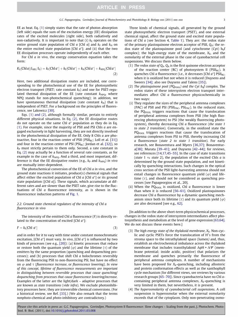
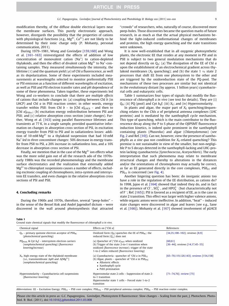
The present Historical Perspective focuses primarily on the fluo-rescence which Chl a in vivo emits and traces the historical evolu-tion of our awareness of its role in the molecular mechanism ofoxygenic photosynthesis, with emphasis up to about the late1970s. As soon as a solution of Chl a, a plant leaf, an alga, or a cya-nobacterium is moved from darkness to light, they start emittingChl a fluorescence. However, while the solution emits constantfluorescence under steady excitation, the intensity of fluorescenceemitted by the photosynthetic organisms changes with time con-tinuously (variable fluorescence), tracing characteristic time pat-terns that are typical of the major taxonomic groups of oxygenicphotosynthesizers (e.g., cyanobacteria, green algae, red algae, vas-cular plants). These Chl a fluorescence time patterns, known also asfluorescence induction, consist of two transients (or waves), a fastone (ls to s; symbolized as OJIPS [2–9] and a slower one (secondsto tens of minutes); symbolized as SMT [10–13]). For backgroundand explanation of these transients, see reviews [14–20]. Fig. 1shows characteristic fast (OJIPS) and slow (SMT) fluorescenceinduction patterns recorded with a plant leaf (Phaseolus vulgaris),suspensions of a green alga (Chlamydomonas reinhardtii), a phyco-bilisome (PBS) – containing cyanoabacterium (Synechocystis sp.)and a PBS-minus cyanobacterium (Acaryochloris marina). Here, Ostands for ‘‘origine’’ or initial fluorescence, J and I for intermediatelevels, P for peak, S for semi-steady state, M for maximum, and Tfor terminal steady state; occasionally, there is an inflection de-noted as D (for dip).
I fluorescence: Slow changes – Scaling from the past, J. Photochem. Photo-
Fig. 1. Chlorophyll a fluorescence induction traces recorded with a higher plant (Phaseolus vulgaris) leaf, a green alga (Chlamydomonas reinhardtii), a phycobilisome (PBS)/Chla-containing cyanobacterium (Synechococcus sp PCC 7942), and with a phycobiliprotein/Chl d/Chl a – containing cyanobacterium that has phycocyanin/allophycocyanin rodsattached to the cytoplasmic side of the thylakoid membrane but no PBS (Acaryochloris marina). Fluorescence data on the left panels are plotted against linear time scales andon the right against logarithmic time scales. All curves were recorded with the Handy PEA fluorometer of Hansatech Instruments, Ltd. (UK). Samples were preadapted todarkness for 20 min before measurements. Fluorescence excitation, k = 650 nm, Dk = 22 nm; fluorescence detection through an RG9 long pass glass filter (transmittance:starting at �690 nm; 50% at �725 nm; and maximal at �780 nm; Schott Glass Technologies, Inc, USA). Excitation intensities in lmol photons m�2 s�1: P. vulgaris, 50; C.reinhardtii, Synechococcus sp. PCC 7942 and A. marina, 1500. See text for explanation of the symbols (OJIPSMT) used; a.u. stands for arbitrary units. Reproduced fromPapageorgiou et al. (2007); the figure used here was produced by Dmitry Shevela.
2 G.C. Papageorgiou, Govindjee / Journal of Photochemistry and Photobiology B: Biology xxx (2011) xxx–xxx
2. Chlorophyll a fluorescence in vivo
For detailed information on various aspects of Chl a fluores-cence in vivo, including its practical applications on both landand marine organisms, see books edited by Govindjee et al. [21],Lichtenthaler [22], DeEll and Toivonen [23], Papageorgiou andGovindjee [24], and Suggett et al. [25]. For information on the de-tails of Photosystem II (PSII) that gives rise to most of constant andvariable Chl a fluorescence, see a book edited by Wydrzynski andSatoh [26], and a review [27]; for details on Photosystem I (PSI)that gives rise to a constant and a low fluorescence, see a book edi-ted by Golbeck [28].
Please cite this article in press as: G.C. Papageorgiou, Govindjee, Photosystem Ibiol. B: Biol. (2011), doi:10.1016/j.jphotobiol.2011.03.008
2.1. Why Chl a fluorescence in vivo is variable under steady excitation?
For Chl a in solution that becomes excited by absorbing light,the law of energy conservation can be expressed as follows (Parson[29]).
KE½Chla�ðIABS=I0Þ ¼ kF½Chla�� þ kH½Chla�� ð1Þ
where IABS and I0 are the absorbed and incident light intensities perunit time. The rate of absorption equals kE[Chl a][IABS/I0], and when[Chl a] does not change, it can be made a part of the rate constant k.Specifically, the first order rate constants considered here are kE forexcitation, kF for fluorescence emission, and kH for the dissipation of
I fluorescence: Slow changes – Scaling from the past, J. Photochem. Photo-
G.C. Papageorgiou, Govindjee / Journal of Photochemistry and Photobiology B: Biology xxx (2011) xxx–xxx 3
EE as heat. Eq. (1) simply states that the rate of photon absorption(left side) equals the sum of the excitation energy (EE) dissipationrates of the excited molecules (right side), both radiatively andnon-radiatively. It is important to note that (i) kE operates on theentire ground state population of Chl a [Chl a] and kF and kH onthe entire excited state population [Chl a⁄]; and (ii) that the twoEE dissipation processes operate independently of each other.
For Chl a in vivo, the energy conservation equation takes theform:
KE½Chla�ðIABS=I0Þ ¼ kF½Chla�� þ kP½Chla�� þ kH½Chla�� þ kNPQ ½Chla��ð2Þ
Here, two additional dissipation routes are included, one corre-sponding to the photochemical use of the EE for photosyntheticelectron transport (PSET; rate constant kP) and one for PSET-regu-lated thermal dissipation of the EE (rate constant kNPQ, whereNPQ stands for non-photochemical quenching); in addition, wehave spontaneous thermal dissipation (rate constant kH) that isindependent of PSET. For a background on the principles of fluores-cence, see Lakowicz [30].
Eqs. (1) and (2), although formally similar, pertain to entirelydifferent physical situations. In Eq. (2), the EE dissipative routesdo not operate on the same Chl a⁄ population as they do in Eq.(1). For example, since the majority of PSII and PSI Chls a are en-gaged exclusively in light harvesting, they are not directly involvedin the photochemical dissipation of the EE. Only 8 Chls a are pho-toactive, four in the reaction center of PSII (PSIIRC; Loll et al. [31])and four in the reaction center of PSI (PSIRC; Jordan et al. [32]), sokP must strictly pertain to them only. Second, a rate constant inEq. (2) may actually be a function of several rate constants, as forexample in the case of kNPQ. And a third, and most important, dif-ference is that the EE dissipative routes (e.g., kP and kNPQ) in vivoare mutually inter-dependent.
The photochemical conversion of the EE of Chl a in vivo, and theground state reactions it initiates, produce(s) chemical signals thataffect either the excited population of Chl a [Chl a⁄] or its groundstate population ([Chl a]). These signals, which accumulate at dif-ferent rates and are slower than the PSET rate, give rise to the fluc-tuations of Chl a fluorescence intensity, as is shown in thefluorescence induction patterns of Fig. 1.
2.2. Ground state chemical regulators of the intensity of Chl afluorescence in vivo
The intensity of the emitted Chl a fluorescence (F) is directly re-lated to the concentration of excited [Chl a⁄]:
F ¼ kF½Chl a�� ð3Þ
and in order for it to vary with time under constant monochromaticexcitation, [Chl a⁄] must vary. In vivo, [Chl a⁄] is influenced by twokinds of processes (see e.g., [20]): (a) kinetic processes that reduceor restore both the quantum yield (u) and the lifetime (s) of theemitters by the same proportion (quenching and dequenching pro-cesses); and (b) processes that shift Chl a holochromes reversiblyfrom the fluorescing PSII to non-fluorescing PSI, but have no effecton u and s (fluorescence increase, or fluorescence lowering). In viewof this concept, lifetime of fluorescence measurements are importantin distinguishing between reversible processes that cause quenching/dequenching from processes that cause fluorescence lowering/increase.Examples of the latter are the light/dark – adaptive processes thatare known as state transitions (vide infra). We exclude photoinhibi-tory processes here; they are irreversible chemical conversions. (Fora historical review, see Ref. [33].) (We also remark that the termsnonphoto-chemical and photo-inhibitory are contradictory.)
Please cite this article in press as: G.C. Papageorgiou, Govindjee, Photosystem Ibiol. B: Biol. (2011), doi:10.1016/j.jphotobiol.2011.03.008
Three kinds of chemical signals, all generated by the groundstate photosynthetic electron transport (PSET), and one externalchemical signal, affect the ground state and excited state popula-tions of Chl a (see Section 4, Table 1). They are: the redox stateof the primary plastoquinone electron acceptor of PSII, QA; the re-dox state of the plastoquinone pool (and cytochrome (Cyt) b6fcomplex); the high-energy state of the membrane, XE, and theosmolarity of the external phase in the case of cyanobacterial cellsuspensions. We discuss them below.
(1) The redox state of QA. QA is the first quinone electron acceptorof the reaction center (RC) of photosystem II (PSIIRC). Itquenches Chl a fluorescence (i.e., it decreases [Chl a⁄] PSIIRC,when it is oxidized but not when it is reduced (Duysens andSweers [34]; also see Duysens and Talens [35]).
(2) The plastoquinone pool (PQPOOL) and the Cyt b6f complex. Theredox states of these intersystem electron transport inter-mediates affect Chl a fluorescence physicochemically intwo ways:
(i) They regulate the sizes of the peripheral antenna complexes(PAC) of PSII and PSI (PSIIPAC, PSIPAC). In the reduced state,the PQPOOL triggers reactions that cause the translocationof peripheral antenna complexes from PSII (the high fluo-rescing photosystem) to PSI (the weakly fluorescing photo-system), thereby decreasing Chl a fluorescence (the state 1to state 2 transition). Conversely, in the oxidized state thePQPOOL triggers reactions that cause the translocation ofantenna complexes from PSI to PSII, thereby increasing Chla fluorescence (state 2 to state 1 transition). For earlyresearch, see Bonaventura and Myers [36,37]; Bonaventur-a[38]; Murata [39–41]; and Duysens [42–44]; for reviews,see references [14,17,45–55]. In the case of state transitions(state 1 ¡ state 2), the population of the excited Chls a isdetermined by the ground state population, and not kineti-cally by quenching interactions. Such changes in the opticalcross section of the PSII light-harvesting antenna should notentail changes in fluorescence quantum yield (u) and life-time (s), and should not be considered as quenching pro-cesses [see Papageorgiou et al., 20].
(ii) When the PQPOOL is oxidized, Chl a fluorescence is lowerthan when it is reduced [56–61]. Oxidized plastoquinonesdecrease Chl a fluorescence by a dynamic quenching mech-anism since both its lifetime (s) and its quantum yield (u)are also decreased [see e.g., 62].
[In addition to the above short-term physicochemical processes,changes in the redox state of intersystem intermediates affect pho-tosynthesis and metabolism at the level of gene expression [63,64].We do not discuss these events here.]
(3) The high energy state of the thylakoid membrane, XE. Non-cyc-lic and cyclic PSETs force the translocation of H+s from thestroma space to the intrathylakoid space (lumen) and, thus,establish an electrochemical imbalance across the thylakoidmembrane that includes transthylakoid DpH + DW (mem-brane potential; inside acidic, positive) that polarizes themembrane and quenches primarily the fluorescence ofperipheral antenna complexes. A number of mechanismshave been proposed for XE-quenching, including allostericand protein conformation effects as well as the xanthophyllcycle mechanism (for different views, see reviews by variousresearch groups [65–70]). Since cyanobacteria have no Chl a-containing peripheral antenna complexes, XE-quenching isvery limited in them, but nevertheless, it is present.
(4) The hyperosmolarity of cyanobacterial cell suspensions. A cellsuspension is hyperosmolar when the external osmolarityexceeds that of the cytoplasm. Only non-penetrating osmo-
I fluorescence: Slow changes – Scaling from the past, J. Photochem. Photo-
4 G.C. Papageorgiou, Govindjee / Journal of Photochemistry and Photobiology B: Biology xxx (2011) xxx–xxx
lytes contribute to the external osmorality that the cell per-ceives. Hyperosmolar media prevent the light-adaptive state2 to state 1 transition of phycobilisome (PBS)-containing cya-nobacteria (i.e., cells are locked in a low fluorescence state)and force an instantaneous transition of state 1 cyanobacte-ria to state 2 (measured as fluorescence lowering). Con-versely, in hypo-osmotic media the light-adaptive statetransitions occur normally. The hyperosmolarity effects oncyanobacterial cells are fully reversible [71–76].
3. Historical evolution of our awareness of the direct and theindirect regulation of Chl a fluorescence by photosyntheticelectron transport (PSET)
3.1. Direct regulators of Chl a fluorescence: The fast changes
Up to the early 1960s, Chl a fluorescence was assayed in orderto answer questions mainly about the light harvesting roles ofthe accessory pigments in photosynthesis and about photosyn-thetic activities. Questions about the light harvesting roles ofaccessory pigments were handled by comparing action spectra ofphotosynthesis and of sensitized Chl a fluorescence (Duysens[77]). Thus, action spectra of Chl a fluorescence were used to proveEE transfer from various accessory pigments to Chl a (from carote-noids, see [78], and reviews [79,80]; from phycobilins, see [81,82];and from Chl b, see [77]). For general reviews on Chl fluorescenceand EE transfer, see [83–86]. In the early days, photosynthesiswas measured in terms of O2 evolution (manometrically), or interms of CO2 uptake (either spectrographically, or by 14C incorpo-ration). With the advent of photoelectric devices, photosynthesiscould be also estimated indirectly, but more conveniently, usingChl a fluorescence as its inverse indicator.
The first compelling evidence for a complementarity relationbetween fluorescence and photosynthesis was obtained, in 1940,by McAlister and Myers [87,88]; they recorded mirror-image kinet-ics for CO2 uptake and Chl a fluorescence (measured with a photo-cell) upon exposing dark-adapted wheat plants and suspensions ofthe green alga Chlorella pyrenoidosa to light. The complementarityrelation is further supported by the fact that while photosyntheticcells emit less than 2–5% of the absorbed light quanta as Chl a fluo-rescence, for Chl a in solution this fraction rises to 20–30% [89–91].
The above information led to the reasonable assumption thatChl a disposes all quanta that cannot be used for photosynthesisas fluorescence, while the balance, namely the fraction of quantalost by thermal de-excitation, was constant and independent ofphotosynthesis. Despite the accumulating evidence to the contrary,the photosynthesis-Chl a fluorescence complementarity was adominant dogma until about the middle of 1970s.
As late as 1971, two of us [14] wrote:
‘‘. . .both Chl a fluorescence and photosynthesis draw on the excitedChl a population, and thus a change in the photosynthetic rate isreflected as a change in the yield of fluorescence;’’
And Myers [92], in 1974, stated:
‘‘a Chl a molecule cannot use the same quantum of energy for bothfluorescence and photochemistry.’’
These statements were, in all likelihood, attempts to simplifythe relationship, but they are historical curiosities considering thatfive years before, the same authors had obtained strong evidenceagainst the absolute dominance of the complementarity dogma(see [10–13,37,92]).
Nineteen sixty was, in a way, the demarcation year between theold photosynthesis and the new photosynthesis (see Myers [92]).By that time, the concept of two pigment systems and two light
Please cite this article in press as: G.C. Papageorgiou, Govindjee, Photosystem Ibiol. B: Biol. (2011), doi:10.1016/j.jphotobiol.2011.03.008
reactions was established (see e.g., Emerson and Rabinowitch[93]) and the photosynthetic electron transfer from water toNADP+ was fit into the framework of a Z-scheme by Hill andBendall [94]; see also Fig. 2 in Stirbet and Govindjee [9], this issue.For the evolution of the current model, and a historical perspective,see [95] and [96]. (Also see Delosme et al. [97].) After these devel-opments, the relation of photosynthesis to Chl a fluorescence couldnot be viewed simply as a direct competition between a photo-chemical act and a photoemissive act, as in the days of McAlisterand Myers [87,88]. The questions asked, the design of the experi-ments, and the interpretations of results were now guided by anew frame of thought, one that involved two pigment systemswith partially overlapping action spectra, and two photoreactions,one reducing and the other oxidizing an intersystem set of electroncarriers. In this spirit, Duysens and Sweers [34] interpreted theirown measurements with algae, cyanobacteria and chloroplasts,and the earlier results of Govindjee et al. [98] and Butler [99] onthe quenching of PSII fluorescence by PSI light, by postulating thatPSIIRC (which they designated as Q for quencher, not for quinone) isa regulator of Chl a fluorescence. Fluorescence was maximal whenall Q was reduced (to QH) by PSII, and minimal when it was all oxi-dized (to Q) by PSI. Their kinetic scheme explained the fast OP fluo-rescence rise both in chloroplasts and in algae by invoking a PSIIagainst PSI competition for the photoactive and redox-activequencher Q.
In the 1960s, evidence started piling up suggesting a more com-plex relation of Chl a fluorescence to photosynthesis. To explain theP to S fluorescence decline (see Fig. 1), which was observed in algaebut not in isolated chloroplasts, Duysens and Sweers [34] invokedthe conversion of QH to another quencher Q’ by means of ‘‘a sidereaction,’’ meaning not directly related to the main PSET. Thus,these authors viewed Q/QH as a direct (that we can also call anon-line) quencher while the QH/Q’ as an indirect (that we can callan off-line) quencher, a first attack on the complementarity dogma.
The strongest argument, however, against the complementaryrelation between fluorescence and photosynthesis, was based onobservations of parallel rise or decay kinetics of Chl a fluorescenceand of O2 evolution. Using actinic light of very high intensity, withor without PSET inhibitors, and fast recording of fluorescencekinetics, Morin [100], and Joliot [101] in the green alga Chlorellaand Delosme [102] in chloroplasts succeeded in resolving the ini-tial OP fluorescence rise in two phases: a first photochemical phase(corresponding to the OI phase; see Fig. 1), and a second thermalphase (corresponding to the IP phase). The first phase was assignedto the destruction of a quencher Q, a primary photoactive and re-dox-active reactant of PSIIRC, and the second phase to a redox-ac-tive intermediate R located between PSII and PSI. (Discovery ofthis ‘‘R’’ has remained elusive until today.) During the photochem-ical phase O2 evolution and Chl a fluorescence rise in parallel, butduring the thermal phase O2 evolution declines while fluorescencecontinues to rise.
3.2. Indirect regulation of chlorophyll a fluorescence: high energy state,protein/membrane conformation, state transitions, quenching
The concept of those early landmark studies was that the regu-lator of Chl a fluorescence is a photoactive and redox-active mole-cule or group of molecules, a link in the noncyclic PSET, which wasidentified either as the PSIIRC, or part of it. Inherent was also theassumption that, at room temperature, Chl a fluorescence origi-nates mostly from PSII, the O2-evolving photosystem. This assump-tion was confirmed, in 1966, after the biochemical separation ofthe PSII and PSI supercomplexes [103–105; also see 106].
The early 1960s saw the formulation of the chemiosmotic theoryby Mitchell [107] and the dramatic demonstration by Hind andJagendorf [108] that photophosphorylation can be split into a light
I fluorescence: Slow changes – Scaling from the past, J. Photochem. Photo-
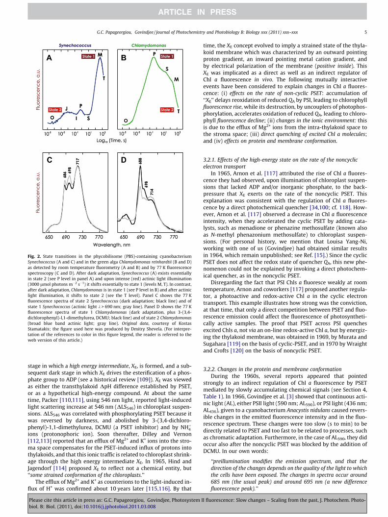
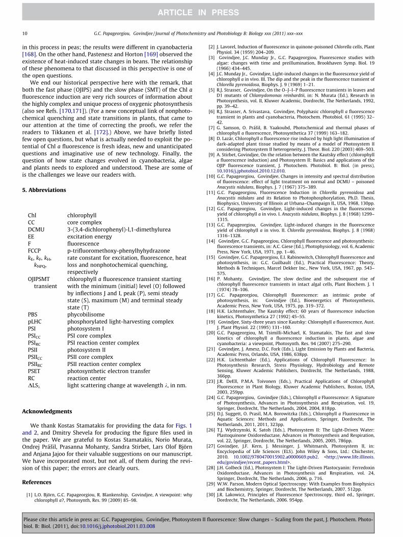
Fig. 2. State transitions in the phycobilisome (PBS)-containing cyanobacteriumSynechococcus (A and C) and in the green alga Chlamydomonas reinhardtii (B and D)as detected by room temperature fluorometry (A and B) and by 77 K fluorescencespectroscopy (C and D). After dark adaptation, Synechococcus (A) exists essentiallyin state 2 (see P level in panel A) and upon intense (red) actinic light illumination(3000 lmol photons m�2 s�1) it shifts essentially to state 1 (levels M, T). In contrast,after dark adaptation, Chlamydomonas is in state 1 (see P level in B) and after actiniclight illumination, it shifts to state 2 (see the T level). Panel C shows the 77 Kfluorescence spectra of state 2 Synechococcus (dark adaptation; black line) and ofstate 1 Synechococcus (actinic light k > 690 nm; gray line). Panel D shows the 77 Kfluorescence spectra of state 1 Chlamydomonas (dark adaptation, plus 3-(3,4-dichlorophenyl)-l,1-dimethylurea, DCMU; black line) and of state 2 Chlamydomonas(broad blue band actinic light; gray line). Original data, courtesy of KostasStamatakis; the figure used here was produced by Dmitry Shevela. (For interpre-tation of the references to color in this figure legend, the reader is referred to theweb version of this article.)
G.C. Papageorgiou, Govindjee / Journal of Photochemistry and Photobiology B: Biology xxx (2011) xxx–xxx 5
stage in which a high energy intermediate, XE, is formed, and a sub-sequent dark stage in which XE drives the esterification of a phos-phate group to ADP (see a historical review [109]). XE was viewedas either the transthylakoid DpH difference established by PSET,or as a hypothetical high-energy compound. At about the sametime, Packer [110,111], using 546 nm light, reported light-inducedlight scattering increase at 546 nm (DLS546) in chloroplast suspen-sions. DLS546 was correlated with phosphorylating PSET because itwas reversed by darkness, and abolished by 3-(3,4-dichloro-phenyl)-1,1-dimethylurea, DCMU (a PSET inhibitor) and by NHþ4ions (protonophoric ion). Soon thereafter, Dilley and Vernon[112,113] reported that an efflux of Mg2+ and K+ ions into the stro-ma space compensates for the PSET-induced influx of protons intothylakoids, and that this ionic traffic is related to chloroplast shrink-age through the high energy intermediate XE. In 1965, Hind andJagendorf [114] proposed XE to reflect not a chemical entity, but‘‘some strained conformation of the chloroplasts.’’
The efflux of Mg2+ and K+ as counterions to the light-induced in-flux of H+ was confirmed about 10 years later [115,116]. By that
Please cite this article in press as: G.C. Papageorgiou, Govindjee, Photosystem Ibiol. B: Biol. (2011), doi:10.1016/j.jphotobiol.2011.03.008
time, the XE concept evolved to imply a strained state of the thyla-koid membrane which was characterized by an outward pointingproton gradient, an inward pointing metal cation gradient, andby electrical polarization of the membrane (positive inside). ThisXE was implicated as a direct as well as an indirect regulator ofChl a fluorescence in vivo. The following mutually interactiveevents have been considered to explain changes in Chl a fluores-cence: (i) effects on the rate of non-cyclic PSET: accumulation of‘‘XE’’ delays reoxidation of reduced QA by PSI, leading to chlorophyllfluorescence rise, while its destruction, by uncouplers of photophos-phorylation, accelerates oxidation of reduced QA, leading to chloro-phyll fluorescence decline; (ii) changes in the ionic environment: thisis due to the efflux of Mg2+ ions from the intra-thylakoid space tothe stroma space; (iii) direct quenching of excited Chl a molecules;and (iv) effects on protein and membrane conformation.
3.2.1. Effects of the high-energy state on the rate of the noncyclicelectron transport
In 1965, Arnon et al. [117] attributed the rise of Chl a fluores-cence they had observed, upon illumination of chloroplast suspen-sions that lacked ADP and/or inorganic phosphate, to the back-pressure that XE exerts on the rate of the noncyclic PSET. Thisexplanation was consistent with the regulation of Chl a fluores-cence by a direct photochemical quencher [34,100; cf. 118]. How-ever, Arnon et al. [117] observed a decrease in Chl a fluorescenceintensity, when they accelerated the cyclic PSET by adding cata-lysts, such as menadione or phenazine methosulfate (known alsoas N-methyl phenazonium methosulfate) to chloroplast suspen-sions. (For personal history, we mention that Louisa Yang-Ni,working with one of us (Govindjee) had obtained similar resultsin 1964, which remain unpublished; see Ref. [15].) Since the cyclicPSET does not affect the redox state of quencher QA, this new phe-nomenon could not be explained by invoking a direct photochem-ical quencher, as in the noncyclic PSET.
Disregarding the fact that PSI Chls a fluoresce weakly at roomtemperature, Arnon and coworkers [117] proposed another regula-tor, a photoactive and redox-active Chl a in the cyclic electrontransport. This example illustrates how strong was the conviction,at that time, that only a direct competition between PSET and fluo-rescence emission could affect the fluorescence of photosyntheti-cally active samples. The proof that PSET across PSI quenchesexcited Chls a, not via an on-line redox-active Chl a, but by energiz-ing the thylakoid membrane, was obtained in 1969, by Murata andSugahara [119] on the basis of cyclic-PSET, and in 1970 by Wraightand Crofts [120] on the basis of noncyclic PSET.
3.2.2. Changes in the protein and membrane conformationDuring the 1960s, several reports appeared that pointed
strongly to an indirect regulation of Chl a fluorescence by PSETmediated by slowly accumulating chemical signals (see Section 4,Table 1). In 1966, Govindjee et al. [3] showed that continuous acti-nic light (AL), either PSII light (590 nm; AL590), or PSI light (436 nm;AL436), given to a cyanobacterium Anacystis nidulans caused revers-ible changes in the emitted fluorescence intensity and in the fluo-rescence spectrum. These changes were too slow (s to min) to bedirectly related to PSET and too fast to be related to processes, suchas chromatic adaptation. Furthermore, in the case of AL590, they didoccur also after the noncyclic PSET was blocked by the addition ofDCMU. In our own words:
‘‘preillumination modifies the emission spectrum, and that thedirection of the changes depends on the quality of the light to whichthe cells have been exposed. The changes in spectra occur around685 nm (the usual peak) and around 695 nm (a new differencefluorescence peak).’’
I fluorescence: Slow changes – Scaling from the past, J. Photochem. Photo-
6 G.C. Papageorgiou, Govindjee / Journal of Photochemistry and Photobiology B: Biology xxx (2011) xxx–xxx
The F695 peak1 had been studied independently in severallaboratories including our own at the University of Illinois atUrbana-Champaign by fluorescence spectroscopy at 77 K (see e.g.,[121–125]). The general consensus was (and is) that F685 andF695 originates in PSII while F715–735 originates in PSI (see 1963papers1 in Kok and Jagendorf [125]; for F720, see Brody [126]).
In 1967, Papageorgiou and Govindjee [10] considered two pos-sible explanations for the slow fluorescence changes. The first, con-sistent with the direct regulation, was the accumulation of reducedQA (or QH) because of (i) the imbalance in the primary photoreactionsin PSIIRC and PSIRC in the case of – DCMU cells, with PSII predominat-ing, and (ii) the inability of PSI to reoxidize reduced QA (QH) inthe + DCMU cells. The second was a novel idea of ‘‘conformationalchanges in the membrane’’ as the cause of the light-induced slowchanges of Chl a fluorescence. It was inspired by the work of Packer[110,111], Dilley and Vernon [112,113] and of Hind and Jagendorf[114] on light-induced light scattering changes by chloroplast sus-pensions, mentioned earlier. This was the first time that light-in-duced changes in Chl a fluorescence were not linked to PSETdirectly, but to PSET-related indirect causes. In 1971, Myers [127]wrote about our work:
‘‘A common explanation has been reached in terms of almost nakedspeculation: that small conformational changes alter distances[between] pigment molecules and thereby provide partial carbure-tor control in transfer of excitation to the reaction centers. The ideais not novel. It was reached previously by Papageorgiou and Gov-indjee (102) from fluorescence time course studies.’’
If the membrane conformational changes, a byproduct of photo-phosphorylating PSET, are indeed behind the slow S to M fluores-cence rise in Anacystis, then photophosphorylation inhibitorsought to block it out. The question was: which one of the twostages that Hind and Jagendorf [114] had described was responsi-ble for it? Was it the photochemical formation of XE, or the darkphosphorylation of ADP? The dilemma was resolved by our obser-vation in 1968 [12,13] that the SM fluorescence rise was blocked(although not quantitatively) by the protonophore carbonyl cya-nide p-trifluoromethoxy phenylhyhydrazone (FCCP), while it wasinsensitive to phlorizin that allows DpH formation across the thy-lakoid membrane but inhibits phosphorylation.
Another striking result, which could not be rationalized interms of the ‘direct’ quenching, was the demonstration that O2
evolution rises in parallel with fluorescence during the SM phasein both algae and cyanobacteria ([11, 12; also see papers by Ban-nister [128] and Bannister and Rice [129]). The two of us [13]attributed it to ‘‘a conversion of a nonphotoactive and nonfluorescentportion of Chl a to the photoactive and fluorescent form’’, whereasBannister and Rice [129] attributed it to the conceptually equiva-lent ‘‘slow activation |that| converts inactive PSII units (IIi) to activeones (IIa).’’ Implicit in the above explanations is the idea that onlythe Chls a of PSII were involved, so these proposed activations wereintra-PSII events. The same phenomenology, namely the parallelkinetics of O2 evolution and of Chl a fluorescence were interpretedone year later by Bonaventura and Myers [36–38] and by Murata[39–41] by a mechanism that involves the Chls a of both photosys-tems, namely by an intersystem regulation, the state transitionmechanism (see Section 3.2.3).
1 The F695 band, at 77 K, had been discovered independently, in 1963, by B. Kok(Fluorescence Studies, pp. 45–55), Govindjee (Emerson Enhancement and Two LightReactions in Photosynthesis, pp 318–334), S. S. Brody and M. Brody (Aggregatedchlorophylls in vivo, pp. 455–478) and J.A, Bergeron (Studies of the Localization,Physicochemical Properties, and Action of Phycocyanin in Anacystis nidulans, pp. 527–536) (see Kok and Jagendorf (Eds.), 1963, Photosynthetic Mechanisms of green Plants,Publication #1145, National Academy of Sciences—National Research Council,Washington, DC [125]. The F720 band, at 77 K, was discovered even earlier by Brody(1958) [126].
Please cite this article in press as: G.C. Papageorgiou, Govindjee, Photosystem Ibiol. B: Biol. (2011), doi:10.1016/j.jphotobiol.2011.03.008
3.2.3. State transitionsThe term state transitions describes a reversible physiological
mechanism that enables plants, algae, and cyanobacteria to opti-mize PSET at rapidly fluctuating light conditions, and additionallyto enable cyanobacteria to dissipate excess EE as heat. In state 1,the PSII light harvesting antenna is larger and the PSI antennasmaller than in state 2. Conversely, in state 2, the PSII antenna issmaller and the PSI antenna larger than in state 1. The light stateof photosynthetic cells can be recognized by kinetic fluorometryat room temperature and by spectrofluorometry at very low tem-peratures. At room temperature, the conversion of photosyntheticcells from state 2 to state 1 is observed by a kinetic rise of Chl afluorescence, whereas the state 1 to 2 conversion by a decline. At77 K, and in state 1, the PSII emission bands, F684 and F696, arestronger and the PSI emission band F720 is weaker than whenthe cells are in state 2 (see examples in Fig. 2).
In the early 1960s, one could have intuitively postulated thenecessity of a valve that would adjust the timing of photoreactionsI and II in changing light conditions by regulating the amount of EEthey receive from peripheral antennae. As mentioned above, such amechanism, that of the state transitions of today, was indeed pos-tulated at the end of the decade, independently by Bonaventuraand Myers [36–38], and by Murata [39–41]. This discovery wasnot based on theoretical reasoning but it was deduced from specif-ically designed experiments.
Going after the events in chronological order, we recognize that,by 1969, at least four groups had independently reported simulta-neous slow rises in Chl a fluorescence and in O2 evolution rate:Duysens and Talens [35] in cyanobacteria, Bonaventura and Myers[36–38], Bannister and Rice [129], and the two of us [11–13] inboth cyanobacteria and green algae. (See also the 1970 paper byMohanty et al. [130] in green algae, and our 1971 review [14].)These parallel rise kinetics of O2 evolution and Chl a fluorescence,which were totally different from the mirror-image kinetics re-ported by McAlister and Myers in 1940 [87,88], constituted thefirst demonstration of what we would call today a state 2 to state1 transition. However, as mentioned above, our research group[12,13] and that of Bannister [128,129] had invoked an intra-PSIIactivation of Chl a while the core of the state transition concept fo-cuses on intersystem EE exchanges and their regulation by light ab-sorbed in PSII and PSI, and by darkness.
The latter idea matured independently in 1968 in two researchgroups, one in the USA and the other in Japan, which, at the time,were unaware of each others’ experiments. At the University ofTexas, at Austin, Texas, Celia Bonaventura and Jack Myers [36] usedthe green alga Chlorella pyrenoidosa for their experiments while atthe University of Tokyo, Norio Murata [39] experimented with thered alga Porphyridium cruentum. Their results were published thefollowing year: by Murata [40] in the January 1969 issue of Biochi-mica et Biophysica Acta, by Bonaventura [38] also in January 1969 inher Ph.D. Thesis at the University of Texas, at Austin, and by Bon-aventura and Myers [37] in the August 1969 issue of Biochimicaet Biophysica Acta.
By preilluminating the red alga Porphyridium cruentum, at roomtemperature, with either actinic light 2 (ALPSII) or with actinic light1 (ALPSI) and by comparing their effects by measuring the 77 K Chla fluorescence emission bands of PSII (F684, F695) and PSI (F712),Murata [40] concluded:
‘‘Upon illumination of pigment system II, a greater amount ofabsorbed light energy is transferred to chlorophyll a in pigment sys-tem I and a lesser amount of light energy is transferred to chloro-phyll a in pigment system II than occurs upon illumination ofpigment system I. Such a change of excitation transfer reducesthe difference between the amounts of excitation energy availablefor photoreactions I and II.’’
I fluorescence: Slow changes – Scaling from the past, J. Photochem. Photo-
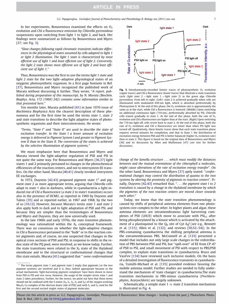
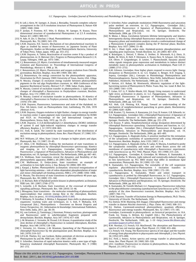
Fig. 3. Simultaneously-recorded kinetic traces of photosynthetic O2 evolution(upper traces) and Chl a fluorescence (lower traces) that illustrate a state transitioncycle (light state 2 ? light state 1 ? light state 2) in the green alga Chlorellapyrenoidosa. From left to right: (Left): state 2 is achieved gradually when cells areilluminated with modulated 650 nm light, which is absorbed preferentially byPhotosystem II. At the end of this phase, the O2 evolution rate is approximately thesame as at the start, while Chl a fluorescence is lowered. (Middle) Upon switchingon additional continuous light (710 nm), preferentially absorbed by PSI, Chlorellacells transit gradually to state 1. At the end of this phase, both the rate of O2
evolution and Chl a fluorescence are higher than at the start. (Right) Upon switchingthe 710 nm light off, cells revert back to state 2. At the end of this phase, both therate of O2 evolution and Chl a fluorescence are lower than when PSI light wasturned off. Qualitatively, these kinetic traces show that each state transition phaserequires several minutes for completion, and that in State 1 the distribution ofexcitation energy between PSII and PSI is better balanced (higher O2 evolution rate)than in state 2. This figure is based on the original data of Bonaventura and Myers[36] and its discussion by Allen and Mullineaux [47] (see text for furtherdiscussion).
G.C. Papageorgiou, Govindjee / Journal of Photochemistry and Photobiology B: Biology xxx (2011) xxx–xxx 7
In her experiments, Bonaventura examined the effects on O2
evolution and Chl a fluorescence emission by Chlorella pyrenoidosasuspensions upon switching from light 1 to light 2, and back. Herfindings were summarized as follows by Bonaventura and Myers[37; see Fig. 3]:
‘‘Slow changes following rapid chromatic transients indicate differ-ences in the physiological states assumed by cells adapted to light 1or light 2 illumination. The light 1 state is characterized by mostefficient use of light 1 and least efficient use of light 2. Conversely,the light 2 state shows most efficient use of light 2 and least effi-cient use of light 1.’’
Thus, Bonaventura was the first to use the terms light 1 state andlight 2 state for the two light–adaptive physiological states of anoxygenic photosynthetic organism. In a first page footnote in Ref.[37], Bonaventura and Myers recognized the published work ofMurata without discussing it further. They wrote, ‘‘A report, pub-lished during preparation of this manuscript, by N. Murata (Biochim.Biophys. Acta, 172 (1969) 242) contains some information similar tothat presented here.’’
Ten months later, Murata published [41] in June 1970 issue ofBiochimica Biophysica Acta the clearest description of these phe-nomena and for the first time he used the terms state 1, state 2and state transitions to describe the light adaptive states of photo-synthetic organisms and their interconversions. He wrote:
‘‘Terms, ‘‘State I’’ and ‘‘State II’’ are used to describe the state ofexcitation transfer. In the State I a lesser amount of excitationenergy is delivered in Pigment System I and greater to Pigment Sys-tem II than in the State II. The conversion of the states is achievedby the selective illumination of pigment systems.’’
We must emphasize here that Bonaventura and Myers andMurata viewed the light-adaptive regulation of PSII and PSI innot quite the same way. For Bonaventura and Myers [36,37] lightstates 1 and 2 primarily pertained to changes in the photochemicalefficiencies of the reaction centers, and not to intersystem EE trans-fers. On the other hand, Murata [40,41] clearly invoked interystemEE exchanges.
In 1972, Duysens [42,43] proposed pigment state 12 and pig-ment state 2 as more appropriate terms, arguing that Chlorella cellsadapt to state 1 also in darkness, while in cyanobacteria a light-in-duced rise of Chl a fluorescence (a state 2 to state1 transition) occursalso in the presence of DCMU, as reported in 1969 by Duysens andTalens [35] and as reported earlier, in 1967 and 1968, by the twoof us [10,12]. However, because Murata’s terms state 1 and state 2also apply both to dark and to light adaptation of PSII and PSI, andbecause they are simpler than the terminologies of Bonaventuraand Myers and Duysens, they are now universally used.
In the late 1960s and early 1970s, the state transition phenom-enology was rationalized within the XE conceptual framework.There was no consensus on whether the light-adaptive changesin Chl a fluorescence pertained to the ‘‘bulk’’ or to the reaction cen-ter pigments and, of course, no one suspected that changes in theoptical cross sections of PSII and PSI, in response to shifts in the re-dox state of the PQ pool, were involved, as we know today. Further,the state transitions were related to the XE state of the thylakoidmembrane and to the structural or conformational changes thatthis state entails. Murata [41] suggested that ‘‘ some conformational
2 The terms pigment state 1 and pigment state 2 imply that pigments (or the twopigment systems) are involved and it is, thus, indeed appropriate because in theactual mechanisms ‘light-harvesting pigment complexes’ have been shown to movefrom PSII to PSI and vice versa. However, the usage of the simpler terms state 1 andstate 2 takes precedence. Further, for the novice, we point out here that these termsshould not be confused with the S-states (S0, S1, S2, S3 and S4) of the oxygen-evolvingMn4O5 Ca complex of the electron donor side of PSII) and with S1 and S2 used for thefirst and the second excited singlet states of pigment molecules.
Please cite this article in press as: G.C. Papageorgiou, Govindjee, Photosystem Ibiol. B: Biol. (2011), doi:10.1016/j.jphotobiol.2011.03.008
change of the lamella structure . . . which must modify the distancesbetween and the mutual orientations of the chlorophyll a molecules,might cause alterations of the rate of excitation energy transfer’’. Onthe other hand, Bonaventura and Myers [37] aptly stated: ‘‘confor-mational changes may control the distribution of quanta to the twosystems by altering the proximity of pigments to reaction centers’’. Fi-nally, Duysens [42,43] remarked that, ‘‘. . . the pigment state I to 2transition is caused by a change in the thylakoid membrane by whichthe pigments of the two reaction centers are moved closer towardseach other.’’
Today, we know that the state transition phenomenology iscaused by shifts of peripheral antenna elements from one photo-system core complex to the other. In higher plants and algae, theseantenna elements are intramembranous light harvesting com-plexes of PSII (LHCII) which move to associate with PSICC afterbeing phosphorylated by a kinase which is activated by the attach-ment of a plastoquinol to the Q0 site of Cyt b6f (see e.g., Bennettet al. [131]; Allen et al. [132]; and reviews [50,52–54]). In thePBS-containing cyanobacteria the shifting peripheral antenna isthe extramembranous PBS. McConnell et al. [133] presented amodel that includes not only large scale changes in the redistribu-tion of PBS between PSII and PSI, but ‘‘spill-over’’ of EE from CP-47of PSII to PSI, and small movement of PSI with respect to PBS/PSIIcomplex, to explain state transitions in cyanobacteria. Bruce andVasil’ev [134] have reviewed such inclusive models. On the basisof a detailed investigation of fluorescence transients in cyanobacte-ria, Tsimilli-Michael et al. [135] provided evidence favoring themobile antenna model. Further studies are needed to fully under-stand the mechanism of ‘state changes’ in cyanobacteria.The statetransition mechanisms in PBS-minus cyanobacteria (Prochloro-phytes, Acaryochloris) are largely unknown.
Schematically, a simple state 1 ¡ state 2 transition mechanismis illustrated in Fig. 4.
I fluorescence: Slow changes – Scaling from the past, J. Photochem. Photo-
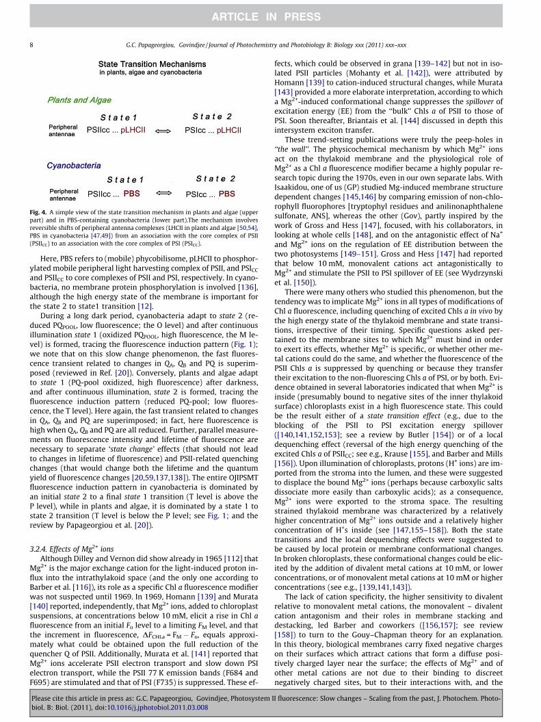
Fig. 4. A simple view of the state transition mechanism in plants and algae (upperpart) and in PBS-containing cyanobacteria (lower part).The mechanism involvesreversible shifts of peripheral antenna complexes (LHCII in plants and algae [50,54],PBS in cyanobacteria [47,49]) from an association with the core complex of PSII(PSIICC) to an association with the core complex of PSI (PSICC).
8 G.C. Papageorgiou, Govindjee / Journal of Photochemistry and Photobiology B: Biology xxx (2011) xxx–xxx
Here, PBS refers to (mobile) phycobilisome, pLHCII to phosphor-ylated mobile peripheral light harvesting complex of PSII, and PSICC
and PSIICC to core complexes of PSII and PSI, respectively. In cyano-bacteria, no membrane protein phosphorylation is involved [136],although the high energy state of the membrane is important forthe state 2 to state1 transition [12].
During a long dark period, cyanobacteria adapt to state 2 (re-duced PQPOOL, low fluorescence; the O level) and after continuousillumination state 1 (oxidized PQPOOL, high fluorescence, the M le-vel) is formed, tracing the fluorescence induction pattern (Fig. 1);we note that on this slow change phenomenon, the fast fluores-cence transient related to changes in QA, QB and PQ is superim-posed (reviewed in Ref. [20]). Conversely, plants and algae adaptto state 1 (PQ-pool oxidized, high fluorescence) after darkness,and after continuous illumination, state 2 is formed, tracing thefluorescence induction pattern (reduced PQ-pool; low fluores-cence, the T level). Here again, the fast transient related to changesin QA, QB and PQ are superimposed; in fact, here fluorescence ishigh when QA, QB and PQ are all reduced. Further, parallel measure-ments on fluorescence intensity and lifetime of fluorescence arenecessary to separate ‘state change’ effects (that should not leadto changes in lifetime of fluorescence) and PSII-related quenchingchanges (that would change both the lifetime and the quantumyield of fluorescence changes [20,59,137,138]). The entire OJIPSMTfluorescence induction pattern in cyanobacteria is dominated byan initial state 2 to a final state 1 transition (T level is above theP level), while in plants and algae, it is dominated by a state 1 tostate 2 transition (T level is below the P level; see Fig. 1; and thereview by Papageorgiou et al. [20]).
3.2.4. Effects of Mg2+ ionsAlthough Dilley and Vernon did show already in 1965 [112] that
Mg2+ is the major exchange cation for the light-induced proton in-flux into the intrathylakoid space (and the only one according toBarber et al. [116]), its role as a specific Chl a fluorescence modifierwas not suspected until 1969. In 1969, Homann [139] and Murata[140] reported, independently, that Mg2+ ions, added to chloroplastsuspensions, at concentrations below 10 mM, elicit a rise in Chl afluorescence from an initial Fo level to a limiting FM level, and thatthe increment in fluorescence, DFCHLa = FM � Fo, equals approxi-mately what could be obtained upon the full reduction of thequencher Q of PSII. Additionally, Murata et al. [141] reported thatMg2+ ions accelerate PSII electron transport and slow down PSIelectron transport, while the PSII 77 K emission bands (F684 andF695) are stimulated and that of PSI (F735) is suppressed. These ef-
Please cite this article in press as: G.C. Papageorgiou, Govindjee, Photosystem Ibiol. B: Biol. (2011), doi:10.1016/j.jphotobiol.2011.03.008
fects, which could be observed in grana [139–142] but not in iso-lated PSII particles (Mohanty et al. [142]), were attributed byHomann [139] to cation-induced structural changes, while Murata[143] provided a more elaborate interpretation, according to whicha Mg2+-induced conformational change suppresses the spillover ofexcitation energy (EE) from the ‘‘bulk’’ Chls a of PSII to those ofPSI. Soon thereafter, Briantais et al. [144] discussed in depth thisintersystem exciton transfer.
These trend-setting publications were truly the peep-holes in‘‘the wall’’. The physicochemical mechanism by which Mg2+ ionsact on the thylakoid membrane and the physiological role ofMg2+ as a Chl a fluorescence modifier became a highly popular re-search topic during the 1970s, even in our own separate labs. WithIsaakidou, one of us (GP) studied Mg-induced membrane structuredependent changes [145,146] by comparing emission of non-chlo-rophyll fluorophores [tryptophyl residues and anilinonaphthalenesulfonate, ANS], whereas the other (Gov), partly inspired by thework of Gross and Hess [147], focused, with his collaborators, inlooking at whole cells [148], and on the antagonistic effect of Na+
and Mg2+ ions on the regulation of EE distribution between thetwo photosystems [149–151]. Gross and Hess [147] had reportedthat below 10 mM, monovalent cations act antagonistically toMg2+ and stimulate the PSII to PSI spillover of EE (see Wydrzynskiet al. [150]).
There were many others who studied this phenomenon, but thetendency was to implicate Mg2+ ions in all types of modifications ofChl a fluorescence, including quenching of excited Chls a in vivo bythe high energy state of the thylakoid membrane and state transi-tions, irrespective of their timing. Specific questions asked per-tained to the membrane sites to which Mg2+ must bind in orderto exert its effects, whether Mg2+ is specific, or whether other me-tal cations could do the same, and whether the fluorescence of thePSII Chls a is suppressed by quenching or because they transfertheir excitation to the non-fluorescing Chls a of PSI, or by both. Evi-dence obtained in several laboratories indicated that when Mg2+ isinside (presumably bound to negative sites of the inner thylakoidsurface) chloroplasts exist in a high fluorescence state. This couldbe the result either of a state transition effect (e.g., due to theblocking of the PSII to PSI excitation energy spillover([140,141,152,153]; see a review by Butler [154]) or of a localdequenching effect (reversal of the high energy quenching of theexcited Chls a of PSIICC; see e.g., Krause [155], and Barber and Mills[156]). Upon illumination of chloroplasts, protons (H+ ions) are im-ported from the stroma into the lumen, and these were suggestedto displace the bound Mg2+ ions (perhaps because carboxylic saltsdissociate more easily than carboxylic acids); as a consequence,Mg2+ ions were exported to the stroma space. The resultingstrained thylakoid membrane was characterized by a relativelyhigher concentration of Mg2+ ions outside and a relatively higherconcentration of H+s inside (see [147,155–158]). Both the statetransitions and the local dequenching effects were suggested tobe caused by local protein or membrane conformational changes.In broken chloroplasts, these conformational changes could be elic-ited by the addition of divalent metal cations at 10 mM, or lowerconcentrations, or of monovalent metal cations at 10 mM or higherconcentrations (see e.g., [139,141,143]).
The lack of cation specificity, the higher sensitivity to divalentrelative to monovalent metal cations, the monovalent – divalentcation antagonism and their roles in membrane stacking anddestacking, led Barber and coworkers ([156,157]; see review[158]) to turn to the Gouy–Chapman theory for an explanation.In this theory, biological membranes carry fixed negative chargeson their surfaces which attract cations that form a diffuse posi-tively charged layer near the surface; the effects of Mg2+ and ofother metal cations are not due to their binding to discreetnegatively charged sites, but to their interactions with, and the
I fluorescence: Slow changes – Scaling from the past, J. Photochem. Photo-
G.C. Papageorgiou, Govindjee / Journal of Photochemistry and Photobiology B: Biology xxx (2011) xxx–xxx 9
modification thereby, of the diffuse double electrical layers nearthe membrane surfaces. This purely electrostatic approach,however, disregards the possibility that the properties of cationswith physiological functions (e.g., Mg2+, Ca2+) are not likely to bedetermined by their ionic charge only (P. Mohanty, personalcommunication, 2011).
During 1979–1981, Wong and Govindjee [159,160] and Wonget al. [161–163] reinvestigated the effects of addition of lowconcentration of monovalent cation (Na+) to cation-depletedthylakoids, and then the effect of divalent cation Mg2+ to Na+-con-taining samples. They measured simultaneously the intensity, thelifetime (s) and the quantum yield (u) of Chl a fluorescence, as wellas its depolarization. Some of these experiments included mea-surements at wavelengths selected to monitor preferentially PSIIor PSI emission as a function of different wavelengths of excitation,as well as PSII and PSI electron transfer rates and pH dependence ofsome of these phenomena. Taken together, these experiments ledWong and co-workers to conclude that there are multiple effectsof cations that include changes in: (a) coupling between Chl b (inLHCP) and Chl a in PSII reaction center; in other words, energytransfer within PSII: from Chl b – to [Chl a]LHCII – and then to[Chl a]PSIIcc; (b) excitation energy redistribution between PSI andPSII; and (c) relative absorption cross section (state changes). Fur-ther, Wong et al. [163] using parallel fluorescence lifetimes andtransients at 77 K, in a single sample, provided quantitative infor-mation on changes in absorption cross section of PSI, in excitationenergy transfer from PSII to PSI and in radiationless losses: addi-tion of 10 mM Mg2+ to a thylakoid suspension that had 10 mMNa+ led to three concomitant changes: 50% decrease in energy trans-fer from PSII to PSI, a 20% increase in radiationless loss, and a 10%decrease in absorption cross section of PSI.
Finally, we mention that in the case of the Mg2+ ion effects whatwas the more solid gain out of all the research ado of the 1970s-early 1980s was the recorded phenomenology and the membranesurface electrostatics and the realization that externally addedMg2+ to chloroplast suspensions causes a number of effects, includ-ing excitonic coupling of chromophores, intra-system and intersys-tem EE transfers, and even changes in the relative absorption crosssections of PSI and PSII.
4. Concluding remarks
During the 1960s and 1970s, therefore, several ‘‘peep-holes’’ –in the sense of the Bessel Kok and André Jagendorf dictum – werediscovered in the wall around photosynthesis that attracted
Table 1Ground state chemical signals that modify the fluorescence of chlorophyll a in vivo.
Chemical signal Effects on [⁄Chl a]
QA – primary quinone electron acceptor of PSIIRC
(photochemical quenching)Oxidized form (QA) qureduced form (Q�A ) doe
PQPOOL & Cyt b6f – intersystem electron carriers(nonphotochemical quenching) (fluorescencelowering/increase)
(a) Quencher of (⁄Chl a(b) Trigger of the stateoxidized (fluorescence1-to-2 when reduced (
XE, high energy state of the thylakoid membrane(i.e., transmembrane DpH and D[Mg2+](nonphotochemical quenching)
(a) Cyanobacteria –que(b) Algae, plants – que� Allosteric effects� Xanthophyll cycl� PsbS protonation
Hyperosmolarity – Cyanobacteria cell suspensions(fluorescence lowering)
Hyperosmolar state 2 cto-1 transitionHyperosmolar state 1transition
Abbreviations: EE – Excitation Energy; PSIICC – PSII core complex; PSIIPAC – PSII peripher
Please cite this article in press as: G.C. Papageorgiou, Govindjee, Photosystem Ibiol. B: Biol. (2011), doi:10.1016/j.jphotobiol.2011.03.008
‘‘crowds’’ of researchers, who, naturally of course, discovered morepeep-holes. Those discoveries became the question marks of futureresearch, in as much as that the actual physical mechanisms be-hind the light-induced conformational changes of membranesand proteins, the high energy quenching and the state transitionswere unknown.
It is now well-established that in all oxygenic photosyntheticplants, the electronic EE that resides at any moment in PSI and inPSII is subject to two general modulation mechanisms that donot depend directly on QA: (a) The dissipation of the EE of Chl aupon the establishment of an electrochemical gradient across thy-lakoid membranes (XE quenching); and (b) the state 1 ¡ state 2processes that shift EE from one photosystem to the other andare triggered by the oxidoreduction state of the PQ-pool. Themechanisms of these two processes are similar but not identicalin the evolutionary distant (by approx. 1 billion years) cyanobacte-rial cells and eukaryotic cells.
Table 1 summarizes four types of signals that modify the fluo-rescence of chlorophyll a in vivo (see text for details). They are (i)QA; (ii) PQ (pool) and Cyt b6f; (iii) XE; and (iv) Hyperosmolarity.
In plants and algae, the major part of XE quenching/dequen-ching relates to the Chls a of peripheral antenna complexes (LHCproteins) and is mediated by the xanthophyll cycle mechanism.This type of quenching, which is the main contributor to the fluo-rescence decay along the P to S phase of the OJIPSMT fluorescenceinduction kinetics, is indeed quite prominent in the xanthophyllcontaining plants (Phaseolus) and algae (Chlamydomonas) (seeFig. 2 and Ref. [10]). Can we, however, view the presence of xantho-phylls as a sine qua non condition for XE-related quenching? Thispremise is not sustainable in view of the smaller, but non-negligi-ble P to S decays detected in the xanthophyll-lacking and LHC-pro-tein lacking cyanobacteria (Synechococcus, Acaryochloris). The earlyinterpretation that such phenomena may relate to membranestructural changes and thereby to alterations in the distancesand/or the orientations of chromophores may actually be correct,as far as EE generated directly in the core complexes, PSIICC andPSICC is concerned (see Fig. 4).
Another lingering question has been: do inorganic anions toohave a role in the regulation of the EE distribution, as cations do?In 1998, Jajoo et al. [164] showed that indeed they do, and in factin the presence of Cl�, SO2�
4 , and HPO2�4 (but characteristically not
of F�; Ref. [165]); PSI is favored as a recipient of EE, as is the case instate 2 transition. This effect was larger with higher valence anions,while organic anions were ineffective. In addition, ‘‘heat’’ – inducedstate changes were discovered in algae and leaves (see e.g., Saneet al. [166]). Mohanty et al. [167] showed involvement of pLHCII
References
enches the EE of PSIIRC; thes not
[34,35,100–102]; reviews [8,9]
)ANT when oxidized [60,61]2-to-1 transition when
increase); trigger of the statefluorescence lowering)
[36–44]; reviews [14,17,45–55]
ncher of ⁄Chl a in PSIICC [65–70,119,120,143]; reviews [154,158]ncher of ⁄Chl a in PSIIPAC
e
ells – Suppression of state 2-
cells – Forced state 1-to-2
[71–74,76]; review [75]
al antenna complex; PSIIRC – PSII reaction center complex.
I fluorescence: Slow changes – Scaling from the past, J. Photochem. Photo-
10 G.C. Papageorgiou, Govindjee / Journal of Photochemistry and Photobiology B: Biology xxx (2011) xxx–xxx
in this process in peas; the results were different in cyanobacteria[168]. On the other hand, Pastenesz and Horton [169] observed theexistence of heat-induced state changes in beans. The relationshipof these phenomena to that discussed in this perspective is one ofthe open questions.
We end our historical perspective here with the remark, thatboth the fast phase (OJIPS) and the slow phase (SMT) of the Chl afluorescence induction are very rich sources of information aboutthe highly complex and unique process of oxygenic photosynthesis(also see Refs. [170,171]). (For a new conceptual link of nonphoto-chemical quenching and state transitions in plants, that came toour attention at the time of correcting the proofs, we refer thereaders to Tikkanen et al. [172].) Above, we have briefly listedfew open questions, but what is actually needed to exploit the po-tential of Chl a fluorescence is fresh ideas, new and unanticipatedquestions and imaginative use of new technology. Finally, thequestion of how state changes evolved in cyanobacteria, algaeand plants needs to explored and understood. These are some ofis the challenges we leave our readers with.
5. Abbreviations
Pb
Chl
lease cite this artiol. B: Biol. (2011
chlorophyll
CC core complex DCMU 3-(3,4-dichlorophenyl)-l,1-dimethylurea EE excitation energy F fluorescence FCCP p-trifluoromethoxy-phenylhyhydrazone kE, kF, kH,kNPQ,
rate constant for excitation, fluorescence, heatloss and nonphotochemical quenching,respectivelyOJIPSMTtransient
chlorophyll a fluorescence transient startingwith the minimum (initial) level (O) followedby inflections J and I, peak (P), semi steadystate (S), maximum (M) and terminal steadystate (T)
PBS
phycobilisome pLHC phosphorylated light-harvesting complex PSI photosystem I PSICC PSI core complex PSIRC PSI reaction center complex PSII photosystem II PSIICC PSII core complex PSIIRC PSII reaction center complex PSET photosynthetic electron transfer RC reaction center DLSk light scattering change at wavelength k, in nm.Acknowledgments
We thank Kostas Stamatakis for providing the data for Figs. 1and 2, and Dmitry Shevela for producing the figure files used inthe paper. We are grateful to Kostas Stamatakis, Norio Murata,Ondrej Prášil, Prasanna Mohanty, Sandra Stirbet, Lars Olof Björnand Anjana Jajoo for their valuable suggestions on our manuscript.We have incorporated most, but not all, of them during the revi-sion of this paper; the errors are clearly ours.
References
[1] L.O. Björn, G.C. Papageorgiou, R. Blankenship, Govindjee, A viewpoint: whychlorophyll a?, Photosynth, Res. 99 (2009) 85–98.
icle in press as: G.C. Papageorgiou, Govindjee, Photosystem I), doi:10.1016/j.jphotobiol.2011.03.008
[2] J. Lavorel, Induction of fluorescence in quinone-poisoned Chlorella cells, PlantPhysiol. 34 (1959) 204–209.
[3] Govindjee, J.C. Munday Jr., G.C. Papageorgiou, Fluorescence studies withalgae: changes with time and preillumination, Brookhaven Symp. Biol. 19(1966) 434–445.
[4] J.C. Munday Jr., Govindjee, Light-induced changes in the fluorescence yield ofchlorophyll a in vivo. III. The dip and the peak in the fluorescence transient ofChlorella pyrenoidosa, Biophys. J. 9 (1969) 1–21.
[5] R.J. Strasser, Govindjee, On the O–J–I–P fluorescence transients in leaves andD1 mutants of Chlamydomonas reinhardtii, in: N. Murata (Ed.), Research inPhotosynthesis, vol. II, Kluwer Academic, Dordrecht, The Netherlands, 1992,pp. 39–42.
[6] R.J. Strasser, A. Srivastava, Govindjee, Polyphasic chlorophyll a fluorescencetransient in plants and cyanobacteria, Photochem. Photobiol. 61 (1995) 32–42.
[7] G. Samson, O. Prášil, B. Yaakoubd, Photochemical and thermal phases ofchlorophyll a fluorescence, Photosynthetica 37 (1999) 163–182.
[8] D. Lazár, Chlorophyll a fluorescence rise induced by high light illumination ofdark-adapted plant tissue studied by means of a model of Photosystem IIconsidering Photosystem II heterogeneity, J. Theor. Biol. 220 (2003) 469–503.
[9] A. Stirbet, Govindjee, On the relation between the Kautsky effect (chlorophylla fluorescence induction) and Photosystem II: Basics and applications of theOJIP fluorescence transient, J. Photochem. Photobiol. B: Biol. (in press),10.1016/j.jphotobiol.2010.12.010.
[10] G.C. Papageorgiou, Govindjee, Changes in intensity and spectral distributionof fluorescence: effect of light treatment on normal and DCMU – poisonedAnacystis nidulans, Biophys. J. 7 (1967) 375–389.
[11] G.C. Papageorgiou, Fluorescence Induction in Chlorella pyrenoidosa andAnacystis nidulans and its Relation to Photophosphorylation, Ph.D. Thesis,Biophysics, University of Illinois at Urbana–Champaign IL, USA, 1968, 130pp.
[12] G.C. Papageorgiou, Govindjee, Light-induced changes in the fluorescenceyield of chlorophyll a in vivo. I. Anacystis nidulans, Biophys. J. 8 (1968) 1299–1315.
[13] G.C. Papageorgiou, Govindjee, Light-induced changes in the fluorescenceyield of chlorophyll a in vivo. II. Chlorella pyrenoidosa, Biophys. J. 8 (1968)1316–1328.
[14] Govindjee, G.C. Papageorgiou, Chlorophyll fluorescence and photosynthesis:fluorescence transients, in: A.C. Giese (Ed.), Photophysiology, vol. 6, AcademicPress, New York, USA, 1971, pp. 1–46.
[15] Govindjee, G.C. Papageorgiou, E.I. Rabinowitch, Chlorophyll fluorescence andphotosynthesis, in: G.C. Guilbault (Ed.), Practical Fluorescence: Theory,Methods & Techniques, Marcel Dekker Inc., New York, USA, 1967, pp. 543–575.
[16] P. Mohanty, Govindjee, The slow decline and the subsequent rise ofchlorophyll fluorescence transients in intact algal cells, Plant Biochem. J. 1(1974) 78–106.
[17] G.C. Papageorgiou, Chlorophyll fluorescence: an intrinsic probe ofphotosynthesis, in: Govindjee (Ed.), Bioenergetics of Photosynthesis,Academic Press, New York, USA, 1975, pp. 319–372.
[18] H.K. Lichtenthaler, The Kautsky effect; 60 years of fluorescence inductionkinetics, Photosynthetica 27 (1992) 45–55.
[19] Govindjee, Sixty-three years since Kautsky: Chlorophyll a fluorescence, Aust.J. Plant Physiol. 22 (1995) 131–160.
[20] G.C. Papageorgiou, M. Tsimilli-Michael, K. Stamatakis, The fast and slowkinetics of chlorophyll a fluorescence induction in plants, algae andcyanobacteria: a viewpoint, Photosynth. Res. 94 (2007) 275–290.
[21] Govindjee, J. Amesz, D.C. Fork (Eds.), Light Emission by Plants and Bacteria,Academic Press, Orlando, USA, 1986, 638pp.
[22] H.K. Lichtenthaler (Ed.), Applications of Chlorophyll Fluorescence: InPhotosynthesis Research, Stress Physiology, Hydrobiology and RemoteSensing, Kluwer Academic Publishers, Dordrecht, The Netherlands, 1988,366pp.
[23] J.R. DeEll, P.M.A. Toivonen (Eds.), Practical Applications of ChlorophyllFluorescence in Plant Biology, Kluwer Academic Publishers, Boston, USA,2003, 259pp.
[24] G.C. Papageorgiou, Govindjee (Eds.), Chlorophyll a Fluorescence: A Signatureof Photosynthesis, Advances in Photosynthesis and Respiration, vol. 19,Springer, Dordrecht, The Netherlands, 2004, 2004, 818pp.
[25] D.J. Suggett, O. Prasil, M.A. Borowitzka (Eds.), Chlorophyll a Fluorescence inAquatic Sciences: Methods and Applications, Springer, Dordrecht, TheNetherlands, 2011, 2011, 323pp.
[26] T.J. Wydrzynski, K. Satoh (Eds.), Photosystem II: The Light-Driven Water:Plastoquinone Oxidoreductase, Advances in Photosynthesis and Respiration,vol. 22, Springer, Dordrecht, The Netherlands, 2005, 2005, 786pp.
[27] Govindjee, J.F. Kern, J. Messinger, J. Whitmarsh, Photosystem II, in:Encyclopedia of Life Sciences (ELS). John Wiley & Sons, Ltd.: Chichester,2010. 10.1002/9780470015902.a0000669.pub2. <http://www.life.illinois.edu/govindjee/recent_papers.html>.
[28] J.H. Golbeck (Ed.), Photosystem I: The Light-Driven Plastocyanin: FerredoxinOxidoreductase, Advances in Photosynthesis and Respiration, vol. 24,Springer, Dordrecht, The Netherlands, 2006, p. 716.
[29] W.W. Parson, Modern Optical Spectroscopy: With Examples from Biophysicsand Biochemistry, Springer, Dordrecht, The Netherlands, 2007. 512pp.
[30] J.R. Lakowicz, Principles of Fluorescence Spectroscopy, third ed., Springer,Dordrecht, The Netherlands, 2006. 954pp.
I fluorescence: Slow changes – Scaling from the past, J. Photochem. Photo-
G.C. Papageorgiou, Govindjee / Journal of Photochemistry and Photobiology B: Biology xxx (2011) xxx–xxx 11
[31] B. Loll, J. Kern, W. Saenger, A. Zouni, J. Biesiadka, Towards complete cofactorarrangement in the 3.0 Å resolution structure of Photosystem II, Nature 438(2005) 1040–1044.
[32] P. Jordan, P. Fromme, H.T. Witt, O. Klukas, W. Saenger, N. Krauss, Threedimensional structure of cyanobacterial Photosystem I at 2.5 Å resolution,Nature 411 (2001) 909–917.
[33] N. Adir, H. Zer, S. Shochat, I. Ohad, Photoinhibition – a historical perspective,Photosynth. Res. 76 (2003) 343–370.
[34] L.N.M. Duysens, H.E. Sweers, Mechanism of two photochemical reactions inalgae as studied by means of fluorescence, in: Japanese Society of PlantPhysiologists, Studies on Microalgae and Photosynthetic Bacteria, Universityof Tokyo Press, Tokyo, Japan, 1963, pp. 353–372.
[35] L.N.M. Duysens, A. Talens, Reactivation of reaction center II by a product ofphotoreaction, in: H. Metzner (Ed.), Progress in Photosynthesis Research 2, H.Laupp, Tübingen, 1969, pp. 1073–1081.
[36] C.J. Bonaventura, J.E. Myers, Correlations of simultaneously measured oxygenevolution and fluorescence, in: Proceedings of International Congress ofPhotobiology, Abstract 4, 1968, p. 36.
[37] C. Bonaventura, J.E. Myers, Fluorescence and oxygen evolution from Chlorellapyrenoidosa, Biochim. Biophys. Acta I89 (1969) 366–383.
[38] C.J. Bonaventura, On energy conversion by the photosynthetic apparatus,Dissertation for Ph.D. (January 1969) University of Texas, Austin, USA, 139pp.
[39] N. Murata, Changes in excitation energy transfer between photosyntheticpigments in the red alga Porphyridium cruentum, in: 40th Conference of theJapanese Biochemical Society, Abstract F-27, 1968, Sakai, Osaka, Japan.
[40] N. Murata, Control of excitation transfer in photosynthesis. I. Light-inducedchanges of chlorophyll a fluorescence in Porphyridium cruentum, Biochim.Biophys. Acta 172 (1969) 242–251.
[41] N. Murata, Control of excitation transfer in photosynthesis. IV.-Kinetics ofchlorophyll a fluorescence in Porphyra yezoensis, Biochim. Biophys. Acta 205(1970) 379–389.
[42] L.N.M. Duysens, Fluorescence, luminescence and state of the thylakoid, in:Proc. 5th Intern. Conf. on Photosynthetic Unit, Gattlinburg, TN, USA, 1970(Abstract A-4).
[43] L.N.M. Duysens, Oxygen evolution in light flashes; changes in energy transferto reaction center 2 upon pigment state transitions and inhibition by DCMUand FCCP, in: Proceedings of the 2nd International Congress onPhotosynthesis, Stresa, Italy, 1972, pp. 19–25.
[44] L.N.M. Duysens, 3-(3,4-dichlorophenyl)-l,1-dimethylurea (DCMU) inhibitionof system II and light-induced regulatory changes in energy transferefficiency, Biophys. J. 12 (1972) 858–863.
[45] D.C. Fork, K. Satoh, The control by state transitions of the distribution ofexcitation energy in photosynthesis, Annu. Rev. Plant Physiol. 37 (1986) 335–361.
[46] W.P. Williams, J.F. Allen, State 1/state 2 changes in higher plants and algae,Photosynth. Res. 13 (1987) 19–45.
[47] J.F. Allen, C.W. Mullineaux, Probing the mechanism of state transitions inoxygenic photosynthesis by chlorophyll fluorescence spectroscopy. Kineticsand imaging, in: G.C. Papageorgiou, Govindjee (Eds.), ChlorophyllFluorescence: a Signature of Photosynthesis, Advances in Photosynthesis andRespiration, vol. 19, Springer, Dordrecht, The Netherlands, 2004, pp. 447–461.
[48] F.A. Wollman, State transitions reveal the dynamics and flexibility of thephotosynthetic apparatus, EMBO J. 20 (2001) 3623–3630.
[49] C.W. Mullineaux, D. Emlyn-Jones, State transitions: an example ofacclimation to low-light stress, J. Exp. Botany 56 (2004) 389–393.
[50] J. Kargul, J. Barber, Photosynthetic acclimation: structural reorganisation oflight harvesting antenna – role of redox-dependent phosphorylation of majorand minor chlorophyll a/b binding proteins, FEBS J. 275 (2008) 1056–1068.
[51] N. Murata, The discovery of state transitions in photosynthesis 40 years ago,Photosynth. Res. 99 (2009) 155–160.
[52] J.-D. Rochaix, Role of thylakoid protein kinases in photosynthetic acclimation,FEBS Lett. 581 (2007) 2768–2775.
[53] S. Lemeille, J.-D. Rochaix, State transitions at the crossroad of thylakoidsignalling pathways, Photosynth. Res. 106 (2010) 33–46.
[54] J. Minagawa, State transitions – the molecular remodelling of photosyntheticsupercomplexes that controls energy flow in the chloroplast, Biochim.Biophys. Acta (in press), 10.1016/j.bbabio.2010.11.005.
[55] P. Mohanty, N. Sreedhar, S. Mishra, S. Rajagopal, State shifts in photosyntheticorganisms: tracking traits and techniques, in: S. Itoh, P. Mohanty, K.N.Guruprasad (Eds.), Photosynthesis – Overviews on Recent Progress andFuture Perspective, I.K. (Information & Knowledge) International Publishers,New Delhi, India, 2011, pp. 38–59.
[56] H.J. van Gorkom, J.J. Tamminga, J. Haveman, Primary reactions, plastoquinoneand fluorescence yield in subchloroplast fragments prepared withdeoxycholate, Biochim. Biophys. Acta 347 (1974) 417–438.
[57] J.-M. Briantais, C. Vernotte, M. Picaud, G.H. Krause, A quantitative study of theslow decline of chlorophyll a fluorescence in isolated chloroplasts, Biochim.Biophys. Acta 548 (1979) 128–138.
[58] C. Vernotte, A.L. Etienne, J.-M. Briantais, Quenching of the Photosystem IIchlorophyll fluorescence by the plastoquinone pool, Biochim. Biophys. Acta545 (1979) 519–527.
[59] A.P.G.M. Thielen, H.J. van Gorkom, Redox potentials of electron acceptors inPhotosystem IIa and IIb, FEBS Lett. 129 (1981) 205–209.
[60] U. Schreiber, Detection of rapid induction kinetics with a new type of high-frequency modulated chlorophyll fluorometer, Photosynth. Res. 9 (1986)261–272.
Please cite this article in press as: G.C. Papageorgiou, Govindjee, Photosystem Ibiol. B: Biol. (2011), doi:10.1016/j.jphotobiol.2011.03.008
[61] U. Schreiber, Pulse–amplitude–modulation (PAM) fluorometry and saturationpulse method: an overview, in: G.C. Papageorgiou, Govindjee (Eds.),Chlorophyll Fluorescence: A Signature of Photosynthesis, Advances inPhotosynthesis and Respiration, vol. 19, Springer, Dordrecht, TheNetherlands, 2004, pp. 279–319.
[62] N. Moise, I. Moya, Correlation between lifetime heterogeneity and kineticsheterogeneity during chlorophyll fluorescence induction in leaves: 1. Mono-frequency phase and modulation analysis reveals a conformational change ofa Photosystem II pigment complex during the IP thermal phase, Biochim.Biophys. Acta 1657 (2004) 33–46.
[63] H. Zer, I. Ohad, Light, redox state, thylakoid-protein phosphorylation andsignaling gene expression, Trends Biochem. Sci. 28 (2003) 467–470.
[64] K. Bräutigam, L. Dietzel, T. Kleine, E. Ströher, D. Wormuth, K.-J. Dietz, D.Radke, M. Wirtz, R. Hell, P. Dörmann, A. Nunes-Nesi, N. Schauer, A.R. Fernie,S.N. Oliver, P. Geigenberger, D. Leister, T. Pfannschmidt, Dynamic plastidredox signals integrate gene expression and metabolism to induce distinctmetabolic states in photosynthetic acclimation in Arabidopsis, Plant Cell 21(2009) 2715–2732.
[65] A.M. Gilmore, Govindjee, How higher plants respond to excess light: Energydissipation in Photosystem II, in: G.S. Singhal, G. Renger, K-D. Irrgang, S.Sopory, Govindjee (Eds.), Concepts in Photobiology: Photosynthesis andPhotomorphogenesis, Narosa Publishers, New Delhi, India/Kluwer AcademicPublishers, Dordrecht, The Netherlands, 1999, pp. 513–548.
[66] P. Horton, A.V. Ruban, M. Wentworth, Allosteric regulation of the light-harvesting system of Photosystem II, Philos. Trans. Roy. Soc. Lond. B. Biol. Sci.355 (2000) 1361–1370.
[67] T. Golan, X.P. Li, P. Muller-Moule, K.K. Niyogi, Using mutants to understandlight stress acclimation in plants, in: G.C. Papageorgiou, Govindjee (Eds.),Chlorophyll Fluorescence: A Signature of Photosynthesis, Advances inPhotosynthesis and Respiration, vol. 19, Springer, Dordrecht, TheNetherlands, 2004, pp. 525–554.
[68] N.E. Holt, G.R. Fleming, K.K. Niyogi, Toward an understanding of themechanism of nonphotochemical quenching in green plants, Biochemistry43 (2004) 8281–8289.
[69] D.M. Kramer, T.J. Avenson, A. Kanazawa, J.A. Cruz, B. Ivanov, G.R. Edwards, in:G.C. Papageorgiou, Govindjee (Eds.), Chlorophyll Fluorescence: A Signature ofPhotosynthesis, Advances in Photosynthesis and Respiration, vol. 19,Springer, Dordrecht, The Netherlands, 2004, pp. 251–278.
[70] G.H. Krause, P. Jahns, Nonphotochemical energy dissipation determined bychlorophyll fluorescence quenching: Characterization and function, in: G.C.Papageorgiou, Govindjee (Eds.), Chlorophyll Fluorescence: A Signature ofPhotosynthesis, Advances in Photosynthesis and Respiration, vol. 19,Springer, Dordrecht, The Netherlands, 2004, pp. 463–495.
[71] G.C. Papageorgiou, A. Alygizaki-Zorba, A sensitive method for the estimationof the cytoplasmic osmolality of cyanobacterial cells using chlorophyll afluorescence, Biochim. Biophys. Acta 1335 (1997) 1–4.
[72] G.C. Papageorgiou, A. Alygizaki-Zorba, N. Ladas, N. Murata, A method to probethe cytoplasmic osmolality and water and solute fluxes across the cellmembrane of cyanobacteria with chlorophyll a fluorescence: experimentswith Synechococcus sp. PCC 7942, Physiol. Plant. 103 (1998) 215–224.
[73] G.C. Papageorgiou, Govindjee, R. Govindjee, M. Mimuro, K. Stamatakis, A.Alygizaki-Zorba, N. Murata, Light-induced and osmotically-induced changesin two Synechocystis sp. PCC 6803 strains that differ in membrane lipidunsaturation, Photosynth. Res. 59 (1999) 125–136.
[74] K. Stamatakis, G.C. Papageorgiou, The osmolality of the cell suspensionregulates phycobilisome-to-Photosystem I excitation transfers incyanobacteria, Biochim. Biophys. Acta 1506 (2001) 172–181.
[75] G.C. Papageorgiou, K. Stamatakis, Water and solute transport incyanobacteria as probed by chlorophyll fluorescence, in: G.C. Papageorgiou,Govindjee (Eds.), Chlorophyll Fluorescence: A Signature of Photosynthesis,Advances in Photosynthesis and Respiration, vol. 19, Springer, Dordrecht, TheNetherlands, 2004, pp. 663–678.
[76] K. Stamatakis, M. Tsimilli-Michael, G.C. Papageorgiou, Fluorescence inductionin the phycobilisome-containing cyanobacterium Synechococcus sp PCC 7942:analysis of the slow fluorescence transient, Biochim. Biophys. Acta 1767(2007) 766–772.
[77] L.N.M. Duysens, Transfer of Excitation Energy in Photosynthesis, Ph.D. Thesis,University of Utrecht, The Netherlands, 1952.
[78] H.J. Dutton, W.M. Manning, B.B. Duggar, Chlorophyll fluorescence and energytransfer in diatom Nitzchia closterium, J. Phys. Chem. 46 (1943) 308–313.
[79] H.J. Dutton, Carotenoid-sensitized photosynthesis: quantum efficiency,fluorescence and energy transfer, Photosynth. Res. 52 (1997) 175–185.
[80] Govindjee, Carotenoids in photosynthesis: an historical perspective, in: H.A.Frank, A.J. Young, G. Britton, R.J. Cogdell (Eds.), The Photochemistry ofCarotenoids, Advances in Photosynthesis and Respiration, vol. 8, Springer,Dordrecht, The Netherlands, 1999, pp. 1–19 <http://www.life.illinois.edu/govindjee/papers/CarFin1.html>.
[81] R.W. Norman, C.S. French, F.D. MacDowall, The absorption and fluorescencespectra of two red marine algae, Plant Physiol. 23 (1948) 455–466.
[82] C.S. French, V.K. Young, The fluorescence spectra of red algae and the transferof energy from phycoerythrin to phycocyanin and chlorophyll, J. Gen. Physiol.36 (1952) 873–890.
[83] D.C. Fork, J. Amesz, Action spectra and energy transfer in photosynthesis,Annu. Rev. Plant Physiol. 20 (1969) 305–328.
[84] J.H.C. Goedheer, Fluorescence in relation to photosynthesis, Annu. Rev. PlantPhysiol. 23 (1972) 87–112.
I fluorescence: Slow changes – Scaling from the past, J. Photochem. Photo-
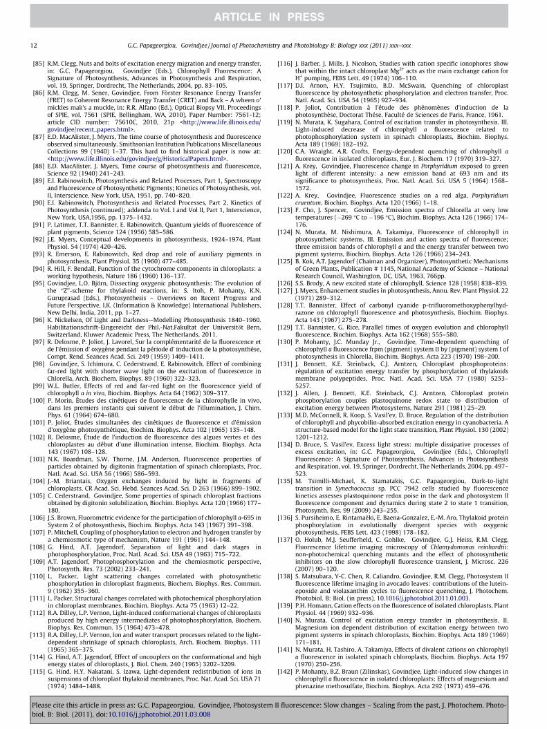
12 G.C. Papageorgiou, Govindjee / Journal of Photochemistry and Photobiology B: Biology xxx (2011) xxx–xxx
[85] R.M. Clegg, Nuts and bolts of excitation energy migration and energy transfer,in: G.C. Papageorgiou, Govindjee (Eds.), Chlorophyll Fluorescence: ASignature of Photosynthesis, Advances in Photosynthesis and Respiration,vol. 19, Springer, Dordrecht, The Netherlands, 2004, pp. 83–105.
[86] R.M. Clegg, M. Sener, Govindjee, From Förster Resonance Energy Transfer(FRET) to Coherent Resonance Energy Transfer (CRET) and Back – A wheen o’mickles mak’s a muckle, in: R.R. Alfano (Ed.), Optical Biopsy VII, Proceedingsof SPIE, vol. 7561 (SPIE, Bellingham, WA, 2010), Paper Number: 7561-12;article CID number: 75610C, 2010, 21p <http://www.life.illinois.edu/govindjee/recent_papers.html>.
[87] E.D. MacAlister, J. Myers, The time course of photosynthesis and fluorescenceobserved simultaneously. Smithsonian Institution Publications MiscellaneousCollections 99 (1940) 1–37. This hard to find historical paper is now at:<http://www.life.illinois.edu/govindjee/g/HistoricalPapers.html>.
[88] E.D. MacAlister, J. Myers, Time course of photosynthesis and fluorescence,Science 92 (1940) 241–243.
[89] E.I. Rabinowitch, Photosynthesis and Related Processes, Part 1, Spectroscopyand Fluorescence of Photosynthetic Pigments; Kinetics of Photosynthesis, vol.II, Interscience, New York, USA, 1951, pp. 740–820.
[90] E.I. Rabinowitch, Photosynthesis and Related Processes, Part 2, Kinetics ofPhotosynthesis (continued); addenda to Vol. I and Vol II, Part 1, Interscience,New York, USA,1956, pp. 1375–1432.
[91] P. Latimer, T.T. Bannister, E. Rabinowitch, Quantum yields of fluorescence ofplant pigments, Science 124 (1956) 585–586.
[92] J.E. Myers, Conceptual developments in photosynthesis, 1924–1974, PlantPhysiol. 54 (1974) 420–426.
[93] R. Emerson, E. Rabinowitch, Red drop and role of auxiliary pigments inphotosynthesis, Plant Physiol. 35 (1960) 477–485.
[94] R. Hill, F. Bendall, Function of the cytochrome components in chloroplasts: aworking hypothesis, Nature 186 (1960) 136–137.
[95] Govindjee, L.O. Björn, Dissecting oxygenic photosynthesis: The evolution ofthe ‘‘Z’’-scheme for thylakoid reactions, in: S. Itoh, P. Mohanty, K.N.Guruprasad (Eds.), Photosynthesis – Overviews on Recent Progress andFuture Perspective, I.K. (Information & Knowledge) International Publishers,New Delhi, India, 2011, pp. 1–27.
[96] K. Nickelsen, Of Light and Darkness—Modelling Photosynthesis 1840–1960.Habilitationschrift-Eingereicht der Phil.-Nat.Fakultat der Universitdt Bern,Switzerland, Kluwer Academic Press, The Netherlands, 2011.
[97] R. Delosme, P. Joliot, J. Lavorel, Sur la complémentarité de la fluorescence etde l’émission d’ oxygène pendant la période d’ induction de la photosynthèse,Compt. Rend. Seances Acad. Sci. 249 (1959) 1409–1411.
[98] Govindjee, S. Ichimura, C. Cederstrand, E. Rabinowitch, Effect of combiningfar-red light with shorter wave light on the excitation of fluorescence inChlorella, Arch. Biochem. Biophys. 89 (1960) 322–323.
[99] W.L. Butler, Effects of red and far-red light on the fluorescence yield ofchlorophyll a in vivo, Biochim. Biophys. Acta 64 (1962) 309–317.
[100] P. Morin, Études des cinétiques de fluorescence de la chlorophylle in vivo,dans les premiers instants qui suivent le début de l’illumination, J. Chim.Phys. 61 (1964) 674–680.
[101] P. Joliot, Études simultanées des cinétiques de fluorescence et d’émissiond’oxygène photosynthétique, Biochim. Biophys. Acta 102 (1965) 135–148.
[102] R. Delosme, Étude de l’induction de fluorescence des algues vertes et deschloroplastes au début d’une illumination intense, Biochim. Biophys. Acta143 (1967) 108–128.
[103] N.K. Boardman, S.W. Thorne, J.M. Anderson, Fluorescence properties ofparticles obtained by digitonin fragmentation of spinach chloroplasts, Proc.Natl. Acad. Sci. USA 56 (1966) 586–593.
[104] J.-M. Briantais, Oxygen exchanges induced by light in fragments ofchloroplasts, CR Acad. Sci. Hebd. Seances Acad. Sci. D 263 (1966) 899–1902.
[105] C. Cederstrand, Govindjee, Some properties of spinach chloroplast fractionsobtained by digitonin solubilization, Biochim. Biophys. Acta 120 (1966) 177–180.
[106] J.S. Brown, Fluorometric evidence for the participation of chlorophyll a-695 inSystem 2 of photosynthesis, Biochim. Biophys. Acta 143 (1967) 391–398.
[107] P. Mitchell, Coupling of phosphorylation to electron and hydrogen transfer bya chemiosmotic type of mechanism, Nature 191 (1961) 144–148.
[108] G. Hind, A.T. Jagendorf, Separation of light and dark stages inphotophosphorylation, Proc. Natl. Acad. Sci. USA 49 (1963) 715–722.
[109] A.T. Jagendorf, Photophosphorylation and the chemiosmotic perspective,Photosynth. Res. 73 (2002) 233–241.
[110] L. Packer, Light scattering changes correlated with photosyntheticphosphorylation in chloroplast fragments, Biochem. Biophys. Res. Commun.9 (1962) 355–360.
[111] L. Packer, Structural changes correlated with photochemical phosphorylationin chloroplast membranes, Biochim. Biophys. Acta 75 (1963) 12–22.
[112] R.A. Dilley, L.P. Vernon, Light-induced conformational changes of chloroplastsproduced by high energy intermediates of photophosphorylation, Biochem.Biophys. Res. Commun. 15 (1964) 473–478.
[113] R.A. Dilley, L.P. Vernon, Ion and water transport processes related to the light-dependent shrinkage of spinach chloroplasts, Arch. Biochem. Biophys. 111(1965) 365–375.
[114] G. Hind, A.T. Jagendorf, Effect of uncouplers on the conformational and highenergy states of chloroplasts, J. Biol. Chem. 240 (1965) 3202–3209.
[115] G. Hind, H.Y. Nakatani, S. Izawa, Light-dependent redistribution of ions insuspensions of chloroplast thylakoid membranes, Proc. Nat. Acad. Sci. USA 71(1974) 1484–1488.
Please cite this article in press as: G.C. Papageorgiou, Govindjee, Photosystem Ibiol. B: Biol. (2011), doi:10.1016/j.jphotobiol.2011.03.008
[116] J. Barber, J. Mills, J. Nicolson, Studies with cation specific ionophores showthat within the intact chloroplast Mg2+ acts as the main exchange cation forH+ pumping, FEBS Lett. 49 (1974) 106–110.
[117] D.I. Arnon, H.Y. Tsujimito, B.D. McSwain, Quenching of chloroplastfluorescence by photosynthetic phosphorylation and electron transfer, Proc.Natl. Acad. Sci. USA 54 (1965) 927–934.
[118] P. Joliot, Contribution à l’étude des phénomènes d’induction de laphotosynthèse, Doctorat Thése, Faculté de Sciences de Paris, France, 1961.
[119] N. Murata, K. Sugahara, Control of excitation transfer in photosynthesis. III.Light-induced decrease of chlorophyll a fluorescence related tophotophosphorylation system in spinach chloroplasts, Biochim. Biophys.Acta 189 (1969) 182–192.
[120] C.A. Wraight, A.R. Crofts, Energy-dependent quenching of chlorophyll afluorescence in isolated chloroplasts, Eur. J. Biochem. 17 (1970) 319–327.
[121] A. Krey, Govindjee, Fluorescence change in Porphyridium exposed to greenlight of different intensity: a new emission band at 693 nm and itssignificance to photosynthesis, Proc. Natl. Acad. Sci. USA 5 (1964) 1568–1572.
[122] A. Krey, Govindjee, Fluorescence studies on a red alga, Porphyridiumcruentum, Biochim. Biophys. Acta 120 (1966) 1–18.
[123] F. Cho, J. Spencer, Govindjee, Emission spectra of Chlorella at very lowtemperatures (�269 �C to �196 �C), Biochim. Biophys. Acta 126 (1966) 174–176.
[124] N. Murata, M. Nishimura, A. Takamiya, Fluorescence of chlorophyll inphotosynthetic systems. III. Emission and action spectra of fluorescence;three emission bands of chlorophyll a and the energy transfer between twopigment systems, Biochim. Biophys. Acta 126 (1966) 234–243.
[125] B. Kok, A.T. Jagendorf (Chaiman and Organizer), Photosynthetic Mechanismsof Green Plants, Publication # 1145, National Academy of Science – NationalResearch Council, Washington, DC, USA, 1963, 766pp.
[126] S.S. Brody, A new excited state of chlorophyll, Science 128 (1958) 838–839.[127] J. Myers, Enhancement studies in photosynthesis, Annu. Rev. Plant Physiol. 22
(1971) 289–312.[128] T.T. Bannister, Effect of carbonyl cyanide p-trifluoromethoxyphenylhyd-
razone on chlorophyll fluorescence and photosynthesis, Biochim. Biophys.Acta 143 (1967) 275–278.
[129] T.T. Bannister, G. Rice, Parallel times of oxygen evolution and chlorophyllfluorescence, Biochim. Biophys. Acta 162 (1968) 555–580.
[130] P. Mohanty, J.C. Munday Jr., Govindjee, Time-dependent quenching ofchlorophyll a fluorescence frpm (pigment) system II by (pigment) system I ofphotosynthesis in Chlorella, Biochim. Biophys. Acta 223 (1970) 198–200.
[131] J. Bennett, K.E. Steinback, C.J. Arntzen, Chloroplast phosphoproteins:régulation of excitation energy transfer by phosphorylation of thylakoidsmembrane polypeptides, Proc. Natl. Acad. Sci. USA 77 (1980) 5253–5257.
[132] J. Allen, J. Bennett, K.E. Steinback, C.J. Arntzen, Chloroplast proteinphosphorylation couples plastoquinone redox state to distribution ofexcitation energy between Photosystems, Nature 291 (1981) 25–29.
[133] M.D. McConnell, R. Koop, S. Vasil’ev, D. Bruce, Regulation of the distributionof chlorophyll and phycobilin-absorbed excitation energy in cyanobacteria. Astructure-based model for the light state transition, Plant Physiol. 130 (2002)1201–1212.
[134] D. Bruce, S. Vasil’ev, Excess light stress: multiple dissipative processes ofexcess excitation, in: G.C. Papageorgiou, Govindjee (Eds.), ChlorophyllFluorescence: A Signature of Photosynthesis, Advances in Photosynthesisand Respiration, vol. 19, Springer, Dordrecht, The Netherlands, 2004, pp. 497–523.
[135] M. Tsimilli-Michael, K. Stamatakis, G.C. Papageorgiou, Dark-to-lighttransition in Synechococcus sp. PCC 7942 cells studied by fluorescencekinetics assesses plastoquinone redox poise in the dark and photosystem IIfluorescence component and dynamics during state 2 to state 1 transition,Photosynth. Res. 99 (2009) 243–255.
[136] S. Pursiheimo, E. Rintamaëki, E. Baena-Gonzalez, E.-M. Aro, Thylakoid proteinphosphorylation in evolutionally divergent species with oxygenicphotosynthesis, FEBS Lett. 423 (1998) 178–182.
[137] O. Holub, M.J. Seufferheld, C. Gohlke, Govindjee, G.J. Heiss, R.M. Clegg,Fluorescence lifetime imaging microscopy of Chlamydomonas reinhardtii:non-photochemical quenching mutants and the effect of photosyntheticinhibitors on the slow chlorophyll fluorescence transient, J. Microsc. 226(2007) 90–120.
[138] S. Matsubara, Y-C. Chen, R. Caliandro, Govindjee, R.M. Clegg, Photosystem IIfluorescence lifetime imaging in avocado leaves: contributions of the lutein-epoxide and violaxanthin cycles to fluorescence quenching, J. Photochem.Photobiol. B: Biol. (in press), 10.1016/j.jphotobiol.2011.01.003.
[139] P.H. Homann, Cation effects on the fluorescence of isolated chloroplasts, PlantPhysiol. 44 (1969) 932–936.
[140] N. Murata, Control of excitation energy transfer in photosynthesis. II.Magnesium ion dependent distribution of excitation energy between twopigment systems in spinach chloroplasts, Biochim. Biophys. Acta 189 (1969)171–181.
[141] N. Murata, H. Tashiro, A. Takamiya, Effects of divalent cations on chlorophylla fluorescence in isolated spinach chloroplasts, Biochim. Biophys. Acta 197(1970) 250–256.
[142] P. Mohanty, B.Z. Braun (Zilinskas), Govindjee, Light-induced slow changes inchlorophyll a fluorescence in isolated chloroplasts: Effects of magnesium andphenazine methosulfate, Biochim. Biophys. Acta 292 (1973) 459–476.
I fluorescence: Slow changes – Scaling from the past, J. Photochem. Photo-
G.C. Papageorgiou, Govindjee / Journal of Photochemistry and Photobiology B: Biology xxx (2011) xxx–xxx 13
[143] N. Murata, Control of excitation transfer in photosynthesis. V. Correlation ofmembrane structure to regulation of excitation transfer between twopigment systems in isolated spinach chloroplasts, Biochim. Biophys. Acta245 (1971) 365–372.
[144] J.-M. Briantais, C. Vernotte, I. Moya, Intersystem exciton transfer in isolatedchloroplasts, Biochim. Biophys. Acta 325 (1973) 530–538.
[145] G. Papageorgiou, J. Isaakidou, C. Argoudelis, Structure-dependent control ofchlorophyll a excitation density: The role of oxygen, FEBS Lett. 25 (1972)139–142.
[146] J. Isaakidou, G. Papageorgiou, A fluorimetric study of the Mg2+-inducedstructural changes in thylakoid membrane protein, Arch. Biochem. Biophys.168 (1975) 266–272.
[147] E.L. Gross, S.C. Hess, Monovalent cation induced inhibition of chlorophyll afluorescence: antagonism by divalent cations, Arch. Biochem. Biophys. 159(1973) 832–836.
[148] P. Mohanty, Govindjee, T. Wydrzynski, Salt-induced alterations of thefluorescence yield and of emission spectra in Chlorella pyrenoidosa, Plant CellPhysiol. 15: (1974) 213–224.
[149] D.L. Vander Meulen, Govindjee, Relation of membrane structural changes toenergy spillover in oats and spinach chloroplasts: use of fluorescence probesand light scattering, Biochim. Biophys. Acta 368 (1974) 61–70.
[150] T. Wydrzynski, E.L. Gross, Govindjee, Effects of sodium and magnesiumcations on the dark and light-induced chlorophyll a fluorescence yields insucrose-washed spinach chloroplasts, Biochim. Biophys. Acta 376 (1975)151–161.
[151] I. Moya, Govindjee, C. Vernotte, J.-M. Briantais, Antagonistic effect of mono-and divalent cations on lifetime t and quantum yield of fluorescence f inisolated chloroplasts, FEBS Lett. 75 (1977) 13–18.
[152] P. Bennoun, Correlation between states I and II in algae and effect ofmagnesium in chloroplasts, Biochim. Biophys. Acta 368 (1974) 141–147.
[153 ] W.L. Butler, M. Kitajima, Fluorescence quenching in Photosystem II ofchloroplasts, Biochim. Biophys. Acta 376 (1975) 116–125.
[154] W.L. Butler, Energy distribution in the photochemical apparatus ofphotosynthesis, Annu. Rev. Plant Physiol. 29 (1978) 345–378.
[155] G.H. Krause, Changes in chlorophyll fluorescence in relation to lightdependent cation transfer across thylakoid membrane, Biochim. Biophys.Acta 333 (1974) 301–313.
[156] J. Barber, J. Mills, Control of chlorophyll fluorescence by the diffuse doublelayer, FEBS Lett. 68 (1976) 288–292.
[157] J. Barber, J. Mills, A. Love, Electrical diffuse layers and their influence onphotosynthetic processes, FEBS Lett. 74 (1977) 174–181.
[158] J. Barber, Membrane surface charges and potentials in relation tophotosynthesis, Biochim. Biophys. Acta 594 (1980) 253–308.
Please cite this article in press as: G.C. Papageorgiou, Govindjee, Photosystem Ibiol. B: Biol. (2011), doi:10.1016/j.jphotobiol.2011.03.008
[159] D. Wong, Govindjee, Antagonistic effects of mono-and divalent cations onpolarization of chlorophyll fluorescence in thylakoids and changes inexcitation energy transfer, FEBS Lett. 97 (1979) 373–377.
[160] D. Wong, Govindjee, Action spectra of cation effects on the fluorescencepolarization and intensity in thylakoids at room temperature, Photochem.Photobiol. 33 (1981) 103–108.
[161] D. Wong, H. Merkelo, Govindjee, Regulation of excitation transfer by cations:wavelength-resolved fluorescence lifetimes and intensities at 77 K inthylakoid membranes of pea chloroplasts, FEBS Lett. 104 (1979) 223–226.
[162] D. Wong, Govindjee, H. Merkelo, Effects of bulk pH and of monovalent anddivalent cations on chlorophyll a fluorescence and electron transport in peathylakoids, Biochim. Biophys. Acta 592 (1980) 546–558.
[163] D. Wong, H. Merkelo, Govindjee, Estimation of energy distribution andredistribution among two Photosystems using parallel measurements offluorescence lifetimes and transients at 77 K, Photochem. Photobiol. 33(1981) 97–101.
[164] A. Jajoo, S. Bharti, Govindjee, Inorganic anion-induced state changes inspinach thylakoid membranes, FEBS Lett. 434 (1998) 193–196.
[165] P. Singh-Rawal, A. Jajoo, S. Bharti, Fluoride affects distribution of absorbedexcitation energy more in favour of photosystem 1, Biol. Plant. 54 (2010)556–560.
[166] P.V. Sane, T.S. Desai, V.G. Tatake, Govindjee, Heat-induced reversible increasein Photosystem I emission in algae, leaves and chloroplasts: spectra,activities, and relation to state changes, Photosynthetica 18 (1984) 439–444.
[167] P. Mohanty, B. Vani, S.S. Prakash, Elevated temperature treatment inducedalteration in thylakoid membrane organization and energy distributionbetween the two systems in Pisum sativum, Z. Naturfosch. 57c (2002) 836–842.
[168] T.S. Babu, S.C. Sabat, P. Mohanty, Heat induced alterations in the photosyntheticelectron transport and emission properties of the cyanobacterium Spirulinaplatensis, J. Photochem. Photobiol. B. Biol. 12 (1992) 161–171.
[169] C. Pastenesz, P. Horton, Effect of high temperature on photosynthesis inbeans. I. Oxygen evolution and chlorophyll fluorescence, Plant Physiol. 112(1996) 1245–1251.
[170] R. Kana, O.Komárek, E. Kotabováa, G.C. Papageorgiou, Govindjee, O. Prášil, Theslow S to M fluorescence rise is missing in the RpaC mutant of Synechocystissp. (PCC6803), in: Proceedings of the 15th International PhotosynthesisCongress, Beijing, China, Springer, Dordrecht, The Netherlands, 2011.
[171] W.J. Vredenberg, Kinetic Analysis and Mathematical Modeling of PrimaryPhotochemical and Photoelectrochemical Processes in Plant Photosystems,BioSystems, Elsevier, 2011, 10.1016/j.biosystems.2010.10.016.
[172] M. Tikkanen, M. Grieco, E.M. Aro, Novel insights into plant light-harvestingcomplex II phosphorylation and state transitions, Trends Plant Sci. 16 (2011)126–131.
I fluorescence: Slow changes – Scaling from the past, J. Photochem. Photo-


![Journal of Photochemistry & Photobiology, B: Biologybose.res.in/~skpal/papers/priya_JPBB1.pdf · Journal of Photochemistry & Photobiology, B: Biology 157 (2016) 105–112 ... [32]τrot](https://static.fdocuments.us/doc/165x107/5f71d8ece961ec0ce1378c74/journal-of-photochemistry-photobiology-b-skpalpaperspriyajpbb1pdf.jpg)