Journal of Nuclear Medicine, published on July 20, 2018 as...
26
1 Performance of 68 Ga-DOTA-Conjugated Somatostatin Receptor Targeting Peptide PET in Detection of Pheochromocytoma and Paraganglioma: A Systematic Review and Meta-Analysis Sangwon Han 1,2,* , Chong Hyun Suh 1,3,* , Sungmin Woo 1,4 , Yeon Joo Kim 5 , Jong Jin Lee 2 1 Meta-analysis for Imaging studies on Diagnostic Accuracy and prognosiS (MIDAS) group 2 Department of Nuclear Medicine, Asan Medical Center, University of Ulsan College of Medicine, Seoul, Korea 3 Department of Radiology and Research Institute of Radiology, Asan Medical Center, University of Ulsan College of Medicine, Seoul, Korea 4 Department of Radiology, Seoul National University College of Medicine, Seoul, Korea 5 Department of Radiation Oncology, Asan Medical Center, University of Ulsan College of Medicine, Seoul, Korea * These authors contributed equally. Corresponding Author: Jong Jin Lee, MD, PhD. Department of Nuclear Medicine, Asan Medical Center, University of Ulsan College of Medicine, 88 Olympic-ro 43-gil, Songpa-gu, Seoul 05505, Korea Tel: 82-2-3010-4596; Fax: 82-2-3010-4588; E-mail: [email protected] First Author: Sangwon Han, M.D. (not under training) Department of Nuclear Medicine, Asan Medical Center, University of Ulsan College of Medicine, 88 Olympic-ro 43-gil, Songpa-gu, Seoul 05505, Korea Tel: 82-2-3010-4590; Fax: 82-2-3010-4588; E-mail: [email protected] Word count: 4921 Financial support: None Short running title: Somatostatin PET in Paraganglioma Journal of Nuclear Medicine, published on July 20, 2018 as doi:10.2967/jnumed.118.211706 by on August 27, 2019. For personal use only. jnm.snmjournals.org Downloaded from
Transcript of Journal of Nuclear Medicine, published on July 20, 2018 as...

1
Performance of 68Ga-DOTA-Conjugated Somatostatin Receptor Targeting Peptide PET in Detection of
Pheochromocytoma and Paraganglioma: A Systematic Review and Meta-Analysis
Sangwon Han1,2,*, Chong Hyun Suh 1,3,*, Sungmin Woo1,4, Yeon Joo Kim5, Jong Jin Lee2
1Meta-analysis for Imaging studies on Diagnostic Accuracy and prognosiS (MIDAS) group
2Department of Nuclear Medicine, Asan Medical Center, University of Ulsan College of Medicine, Seoul, Korea
3 Department of Radiology and Research Institute of Radiology, Asan Medical Center, University of Ulsan College
of Medicine, Seoul, Korea
4Department of Radiology, Seoul National University College of Medicine, Seoul, Korea
5Department of Radiation Oncology, Asan Medical Center, University of Ulsan College of Medicine, Seoul, Korea
*These authors contributed equally.
Corresponding Author: Jong Jin Lee, MD, PhD.
Department of Nuclear Medicine, Asan Medical Center, University of Ulsan College of Medicine, 88 Olympic-ro
43-gil, Songpa-gu, Seoul 05505, Korea
Tel: 82-2-3010-4596; Fax: 82-2-3010-4588; E-mail: [email protected]
First Author: Sangwon Han, M.D. (not under training)
Department of Nuclear Medicine, Asan Medical Center, University of Ulsan College of Medicine, 88 Olympic-ro
43-gil, Songpa-gu, Seoul 05505, Korea
Tel: 82-2-3010-4590; Fax: 82-2-3010-4588; E-mail: [email protected]
Word count: 4921
Financial support: None
Short running title: Somatostatin PET in Paraganglioma
Journal of Nuclear Medicine, published on July 20, 2018 as doi:10.2967/jnumed.118.211706by on August 27, 2019. For personal use only. jnm.snmjournals.org Downloaded from
2
Abstract
We performed a systematic review and meta-analysis of the performance of 68Ga-DOTA-conjugated somatostatin
receptor targeting peptides (68Ga-DOTA-SST) PET in the detection of pheochromocytomas and paragangliomas
(PPGLs). Methods: PubMed and Embase were searched until 08 May 2018. We included studies that reported the
detection rate of 68Ga-DOTA-SST PET in patients with PPGLs. Detection rates were pooled using a random-effects
model. Subgroup analyses and meta-regression were performed to explore the cause of heterogeneity. Results:
Thirteen studies were included for qualitative synthesis. Per-lesion detection rates of 68Ga-DOTA-SST PET were
consistently higher (ranging from 92 to 100%) than other imaging modalities, including 18F-FDOPA PET, 18F-FDG
PET, and 123/131I-MIBG scintigraphy. However, in patients with polycythemia/paraganglioma syndrome, the
detection rate of 68Ga-DOTA-DOTATATE PET was 35%. Nine studies (215 patients) with no specific inclusion
criteria for subtype were quantitatively synthesized. The pooled detection rate was 93% (95% confidence interval
[CI] 91–95%), which was significantly higher than that of 18F-FDOPA PET (80% [95% CI 69–88%]), 18F-FDG PET
(74% [95% CI 46–91%]), and 123/131I-MIBG scan (38% [95% CI 20–59%], p < 0.001 for all). A greater prevalence
of head and neck paragangliomas was associated with higher detection rates of 68Ga-DOTA-SST PET (p = 0.0002).
Conclusion: 68Ga-DOTA-SST PET exhibited superior performance for lesion detection, over other functional
imaging modalities, in patients with PPGLs, with the exception of polycythemia/paraganglioma syndrome. This
might suggest 68Ga-DOTA-SST PET as a first-line imaging modality for the primary staging of PPGL or the re-
staging of PPGL with unknown genetic status.
Keywords: 68Ga-DOTATATE; 68Ga-DOTATOC; 68Ga-DOTANOC; Pheochromocytoma; Paraganglioma
by on August 27, 2019. For personal use only. jnm.snmjournals.org Downloaded from
3
INTRODUCTION
Pheochromocytomas and paragangliomas (PPGLs) are tumors arising from sympathetic lineage-derived cells in
adrenal medulla and extra-adrenal thoracic and abdominal paraganglia or from the parasympathetic nervous system
in the head and neck (1). Functional imaging plays an important role in the confirmation of diagnosis, staging or re-
staging, selection of targeted radionuclide therapy, and response evaluation in patients with PPGLs (2). 18F-
fluorohydroxyphenylalanine (18F-FDOPA) PET is one of the standard diagnostic work-up for non-metastatic PPGLs
in the current guidelines (2-4). In a meta-analysis, the pooled lesion-based sensitivity and specificity of 18F-FDOPA
PET were 79% and 95%, respectively (5). However, the diagnostic performance of 18F-FDOPA PET is largely
influenced by tumor location and genetic status (6). 18F-fluorodeoxyglucose (FDG) PET is recommended in
metastatic PPGLs with succinate dehydrogenase A–D (collectively, SDHx) mutation and unknown or negative
genetic mutations (2,3). A previous meta-analysis showed that the pooled sensitivity and specificity of 18F-FDG PET
for metastatic PPGLs at a per-lesion level is 83% and 74%, respectively (7). 123I-metaiodobenzylguanidine (MIBG)
scintigraphy has excellent sensitivity and specificity on a per-patient basis (8,9); however, its lesion-based
diagnostic accuracy is limited (2).
Because PPGLs express high levels of somatostatin receptor (SSTR) (10-12), 68Ga-DOTA-conjugated
somatostatin receptor targeting peptides (68Ga-DOTA-SST) PET have shown an excellent lesion-based accuracy in
detection of PPGLs (13-21). Recent publications suggest that 68Ga-DOTA-SST PET provides a high detection rate
across a wide range of mutations (22-25). However, because of the small number of subjects in individual studies, it
is difficult to conclude a higher level of evidence.
Therefore, we performed a systematic review and meta-analysis to evaluate the performance of 68Ga-DOTA-SST
PET for lesion detection in patients with PPGLs.
MATERIALS AND METHODS
This systematic review and meta-analysis adhered to the Preferred Reporting Items for Systematic Reviews and
Meta-Analyses guidelines (26). The protocol was registered to the International Prospective Register of Systematic
Reviews (registration no. CRD42018085906). The research question for this meta-analysis was as follows: “What is
the performance of 68Ga-DOTA-SST PET for lesion detection in patients with PPGL, compared with
by on August 27, 2019. For personal use only. jnm.snmjournals.org Downloaded from
4
histopathological results or best value comparator (BVC; a combination of imaging, clinical, and/or biological
studies)?”
Search Strategy
A computerized search on PubMed and Embase databases was performed until 08 May, 2018. The search query
included keywords of “pheochromocytoma/paraganglioma,” “68Ga-DOTA-SST PET,” and their related terms, as
follows: (paraganglioma OR paragangliomas OR paragangliom* OR pheochromocytoma OR pheochromocytomas
OR pheochromocytoma* OR feochromocytoma*) AND (Gallium OR Ga) AND (DOTA* OR somatostatin) AND
(“positron emission tomography” OR PET). Reference lists of the retrieved articles were also checked to identify
additional relevant articles. The search was not limited to any particular language.
Study Selection
Studies were included based on “Patient/Intervention/Comparator/Outcome/Study design” (PICOS) criteria (26):
(1) “patients” with PPGL, (2) 68Ga-DOTA-SST PET as “intervention,” (3) histopathology or BVC as “comparator,”
(4) detection rate as “outcome,” and (5) “study design” as original articles. The following exclusion criteria were
applied: (1) population ≤5; (2) non-original articles; (3) papers irrelevant to the research question; and (4)
overlapping study populations. When study populations overlapped, we selected the publication with the largest
population for the meta-analysis. Two independent reviewers performed the literature search and selection process.
Disagreement was resolved via discussion.
Data Extraction and Quality Assessment
Study and clinicopathological characteristics were extracted using a standardised form. The methodologic quality
of included studies was assessed using the Quality Assessment of Diagnostic Accuracy Studies-2 tool (27). Data
extraction and quality assessment were independently performed by two reviewers; any disagreements were
resolved by discussion.
by on August 27, 2019. For personal use only. jnm.snmjournals.org Downloaded from

5
Data Synthesis and Analysis
The primary outcome was per-lesion detection rate of 68Ga-DOTA-SST PET in patients with PPGLs. The
secondary outcome was a comparison of the pooled estimates with those of other functional imaging modalities
(18F-FDOPA PET, 18F-FDG PET, or MIBG scintigraphy) and to assess heterogeneity among the included articles.
The detection rate for each study was based on proportions reported in the study, or calculated based on the
number of total lesions and number of lesions detected on PET. Of note, we re-calculated the detection rate in one
study after excluding one patient with medullary thyroid cancer (18). One study assessed metastasis on a per-site
basis; it was analysed on a per-lesion basis because the sites were subdivided into abdomen, bones, liver, lungs, and
mediastinum (15).
The proportions were meta-analytically pooled using random-effects models with logit transformation.
Statistical analyses were performed using “meta” and “metafor” packages in R software (version 3.4.3; R
Foundation for Statistical Computing, Vienna, Austria). Publication bias was evaluated with the funnel plot and
Egger’s test (28). Heterogeneity was evaluated by the Higgins I2 test (29). Subgroup analyses and meta-regression
were performed to investigate the possible causes of heterogeneity using several clinically relevant covariates.
RESULTS
Literature Search
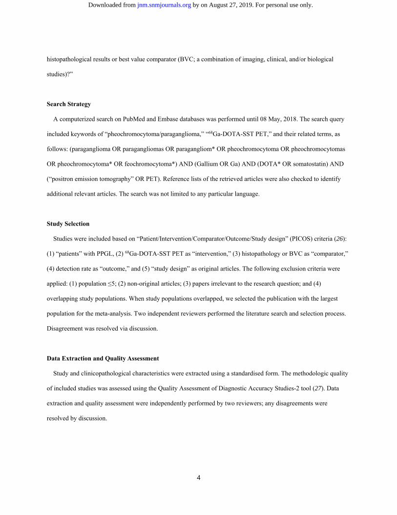
The detailed study selection process is shown in Figure 1. A total of 382 articles were retrieved by the initial
systematic search. After the removal of 93 duplicate articles and exclusion of 261 papers during screening of the
titles and abstracts, there were 28 potentially eligible articles. Full-text reviews were performed and 15 were
excluded for the following reasons: neuroendocrine tumor other than PPGL (n = 7) (30-35), population ≤ 5 (n = 3)
(36-38), overlapping study population (n = 2) (39,40), insufficient information for detection rate (n = 1) (41), and
non-original articles (n = 3) (42-44). Thus, 13 studies were included in the qualitative synthesis. We further
excluded four studies that had exclusive patient populations: SDHB mutation (22), SDHx mutation in pediatric
patients (24), sporadic type (23), and polycythemia/paraganglioma syndrome (45); inclusion of those studies might
hinder generalization of the results. Therefore, nine studies (215 patients) with no specific inclusion criteria for
subtype were included in the meta-analysis (13-21), with the assumption that this pooled population might reflect
patients with unknown genetic status in clinical practice.
by on August 27, 2019. For personal use only. jnm.snmjournals.org Downloaded from

6
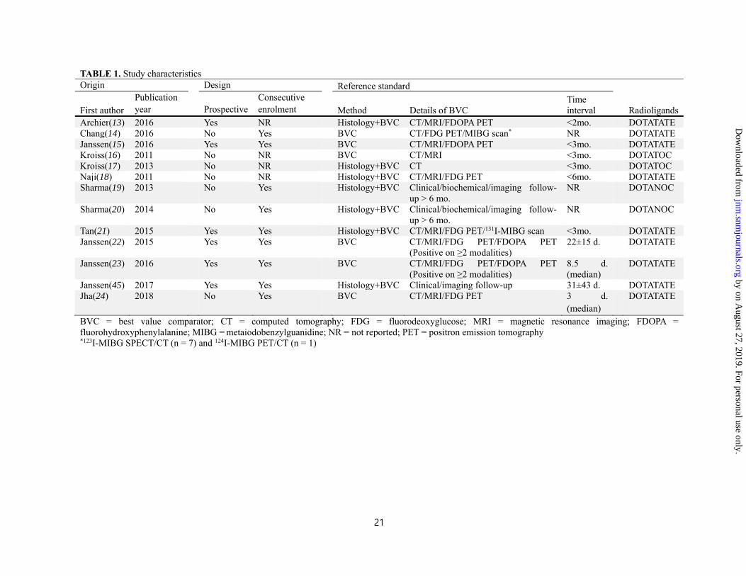
Characteristics of Included Studies
Study and clinicopathological characteristics are described in Tables 1 and 2, respectively. Seven studies used
histopathology and BVC as the reference standard (13,17-21,45), while six used only BVC (14-16,22-24). The
imaging modalities used for BVC included CT, MRI, 18F-FDG PET, 18F-FDOPA PET, and MIBG scintigraphy.
68Ga-DOTA-SST PET was performed for primary staging in four (15,18-20), re-staging in two (22,45), and staging
or re-staging in seven studies (13,14,16,17,21,23,24). Radioligands were DOTATATE in nine (13-15,18,21-24,45),
DOTATOC in two (16,17), and DOTANOC in two studies (19,20).
Quality Assessment
The quality of the studies was considered moderate to good, with 12 of 13 studies satisfying at least four of the
seven QUADAS-2 domains (Fig. 2). Regarding the patient selection domain, three studies had an unclear risk of
bias because they were retrospective and it was not reported whether patients were consecutively enrolled (16-18).
There was a high concern of applicability in four studies, as they only included patients with a specific genetic status
or phenotypic subtype (15,22,24,45). Regarding the index test domain, there was an unclear risk of bias in three
studies, as it was unclear whether the index test was interpreted without knowledge of the reference standard
(14,18,21). For all studies, the concern for applicability was low. Regarding the reference standard domain, four
studies showed an unclear risk of bias, as it was unclear whether reference standard interpretation was blinded to the
index test results (14,19-21). There was an unclear concern for applicability in 10 studies because the BVCs were
solely based on imaging modalities, without clinical or biochemical follow-up (13-18,21-24). Regarding the flow
and timing domain, three studies had an unclear risk of bias, as the PET–reference standard interval was not
provided (14,19,20).
Qualitative Synthesis
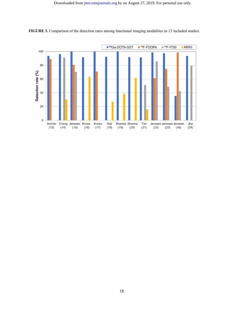
The detection rates of 68Ga-DOTA-SST PET and other imaging modalities (18F-FDOPA PET, 18F-FDG PET, and
123/131I-MIBG scan) are illustrated in Figure 3. 68Ga-DOTA-SST PET consistently showed a higher detection rate
than 18F-FDOPA PET, 18F-FDG PET, and 123/131I-MIBG scintigraphy, with the exception of one study regarding
polycythemia/paraganglioma syndrome (45). In that study, 68Ga-DOTA-SST PET showed the lowest detection rate
by on August 27, 2019. For personal use only. jnm.snmjournals.org Downloaded from

7
of 35% (95% CI 24–48%), whereas the detection rate for 18F-FDOPA PET was 99% (95% CI 93–100%). In the
studies included patients with SDHx mutation (22,24) and sporadic type (15), 68Ga-DOTA-SST PET showed the
highest detection rates among the functional imaging modalities.
Quantitative Synthesis
The per-lesion detection rate in nine studies included in the quantitative synthesis ranged from 92% to 100%, with
a pooled estimate of 93% (95% CI 91–95%) (Fig. 4). Based on the Higgins I2 statistics (I2 = 26%), no significant
heterogeneity was present. There was significant publication bias, according to the funnel plot and Egger’s test (p =
0.0809) (Supplemental Fig. 1). The pooled detection rate of 68Ga-DOTA-SST PET was significantly higher than that
of 18F-FDOPA PET (80% [95% CI 69–88%], p = 0.0003), 18F-FDG PET (74% [95% CI 46–91%], p < 0.0001), or
123/131I-MIBG scintigraphy (38% [95% CI 20–59%], p < 0.0001). There was no difference in the detection rates of
68Ga-DOTA-SST PET among the multiple subgroups stratified by reference standard, clinical setting, or radioligand
(Table 3). A greater proportion of head and neck paragangliomas was significantly associated with higher detection
rates of 68Ga-DOTA-SST PET (p = 0.0002), whereas other variables, including the proportions of multifocal or
metastatic disease, SDHx mutation, sporadic type, catecholamine-secretory PPGLs, age, and tumor size, were not
significant in meta-regression analyses (Table 4).
DISCUSSION
In the present systematic review and meta-analysis, we evaluated the performance of 68Ga-DOTA-SST PET for
lesion detection in patients with PPGLs. The pooled detection rate was 93%, which was significantly higher than the
detection rates of other functional imaging modalities. Accurate lesion detection is important for PPGLs, as these are
typically surgically amenable; complete resection of lesions is needed, especially for catecholamine-secreting
tumors.
18F-FDOPA PET is one of the standard imaging modalities in non-metastatic PPGLs (2-4). However, the difficulty
in synthesis and the requirement of a nearby cyclotron precludes the wider use of 18F-FDOPA. Furthermore, the
diagnostic performance of 18F-FDOPA PET is lower in extra-adrenal paraganglioma and SDHx-related metastatic
disease (6). The role of 18F-FDG PET in PPGLs is limited for metastatic disease. MIBG scintigraphy requires
complicated patient preparation (including thyroid blockade and discontinuation of certain drugs) and a long delay
by on August 27, 2019. For personal use only. jnm.snmjournals.org Downloaded from

8
between injection and imaging. 123I might not be available in every facility, whereas 131I suffers from low image
quality and unfavorable dosimetry. In contrast, 68Ga-DOTA-SST PET imaging exhibits both practical advantages (no
patient preparation, easy synthesis, and wide availability due to 68Ge/68Ga generator) and superior detection rates,
relative to any other functional imaging modalities. The high cost of 68Ge/68Ga generators can be a potential
drawback of 68Ga-DOTA-SST PET imaging. However, increasing demand for 68Ga-labelled radiotracers and recent
approval of the SST analogue kit by the United States Food and Drug Administration will make 68Ge/68Ga
generators more readily available. Further, more effective planning, such as imaging centralization and a referral
system, would help reduce the cost of 68Ga imaging.
For meta-analysis, we excluded four studies that exclusively included patients with specific subtypes. If we
assume that the study samples included in our quantitative synthesis are representative of a PPGL population with
unknown genetic status, it may be suggested that 68Ga-DOTA-SST PET can serve as a first-line imaging modality
for the primary staging of PPGLs, or the re-staging of PPGLs with unknown genetic status. However, in four of the
included studies (13-15,18), a substantial portion of patients were found to have the SDHx mutation; these
proportions ranged from 27%‒80%, which are higher than the proportions in general PPGL populations (46). A
higher prevalence of multifocal or metastatic disease, which is related to SDHx mutation, was also observed.
Therefore, caution is necessary regarding the general application of our pooled estimate. Based on our meta-
regression analyses, the performance of 68Ga-DOTA-SST PET may not be affected by the prevalence of metastasis,
SDHx mutation, or sporadic type. Our study also suggested that 68Ga-DOTA-SST PET might exhibit a superior
detection rate relative to 18F-FDOPA or 18F-FDG PET and serve as a functional imaging modality of choice in
PPGLs with metastasis, SDHx mutation or sporadic type.
68Ga-DOTA-SST ligands have the highest affinity for SSTR2, with different affinities for other SSTR subtypes
(12). 68Ga-DOTATATE predominantly binds to SSTR2, 68Ga-DOTATOC binds to SSTR2 and SSTR5, and 68Ga-
DOTANOC has a high affinity throughout SSTR2–5. No difference in detection performance was observed between
the radioligands in our subgroup analysis; however, the low number of studies limited its significance. Of note,
higher detection rates of 68Ga-DOTA-SST PET were reported in studies that showed greater prevalence of head and
neck paragangliomas. These tumors are parasympathetic in origin and usually do not secrete catecholamine; thus,
they differ from pheochromocytomas or paragangliomas in the thorax and abdomen (1). Our findings are consistent
with the recent guideline that recommends 68Ga-DOTA-SST PET as the first-line imaging tool for head and neck
by on August 27, 2019. For personal use only. jnm.snmjournals.org Downloaded from
9
paraganglioma (4). We suspect that the difference in overexpressed SSTR subtypes between the two kinds of PPGLs
might affect the diagnostic performance of 68Ga-DOTA-SST PET. Paragangliomas overexpress SSTR2
predominantly (11,12), whereas a single in vitro study showed that pheochromocytomas overexpress SSTR3
predominantly and SSTR2 to a lesser extent (10).
It should be noted that 68Ga-DOTATATE showed poor diagnostic performance in patients presenting with
polycythemia/paraganglioma syndrome, whereas 18F-FDOPA PET exhibited the highest detection rate (45). The
reason for this disparate diagnostic performance remains unclear; however, we speculate that a lack of SSTR
expression, inactivation of SSTR, or overexpression of other SSTR subtypes (non-SSTR2) could explain such
behaviour. Similarly, in a recent study by Taieb et al.(38) , 68Ga-DOTATATE PET showed an inferior lesion
detection rate, compared to 18F-FDOPA PET, in MYC-associated factor X-related pheochromocytoma; however,
only three subjects were evaluated. Further research is needed to clarify these discrepancies.
There are some limitations in our review. First, the number of included studies is small. Even after a systematic
search without any language restriction, we could only identify eight suitable studies for quantitative synthesis.
Nevertheless, meta-analysis is an appropriate method to generate a higher level of evidence in rare diseases, such as
PPGLs, for which large cohort studies are not feasible. Second, approximately half of the included studies were
retrospective in nature. Pooling results based on predominantly retrospective studies might lead to overestimation of
the outcomes. Third, there were heterogeneities in scanners, image acquisition and reconstruction protocols among
the studies. Lastly, our pooled estimates were not based on studies that assessed patients with specific genetic
mutations. No genetic test was performed in half of the included studies in our quantitative synthesis. Therefore, our
results might not be applicable to specific genetic subtypes of PPGLs.
CONCLUSION
68Ga-DOTA-SST PET demonstrated an excellent lesion detection rate in patients with PPGLs. The pooled
detection rate of the eight included articles was 93%, which was significantly higher than the detection rate of other
functional imaging modalities. Greater prevalence of head and neck paragangliomas was associated with higher
detection rates of 68Ga-DOTA-SST PET. However, in patients with polycythemia/paraganglioma syndrome, 68Ga-
DOTA-SST PET exhibited a poor detection rate.
by on August 27, 2019. For personal use only. jnm.snmjournals.org Downloaded from

10
DISCLOSURE
The authors have no potential conflict of interest. This research did not receive any specific grant from funding
agencies in the public, commercial, or not-for-profit sectors.
by on August 27, 2019. For personal use only. jnm.snmjournals.org Downloaded from

11
REFERENCES 1. Dahia PL. Pheochromocytoma and paraganglioma pathogenesis: learning from genetic heterogeneity. Nat
Rev Cancer. 2014;14:108-119.
2. Taïeb D, Timmers HJ, Hindié E, et al. EANM 2012 guidelines for radionuclide imaging of
phaeochromocytoma and paraganglioma. Eur J Nucl Med Mol Imaging. 2012;39:1977-1995.
3. Lenders JW, Duh QY, Eisenhofer G, et al. Pheochromocytoma and paraganglioma: an endocrine society
clinical practice guideline. J Clin Endocrinol Metab. 2014;99:1915-1942.
4. Bozkurt MF, Virgolini I, Balogova S, et al. Guideline for PET/CT imaging of neuroendocrine neoplasms
with 68Ga-DOTA-conjugated somatostatin receptor targeting peptides and 18F–DOPA. Eur J Nucl Med Mol Imaging.
2017;44:1588-1601.
5. Treglia G, Cocciolillo F, De Waure C, et al. Diagnostic performance of 18F-dihydroxyphenylalanine
positron emission tomography in patients with paraganglioma: a meta-analysis. Eur J Nucl Med Mol Imaging.
2012;39:1144-1153.
6. Taïeb D, Tessonnier L, Sebag F, et al. The role of 18F-FDOPA and 18F-FDG-PET in the management of
malignant and multifocal phaeochromocytomas. Clin Endocrinol (Oxf). 2008;69:580-586.
7. Kan Y, Zhang S, Wang W, Liu J, Yang J, Wang Z. 68Ga-somatostatin receptor analogs and 18F-FDG
PET/CT in the localization of metastatic pheochromocytomas and paragangliomas with germline mutations: a meta-
analysis. Acta Radiol. January 1, 2018 [Epub ahead of print].
8. Van Der Horst-Schrivers AN, Jager PL, Boezen HM, Schouten JP, Kema IP, Links TP. Iodine-123
metaiodobenzylguanidine scintigraphy in localising phaeochromocytomas--experience and meta-analysis.
Anticancer Res. 2006;26:1599-1604.
9. Jacobson AF, Deng H, Lombard J, Lessig HJ, Black RR. 123I-meta-iodobenzylguanidine scintigraphy for
the detection of neuroblastoma and pheochromocytoma: results of a meta-analysis. J Clin Endocrinol Metab.
2010;95:2596-2606.
10. Mundschenk J, Unger N, Schulz S, et al. Somatostatin receptor subtypes in human pheochromocytoma:
subcellular expression pattern and functional relevance for octreotide scintigraphy. J Clin Endocrinol Metab.
by on August 27, 2019. For personal use only. jnm.snmjournals.org Downloaded from

12
2003;88:5150-5157.
11. Reubi JC, Waser B, Schaer JC, Laissue JA. Somatostatin receptor sst1-sst5 expression in normal and
neoplastic human tissues using receptor autoradiography with subtype-selective ligands. Eur J Nucl Med.
2001;28:836-846.
12. Reubi JC. Peptide receptors as molecular targets for cancer diagnosis and therapy. Endocr Rev.
2003;24:389-427.
13. Archier A, Varoquaux A, Garrigue P, et al. Prospective comparison of 68Ga-DOTATATE and 18F-FDOPA
PET/CT in patients with various pheochromocytomas and paragangliomas with emphasis on sporadic cases. Eur J
Nucl Med Mol Imaging. 2016;43:1248-1257.
14. Chang CA, Pattison DA, Tothill RW, et al. 68Ga-DOTATATE and 18F-FDG PET/CT in paraganglioma and
pheochromocytoma: utility, patterns and heterogeneity. Cancer Imaging. 2016;16:22.
15. Janssen I, Chen CC, Taieb D, et al. 68Ga-DOTATATE PET/CT in the localization of head and neck
paragangliomas compared with other functional imaging modalities and CT/MRI. J Nucl Med. 2016;57:186-191.
16. Kroiss A, Putzer D, Uprimny C, et al. Functional imaging in phaeochromocytoma and neuroblastoma with 68Ga-DOTA-Tyr 3-octreotide positron emission tomography and 123I-metaiodobenzylguanidine. Eur J Nucl Med Mol
Imaging. 2011;38:865-873.
17. Kroiss A, Putzer D, Frech A, et al. A retrospective comparison between 68Ga-DOTA-TOC PET/CT and 18F-DOPA PET/CT in patients with extra-adrenal paraganglioma. Eur J Nucl Med Mol Imaging. 2013;40:1800-1808.
18. Naji M, Zhao C, Welsh SJ, et al. 68Ga-DOTA-TATE PET vs. 123I-MIBG in identifying malignant neural
crest tumours. Mol Imaging Biol. 2011;13:769-775.
19. Sharma P, Thakar A, KC SS, et al. 68Ga-DOTANOC PET/CT for baseline evaluation of patients with head
and neck paraganglioma. J Nucl Med. 2013;54:841-847.
20. Sharma P, Dhull VS, Arora S, et al. Diagnostic accuracy of 68Ga-DOTANOC PET/CT imaging in
pheochromocytoma. Eur J Nucl Med Mol Imaging. 2014;41:494-504.
21. Tan TH, Hussein Z, Saad FF, Shuaib IL. Diagnostic performance of 68Ga-DOTATATE PET/CT, 18F-FDG
PET/CT and 131I-MIBG scintigraphy in mapping metastatic pheochromocytoma and paraganglioma. Nucl Med Mol
by on August 27, 2019. For personal use only. jnm.snmjournals.org Downloaded from

13
Imaging. 2015;49:143-151.
22. Janssen I, Blanchet EM, Adams K, et al. Superiority of [68Ga]-DOTATATE PET/CT to other functional
imaging modalities in the localization of SDHB-associated metastatic pheochromocytoma and paraganglioma. Clin
Cancer Res. 2015;21:3888-3895.
23. Janssen I, Chen CC, Millo CM, et al. PET/CT comparing 68Ga-DOTATATE and other
radiopharmaceuticals and in comparison with CT/MRI for the localization of sporadic metastatic
pheochromocytoma and paraganglioma. Eur J Nucl Med Mol Imaging. 2016;43:1784-1791.
24. Jha A, Ling A, Millo C, et al. Superiority of 68Ga-DOTATATE over 18F-FDG and anatomic imaging in the
detection of succinate dehydrogenase mutation (SDHx)-related pheochromocytoma and paraganglioma in the
pediatric population. Eur J Nucl Med Mol Imaging. 2018;45:787-797.
25. Hofman MS, Lau WFE, Hicks RJ. Somatostatin receptor imaging with 68Ga DOTATATE PET/CT: clinical
utility, normal patterns, pearls, and pitfalls in interpretation. Radiographics. 2015;35:500-516.
26. Moher D, Liberati A, Tetzlaff J, Altman DG. Preferred reporting items for systematic reviews and meta-
analyses: the PRISMA statement. PLoS Med. 2009;6:e1000097.
27. Whiting PF, Rutjes AW, Westwood ME, et al. QUADAS-2: a revised tool for the quality assessment of
diagnostic accuracy studies. Ann Intern Med. 2011;155:529-536.
28. Egger M, Smith GD, Schneider M, Minder C. Bias in meta-analysis detected by a simple, graphical test.
BMJ. 1997;315:629-634.
29. Higgins JP, Thompson SG, Deeks JJ, Altman DG. Measuring inconsistency in meta-analyses. BMJ.
2003;327:557-560.
30. Abongwa C, Mott S, Schafer B, et al. Safety and accuracy of 68Ga-DOTATOC PET/CT in children and
young adults with solid tumors. Am J Nucl Med Mol Imaging. 2017;7:228-235.
31. Berzaczy D, Giraudo C, Haug AR, et al. Whole-Body 68Ga-DOTANOC PET/MRI versus 68Ga-
DOTANOC PET/CT in patients with neuroendocrine tumors: a prospective study in 28 patients. Clin Nucl Med.
2017;42:669-674.
32. Goel R, Shukla J, Bansal D, et al. 68Ga-DOTATATE positron emission tomography/computed tomography
by on August 27, 2019. For personal use only. jnm.snmjournals.org Downloaded from

14
scan in the detection of bone metastases in pediatric neuroendocrine tumors. Indian J Nucl Med. 2014;29:13-17.
33. Lawal IO, Ololade KO, Lengana T, et al. Gallium-68-dotatate PET/CT is better than CT in the
management of somatostatin expressing tumors: first experience in Africa. Hell J Nucl Med. 2017;20:128-133.
34. Sharma P, Arora S, Mukherjee A, et al. Predictive value of 68Ga-DOTANOC PET/CT in patients with
suspicion of neuroendocrine tumors: is its routine use justified? Clin Nucl Med. 2014;39:37-43.
35. Sharma P, Mukherjee A, Karunanithi S, et al. Accuracy of 68Ga DOTANOC PET/CT imaging in patients
with multiple endocrine neoplasia syndromes. Clin Nucl Med. 2015;40:e351-356.
36. Kornaczewski ER, Pointon OP, Burgess JR. Utility of FDG-PET imaging in screening for succinate
dehydrogenase B and D mutation-related lesions. Clin Endocrinol (Oxf). 2016;85:172-179.
37. Win Z, Al-Nahhas A, Towey D, et al. 68Ga-DOTATATE PET in neuroectodermal tumours: first experience.
Nucl Med Commun. 2007;28:359-363.
38. Taïeb D, Jha A, Guerin C, et al. 18F-FDOPA PET/CT imaging of MAX-related pheochromocytoma. J Clin
Endocrinol Metab. 2018;103:1574-1582.
39. Kroiss A, Shulkin BL, Uprimny C, et al. 68Ga-DOTATOC PET/CT provides accurate tumour extent in
patients with extraadrenal paraganglioma compared to 123I-MIBG SPECT/CT. Eur J Nucl Med Mol Imaging.
2015;42:33-41.
40. Naswa N, Sharma P, Nazar AH, et al. Prospective evaluation of 68Ga-DOTA-NOC PET-CT in
phaeochromocytoma and paraganglioma: preliminary results from a single centre study. Eur Radiol. 2012;22:710-
719.
41. Jing H, Li F, Wang L, et al. Comparison of the 68Ga-DOTATATA PET/CT, FDG PET/CT, and MIBG
SPECT/CT in the evaluation of suspected primary pheochromocytomas and paragangliomas. Clin Nucl Med.
2017;42:525-529.
42. Maurice JB, Troke R, Win Z, et al. A comparison of the performance of 68Ga-DOTATATE PET/CT and
123I-MIBG SPECT in the diagnosis and follow-up of phaeochromocytoma and paraganglioma. Eur J Nucl Med Mol
Imaging. 2012;39:1266-1270.
43. Naswa N, Kumar A, Sharma P, Bal C, Malhotra A, Kumar R. Imaging carotid body chemodectomas with
by on August 27, 2019. For personal use only. jnm.snmjournals.org Downloaded from

15
68Ga-DOTA-NOC PET-CT. Br J Radiol. 2012;85:1140-1145.
44. Şimşek DH, Şanlı Y, Kuyumcu S, Başaran B, Mudun A. 68Ga-DOTATATE PET–CT imaging in carotid
body paragangliomas. Ann Nucl Med. 2018:32:297-301.
45. Janssen I, Chen CC, Zhuang Z, et al. Functional imaging signature of patients presenting with
polycythemia/paraganglioma syndromes. J Nucl Med. 2017;58:1236-1242.
46. Lenders JW, Eisenhofer G, Mannelli M, Pacak K. Phaeochromocytoma. Lancet. 2005;366:665-675.
by on August 27, 2019. For personal use only. jnm.snmjournals.org Downloaded from
16
FIGURE LEGENDS
FIGURE 1. Flow diagram showing the study selection process
by on August 27, 2019. For personal use only. jnm.snmjournals.org Downloaded from
17
FIGURE 2. Quality assessment of 13 included studies
by on August 27, 2019. For personal use only. jnm.snmjournals.org Downloaded from
18
FIGURE 3. Comparison of the detection rates among functional imaging modalities in 13 included studies.
by on August 27, 2019. For personal use only. jnm.snmjournals.org Downloaded from

19
FIGURE 4. Forest plot showing the pooled proportion of detection rate of 68Ga-DOTA-SST PET
by on August 27, 2019. For personal use only. jnm.snmjournals.org Downloaded from

20
FIGURE 5. Bubble plot for the detection rate of 68Ga-DOTA-SST PET and the proportion of head and neck
paragangliomas shows that it is a significant factor affecting heterogeneity (p = 0.0002)
by on August 27, 2019. For personal use only. jnm.snmjournals.org Downloaded from
21
TABLE 1. Study characteristics Origin Design Reference standard
Radioligands First author Publication year Prospective
Consecutive enrolment Method Details of BVC
Time interval
Archier(13) 2016 Yes NR Histology+BVC CT/MRI/FDOPA PET <2mo. DOTATATE Chang(14) 2016 No Yes BVC CT/FDG PET/MIBG scan* NR DOTATATE Janssen(15) 2016 Yes Yes BVC CT/MRI/FDOPA PET <3mo. DOTATATE Kroiss(16) 2011 No NR BVC CT/MRI <3mo. DOTATOC Kroiss(17) 2013 No NR Histology+BVC CT <3mo. DOTATOC Naji(18) 2011 No NR Histology+BVC CT/MRI/FDG PET <6mo. DOTATATE Sharma(19) 2013 No Yes Histology+BVC Clinical/biochemical/imaging follow-
up > 6 mo. NR DOTANOC
Sharma(20) 2014 No Yes Histology+BVC Clinical/biochemical/imaging follow-up > 6 mo.
NR DOTANOC
Tan(21) 2015 Yes Yes Histology+BVC CT/MRI/FDG PET/131I-MIBG scan <3mo. DOTATATE Janssen(22) 2015 Yes Yes BVC CT/MRI/FDG PET/FDOPA PET
(Positive on ≥2 modalities) 22±15 d. DOTATATE
Janssen(23) 2016 Yes Yes BVC CT/MRI/FDG PET/FDOPA PET (Positive on ≥2 modalities)
8.5 d. (median)
DOTATATE
Janssen(45) 2017 Yes Yes Histology+BVC Clinical/imaging follow-up 31±43 d. DOTATATE Jha(24) 2018 No Yes BVC CT/MRI/FDG PET 3 d.
(median) DOTATATE
BVC = best value comparator; CT = computed tomography; FDG = fluorodeoxyglucose; MRI = magnetic resonance imaging; FDOPA = fluorohydroxyphenylalanine; MIBG = metaiodobenzylguanidine; NR = not reported; PET = positron emission tomography *123I-MIBG SPECT/CT (n = 7) and 124I-MIBG PET/CT (n = 1)
by on August 27, 2019. For personal use only.
jnm.snm
journals.org D
ownloaded from

22
TABLE 2. Clinicopathologic characteristics
First author
Patients (n)
Mean age (yr) Subtype Setting
Pheochromocytoma (n)
Paraganglioma (n) Multifocal/Metastatic (n)
Catecholamine-secretory (n)
Genetic mutation (n) Tumor size (cm)
T&A H&N SDHx Others Sporadic
Not tested
Archier(13)
30 53 No S+R 11 0 20 7 NR 8 MAX: 1 21 0 2.0
Chang(14)
23 43 No S+R 8 7 8 19 12 10 0 3 9 NR
Janssen(15)
20 48 No S 0 0 20 17 12 16 HIF2A: 1 3 0 2.2
Kroiss(16)
6 46 No S+R 5 1 0 6 NR NR NR NR 6 NR
Kroiss(17)
20 50 No S+R 0 3 19 5 NR NR NR NR 20 NR
Naji(18) 11 NR No S 7 2 2 2 NR 4 0 1 6 1.8 Sharma(19)
26 34 No S 0 0 26 15 4 NR NR NR 26 3.4
Sharma(20)
62 34 No S 62 0 0 7 54 NR RET†: 14 NR 62 4.1
Tan(21) 17 40* No S+R 10 NR NR 15 9 NR NR NR 17 NR Janssen(22)
17 40 SDHB mutation R 2 10 5 17 11 17 0 0 0 NR
Janssen(23)
22 50 Sporadic S+R 13 9 0 22 19 0 0 22 0 NR
Janssen(45)
13 37 PPGL–polycythemia
R 7 7 0 4 13 0 HIF2A: 6 PHD: 2
6 0 NR
Jha(24) 9 17 SDHx mutation S+R 1 6 2 9 7 9 0 0 0 NR HIF2A = hypoxia-inducible factor 2A; H&N = Head and neck; MAX = MYC-associated factor X; NR = not reported; PHD = prolyl hydroxylase; PPGL = pheochromocytoma and paraganglioma; R = re-staging; S = staging; SDH = succinate dehydrogenase; T&A = thorax and abdomen *median †multiple endocrine neoplasia type 2
by on August 27, 2019. For personal use only.
jnm.snm
journals.org D
ownloaded from

23
TABLE 3. Subgroup analyses for the detection rates Variable No. of studies Detection rate (%) 95% CI (%) I2 (%) p Reference 0.7842
Histology+BVC 6 93 89–95 14 BVC 3 95 88–98 52
Setting 0.1412 Staging 4 96 88–99 44 Staging+Re-staging 5 93 90–95 14
Radioligands 0.5924 DOTATATE 5 94 90–96 27 DOTATOC 2 95 76–99 52 DOTANOC 2 97 72–100 67
BVC = best value comparator; CI = confidence interval; NA = not applicable
by on August 27, 2019. For personal use only.
jnm.snm
journals.org D
ownloaded from

24
TABLE 4. Results of meta-regression analyses Variable No. of studies Regression coefficient 95% CI p Multifocal/Metastatic disease (%) 9 ‒0.0001 ‒0.0010–0.0008 0.8299 Head and neck paragangliomas (%) 8 0.0007 0.0003–0.0011 0.0002 SDHx mutation (%) 4 0.0010 ‒0.0006–0.0025 0.2146 Sporadic type (%) 4 ‒0.0010 ‒0.0025–0.0004 0.1748 Catecholamine-secretory tumors (%) 5 ‒0.0009 ‒0.0027–0.0009 0.3164 Mean age (yr) 8 0.0006 ‒0.0045–0.0056 0.8248 Mean tumor size (cm) 5 0.0118 ‒0.0301–0.0537 0.5522 CI = confidence interval; SDH = succinate dehydrogenase
by on August 27, 2019. For personal use only.
jnm.snm
journals.org D
ownloaded from
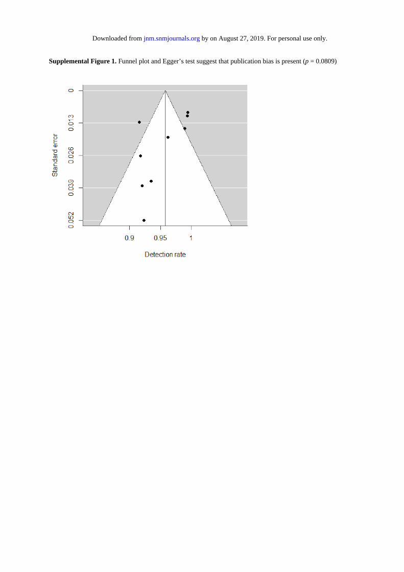
Supplemental Figure 1. Funnel plot and Egger’s test suggest that publication bias is present (p = 0.0809)
by on August 27, 2019. For personal use only. jnm.snmjournals.org Downloaded from
Doi: 10.2967/jnumed.118.211706Published online: July 20, 2018.J Nucl Med. Sangwon Han, Chong Hyun Suh, Sungmin Woo, Yeon Joo Kim and Jong Jin Lee Meta-Analysisin Detection of Pheochromocytoma and Paraganglioma: A Systematic Review and
Ga-DOTA-Conjugated Somatostatin Receptor Targeting Peptide PET68Performance of
http://jnm.snmjournals.org/content/early/2018/07/19/jnumed.118.211706This article and updated information are available at:
http://jnm.snmjournals.org/site/subscriptions/online.xhtml
Information about subscriptions to JNM can be found at:
http://jnm.snmjournals.org/site/misc/permission.xhtmlInformation about reproducing figures, tables, or other portions of this article can be found online at:
and the final, published version.proofreading, and author review. This process may lead to differences between the accepted version of the manuscript
ahead of print area, they will be prepared for print and online publication, which includes copyediting, typesetting,JNMcopyedited, nor have they appeared in a print or online issue of the journal. Once the accepted manuscripts appear in the
. They have not beenJNM ahead of print articles have been peer reviewed and accepted for publication in JNM
(Print ISSN: 0161-5505, Online ISSN: 2159-662X)1850 Samuel Morse Drive, Reston, VA 20190.SNMMI | Society of Nuclear Medicine and Molecular Imaging
is published monthly.The Journal of Nuclear Medicine
© Copyright 2018 SNMMI; all rights reserved.
by on August 27, 2019. For personal use only. jnm.snmjournals.org Downloaded from


![0&0jb.asm.org/content/early/2015/08/11/JB.00496-15.full.pdf3 49 VXJJHVWWKDW Sulfolobus *,16PD\VWDELOL]HWKHLQWH UDFWLRQRI0&0ZLWKWKHPRYLQJ 50 UHSOLFDWLRQIRUN IDFLOLWDWL QJSURFHVVLYH'1$XQZLQGLQJ](https://static.fdocuments.us/doc/165x107/5ab7add17f8b9ad5338bdeeb/00jbasmorgcontentearly20150811jb00496-15fullpdf3-49-vxjjhvwwkdw-sulfolobus.jpg)