Emergency Medicine/ Emergency Medicine-Internal Medicine ...

Introduction
Extracorporeal Life Support is a complex modality requir-ing cannulation of major arteries of the body such as the femoral artery with large bore cannulae, usually 15-25 Fr. Also withdrawing these arterial cannulae when ECLS is no longer needed might be a challenging process because of the potential arterial bleeding following the decannulation. In addition, a “bloodless area” at least for a short time is man-datory for repairing the femoral arterial wall. We present a case in which REBOA was used for temporary bleeding control during the decannulation of a 21 Fr femoral arterial cannula and femoral arterial wall repair in addition to a con-ventional femoral cut-down.
Case Report
A 42-year-old male patient was admitted to the intensive care unit with acute respiratory distress syndrome (ARDS) associated with cardiogenic shock in whom the return of spontaneous circulation (ROSC) was achieved after con-
ventional cardiopulmonary resuscitation for 8 minutes. A veno-arterial ECMO with a 21 Fr arterial in the common femoral artery and a 7 Fr backflow cannula in the superficial femoral artery was then initiated regarding the acute heart failure (Left ventricle ejection fraction: 20%) unresponsive to fluid and positive inotropic agent infusions. On the 4th day of the follow up the native cardiac functions of the pa-tient improved with LVEF of 60% in bedside transthoracic echocardiographic re-evaluation, and the positive inotropic agents were no longer needed. Due to persistent respiratory failure and increased native cardiac performance, we have planned to convert VA-ECMO to VV-ECMO. An additional infusion cannula was placed through the right internal jug-ular vein, and a veno-arterio-venous (VAV) ECMO was es-tablished. The femoral infusion cannula which was already in place for VA ECMO was then clamped. Following the confirmation of the patient did not hemodynamically dete-riorate for one hour, the right femoral arterial cannula was planned to be withdrawn.
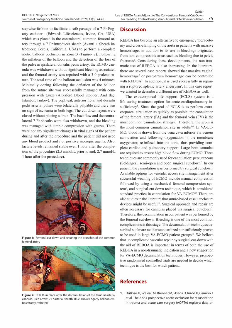
A femoral cut-down was performed and the common femoral artery and its branches superficial and deep femo-ral arteries were secured proximally and distally (Figure-1). The cannula was slightly withdrawn for about 10 cm in a
Corresponding Author: Yunus Emre ÖZLÜER e-mail: [email protected]: 02.06.2020 • Accepted: 03.10.2020DOI: 10.33706/jemcr.747023©Copyright 2020 by Emergency Physicians Association of Turkey - Available online at www.jemcr.com
Abstract
Introduction: One of the potential complications of Extracorporeal Life Support is the arterial bleeding during decannulation. We present a case that de-scribes the use of REBOA for hemorrhage control during femoral decannulation.
Case report: 42-old-male who was suffering from ARDS and cardiogenic shock was admitted to the Intensive Care Unit and Veno-arterial (VA) ECMO was commenced. On day 4, his cardiac performance improved and we decided that there was no longer a need for cardiac support with ECMO. Thus, a conversion from VA to Veno-venous (VV) ECMO was planned. An additional venous cannula was placed and the arterial cannula in the femoral artery was withdrawn and the femoral artery wall was repaired just after a Zone III total REBOA was achieved from the contralateral femoral artery. There were not any significant changes in blood pressure, heart rate, and lactate levels before and after the procedure.
Conclusion: REBOA might be a useful tool in the hands of non-vascular surgeons to avoid unintended bleeding during the decannulation of a large-bore arterial cannulae.
Yunus Emre ÖZLÜER1, Mücahit AVCİL1, Çağaç YETİŞ1, Kezban ŞEKER YAŞAR1
1Adnan Menderes University Hospital Department of Emergency Medicine Aytepe Mevkii, 09100, Aydın
Use of REBOA As an Adjunct to The Conventional Femoral Cut-Down For Bleeding Control During Veno-Arterial ECMO Decannulation
Case ReportJournal of Emergency
Medicine Case Reports
ÖzlüerUse of REBOA As an Adjunct to The Conventional Femoral Cut-Down
For Bleeding Control During Veno-Arterial ECMO Decannulation Journal of Emergency Medicine Case Reports 2020; 11(3): 74-76DOI: 10.33706/jemcr.747023
75
stepwise fashion to facilitate a safe passage of a 7 Fr Fog-arty catheter (Edwards Lifesciences, Irvine, CA, USA) which was placed in the contralateral common femoral ar-tery through a 7 Fr introducer sheath (Avanti + Sheath in-troducer; Cordis, California, USA) to perform a complete aortic balloon occlusion in Zone 3 (Figure- 2). Following the inflation of the balloon and the detection of the loss of the pulse in ipsilateral dorsalis pedis artery, the ECMO can-nula was withdrawn without significant bleeding associated and the femoral artery was repaired with a 3-0 prolene su-ture. The total time of the balloon occlusion was 4 minutes. Minimally oozing following the deflation of the balloon from the suture site was successfully managed with com-pression with gauze (Ankaferd Blood Stopper; And Ilaç, Istanbul, Turkey). The popliteal, anterior tibial and dorsalis pedis arterial pulses were bilaterally palpable and there was no sign of ischemia in both legs. The cut down layers were closed without placing a drain. The backflow and the contra-lateral 7 Fr sheaths were also withdrawn, and the bleeding was managed with simple compression with gauzes. There were not any significant changes in vital signs of the patient during and after the procedure and the patient did not need any blood product and / or positive inotropic agents. Also, lactate levels remained stable even 1 hour after the comple-tion of the procedure (2,5 mmol/L prior to and, 2,7 mmol/L 1 hour after the procedure).
Discussion
REBOA has become an alternative to emergency thoracoto-my and cross-clamping of the aorta in patients with massive hemorrhage, in addition to its use in bleedings originated from non-compressible areas such as bleeding due to pelvic fractures1. Considering these developments, the non-trau-matic use of REBOA is also increasing. In the literature, there are several case reports showed that massive vaginal hemorrhage2 or postpartum hemorrhage can be controlled with REBOA3. In addition, it is used successfully in repair-ing a ruptured splenic artery aneurysm4. In this case report, we wanted to describe a different use of REBOA as well.
The extracorporeal life support (ECLS) system is a life-saving treatment option for acute cardiopulmonary in-sufficiency5. Since the goal of ECLS is to perform extra-corporeal circulation as quickly as possible, the cannulation of the femoral artery (FA) and the femoral vein (FV) is the most common cannulation strategy. Therefore, the groin is the most common cannulation site in adults5,6. In VA-EC-MO, blood is drawn from the vena cava inferior via venous cannulation and following oxygenation in the membrane oxygenator, re-infused into the aorta, thus providing com-plete cardiac and pulmonary support. Large bore cannulae are required to ensure high blood flow during ECMO. Three techniques are commonly used for cannulation: percutaneous (Seldinger), semi-open and open surgical cut-down7. In our patient, the cannulation was performed by surgical cut-down. Available options for vascular access site management after successful weaning of ECMO include manual compression followed by using a mechanical femoral compression sys-tem8, and surgical cut-down technique, which is considered standard practice in cannulation for VA-ECMO8,9 There are also studies in the literature that suture-based vascular closure devices might be useful10. Surgical approach and repair are often necessary for cannulas placed via surgical cut-down7. Therefore, the decannulation in our patient was performed by the femoral cut-down. Bleeding is one of the most common complications at this stage. The decannulation techniques de-scribed so far are neither standardized nor sufficiently proven to be used in large VA-ECMO patient groups10. We believe that uncomplicated vascular repair by surgical cut-down with the aid of REBOA is important in terms of both the use of REBOA in a non-traumatic indication and a new suggestion for VA-ECMO decannulation techniques. However, prospec-tive randomized controlled trials are needed to decide which technique is the best for which patient.
References
1. DuBose JJ, Scalea TM, Brenner M, Skiada D, Inaba K, Cannon J, et al. The AAST prospective aortic occlusion for resuscitation in trauma and acute care surgery (AORTA) registry: data on
Figure 1: Femoral cut down and securing the branches of the common femoral artery
Figure 2: REBOA in place after the decannulation of the femoral arterial cannula. (Red arrow: 7 Fr arterial sheath; Blue arrow: Fogarty balloon em-bolectomy catheter)
76 Journal of Emergency Medicine Case Reports 2020; 11(3): 74-76DOI: 10.33706/jemcr.747023
ÖzlüerUse of REBOA As an Adjunct to The Conventional Femoral Cut-Down For Bleeding Control During Veno-Arterial ECMO Decannulation
contemporary utilization and outcomes of aortic occlusion and resuscitative balloon occlusion of the aorta (REBOA). J Trauma Acute Care Surg 2016; 81: 409–19.
2. Özlüer YE, Yetis Ç, Sayın E, Avcil M. Successful control of mas-sive vaginal bleeding with resuscitative endovascular bal-loon occlusion of the aorta and pelvic packing. J Endovasc Resusc Trauma Manag 2019; 3(3): 131-2.
3. Stensaeth KH, Sovik E, Haig INY, Skomedal E, Jorgensen A. Fluoroscopy-free resuscitative endovascular balloon occlu-sion of the aorta (REBOA) for controlling life-threatening postpartum hemorrhage. PLoS One 2017; 12: e174520.
4. Ologun G, Sharpton K, Granet P. Successful use of resus-citative endovascular balloon occlusion of the aorta in the treatment of ruptured 8.5-cm splenic artery aneurysm. J Vasc Surg 2017; 66:1873–5.
5. Xie A, Phan K, Tsai YC, Yan TD, Forrest P. Venoarterial extra-corporeal membrane oxygenation for cardiogenic shock and cardiac arrest: a meta-analysis. J Cardiothorac Vasc Anesth 2015; 29(3): 637–45.
6. Clark JB, Wang S, Palanzo DA, Wise R, Baer LD, Brehm C, et al. Current techniques and outcomes in extracorporeal life sup-port. Artif Organs 2015; 39 (11): 926–30.
7. Conrad SA. Vascular Access for ECLS. In Schmidt GA, editor. Extracorporeal Life Support for Adults. New York: Spring-er; 2016. p.133-46.
8. Bisdas T, Beutel G, Warnecke G. Vascular complications in patients undergoing femoral cannulation for extracorporeal membrane oxygenation support. Ann Thorac Surg 2011; 92: 626-31.
9. Aziz F, Brehm CE, El-Banyosy A, Han DC, Atnip RG, Reed AB. Arterial complications in patients undergoing extracorpore-al membrane oxygenation via femoral cannulation. Ann Vasc Surg 2014; 28: 178-83.
10. Hwang JW, Yang JH, Sung K, Song YB, Hahn JY, Choi JH, et al. Percutaneous removal using perclose ProGlide closure de-vices versus surgical removal for weaning after percutaneous cannulation for venoarterial extracorporeal membrane oxy-genation. J.Vasc Surg 2016; 63: 998–1003.e1.