Johne’s Disease in Pygmy Goats (Part 1) Disease in Pygmy Go… · Johne’s Disease in Pygmy...
13
1 Johne’s Disease in Pygmy Goats (Part 1) Elaine Krieg, DVM and Nic Everett, Ph.D. Summary Johne’s Disease (JD) is a contagious bacterial disease of goats and other ruminants that can be fatal and for which there is no cure Young kids are the most susceptible to infection by ingestion of feces, colostrum or milk from infected adults There is usually a long delay (more than 6 months) between infection, becoming infectious to others, and development of clinical symptoms of rapid weight loss – the disease can spread before clinical signs are evident – regular testing and herd security and sanitation are important Tests detect either the bacteria in feces (by PCR or culture) or antibodies to the bacteria in blood or milk (ELISA). No single test is conclusive of either a positive or negative diagnosis. The available tests were originally developed for cattle. Testing strategies and interpretations may need adaptation for pygmy goats. Studies in progress will provide this information. Some discrepancies between test laboratories have been detected with pygmy goat samples. These are being investigated further with samples from multiple herds using multiple tests at multiple test labs and will be discussed in Part 2 of this article. Introduction Johne’s Disease (JD; paratuberculosis) is a worldwide chronic, debilitating, contagious disease which affects primarily the small intestine of all ruminants including cattle, sheep, goats, llamas, and alpacas and was first observed in the United States in the early 1900s. The disease is caused by Mycobacterium avium subsp. paratuberculosis (MAP) which is a bacterium that grows slowly in animals and in laboratory culture, and can survive in soil for many months. Most of the literature on JD is directed toward commercial dairy cattle and sheep (Australia) herds and care must be exercised in extrapolating this information to goats because of differences in infection rates, symptoms, and strategies for diagnosis and management (Robbe-Austerman, 2011; Sweeney, 2011). For this reason it is advisable to consult with a veterinarian experienced in small ruminants to provide advice on testing and management strategies that are suitable for a particular herd situation. During the last ten years there have been significant improvements in disease testing and diagnosis, but there can be significant differences between individual tests and testing centers. This article represents a review of the most recent JD literature, opinions of leading academic JD research experts, and ongoing case studies of multiple herds, tests and test centers with samples from pygmy goats and sheep. The cited references provide a route to earlier publications. JD is caused by a bacterium that infects primarily young animals but generally does not show symptoms until the animals are older than 12 months. This is why the disease is insidious and may have spread in the herd before it is detected. There is no cure for JD and no drugs approved for use in the United States (Fecteau and Whitlock, 2011). Vaccination is not approved in California and is not recommended
Transcript of Johne’s Disease in Pygmy Goats (Part 1) Disease in Pygmy Go… · Johne’s Disease in Pygmy...

1
Johne’s Disease in Pygmy Goats (Part 1)
Elaine Krieg, DVM and Nic Everett, Ph.D.
Summary
Johne’s Disease (JD) is a contagious bacterial disease of goats and other ruminants that can be
fatal and for which there is no cure
Young kids are the most susceptible to infection by ingestion of feces, colostrum or milk from
infected adults
There is usually a long delay (more than 6 months) between infection, becoming infectious to
others, and development of clinical symptoms of rapid weight loss – the disease can spread
before clinical signs are evident – regular testing and herd security and sanitation are important
Tests detect either the bacteria in feces (by PCR or culture) or antibodies to the bacteria in blood
or milk (ELISA). No single test is conclusive of either a positive or negative diagnosis.
The available tests were originally developed for cattle. Testing strategies and interpretations
may need adaptation for pygmy goats. Studies in progress will provide this information.
Some discrepancies between test laboratories have been detected with pygmy goat samples.
These are being investigated further with samples from multiple herds using multiple tests at
multiple test labs and will be discussed in Part 2 of this article.
Introduction
Johne’s Disease (JD; paratuberculosis) is a worldwide chronic, debilitating, contagious disease which
affects primarily the small intestine of all ruminants including cattle, sheep, goats, llamas, and alpacas
and was first observed in the United States in the early 1900s. The disease is caused by Mycobacterium
avium subsp. paratuberculosis (MAP) which is a bacterium that grows slowly in animals and in
laboratory culture, and can survive in soil for many months. Most of the literature on JD is directed
toward commercial dairy cattle and sheep (Australia) herds and care must be exercised in extrapolating
this information to goats because of differences in infection rates, symptoms, and strategies for
diagnosis and management (Robbe-Austerman, 2011; Sweeney, 2011). For this reason it is advisable to
consult with a veterinarian experienced in small ruminants to provide advice on testing and
management strategies that are suitable for a particular herd situation. During the last ten years there
have been significant improvements in disease testing and diagnosis, but there can be significant
differences between individual tests and testing centers. This article represents a review of the most
recent JD literature, opinions of leading academic JD research experts, and ongoing case studies of
multiple herds, tests and test centers with samples from pygmy goats and sheep. The cited references
provide a route to earlier publications.
JD is caused by a bacterium that infects primarily young animals but generally does not show symptoms
until the animals are older than 12 months. This is why the disease is insidious and may have spread in
the herd before it is detected. There is no cure for JD and no drugs approved for use in the United States
(Fecteau and Whitlock, 2011). Vaccination is not approved in California and is not recommended

2
because its use precludes subsequent ELISA testing for infection. Adult goats with advanced disease lose
weight and body condition and are intermittent shedders of the bacteria in feces, which can then
provide a route of infection to the rest of the herd. Bacterial shedding can be variable and is elevated in
infected does immediately after kidding, so this exposure to young kids can be a primary route of
infection. Adult goats are less susceptible to infection. Infected pygmy goats shed less bacteria than
cattle and other goat breeds, but it has also been suggested (but not proven) that pygmies may be more
susceptible than other goats to acquiring infection (Collins, 2011; Robbe-Austerman, 2011). Goats are
reported to have a stronger, earlier antibody response than sheep, suggesting that serological tests may
be more sensitive in goats than sheep (Robbe-Austerman, 2011). Goats can be infected with either the
cattle strains or sheep strains of MAP. In the United States, cattle MAP strains are most common in
goats (Robbe-Austerman, 2011).
Unlike cattle, goats do not usually develop extreme diarrhea, and the more subtle clinical signs of loss of
weight and body condition are also symptoms of other health problems. This places even more
emphasis on appropriate laboratory testing and interpretation to monitor herds for any early signs of
infection, to identify individual animals that represent high and low risk, and support herd management
strategies if a suspect animal is detected. Accordingly, the following sections include considerable detail
to enable informed decision-making, as well as citations and links to primary information sources for
additional reading.
Potential Sources of Infection
Mycobacteria can be found in soil but will usually present a very low risk of infection unless recently
contaminated by an adult animal that is shedding significant numbers of MAP bacteria in its feces. Once
in the soil, the bacteria will not multiply but can survive in a dormant state for months. After about a
year without new contamination, the soil may again represent a low risk of infection, particularly to
adult animals (Whittington et al., 2003).
Does that are shedding MAP in their feces may also shed infectious MAP in their milk and colostrum. In
cows, shedding of MAP in colostrum (22.2% of infected cows) was 3 times greater than in milk (8.3%)
(Lombard, 2011). Similarly, infected bucks may transmit MAP infection in their semen, as shown in
cattle (Ayele et al., 2004). In utero infection of kids from infected does appears to be low (less than 10%)
unless the doe is a particularly high shedder or showing signs of clinical disease. However, as also occurs
with Coccidia, does often shed more bacteria and parasites in their feces immediately after kidding.
Unfortunately, this coincides with newborns being the most susceptible to infection because the
permeability of their intestines allows absorption of antibodies from colostrum and facilitates infection
by MAP bacteria. This identifies the kidding barn as a particularly high risk environment that should
receive special attention. It should be thoroughly cleaned and disinfected between kiddings. If a
pregnant doe tests potentially positive for MAP infection, it would be wise to separate the kids
immediately after birth and feed colostrum only from verified Johne’s-free does to minimize the risk of
the kids becoming infected. Powdered colostrum is an alternative to consider, except that it may not
provide as much general benefit as fresh colostrum.

3
The risk of infection from breeding to an infected buck is not well documented. But if the buck is
infected it may not only produce infected semen (Ayele et al., 2004) but may also be shedding MAP
bacteria in its feces. Interestingly, MAP was identified by PCR in the semen of a Holstein bull that had
tested ELISA-positive but negative by fecal culture (Buergelt et al., 2004). Breeding to a buck that has
tested positive by either ELISA or fecal testing would seem to be an unnecessary risk that should be
avoided.
Although the main focus of attention is on the goat herd itself, it should not be overlooked that
companion llamas, sheep and other ruminants could also be sources of infection as well as other goats,
sheep or cattle in neighboring fields.
It is the opinion of JD experts that the risk of infection by judges checking teeth and teats at a show is
extremely low. But it would seem logical to minimize the potential sources and spread of all contagious
diseases at shows. The NPGA HER committee is currently suggesting that the oral check at shows be
limited to a visual check. Only if a bite problem is suspected will the judge probe the mouth, using fresh
examination gloves or a hand sanitizer between each goat. Adult animals with advanced clinical JD are
likely to be in poor show condition and so not be shown. Animals that are sub-clinically infected (and not
shedding) are unlikely to infect other animals at a show. The highest risk animals are does that have
previously produced a potentially positive blood or fecal test result and have recently kidded. It is hoped
that responsible exhibitors would not bring such animals to a show
What Tests, When and Where?
By 2006, a number of different tests had become available for JD and many producers and practitioners
were confused about which test(s) to use. This confusion was resolved by a panel of five U.S. JD experts
from University of Wisconsin, University of California-Davis, Colorado State University, Texas A&M, and
University of Minnesota. They produced consensus recommendations for cattle that were reviewed by
experts at the USDA and academic centers as well as stakeholders. These recommendations were
accepted by the National Johne’s Working Group and Johne’s Disease Committee of the US Animal
Health Association at the end of 2006 (Collins et al., 2006).
(a) Culture Tests
The classical test for infectious diseases has been the culture, isolation and identification of the
organism from infected tissues or fluids. This represents a challenge for JD because MAP is a slow-
growing organism and requires a minimum of 8 weeks to report a positive result and 13 weeks or more
to report a negative sample (Washington Animal Disease Diagnostic Lab; WADDL, 2011). For this reason,
fecal culture is being replaced by fecal PCR for routine testing. It should be remembered that a negative
culture result does not necessarily mean a non-infected animal because an early stage infected animal
will not be shedding viable bacteria in feces and milk. The value of culture tests is that they produce a
pure culture of the pathogen that can then be subjected to multiple genetic tests to define its
relatedness to common strains from cattle, sheep or other sources. It should be reserved for animals
that have shown positive signs of advanced disease by other tests. Previously untested animals dying of
a wasting disease should be submitted for necropsy and culture testing of samples from mesenteric
4
lymph nodes. Unlike cattle, goats often do not show any remarkable thickening of the ileum wall that
would otherwise be indicative of JD (Robbe-Austerman, 2011).
(b) Fecal PCR Tests
Molecular biology tests (polymerase chain reaction; PCR) can now specifically test for the presence of
MAP DNA in fecal samples without requiring culture. This provides a more rapid test result than culture
and has been shown to have comparable sensitivity and high specificity in cattle (Washington Animal
Disease Diagnostic Lab; WADDL,2011). A positive PCR result should therefore provide a high confidence
of an infected animal, but a negative result only means that it was not shedding MAP at the time of
sampling – it does not guarantee a non-infected animal. PCR tests can be performed on pooled (e.g. 5
animals) fecal samples to reduce costs on the understanding that a positive pooled sample then requires
retesting of the components of the pool to identify the infected individual(s). Direct fecal PCR costs
about $25-$35 per sample. Washington charges $55 for a fecal PCR test based on the Applied
Biosystems kit. The Johne’s Testing Center (Wisconsin) offers a combined culture and PCR test of fecal
samples pooled from 5 goats for $35.
The real-time PCR test used by both UC Davis and Wisconsin for direct fecal PCR testing is one
developed, patented and licensed from the USDA and marketed as a VetAlert™ kit by by Tetracore
(www.tetracore.com). In validation tests of samples from cattle that included both infected and non-
infected animals, the PCR test kit correctly identified 75/75 culture-positive samples and 88/88 culture-
negative samples. It was only at very low levels of MAP shedding that both the culture and PCR tests
produced more variable results. Direct fecal PCR testing is beginning to become the test of choice for
cattle. It is currently not licensed for goats and not proven whether pygmy goats become ELISA-positive
before shedding bacteria in feces (and so becoming PCR-positive) or vice-versa. Tests comparing fecal
PCR testing with ELISA blood tests are in progress using pygmy goat samples from multiple herds and
will be reported in Part 2 of this article.
(c) Blood Tests
A less expensive alternative to fecal PCR testing is blood testing using immunoassays. Instead of testing
directly for the presence of the MAP bacteria, the immunoassays (ELISA) test for the presence of
antibodies that the animal has produced in response to MAP infection. In general, a high positive ELISA
test has shown a good correlation with positive fecal PCR results from high shedders. It has been
suggested that goats may show an ELISA-positive result before they become fecal-positive (Robb-
Austerman, 2011). But there may be important differences between different ELISA test kits and the
way different test centers report their results that need to be considered. As with other tests, an
infected (young) animal may produce a negative ELISA test result and still represent a future disease
risk. Remember the incubation period can be 6-12 months or more, especially for animals in a low stress
environment.
There have recently been multiple variations and improvements to ELISA tests used to diagnose animals
infected with MAP. In 2005, Dr. Collins at University of Wisconsin-Madison School of Veterinary
Medicine published a thorough evaluation of five antibody detection tests for diagnosis of bovine

5
paratuberculosis (Collins et al., 2005). This study evaluated individual serum or milk samples from 359
dairy cattle in seven paratuberculosis-free herds and 2,094 dairy cattle in seven paratuberculosis-
infected herds. Fecal culture tests (by three independent laboratories) were used to identify the 417
individual animals that were shedding bacteria because at that time the fecal direct PCR test was
unproven. The results of using the ELISA test kits according to the manufacturers’ recommendations at
three different testing laboratories were then compared. If the data from all tests at all test centers
were combined, fewer than one third of all paratuberculosis-infected and excreting (culture positive)
dairy cattle were detected by the tests available at that time. If the determinations were based on the
culture results from a single laboratory instead of pooled results, about 50% of infected animals were
detected by ELISA. Cattle with large numbers of MAP per gram of feces (“heavy shedders”) were
detected by ELISA more than 72% of the time. These are the animals that are most important to identify,
cull or isolate. The overall results obtained with the Pourquier ELISA test (now marketed by IDEXX) and
the Parachek ELISA (now marketed by Prionics) were almost identical in this study.
While there was a good correlation between the magnitude of the ELISA result and the odds of an
animal shedding MAP in its feces, there was a poor numerical correlation between different assays. Thus
ELISA results for some cows were high in one assay and low in another. This suggested that different
ELISA kits were detecting different subsets of antibodies and that this could particularly affect test
interpretations for animals with early-stage infections. Also the detection of early-stage infections is
sensitive to the cut-off value used to distinguish between a positive and negative result. It is
understandable that the manufacturer might suggest a high cut-off to minimize the risk of false-positive
results that might cause the unnecessary culling of animals that were not infected. However, pygmy
goat owners are more likely to want to take a more aggressive management stance and isolate any
suspect animals with a low positive or high negative result while retests are performed. Also, the
monensin in medicated goat feed intended to control coccidiosis has been reported to reduce the odds
of a cow testing MAP- positive by milk-ELISA test or fecal culture (Fecteau and Whitlock, 2011). It is
therefore reasonable to expect that infected goats on medicated feed may produce lower test scores, so
it is important that the tests results are reported quantitatively rather than just positive/negative, so
that the veterinarian and herd owner can decide the appropriate course of action. ELISA tests are cost-
effective to identify which animals to submit for follow-up testing by fecal PCR and/or fecal culture.
In 2006, an Australian research group (Gumber et al., 2006) evaluated a Pourquier ELISA kit (which is
IDEXX kit now used at UC Davis) in sheep and goats. They reported a sensitivity of detecting 56.4% of
goats already known to be infected with MAP but used a relatively high cut-off value of 0.70. Using a
lower cut-off (0.30) would have captured more of the infected goats (sensitivity increased to 67%) for
subsequent confirmatory testing (see also Case Study Example, below).
In 2008, the research group of Collins in Wisconsin (Shin et al., 2008) reported a new ELISA assay (JTC-
ELISA) that had twice the sensitivity of detecting early stage MAP infections when compared against
commercial kits that were in widespread use (Table 1). The difference is that it detects antibodies raised
against antigens secreted by the bacterium which may be more likely to be present early in the
infection cycle. Unfortunately, this test is currently not available as a commercial kit.
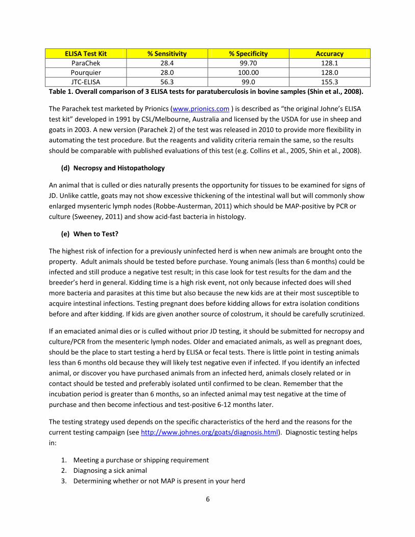
6
ELISA Test Kit % Sensitivity % Specificity Accuracy
ParaChek 28.4 99.70 128.1
Pourquier 28.0 100.00 128.0
JTC-ELISA 56.3 99.0 155.3
Table 1. Overall comparison of 3 ELISA tests for paratuberculosis in bovine samples (Shin et al., 2008).
The Parachek test marketed by Prionics (www.prionics.com ) is described as “the original Johne’s ELISA
test kit” developed in 1991 by CSL/Melbourne, Australia and licensed by the USDA for use in sheep and
goats in 2003. A new version (Parachek 2) of the test was released in 2010 to provide more flexibility in
automating the test procedure. But the reagents and validity criteria remain the same, so the results
should be comparable with published evaluations of this test (e.g. Collins et al., 2005, Shin et al., 2008).
(d) Necropsy and Histopathology
An animal that is culled or dies naturally presents the opportunity for tissues to be examined for signs of
JD. Unlike cattle, goats may not show excessive thickening of the intestinal wall but will commonly show
enlarged mysenteric lymph nodes (Robbe-Austerman, 2011) which should be MAP-positive by PCR or
culture (Sweeney, 2011) and show acid-fast bacteria in histology.
(e) When to Test?
The highest risk of infection for a previously uninfected herd is when new animals are brought onto the
property. Adult animals should be tested before purchase. Young animals (less than 6 months) could be
infected and still produce a negative test result; in this case look for test results for the dam and the
breeder’s herd in general. Kidding time is a high risk event, not only because infected does will shed
more bacteria and parasites at this time but also because the new kids are at their most susceptible to
acquire intestinal infections. Testing pregnant does before kidding allows for extra isolation conditions
before and after kidding. If kids are given another source of colostrum, it should be carefully scrutinized.
If an emaciated animal dies or is culled without prior JD testing, it should be submitted for necropsy and
culture/PCR from the mesenteric lymph nodes. Older and emaciated animals, as well as pregnant does,
should be the place to start testing a herd by ELISA or fecal tests. There is little point in testing animals
less than 6 months old because they will likely test negative even if infected. If you identify an infected
animal, or discover you have purchased animals from an infected herd, animals closely related or in
contact should be tested and preferably isolated until confirmed to be clean. Remember that the
incubation period is greater than 6 months, so an infected animal may test negative at the time of
purchase and then become infectious and test-positive 6-12 months later.
The testing strategy used depends on the specific characteristics of the herd and the reasons for the
current testing campaign (see http://www.johnes.org/goats/diagnosis.html). Diagnostic testing helps
in:
1. Meeting a purchase or shipping requirement
2. Diagnosing a sick animal
3. Determining whether or not MAP is present in your herd

7
4. Controlling MAP in an infected herd
5. Eradicating MAP from an infected herd
For a small herd wanting to determine if any MAP is present in the herd, it may be prudent to test all
animals individually by ELISA and fecal PCR. For larger herds it may be more cost-effective to start by
testing a subset of the herd that represents the highest risk of infection and highest probability of
testing positive if infected (i.e. older animals, does recently kidded, animals with contact with an
infected herd). ELISA tests can be used to prioritize which animals to submit for fecal PCR, or pooled
fecal testing can be used to mitigate costs until an infected animal is detected. If a herd has received a
positive test result, all pregnant does should be tested 2 weeks before kidding to identify those in need
of special isolation from other does and kids. For eradication of MAP from an infected herd, testing of
the entire herd every 6 months until there are 18 months of negative tests is required.
(f) Where to Test?
So, where to send samples for testing? This may depend on the test being used, the way the results are
reported, your location, and the cost (out-of-state centers often have a surcharge). The USDA provides a
list of approved laboratories that have voluntarily taken and passed an annual test of their capabilities
at http://www.aphis.usda.gov/animal_health/lab_info_services/approved_labs.shtml. Regarding ELISA
tests, both the Prionics test (Parachek) and the IDEXX-Pourquier test (HerdCheck) are suitable for use in
goats because, unlike some other tests, they have both been shown not to cross-react with antibodies
to the common disease caseous lymphadenitis (CLA) caused by Corynebacterium
pseudoparatuberculosis (Collins, 2011). Also both test kits include a step of pre-binding to
Mycobacterium phlei, which reduces false positives from mycobacteria that do not cause JD. The Johne’s
Information Center web site (Wisconsin) considers the Prionics test preferred for use in goats because it
has been licensed for use in goats and sheep in the USA after having been developed and licensed for
cattle (www.johnes.org/goats/diagnosis.html).
Many West Coast herds have been sending samples to Washington because they use the Prionics test,
but they currently report results only as positive or negative rather than quantitative results that can
better guide herd management decisions. UC Davis reports quantitative results but uses the IDEXX-
Pourquier test. As shown by Collins et al. (2005) these tests should give similar results and conclusions
but recent results for pygmy goat samples have produced more variability than expected between these
two test centers (see Case Study Example below). A third option is the University of Wisconsin Johne’s
Test Center which uses the Prionics test and reports quantitative results. This question of variable test
results is currently being actively investigated with samples being sent to all three test centers.
Whatever test is used, a positive ELISA test should not be considered definitive until confirmed by either
fecal PCR or fecal culture. Conversely, a negative fecal PCR or culture test after a potentially positive
ELISA test should still be considered a high risk animal that should be isolated and retested. Negative
herd status should not be concluded without comprehensive testing with no positive results for at least
18 months.
8
Herd Management Practices to Minimize Infection Risk
Maintaining a closed herd or carefully scrutinizing the purchase of animals is the best protection for
maintaining a herd free of JD. Visiting other herds’ kidding barns (either you or your animals) should also
be considered a potentially high risk opportunity for transmitting communicable diseases, both to and
from your herd. Disinfecting shoes and boots before and after the visit, like your veterinarian, should be
standard practice. Mycobacteria are relatively resistant to some disinfectants. The USDA recommends
sodium orthophenylphenate for premises contaminated by Mycobacteria. A commercial disinfectant in
this class is 1-Stroke Environ® made by Steris Corporation (www.steris.com ). Disinfectants can be
effective on hard surfaces (cement floors, feeders, water buckets etc.) but not on organic matter like
soil.
Purchased animals that have tested negative by ELISA and fecal PCR or culture may still be infected by
MAP and just be at an early stage of infection (especially young animals under 9 months). The stress of
moving to a new property itself may be sufficient to trigger the development of infectious shedding in
such animals. Therefore it would be wise to set aside a pen that is reserved for new animals to minimize
their exposure to the main herd until they have been retested a few months later. If you have any
reason to believe you may have suspect animals already in your herd, it would be good to separate
“high risk” and “low risk” animals while further testing is being performed. Using feeders and water
supplies that reduce the risk of fecal contamination is also a good precaution that applies to coccidiosis
as well as JD. Creep feeders for kids and fence-mounted feeders for adults can reduce kids sharing food
that may have been contaminated by adults.
Interpreting Test Results and Case Study Examples
Everyone would like to think that there is a single test that can definitively and unambiguously identify
an animal as either positive or negative for MAP infection with a minimum of delay and at low cost.
Unfortunately, the reality of JD is not that simple. While a positive fecal PCR or culture result can be
considered reasonably conclusive of a MAP-infected animal that is shedding bacteria that may infect
others, a negative result does not necessarily prove lack of infection. There are a number of factors that
can contribute to a false-negative test result. These include early stage infected animals not shedding,
low and variable shedding that may be influenced by diet, antibiotics, general health, quality of fecal
sample, and efficiency/sensitivity of the test. Fecal samples should be as fresh as possible and
transported to the testing lab quickly without excessive heating or multiple freeze/thaw cycles.
Blood/serum samples are usually refrigerated and shipped to the testing laboratory without delay. The
variability of ELISA test results is therefore related to the particular ELISA test being used, the test center
performing the test, and how the resulting data are processed, reported and interpreted. The ELISA
tests produce a color reaction such that the amount of color is proportional to the amount of antibody
in the blood sample. The color is measured as optical density (OD) and compared with positive and
negative control samples that are included in the kit. If the test laboratory usually reports results as only
positive or negative, your veterinarian should request a copy of the quantitative data. If these data are
9
reported as OD values, you need the values for the positive and negative controls as well as your test
samples.
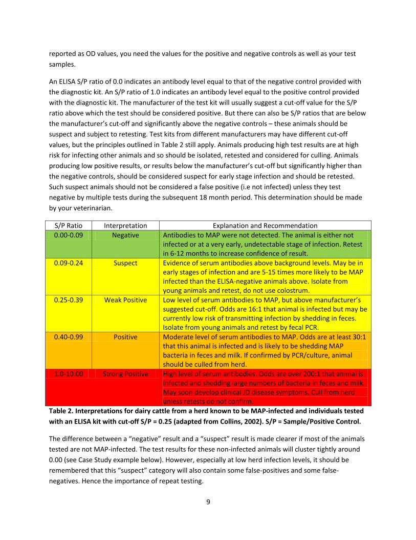
An ELISA S/P ratio of 0.0 indicates an antibody level equal to that of the negative control provided with
the diagnostic kit. An S/P ratio of 1.0 indicates an antibody level equal to the positive control provided
with the diagnostic kit. The manufacturer of the test kit will usually suggest a cut-off value for the S/P
ratio above which the test should be considered positive. But there can also be S/P ratios that are below
the manufacturer’s cut-off and significantly above the negative controls – these animals should be
suspect and subject to retesting. Test kits from different manufacturers may have different cut-off
values, but the principles outlined in Table 2 still apply. Animals producing high test results are at high
risk for infecting other animals and so should be isolated, retested and considered for culling. Animals
producing low positive results, or results below the manufacturer’s cut-off but significantly higher than
the negative controls, should be considered suspect for early stage infection and should be retested.
Such suspect animals should not be considered a false positive (i.e not infected) unless they test
negative by multiple tests during the subsequent 18 month period. This determination should be made
by your veterinarian.
S/P Ratio Interpretation Explanation and Recommendation
0.00-0.09 Negative Antibodies to MAP were not detected. The animal is either not infected or at a very early, undetectable stage of infection. Retest in 6-12 months to increase confidence of result.
0.09-0.24 Suspect Evidence of serum antibodies above background levels. May be in early stages of infection and are 5-15 times more likely to be MAP infected than the ELISA-negative animals above. Isolate from young animals and retest, do not use colostrum.
0.25-0.39 Weak Positive Low level of serum antibodies to MAP, but above manufacturer’s suggested cut-off. Odds are 16:1 that animal is infected but may be currently low risk of transmitting infection by shedding in feces. Isolate from young animals and retest by fecal PCR.
0.40-0.99 Positive Moderate level of serum antibodies to MAP. Odds are at least 30:1 that this animal is infected and is likely to be shedding MAP bacteria in feces and milk. If confirmed by PCR/culture, animal should be culled from herd.
1.0-10.00 Strong Positive High level of serum antibodies. Odds are over 200:1 that animal is infected and shedding large numbers of bacteria in feces and milk. May soon develop clinical JD disease symptoms. Cull from herd unless retests do not confirm.
Table 2. Interpretations for dairy cattle from a herd known to be MAP-infected and individuals tested
with an ELISA kit with cut-off S/P = 0.25 (adapted from Collins, 2002). S/P = Sample/Positive Control.
The difference between a “negative” result and a “suspect” result is made clearer if most of the animals
tested are not MAP-infected. The test results for these non-infected animals will cluster tightly around
0.00 (see Case Study example below). However, especially at low herd infection levels, it should be
remembered that this “suspect” category will also contain some false-positives and some false-
negatives. Hence the importance of repeat testing.
10
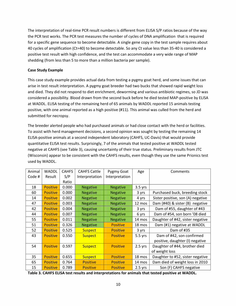
The interpretation of real-time PCR result numbers is different from ELISA S/P ratios because of the way
the PCR test works. The PCR test measures the number of cycles of DNA amplification that is required
for a specific gene sequence to become detectable. A single gene copy in the test sample requires about
40 cycles of amplification (Ct=40) to become detectable. So any Ct value less than 35-40 is considered a
positive test result with high confidence, and the test can accommodate a very wide range of MAP
shedding (from less than 5 to more than a million bacteria per sample).
Case Study Example
This case study example provides actual data from testing a pygmy goat herd, and some issues that can
arise in test result interpretation. A pygmy goat breeder had two bucks that showed rapid weight loss
and died. They did not respond to diet enrichment, deworming and various antibiotic regimes, so JD was
considered a possibility. Blood drawn from the second buck before he died tested MAP-positive by ELISA
at WADDL. ELISA testing of the remaining herd of 65 animals by WADDL reported 15 animals testing
positive, with one animal reported as a high positive (#11). This animal was culled from the herd and
submitted for necropsy.
The breeder alerted people who had purchased animals or had close contact with the herd or facilities.
To assist with herd management decisions, a second opinion was sought by testing the remaining 14
ELISA-positive animals at a second independent laboratory (CAHFS, UC-Davis) that would provide
quantitative ELISA test results. Surprisingly, 7 of the animals that tested positive at WADDL tested
negative at CAHFS (see Table 3), causing uncertainty of their true status. Preliminary results from JTC
(Wisconsin) appear to be consistent with the CAHFS results, even though they use the same Prionics test
used by WADDL.
Animal Code #
WADDL Result
CAHFS S/P
Ratio
CAHFS Cattle Interpretation
Pygmy Goat Interpretation
Age Comments
18 Positive 0.000 Negative Negative 3.5 yrs
60 Positive 0.000 Negative Negative 3 yrs Purchased buck, breeding stock
14 Positive 0.002 Negative Negative 4 yrs Sister positive, son (A) negative
47 Positive 0.003 Negative Negative 12 mos Dam (#40) & sister (B) negative
42 Positive 0.004 Negative Negative 3 yrs Dam of #55, daughter of #43
44 Positive 0.007 Negative Negative 6 yrs Dam of #54, son born ’08 died
55 Positive 0.011 Negative Negative 14 mos Daughter of #42, sister negative
51 Positive 0.326 Negative Positive 18 mos Dam (#1) negative at WADDL
52 Positive 0.525 Suspect Positive 3 yrs Dam of #35
43 Positive 0.550 Suspect Positive 5.5 yrs Dam of #42, son confirmed positive, daughter (I) negative
54 Positive 0.597 Suspect Positive 2.5 yrs Daughter of #44, brother died of weight loss
35 Positive 0.655 Suspect Positive 18 mos Daughter to #52, sister negative
65 Positive 0.764 Positive Positive 14 mos Dam died of weight loss in 2010
15 Positive 0.789 Positive Positive 2.5 yrs Son (F) CAHFS negative
Table 3. CAHFS ELISA test results and interpretations for animals that tested positive at WADDL.
11
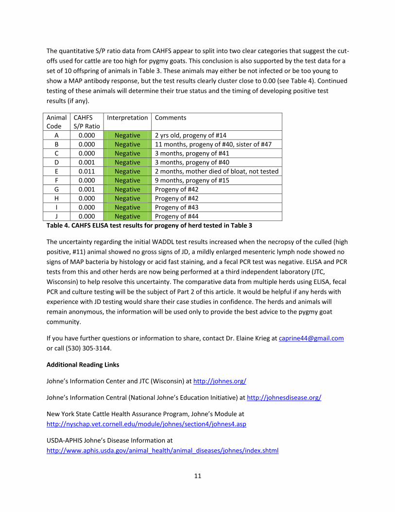
The quantitative S/P ratio data from CAHFS appear to split into two clear categories that suggest the cut-
offs used for cattle are too high for pygmy goats. This conclusion is also supported by the test data for a
set of 10 offspring of animals in Table 3. These animals may either be not infected or be too young to
show a MAP antibody response, but the test results clearly cluster close to 0.00 (see Table 4). Continued
testing of these animals will determine their true status and the timing of developing positive test
results (if any).
Animal Code
CAHFS S/P Ratio
Interpretation Comments
A 0.000 Negative 2 yrs old, progeny of #14
B 0.000 Negative 11 months, progeny of #40, sister of #47
C 0.000 Negative 3 months, progeny of #41
D 0.001 Negative 3 months, progeny of #40
E 0.011 Negative 2 months, mother died of bloat, not tested
F 0.000 Negative 9 months, progeny of #15
G 0.001 Negative Progeny of #42
H 0.000 Negative Progeny of #42
I 0.000 Negative Progeny of #43
J 0.000 Negative Progeny of #44
Table 4. CAHFS ELISA test results for progeny of herd tested in Table 3
The uncertainty regarding the initial WADDL test results increased when the necropsy of the culled (high
positive, #11) animal showed no gross signs of JD, a mildly enlarged mesenteric lymph node showed no
signs of MAP bacteria by histology or acid fast staining, and a fecal PCR test was negative. ELISA and PCR
tests from this and other herds are now being performed at a third independent laboratory (JTC,
Wisconsin) to help resolve this uncertainty. The comparative data from multiple herds using ELISA, fecal
PCR and culture testing will be the subject of Part 2 of this article. It would be helpful if any herds with
experience with JD testing would share their case studies in confidence. The herds and animals will
remain anonymous, the information will be used only to provide the best advice to the pygmy goat
community.
If you have further questions or information to share, contact Dr. Elaine Krieg at [email protected]
or call (530) 305-3144.
Additional Reading Links
Johne’s Information Center and JTC (Wisconsin) at http://johnes.org/
Johne’s Information Central (National Johne’s Education Initiative) at http://johnesdisease.org/
New York State Cattle Health Assurance Program, Johne’s Module at
http://nyschap.vet.cornell.edu/module/johnes/section4/johnes4.asp
USDA-APHIS Johne’s Disease Information at
http://www.aphis.usda.gov/animal_health/animal_diseases/johnes/index.shtml
12
USDA-approved Testing Laboratories at
http://www.aphis.usda.gov/animal_health/lab_info_services/approved_labs.shtml
Parachek ELISA test at http://www.prionics.com/diseases-solutions/paratuberculosis/PARACHEK/
IDEXX-Pourquier ELISA test at http://www.idexx.com/view/xhtml/en_us/livestock-
poultry/ruminant/map.jsf
Tetracore fecal PCR test kit at http://www.tetracore.com
Cited References
Ayele, W.Y., Bartos, M., Svastova, P., and Pavlik, I., 2004.Distribution of Mycobacterium avium subsp.
paratuberculosis in organs of naturally infected bull-calves and breeding bulls. Vet Microbiol. 103 (3-4)
209-17.
Buergelt, C.D., Donovan, G.A., and Williams, J.E., 2004.Identification of Mycobacterium avium
subspecies paratuberculosis by Polymerase Chain Reaction in Blood and Semen of a Bull with Clinical
Paratuberculosis. Intern J Appl Res Vet Med 2 (2): 130-134. Article available free of charge at
www.jarvm.com/articles/Vol21ss2/BUERGELTJARVMVol2No2.pdf
Collins, M.T., 2002. Interpretation of a Commercial Bovine Paratuberculosis Enzyme-Linked
Immunoabsorbent Assay by Using Likelihood Ratios. Clinical and Diagnostic Laboratory Immunology
9(6): 1367-1371. This article is available free of charge at
http://www.ncbi.nlm.nih.gov/pmc/articles/PMC130105/?tool=pubmed
Collins, M.T., Wells, S.J., Petrini, K.R., Collins, J.E., Schultz, R.D., and Whitlock, R.H., 2005. Evaluation of
Five Antibody Detection Tests for Diagnosis of Bovine Paratuberculosis. Clinical and Diagnostic
Laboratory Immunology 12 (6): 685-692. This article is available free of charge at
www.ncbi.nlm.nih.gov/pmc/articles/PMC1151972/?tool=pubmed
Collins, M.T., Gardner, I.A., Garry, F.B., Roussel, A.J., and Wells, S.J., 2006.Consensus recommendations
on diagnostic testing for the detection of paratuberculosis in cattle in the United States. JAVMA 229
(12):1912-1919. Article available free of charge at
http://avmajournals.avma.org/doi/abs/10.2460/javma.229.12.1912
Collins, M.T., 2011. Diagnosis of Paratuberculosis. Vet Clin Food Anim 27: 581-591. Note: this article is
part of a special issue on Johne’s Disease, a 12-month subscription to this journal costs $209, or $97 for
students; see vetfood.theclinics.com
Fecteau, M.-E., and Whitlock, R.H., 2011.Treatment and Chemoprophylaxis for Paratuberculosis. Vet Clin
Food Anim 27:547-557. Note: this article is part of a special issue on Johne’s Disease, a 12-month
subscription to this journal costs $209, or $97 for students; see vetfood.theclinics.com

13
Gumber, S., Eamans, G. and Whittington, R.J., 2006.Evaluation of a Pourquier ELISA kit in relation to agar
gel immunodiffusion (AGID) test for assessment of the humoral immune response in sheep and goats
with and without Mycobacterium paratuberculosis infection. Veterinary Microbiology 115: 91-101.
Lombard, J.E., 2011. Epidemiology and Economics of Paratuberculosis. Vet Clin Food Anim 27: 525-535.
Note: this article is part of a special issue on Johne’s Disease, a 12-month subscription to this journal
costs $209, or $97 for students; see vetfood.theclinics.com
Robbe-Austerman, S., 2011.Control of Paratuberculosis in Small Ruminants. Vet Clin Food Anim 27: 609-
620. Note: this article is part of a special issue on Johne’s Disease, a 12-month subscription to this journal
costs $209, or $97 for students see vetfood.theclinics.com
Shin, S.J., Cho, D., and Collins, M.T., 2008.Diagnosis of Bovine Paratuberculosis by a Novel Enzyme-
Linked Immunosorbent Assay Based on Early Secreted Antigens of Mycobacterium avium subsp.
paratuberculosis. Clinical and Vaccine Immunology 15 (8): 1277-1281. This article is available free of
charge at www.ncbi.nlm.nih.gov/pmc/articles/PMC2519299/?tool=pubmed
Sweeney, R.W., 2011. Pathogenesis of Paratuberculosis. Vet Clin Food Anim 27: 537-546. Note: this
article is part of a special issue on Johne’s Disease, a 12-month subscription to this journal costs $209, or
$97 for students; see vetfood.theclinics.com
Washington Animal Disease Diagnostic Lab; WADDL, 2011. New Strategies for Johne’s Disease Testing.
http://www.vetmed.wsu.edu/depts_waddl/dx/johnes.aspx
Whittington, R.J., Marsh, I.B., Taylor, P.J., Marshall, D.J., Taragel, C., and Reddacliff, L.A., 2003. Isolation
of Mycobacterium avium subsp. paratuberculosis from environmental samples collected from farms
before and after destocking sheep with paratuberculosis. Aust. Vet. J. 81(9): 559-563.