
J.Lopez_Research
15
Structural Basis of Substrate Specificity and Selectivity of Murine Cytosolic 5′-Nucleotidase III Christina L. Grobosky 1 , Jennifer B. Lopez 1 , SarahBeth Rennie 1 , Dionysus J. Skopelitis 1 , Amanda T. Wiest 1 , Craig A. Bingman 2 and Eduard Bitto 1 ⁎ 1 Department of Chemistry and Biochemistry, Georgian Court University, Lakewood, NJ 08701, USA 2 Department of Biochemistry, University of Wisconsin-Madison, Madison, WI 53706, USA Received 13 June 2012; received in revised form 14 August 2012; accepted 19 August 2012 Available online 25 August 2012 Edited by G. Schulz Keywords: pyrimidine nucleotidase; HAD superfamily; substrate complex; induced fit; nonspherocytic hemolytic anemia Cytosolic 5′-nucleotidase III (cN-III) is responsible for selective degradation of pyrimidine 5′-monoribonucleotides during maturation of reticulocytes to erythrocytes. The lack of this enzymatic activity due to genetic aberrations or lead poisoning results in a mild to moderate nonspherocytic hemolytic anemia. In affected individuals, pyrimidine nucleotides as well as their precursor polymers and their off-path metabolites accumulate in erythro- cytes, interfering with their proper function in ways that are not yet fully understood. This report describes the first X-ray structure of a catalytically inactivated variant of murine cN-III with a natural substrate, uridine 5′-monophosphate, in the active site at 1.74 Å resolution. The structure captures in an atomic detail the closed conformation that cN-III adopts upon substrate binding. Structure and sequence analysis coupled with enzymatic characterization of several mutants confirmed that the aromatic ring of a nitrogenous base of substrate nucleotide is stabilized by parallel π-stacking interactions with conserved aromatic rings of Trp113 and His68. The nitrogenous base is further stabilized by T-shaped stacking with the conserved aromatic ring of Tyr114, as well as by polar contacts with side chains of Thr66 and Ser117. Two water molecules help to stabilize the nucleotide binding by bridging it to protein residues Asp72 and His68 via hydrogen bonds. Finally, fully conserved Glu96 is responsible for recognition of ribose ring via two hydrogen bonds. The presented substrate complex structure elucidates how cN-III achieves specificity for pyrimidine 5′-nucleotides and how it selects against purine 5′-nucleotides. © 2012 Elsevier Ltd. All rights reserved. Introduction The 5′-nucleotidases constitute a family of en- zymes involved in degradation pathways of nucleotides. 1 They catalyze the enzymatic removal of the phosphate group of mononucleotides to yield respective nucleosides. To date, seven different 5′-nucleotidases with differential specificities and selectivities have been described in humans and higher animals. 2 Six of these enzymes are members of the haloacid dehalogenase (HAD) superfamily of Mg 2+ -dependent enzymes characterized by a *Corresponding author. E-mail address: [email protected]. Abbreviations used: cN-III, cytosolic 5′-nucleotidase III; mcN-III, murine cN-III; UMP, uridine 5′-monophosphate; GMP, guanidine 5′-monophosphate; mdN, mitochondrial deoxyribonuclease; cdN, cytosolic deoxyribonuclease; HAD, haloacid dehalogenase; TEV, tobacco etch virus; PEG, polyethylene glycol; PDB, Protein Data Bank. http://dx.doi.org/10.1016/j.jmb.2012.08.014 J. Mol. Biol. (2012) 423, 540–554 Contents lists available at www.sciencedirect.com Journal of Molecular Biology journal homepage: http://ees.elsevier.com.jmb 0022-2836/$ - see front matter © 2012 Elsevier Ltd. All rights reserved.
-
Upload
jennifer-lopez -
Category
Documents
-
view
5 -
download
0
Transcript of J.Lopez_Research

http://dx.doi.org/10.1016/j.jmb.2012.08.014 J. Mol. Biol. (2012) 423, 540–554
Contents lists available at www.sciencedirect.com
Journal of Molecular Biologyj ourna l homepage: ht tp : / /ees .e lsev ie r.com. jmb
Structural Basis of Substrate Specificity and Selectivityof Murine Cytosolic 5′-Nucleotidase III
Christina L. Grobosky 1, Jennifer B. Lopez 1, SarahBeth Rennie 1,Dionysus J. Skopelitis 1, Amanda T. Wiest 1,Craig A. Bingman2 and Eduard Bitto 1⁎1Department of Chemistry and Biochemistry, Georgian Court University, Lakewood, NJ 08701, USA2Department of Biochemistry, University of Wisconsin-Madison, Madison, WI 53706, USA
Received 13 June 2012;received in revised form14 August 2012;accepted 19 August 2012Available online25 August 2012
Edited by G. Schulz
Keywords:pyrimidine nucleotidase;HAD superfamily;substrate complex;induced fit;nonspherocytic hemolyticanemia
*Corresponding author. E-mail [email protected] used: cN-III, cytoso
mcN-III, murine cN-III; UMP, uridinGMP, guanidine 5′-monophosphatedeoxyribonuclease; cdN, cytosolic dHAD, haloacid dehalogenase; TEV,PEG, polyethylene glycol; PDB, Pro
0022-2836/$ - see front matter © 2012 E
Cytosolic 5′-nucleotidase III (cN-III) is responsible for selective degradationof pyrimidine 5′-monoribonucleotides during maturation of reticulocytes toerythrocytes. The lack of this enzymatic activity due to genetic aberrationsor lead poisoning results in a mild to moderate nonspherocytic hemolyticanemia. In affected individuals, pyrimidine nucleotides as well as theirprecursor polymers and their off-path metabolites accumulate in erythro-cytes, interfering with their proper function in ways that are not yet fullyunderstood. This report describes the first X-ray structure of a catalyticallyinactivated variant of murine cN-III with a natural substrate, uridine5′-monophosphate, in the active site at 1.74 Å resolution. The structurecaptures in an atomic detail the closed conformation that cN-III adoptsupon substrate binding. Structure and sequence analysis coupled withenzymatic characterization of several mutants confirmed that the aromaticring of a nitrogenous base of substrate nucleotide is stabilized by parallelπ-stacking interactions with conserved aromatic rings of Trp113 and His68.The nitrogenous base is further stabilized by T-shaped stacking with theconserved aromatic ring of Tyr114, as well as by polar contacts with sidechains of Thr66 and Ser117. Two water molecules help to stabilize thenucleotide binding by bridging it to protein residues Asp72 and His68 viahydrogen bonds. Finally, fully conserved Glu96 is responsible forrecognition of ribose ring via two hydrogen bonds. The presented substratecomplex structure elucidates how cN-III achieves specificity for pyrimidine5′-nucleotides and how it selects against purine 5′-nucleotides.
© 2012 Elsevier Ltd. All rights reserved.
ress:
lic 5′-nucleotidase III;e 5′-monophosphate;; mdN, mitochondrialeoxyribonuclease;tobacco etch virus;tein Data Bank.
lsevier Ltd. All rights reserve
Introduction
The 5′-nucleotidases constitute a family of en-zymes involved in degradation pathways ofnucleotides.1 They catalyze the enzymatic removalof the phosphate group of mononucleotides to yieldrespective nucleosides. To date, seven different5′-nucleotidases with differential specificities andselectivities have been described in humans andhigher animals.2 Six of these enzymes are membersof the haloacid dehalogenase (HAD) superfamily ofMg2 +-dependent enzymes characterized by a
d.

541Substrate Specificity and Selectivity of mcN‐III
consensus motif [DxDx(T/V)] that harbors thecatalytic aspartate (the first “D” in the consensusmotif) as well as a second aspartate that is involvedin general acid/base catalysis.3,4 During catalysis,the catalytic aspartate attacks the phosphorus of abound substrate, while the second aspartate of theconservedmotif protonates the leaving group.5,6 Theenzyme becomes transiently phosphorylated duringthe process.7 The covalent phosphoenzyme inter-mediate is usually resolved by hydrolysis; however,for some 5′-nucleotidases, another nucleoside canresolve the intermediate. This chemistry explainsphosphotransferase activity that has been describedin some 5′-nucleotidases.8,9 This activity is ofimportance for pharmacokinetics of several chemo-therapeutic agents based on nucleoside chemistry.2
The structural knowledge of 5′-nucleotidases fromthe HAD superfamily has rapidly expanded in thelast decade. The structures of enzymes and theirvarious complexes with substrates, products, ana-logs, and effectors known to date include those ofhumanmitochondrial deoxynucleotidase,5,10 humanandmurine cytosolic 5′(3′)-deoxyribonucleotidase,11
human and murine cytosolic 5′-nucleotidase III(cN-III),12,13 human cytosolic 5′-nucleotidase II,13,14
5′-nucleotidase SDT1 from Saccharomyces cerevisiae,15
and cytosolic IMP–guanidine 5′‐monophosphate(GMP)‐specific 5′-nucleotidase from Legionellapneumophila [unpublished; Protein Data Bank (PDB)ID: 2BDE].cN-III (UniProt ID: Q9D020, OMIM ID: 606224),
which is also known as pyrimidine 5′-nucleotidasetype 1, shows specificity for pyrimidine 5′-nucleo-tides. The enzyme can efficiently hydrolyze CMPand uridine 5′‐monophosphate (UMP) as well asdUMP, dTMP, and dCMP with a lower specificactivity; however, it selects against and will nothydrolyze purine nucleotides.16 In humans, theenzyme has a specific role in reticulocytes where ithelps with degradation of translational machineryduring maturation to erythrocytes.17,18 A lack of thisactivity during erythropoiesis leads to accumulationof pyrimidine nucleotides and precipitation ofunhydrolyzed ribosomal RNA.19 Formation oflarge aggregates can be clinically visualized byWright's staining and manifests as a basophilicstippling on red blood cell smears.17 Clinically, lackof pyrimidine 5′-nucleotidase activity ultimatelymanifests as a mild nonspherocytic anemia (OMIMID: 266120).20 The deficiency may be familial or itcan also be acquired during severe lead poisoning,because Pb2+, as well as some other heavy metalions, inhibits activity of this enzyme.21 Severaldozen mutations in the gene encoding cN-III havebeen described in literature and biochemical defectsof many missense mutations have been studied atthe protein level.20,22–24
The structures of murine cN-III (mcN-III; UniProtID: Q9D020) as well as human cN-III that provide
snapshots of several states of the catalytic cycle havebeen previously reported.12,13 These studies so fardid not address important questions such as how thesubstrate is recognized by cN-III and how thisenzyme at a structural level achieves the selectivityagainst purine nucleotides. To fill this gap in ourknowledge, we present and discuss in this article thefirst ever X-ray structure of catalytically inactivatedmcN-III in complex with its natural substrate, UMP.
Results
Experimental objective
The objective of this study was to provide astructural insight into how cN-III binds pyrimidine5′-mononucleotides. Structural information isneeded to rationalize how specificity for pyrimi-dine 5′-mononucleotides and selectivity againstpurine 5′-mononucleotides, both observed at bio-chemical level,9 are achieved by this nucleotidase.The physiological reason for utilization of nucleo-tidase with specificity for pyrimidine 5′-nucleotidesduring maturation of erythrocytes (and simulta-neous down-regulation of less selective and/orpurine-specific nucleotidases) is to preserve purine5′-nucleotide pool derived from degradation ofribosomes and mRNA in order to support laterenergy metabolism of erythrocytes that lack theability to resynthesize these nucleotides de novo.17
Catalytically inactive mcN-III variant
To prepare a catalytically inactive variant of mcN-III, we mutated the general acid–base residue Asp51of mcN-III to asparagine. Asparagine has a similarshape as aspartate but is not able to support generalacid–base chemistry. Recombinant human cN-III-D51N has been reported to show no detectablehydrolytic activity.25 Before crystallization trials,recombinant mcN-III-D51N protein was assayedand confirmed to show no detectable activity at upto 20 mM UMP concentration (data not shown).
Structure solution
Crystals of the substrate complex of mcN-III-D51N with UMP (mcN-III-D51N ∙UMP) were pre-pared and diffraction data were collected asdescribed in Materials and Methods. Diffractiondata statistics are summarized in Table 1. The spacegroup and unit cell parameters of the crystal formobtained for the complex differ from those reportedoriginally for mcN-III (P4322 versus P32);
12 thus, anew crystal form was obtained for the substratecomplex. The diffraction limit of the data set was1.74 Å and extended the previously achieved

Table 1. Statistics for data collection, phasing, modelrefinement, and validation
Data collection statisticsa
Space group P4322Unit cell dimensionsa, b, c (Å) 46.88, 46.88, 287.10α, β, γ (°) 90.00, 90.00, 90.00X-ray wavelength (Å) 0.9800Resolution (Å)b 50.00–1.74 (1.77–1.74)Total/unique reflections 607,246/33,980Completeness (%) 98.3 (97.1)Rmerge
c 0.080 (0.555)Average I/σ(I) 32.3 (4.8)Data redundancy 17.9 (12.8)B-factor from Wilson plot (Å2) 21.80
Phasing statisticsMolecular replacement model A monomer from
PDB entry 2G09R-factor for the top solutiond 0.461
Refinement statisticsResolution (Å) 50–1.74 (1.784–1.739)Rwork
e/Rfreef 0.169/0.218
(0.286/0.355)Macromolecules in asymmetric unit 1 mcN-III-D51NOther entities in asymmetric unitg 335 H2O, 1 Mg2+,
1 Na+, 2 BME, 1 UMPNo. of protein atoms/mean B‐factor (Å2) 2340/23.5No. of solvent atoms/mean B‐factor (Å2) 335/37.1No. of ligand atoms/mean B‐factor (Å2) 31/25.9RMSD from ideal geometryBond lengths (Å) 0.010Bond angles (°) 1.356
Validation statisticsh
Ramachandran outliers (%) 0.00Ramachandran favored (%) 99.31Poor rotamers (%) 0.38PDB ID of the deposited structure 4FE3
a Data collected at Sector 24-ID-D of the Advanced PhotonSource.
b Numbers in parentheses indicate the highest‐resolution shellof 20.
c Rmerge =∑h∑i|I(h)i − ⟨I(h)⟩|/∑h∑iI(h)i, where I(h) is theobserved intensity of reflection h, and ⟨I(h)⟩ is the averageintensity obtained from multiple measurements.
d Reported by the program REFMAC as wRfac parameter.e Rwork =∑||Fo| − |Fc||/∑|Fo|, where |Fo| is the observed
structure factor amplitude and |Fc| is the calculated structurefactor amplitude.
f Rfree =R-factor based on 1710 or 5.1% of the reflectionsexcluded from refinement.
g BME stands for 2-mercaptoethanol.h Validation was performed by MolProbity software.
UMPW113
K89
Mg2+
Y93
D238
D49
H118
Fig. 1. Electron density quality. The blue mesh repre-sents a composite 2Fo − Fc omit electron density map of themcN-III-D51N ∙UMP complex contoured at the 1.0 σlevel. The model is depicted in stick representation.Carbons are in yellow, oxygens are in red, nitrogens arein blue, and phosphorus is in orange.
542 Substrate Specificity and Selectivity of mcN‐III
resolution range of 2.1–2.5 Å. The structure wassolved by molecular replacement methodologyusing mcN-III protomer as a search model (basedon coordinates of the chain A from PDB ID 2G09).During the process, the space group assignment ofthe data set was finalized to P4322 (as opposed toP4122). The electron density map at this stagerevealed a well-defined unexplained electron densi-ty in the active site. The molecular model wasrefined as described in Materials and Methods to anR‐factor and Rfree of 0.169 and 0.218, respectively.
The relevant refinement and validation statistics forthe final model are summarized in Table 1.The final refined structure defines a continuous
segment of mcN-III-D51N spanning residues 7 to297. The electron density was well defined for themain chain of the entire structure and most sidechains; however, it was occasionally less clear forside chains of solvent‐exposed surface residues. Inthe active site, a clear density was observed for anoctahedrally coordinatedmagnesium ion that servesas a cofactor for this enzyme, as well as for boundUMP. The quality of the model and active‐siteelectron density can be judged from the compositeomit electron density map shown in Fig. 1. Inaddition to the protein chain, the final model alsoincludes 335 water molecules. One of the solventpeaks proved to be of larger intensity than could beexplained by water; this density was ultimatelymodeled as a sodium ion. Two surface‐exposedcysteine residues (Cys34 and Cys64) were covalent-ly modified. The observed density was interpretedand modeled as β-mercaptoethanol, an agent usedduring protein purification.
Global conformational differences
The structure of mcN-III consists of two distinctdomains: the globular core domain (residues 7–55,142–189, and 212–297) and the smaller cap domain(residues 56–142 and 190–211). The core domain hasα/β-Rossmannoid fold and harbors a portion of thecatalytic site including residues that coordinate the
543Substrate Specificity and Selectivity of mcN‐III
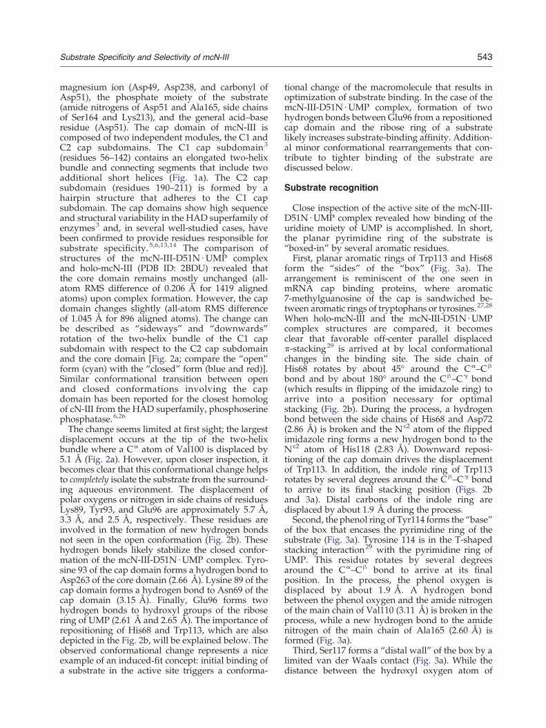
magnesium ion (Asp49, Asp238, and carbonyl ofAsp51), the phosphate moiety of the substrate(amide nitrogens of Asp51 and Ala165, side chainsof Ser164 and Lys213), and the general acid–baseresidue (Asp51). The cap domain of mcN-III iscomposed of two independent modules, the C1 andC2 cap subdomains. The C1 cap subdomain3
(residues 56–142) contains an elongated two-helixbundle and connecting segments that include twoadditional short helices (Fig. 1a). The C2 capsubdomain (residues 190–211) is formed by ahairpin structure that adheres to the C1 capsubdomain. The cap domains show high sequenceand structural variability in the HAD superfamily ofenzymes3 and, in several well-studied cases, havebeen confirmed to provide residues responsible forsubstrate specificity.5,6,13,14 The comparison ofstructures of the mcN-III-D51N ∙UMP complexand holo-mcN-III (PDB ID: 2BDU) revealed thatthe core domain remains mostly unchanged (all-atom RMS difference of 0.206 Å for 1419 alignedatoms) upon complex formation. However, the capdomain changes slightly (all-atom RMS differenceof 1.045 Å for 896 aligned atoms). The change canbe described as “sideways” and “downwards”rotation of the two-helix bundle of the C1 capsubdomain with respect to the C2 cap subdomainand the core domain [Fig. 2a; compare the “open”form (cyan) with the “closed” form (blue and red)].Similar conformational transition between openand closed conformations involving the capdomain has been reported for the closest homologof cN-III from the HAD superfamily, phosphoserinephosphatase.6,26
The change seems limited at first sight; the largestdisplacement occurs at the tip of the two-helixbundle where a Cα atom of Val100 is displaced by5.1 Å (Fig. 2a). However, upon closer inspection, itbecomes clear that this conformational change helpsto completely isolate the substrate from the surround-ing aqueous environment. The displacement ofpolar oxygens or nitrogen in side chains of residuesLys89, Tyr93, and Glu96 are approximately 5.7 Å,3.3 Å, and 2.5 Å, respectively. These residues areinvolved in the formation of new hydrogen bondsnot seen in the open conformation (Fig. 2b). Thesehydrogen bonds likely stabilize the closed confor-mation of the mcN-III-D51N ∙UMP complex. Tyro-sine 93 of the cap domain forms a hydrogen bond toAsp263 of the core domain (2.66 Å). Lysine 89 of thecap domain forms a hydrogen bond to Asn69 of thecap domain (3.15 Å). Finally, Glu96 forms twohydrogen bonds to hydroxyl groups of the ribosering of UMP (2.61 Å and 2.65 Å). The importance ofrepositioning of His68 and Trp113, which are alsodepicted in the Fig. 2b, will be explained below. Theobserved conformational change represents a niceexample of an induced‐fit concept: initial binding ofa substrate in the active site triggers a conforma-
tional change of the macromolecule that results inoptimization of substrate binding. In the case of themcN-III-D51N ∙UMP complex, formation of twohydrogen bonds between Glu96 from a repositionedcap domain and the ribose ring of a substratelikely increases substrate-binding affinity. Addition-al minor conformational rearrangements that con-tribute to tighter binding of the substrate arediscussed below.
Substrate recognition
Close inspection of the active site of the mcN-III-D51N ∙UMP complex revealed how binding of theuridine moiety of UMP is accomplished. In short,the planar pyrimidine ring of the substrate is“boxed-in” by several aromatic residues.First, planar aromatic rings of Trp113 and His68
form the “sides” of the “box” (Fig. 3a). Thearrangement is reminiscent of the one seen inmRNA cap binding proteins, where aromatic7-methylguanosine of the cap is sandwiched be-tween aromatic rings of tryptophans or tyrosines.27,28
When holo-mcN-III and the mcN-III-D51N ∙UMPcomplex structures are compared, it becomesclear that favorable off-center parallel displacedπ-stacking29 is arrived at by local conformationalchanges in the binding site. The side chain ofHis68 rotates by about 45° around the Cα–Cβ
bond and by about 180° around the Cβ–Cγ bond(which results in flipping of the imidazole ring) toarrive into a position necessary for optimalstacking (Fig. 2b). During the process, a hydrogenbond between the side chains of His68 and Asp72(2.86 Å) is broken and the Nε2 atom of the flippedimidazole ring forms a new hydrogen bond to theNε2 atom of His118 (2.83 Å). Downward reposi-tioning of the cap domain drives the displacementof Trp113. In addition, the indole ring of Trp113rotates by several degrees around the Cβ–Cγ bondto arrive to its final stacking position (Figs. 2band 3a). Distal carbons of the indole ring aredisplaced by about 1.9 Å during the process.Second, the phenol ring of Tyr114 forms the “base”
of the box that encases the pyrimidine ring of thesubstrate (Fig. 3a). Tyrosine 114 is in the T-shapedstacking interaction29 with the pyrimidine ring ofUMP. This residue rotates by several degreesaround the Cα–Cβ bond to arrive at its finalposition. In the process, the phenol oxygen isdisplaced by about 1.9 Å. A hydrogen bondbetween the phenol oxygen and the amide nitrogenof the main chain of Val110 (3.11 Å) is broken in theprocess, while a new hydrogen bond to the amidenitrogen of the main chain of Ala165 (2.60 Å) isformed (Fig. 3a).Third, Ser117 forms a “distal wall” of the box by a
limited van der Waals contact (Fig. 3a). While thedistance between the hydroxyl oxygen atom of

Fig. 2. Conformational changes of mcN-III. (a) A stereo representation reveals global differences between the open andclosed conformations of mcN-III. The cyan ribbon connects Cα atoms of the main chain of holo-mcN-III (from PDB ID:2G09), which is in the open conformation. Blue and red ribbons connect Cα atoms of the main chain of the core and the capdomains of mcN-III-D51N ∙UMP complex, respectively. The complex structure is in the closed conformation. Thesubstrate is depicted in stick representation. Carbons are in yellow, oxygens are in red, nitrogens are in blue, andphosphorus is in orange. (b) A stereo representation detailing structural changes during open-to-closed conformationtransition. Side chains of selected residues are depicted in stick representation. The color scheme is identical to that used in(a). Black broken lines represent hydrogen bonds.
544 Substrate Specificity and Selectivity of mcN‐III
Ser117 and the O4 oxygen of the keto group of theuracil ring seems optimal (2.8 Å), the local geometryis not conducive to formation of a favorablehydrogen bond due to nearly perpendicular place-ment of sp2 atomic orbitals of the O4 oxygen withrespect to the hydroxyl group of Ser117 side chain.Fourth, the “top” of the box seems relatively open;
the two closest residues include Thr66 and Asn69(Fig. 3a). Again, the side chain of Thr66 is in the vander Waals contact with the uracil; however, forma-tion of a strong hydrogen bond is unlikely due tomisalignment of sp2 orbitals on the O2 atom of theuracil ring and the hydroxyl group of Thr66.
Residue Asn69 is located even further away, withthe side‐chain amide nitrogen located 3.6 Å awayfrom the O2 atom of the uracil ring. One additionalinteraction that has a potential to stabilize binding ofthe uracil ring in the active‐site cavity involves awater molecule (w1 in Fig. 3a) that forms ahydrogen bond to both the N3 atom of the uracilring of the substrate (2.78 Å) and the carboxyloxygen of Asp72 (2.57 Å).The ribose ring of UMP is stabilized by the above-
mentioned Glu96, which forms two favorablehydrogen bonds to hydroxyl groups of the ribosering (2.61 Å and 2.65 Å, Fig. 3a). In addition, two

Fig. 3. UMP binding in the active site of mcN-III. (a) A stereo representation of the active‐site residues of the mcN-III-D51N ∙UMP complex involved in recognition of the uridine moiety of UMP. The protein main chain is shown in linerepresentation and is highlighted by a gray ribbon connecting Cα atoms. Selected residue side chains are shown in stickrepresentation and properly annotated. Carbons of these side chains are in light gray, oxygens are in red, nitrogens are inblue, and phosphorus is in orange. Red spheres representwatermolecules directly in contact with the substrate. Gray three-dimensional crosses represent other water molecules present in the active site. Black broken lines represent well-formedhydrogen bonds; yellow broken lines represent polar contacts that are unlikely to form strong hydrogen bonds due toimproper geometry. Green sphere represents a magnesium ion; green broken lines represent coordination bonds betweenthe magnesium ion and neighboring polar atoms. (b) Schematic diagram of the hydrogen bonds (broken lines) and polarcontacts (crossing arcs) between UMP and the active‐site residues of mcN-III-D51N. The distances are given in angstroms.
545Substrate Specificity and Selectivity of mcN‐III

546 Substrate Specificity and Selectivity of mcN‐III
water molecules play a role in the stabilization of theribose ring. The water molecule w2 forms twohydrogen bonds to the main chain and side chain ofHis68 (3.00 Å and 2.78 Å, respectively; Fig. 3a).Furthermore, the w2 water molecule forms ahydrogen bond to the O4′ atom of the ribose ring(3.03 Å). This water is in a polar contact with residueAsp51 (3.08 Å; in our case, Asn51, as this residuewas mutated to solve the structure of the complex);however, due to local arrangements of atoms andorbitals, this contact is unlikely to result in afavorable hydrogen bond. Finally, 3′-hydroxylgroup of the ribose ring is stabilized by a hydrogenbond (2.93 Å) to the water molecule w3 coordinatedto a magnesium ion coordinated in the active site(Fig. 3a). All polar contacts observed between UMPand the active‐site residues are presented in aschematic form in Fig. 3b. The aromatic rings ofTrp113 and Tyr114 were omitted from this figurefor clarity.
Analysis of site-directed mutants
To provide biochemical evidence for involvementof key residues from the C1 cap subdomain inbinding of the uridine moiety of UMP, we generateda set of site-directed mutants of mcN-III. Thetargeted residues included all residues identified tobe in direct contact with the substrate. Namely,T66G, N69G, H68A, E96S, W113A, Y114A, andS117G variants of mcN-III were prepared andcharacterized using an endpoint kinetic assay thatused UMP as a substrate. The results are summa-rized in Table 2. All mutants retained measurablecatalytic activity. The W113A mutant had signifi-cantly lower catalytic ability compared to that of thewild‐type mcN-III as can be judged from the over100-fold drop in the specificity constant (i.e., kcat/Kmratio). The effect comes mostly from an increase inMichaelis constant (i.e., Km) value, which, to someextent, reflects significantly lowered binding affinityfor UMP. Mutants H68A, Y114A, and E96S also hadsignificantly lower catalytic ability as can be judgedfrom their 13-fold to 30-fold decreases in specificityconstants. Interestingly, both H68A and Y114Ashowed a large decrease of the turnover number
Table 2. Kinetic parameters of mcN-III and its mutantsduring hydrolysis of UMP
Protein Km (mM) kcat (s− 1) kcat/Km (M− 1 s− 1)
Wild type 1.1 ± 0.1 1.2 ± 0.2 1100 ± 200T66G 1.1 ± 0.1 1.9 ± 0.3 1700 ± 400H68A 3.2 ± 0.3 0.12 ± 0.02 37 ± 7N69G 2.1 ± 0.2 1.1 ± 0.2 550 ± 100E96S 4.8 ± 0.9 0.38 ± 0.06 80 ± 20W113A 29 ± 7 0.29 ± 0.06 10 ± 3Y114A 2.3 ± 0.3 0.14 ± 0.02 60 ± 10S117G 1.1 ± 0.4 1.7 ± 0.3 1500 ± 300
(i.e., kcat constant) that characterizes the rate of thechemical step of a catalytic conversion. This issomewhat puzzling and suggests that the substratemay become misaligned in the larger active‐sitecavity of these mutants and compromise properalignment of the phosphate moiety in the active site,thus lowering the efficiency of the catalytic step. Onthe other hand, the E96S mutant showed anintermediate increase in its Km value; this observa-tion is consistent with the expected loss of twostabilizing hydrogen bonds. Mutants T66G andS117G did not significantly differ from wild‐typemcN-III. The structure of the mcN-III-D51N ∙UMPcomplex indicates that neither residue forms favor-able hydrogen bonds to the substrate. Our kineticresults seem to be consistent with this view andsuggest that a loss of small amino acid side chains atthese positions is well tolerated and does notinfluence catalytic activity towards UMP. TheN69G mutant showed slightly higher Km comparedto the wild‐type form. Based on the structure ofthe mcN-III-D51N ∙UMP complex, this residue maybe involved in the stabilization of the closedconformation of mcN-III by forming a hydrogenbond to Lys89.
Sequence conservation analysis
To gain a deeper insight into the evolutionaryconservation of residues within the C1 cap sub-domain of the cN-III family of proteins, weperformed a profile–profile sequence search usingFFAS0330 and constructed multiple sequence align-ment. The analysis focused on residues 50–125 ofmcN-III that include the active‐site consensusDxDx(T/V) motif and the C1 cap subdomain. Thesearch identified 147 sequences from the nr85sdatabase (derived for use by FFAS03 from theNational Center for Biotechnology Informationnonredundant database by clustering sequences at85% identity level). Eighteen of these sequences aswell as the sequence of human cN-III were selectedand are shown in the multiple sequence alignmentin Fig. 4a to demonstrate the observed pattern ofamino acid conservation. The representative se-quences were selected to cover a wide range ofsequence identities, spanning from 92% to 23%. Thespecies ranged from mammals, fish, plants, worms,insects, to single‐celled ciliated protozoans. Resi-dues of the signature motif of the HAD superfamilyDxDx(T/V) were fully conserved in the alignment(residues 49–53), confirming that all selected se-quences came from the HAD superfamily ofenzymes. Several residues from the lid domainwere very highly conserved, namely, Tyr92, Glu96,Lys106, Met110, Glu112, and Trp113 (Fig. 4a).Importantly, all three aromatic residues (His68,
Trp113, and Tyr114) identified as the componentsof the binding pocket for pyrimidine ring in

50 60 70 80 90 100 110 120
.|....:... .|....:... .|....:... .|....:... .|....:... .|....:... .|....:... .|....: mcN-III DFDMTLSRFS YNGKRCPTCH NIIDNCKLVT DECRRKLLQL KEQYYAIEVD PVLTVEEKFP YMVEWYTKSH GLLIEQG hcN-III DFDMTLSRFS YKGKRCPTCH NIIDNCKLVT DECRKKLLQL KEKYYAIEVD PVLTVEEKYP YMVEWYTKSH GLLVQQA92%-GI90083362 DFDMTLSRFS YKGKRCPTCH NIIDNCKLVT DECRKKLLQL KEKYYAIEVD PVLTVEEKYP YMVEWYTKSH GLLVEQA90%-GI332260319 DFDMTLSRFS YKGKRCPTCH NIIDNCKLVT DECRKKLLQL KEKYYAIEVD PVLTVEEKYP YMVEWYTKSH GLLVQQA78%-GI222088009 DFDMTLSKFA VNGKRCPTCH NIIDNCKLVT EECRQKLLQL KNKYYPIEID PHLTMEEKYP FMVEWYFKSH SLLVEQ 77%-GI317667937 DFDMTLSKFS INGKRCPTCH NIIDNCKLVT EECRQKLYKL KDTYYPIEID PHLTMEEKYP FMVEWYFKSH TLLVEQ 65%-GI47206341 DFDMTLTRFA YKGKRCPTCH NILDNSTLIS DDCKLKLKQL LDTYYPIEID TSLSVEEKLP LMVEWWTKAH ELLVEQ 63%-GI327275435 DFDMTLTRFG FNGKRCPTSH NIIDNSRVIN EEGRRKLKDL LHYYYPIEID PYRTVEDKLP LMVEWWTKAH NLLTQQ 60%-GI338711434 DFDMTLSRFA YNGKRCPSSH NILDDSKIIS EECRKELKML LHHYYPIEID PHRTIKEKLP HMVEWWTKAH NLLCQQ 60%–GI148236428 DFDMTLSRFS RNGERCPTCY NIIDNSNIIS DEGRKKLKCL FDIYYPLEID PKKSIEEKYP LMVEWWSKAH DLFYEQ 55%-GI317419431 DFDMTLTKFA HNGKRVPTTH NILDTRLLIN EDCTKKMREL LNTYYPIEID PGRSAEEKLP LMVEWWTKVH ELLIQQ 45%-GI320168041 DFDMTLTRFL YNGKRGASSH GIIEHSHMLE EAYHEATHNL FSTYYPMEVD PTLTQAEKIP HMVAWWESAH ELLIKH 40%-GI53370705 DFDGTLTRYW YDGSRGQSSH GLLRQG---N EEYDAKREEL FEHYHPIEIC PDIPLPEKAK LMEEWWEKTH ALLIEGG37%-GI289741479 DFDYTITKQR TDGTPVLSSF GIFNSCKSLP KGYVEETEKL YQKYRPIEVD PHLPIQEKIK YMVEWWTKSG ELLV 35%-GI348667774 DFDYTLTPYK PTDDQALSTH RLLMESEALG PDAEDVAREI FEKYYPIEQS ATLTMEEKLP YMIEWWAKTH GLMIQHG31%-GI226468492 DFDHTLSKYT HNAERVPVCH GLFIKNSRIP EVCRLRLKEL SDFYSPFETS PDMYAYEKSC LMKEFWNLAH KALVDG 30%-GI301110074 DFDRTLTPYY KQRRDP-SSH GLLMTSSVLQ PQVCAGEQEL FAKFYPVEMS PTLSAAEKLP FMEQWWNSAH ALLVE 28%-GI340509290 DFDHTITDFY YNNQPVSPSF GILNRSQHVT IEMENQAKEI FEIYSKYENT KQYTFEFKYQ KMEEWVKLTA ELFVK 26%-GI322796933 DFDLTVTKQH INGTHVLSSF GMFRKCKQLP QHYLDSAKLL YDKYRPIEID PYVPLNKKAD AMHDWMSIAN NLL23%-GI118364864 DYDQTITAFG CPNEQEISTF GVIAKSKRVS QKYLDDMTAL YQHYSPYEKD STIDKETLVK YVTEWYSKTA E + + + + + ++ + Consensus DFD.Tlt.f. .ng.r..ssh gi...s.... .........l ...YypiE.d p....eeK.p .M.#Ww.k.h .ll....
(a)
(b) 211E211E
Y92 Y92
311W311W
PMUPMU
69E69E
K106K106
011M011M
Fig. 4. Sequence alignment of the cN-III lid domain. (a) The alignment shows consensus motif of the HADsuperfamily and the C1 cap subdomain of the cN-III family. The topmost line gives residue numbering of mcN-III; thesecond line serves as a ruler to facilitate residue identification. The second sequence is that of human cN-III (UniProt ID:Q9H0P0). The remaining sequences are identified by percent identity to mcN-III in the aligned region followed by theGenBank GI accession number (which can be looked up at http://www.ncbi.nlm.nih.gov/protein). Red color indicatesmore than 90% conservation; blue color indicates more than 50% conservation at a given position. In the consensussequence, uppercase red letters signify overall conservation (N 90%) at a given position, while blue lowercase lettersindicate intermediate level of conservation (50% to 90%). Plus signs identify the position of residues that are in closecontact with the substrate based on the structure of the mcN-III-D51N ∙UMP complex. Alignment was produced withthe aid of the MultAlin server.31 (b) A stereo representation of spatial clustering of highly conserved residues in the liddomain. The protein main chain is shown in line representation and highlighted by a gray ribbon connecting Cα atoms.Selected residue side chains are shown in stick representation and are annotated. Carbons of highlighted side chains arein light gray, oxygens are in red, nitrogens are in blue, and phosphorus is in orange. The black broken line represents ahydrogen bond.
547Substrate Specificity and Selectivity of mcN‐III
the previous section showed a high level ofconservation: a residue at position 113 shows nearlyabsolute preference for tryptophan (Fig. 4a), aresidue at position 114 shows a strong preferencefor tryptophan or tyrosine, and finally a residue atposition 68 shows an absolute preference for anaromatic residue, either histidine, phenylalanine, ortyrosine (Fig. 4a). The high level of conservation at
these positions is consistent with the role thearomatic residues play in the formation of the cavitythat binds the aromatic pyrimidine ring of thenucleotide substrate.A residue at position 96 (corresponding to Glu96)
is also absolutely conserved in the alignment,underscoring the role of this residue in recognitionof the ribose ring, as discussed above.

548 Substrate Specificity and Selectivity of mcN‐III
Positions 66 and 117 (corresponding to Thr66 andSer117) show a preference for residues with smallside chains. Positions 69 and 72 (corresponding toAsn69 and Asp72) are not universally conserved,although these residues were conserved in thealignment for all sequences with more than 55%sequence identity. The more distant homologs ofmcN-III likely use a different set of residues tostabilize the closed conformation.Additional highly conserved residues identified in
the alignment may be important for maintaining thecorrect fold of the C1 cap subdomain. Lysine 106 islocated in the tip of the lid domain and is likelyrequired for retaining the structural integrity of thisportion of the molecule. Remaining residues clusterinto the same area (Fig. 4b). Methionine 110 is in vander Waals contact with both Glu96 and Trp113,likely contributing to the correct positioning of theseresidues in the active site of cN-III proteins. Glu112forms a stabilizing hydrogen bond to Tyr92 (2.94 Å),which, in turn, is in van der Waals contact with thearomatic ring of Trp113 (Fig. 4b).
Discussion
Substrate specificity
The key result of this study is the elucidation ofthe X-ray structure of the substrate complex ofmouse cytosolic 5′-nucleotidase III (mcN-III) withUMP in the active site at a high resolution of1.74 Å. The structure and accompanying mutagen-esis study gives a clear answer to questions abouthow the substrate specificity is accomplished. mcN-III in its open conformation possesses a deep active‐site cavity easily accessible from the solution(Fig. 5a). As UMP enters the active site, mcN-IIIundergoes a conformational change, which ulti-mately results in a complete enclosure of thesubstrate in the tight binding pocket (Fig. 5b),which can be visualized by rendering a molecularsurface. Several residues identified in this study areresponsible for recognition of the nucleoside por-tion of the substrate. In order of importance, theseresidues are Trp113, His68, Glu96, and Tyr117. Allof these residues are functionally well conserved inthe family of proteins closely related to mcN-III(Fig. 4a). Site‐directed mutants of mcN-III thattargeted these residues showed severely limitedcatalytic competencies in in vitro kinetic assays(Table 2).
Substrate selectivity
The structure of the mcN-III-D51N ∙UMP complexallows us to further speculate about origins ofsubstrate selectivity against purine nucleotides
observed and reported for cN-III.9 As seen inFig. 5c, the residues Tyr114, Ser117, Asn69, andLys89 modulate the size of the cavity available forthe nitrogenous base. For uracil of UMP, an optimalfit is achieved (Fig. 5c). For cytosine of CMP, only aminimal change is expected, which involves reposi-tioning of the side chain of Ser117 to form a newhydrogen bond. The nitrogenous base is constrainedby π-stacking between aromatic rings of His68 andTrp113. Assuming catalytically competent align-ment of the phosphate moiety of a purine nucleotidein the active site, the purine ring would have toassume one of two conformations. These conforma-tions were modeled by placing GMP into the activesite of the mcN-III-D51N ∙UMP complex using themodel-building program Coot. The positions ofphosphate and ribose were constrained to thoseobserved for UMP. The resulting “up” conformationof the purine ring results in a direct steric clash withAsn69 (Fig. 5d). Also, a bridging water moleculelocated in this area would have to be displaced toaccommodate the ring. In the “down” conformation,the purine ring would severely clash with Tyr114(Fig. 5e). While trying to avoid these steric clashes,the enzyme would not fully close. This wouldlower the substrate affinity due to misalignmentof Glu96 that forms two hydrogen bonds tohydroxyl groups of the ribose ring. Importantly,the phosphate moiety would likely become mis-aligned in the active site. As a result of these effects,the enzyme does not show activity against purine5′-mononucleotides.The structure of the mcN-III-D51N ∙UMP complex
allows us to rationalize the preference for ribonu-cleotides over deoxyribonucleotides that has beenreported for cN-III.16 In the complex, Glu96 formstwo hydrogen bonds to the ribose ring of a substrate(Fig. 3a). When a deoxyribonucleotide is bound inthe active site, one of these hydrogen bonds cannotbe formed. Due to this loss, it is expected thatsubstrate affinity would be lowered compared tothat of an analogous ribonucleotide. Residue Glu96is absolutely conserved in the family of proteinsrelated to mcN-III (Fig. 4a). In addition, our analysisof the E96S mutant confirmed that loss of thehydrogen bonds at this position leads to thelowering of catalytic activity of a resulting protein(Table 2).
Comparison of erythrocyte nucleotidases
Two nucleotidases, cN-III and cytosolic 5′(3′)-deoxyribonuclease (cdN), have been reportedto function in erythrocytes. 9,32 Due to theirsubstrate specificities and selectivities, these nucle-otidases spare purine ribonucleotides that arenecessary for energy metabolism of terminallydifferentiated erythrocytes that lack the capacity tomake these nucleotides de novo. Structures of

UMP
active sitecavity
Y114
S117
N69K89
UMP
(b)(a)
(e)(d)
(c)
clash
docked GMP
clash
docked GMP
or
Fig. 5. Origins of mcN-III selectivity against purine nucleotides. (a) Molecular surface of the open form of mcN-III (PDBID: 2G09) is shown in light orange. The clipping plane has been adjusted to reveal the accessible internal active‐site cavity.(b) Molecular surface of the closed form of mcN-III (based on coordinates of the mcN-III-D51N ∙UMP complex) renderedin an orientation identical to that in (a). The substrate (shown in sticks) is completely isolated from the aqueousenvironment. (c) Side chains of selected residues that limit the size of the substrate cavity are shown in cyan sticks. (d)A model of GMP in the active site of the mcN-III-D51N ∙UMP complex. The purine ring of GMP is stacked betweenaromatic rings of Trp113 and His68 (not shown). (e) An alternate model of GMP in the active site of the mcN-III-D51N ∙UMP complex.
549Substrate Specificity and Selectivity of mcN‐III
complexes of cytosolic and mitochondrial 5′(3′)-deoxyribonuleases (mdN, 52% identical to cdN)with various mononucleotides have been reportedpreviously.10,11 Based on a structural alignmentby the DaliLite server,33 cdN/mdN and mcN-III
share only ~ 15% sequence identity and show alimited structural similarity (Fig. 6a); however,these proteins preserve a structurally conservedportion of the active site that is responsible forchemical catalysis (compare Fig. 6b and c). In the

550 Substrate Specificity and Selectivity of mcN‐III
remaining paragraphs, we point out several keydifferences between the cdN/mdN and mcN-IIIsubstrate complexes.First, when these proteins are structurally aligned,
it becomes clear that the active‐site cavities in cdN/
Fig. 6. Structural comparison of mdN and mcN-III. (a) Str(cyan Cα‐trace) and the mdN-D41N ∙UMP complex (PDB IDstructural similarity limited to the active site and several nD41N ∙UMP complex is depicted in stick representation and th(b) The active site of the catalytically inactive mdN-D41N ∙UMa gray ribbon connecting Cα atoms. Selected residue side channotated. The carbons of these side chains are in light gray, oin orange. Carbons of UMP are yellow. The green sphere reprD51N ∙UMP complex in the same orientation and using the samis depicted. mcN-III-D51N binds the phosphate moiety of UMcontrast, the nucleoside moiety of UMP shows a nearly 1conformation observed in the mdN-D41N ∙UMP complex. Bothdifferent set of strategically located residues.
mdN and mcN-III are accessible from nearlyopposite directions. The cap domains of cdN/mdN and mcN-III are structurally dissimilar.Based on the structural alignments,34 mcN-III ismost similar to phosphoserine phosphatases that
uctural superposition of the cN-III-D51N ∙UMP complex1Z4M, red Cα‐trace) suggests that these proteins show
earby secondary‐structure segments. UMP of the mdN-e green sphere shows the position of the magnesium ion.P complex is shown. The protein main chain is depicted asains are shown in stick representation and are properlyxygens are in red, nitrogens are in blue, and phosphorus isesents a magnesium ion. (c) The active site of the mcN-III-e color scheme as for themdN-D41N ∙UMP complex in (b)P in a way closely analogous to that of mdN-D41N. In80° rotation around the C4′–C5′ bond compared to aproteins recognize the nucleoside moiety by a completely
551Substrate Specificity and Selectivity of mcN‐III
recognize phosphoserine via the cap domain ofsimilar topology and structural design.6,26
Second, cdN and mdN are capable of binding topyrimidine (T,U,C) or even purine (G) deoxyribo- orribonucleotides with various locations of phosphategroups (5′, 3′, or 2′). Based on structural studies,substrate binding in the active site of mdN showssignificant promiscuity: the planar nitrogenousbases fit into a large planar cavity formed by aconserved cassette of T-stacked aromatic residues(Phe75 and Trp76 as well as Phe49 and Trp96,Fig. 6a).10,11 Specific positioning of a given nitrog-enous base depends on the positioning of sugar inthe active site, which in turn depends on where thephosphate is attached to the furanose ring. The baseis anchored within the planar cavity by stabilizinghydrogen bonds to the protein main chain orneighboring polar side chains. On the other hand,mcN-III forms a small planar cavity that perfectlyaccommodates pyrimidine ring(s) [via close paralleland T-shaped stacking with highly conservedTrp113, His68, and Tyr114 (Fig. 6c) and van derWaals contact of Ser117 and Asn69 stabilized byLys89]. Based on our analysis, this structural designdoes not leave enough space to accommodate alarger purine ring (Fig. 5d).Third, cdN and mdN control the position of the
sugar of 5′-nucleotides via specific hydrogenbonding,10,11 while only van der Waals contactremains for 3′-nucleotides. Instead, polar hydroxylsof furanoses of 3′-nucleotides displace the cluster ofsolvent molecules normally present in the solvent-exposed portion of the active site. HydrophobicPhe49, Phe102, and Ile133 of mdN that come intocontact with the furanose ring help rationalize thepreference for deoxyribonucleotides.10,11 In the caseof mcN-III, we detected a specific recognition of theribose ring: two favorable hydrogen bonds areformed from conserved Glu96 to ribose hydroxylgroups; in addition, the ribose ring oxygen isstabilized via hydrogen bonding to a structurallyconserved water molecule (Fig. 3a).Fourth, in mdN, there is no conformational
change during the formation of the substratecomplex.5,10 In mcN-III, a conformational changeoccurs upon substrate binding that completelyisolates the substrate from the aqueous environ-ment, as discussed in the preceding paragraphs.
†http://plasmid.med.harvard.edu/
Conclusion
In summary, the substrate complex structurepresented in this study elucidates how cN-IIIachieves specificity for pyrimidine 5′-nucleotidesand how it selects against purine 5′-nucleotides.This insight extends our knowledge about howpurine nucleotides, needed for energy metabolism
of terminally differentiated erythrocytes, are pro-tected from degradation during erythropoiesis.
Materials and Methods
Plasmids
The plasmids encoding murine cN-III and tobacco etchvirus (TEV) protease used in this study were previouslycloned by researchers at the Center for EukaryoticStructural Genomics. 35 These plasmids (clonesMmCD00084197 and TvCD00084286, respectively) wereobtained from the Harvard Plasmid Repository†. TheMmCD00084197 plasmid encodes a fusion protein con-sisting of the N-terminal His‐tag, maltose-binding pro-tein, TEV protease cleavage site, and mcN-III protein. Thefull sequence of the encoded fusion protein isMGHHHHHHHH-(SKIEE-Maltose-Binding-Protein-KDAQT) -NSSSNNNNNNNNNNLGIDENLYL -ENLYFQS-(TNQES-mcN-III-LQKTL); the TEV cleavagesite is underlined. The plasmid also encodes an ampicillinresistance gene. The TvCD00084286 plasmid encodes afusion protein consisting of the maltose-binding protein,TEV protease cleavage site, His‐tag, and a modified TEVprotease.36 The plasmid also encodes a kanamycinresistance gene. mcN-III mutant clones (D51N, T66G,N69G, H68A, E96S, W113A, Tyr114A, and Ser117G) weregenerated from the MmCD00084197 plasmid usingQuikChangeTM II Site-Directed Mutagenesis Kit (Strata-gene, California) following the manufacturer's protocol.Selected clones were verified by DNA sequencing.
Recombinant protein expression
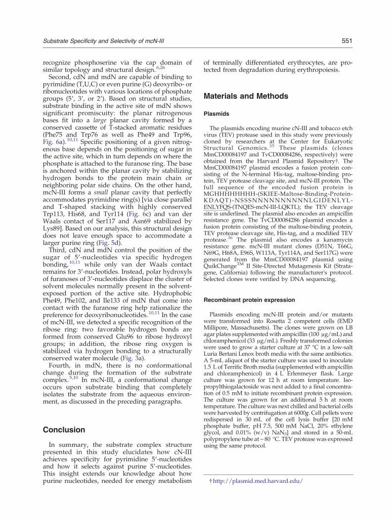
Plasmids encoding mcN-III protein and/or mutantswere transformed into Rosetta 2 competent cells (EMDMillipore, Massachusetts). The clones were grown on LBagar plates supplementedwith ampicillin (100 μg/mL) andchloramphenicol (33 μg/mL). Freshly transformed colonieswere used to grow a starter culture at 37 °C in a low‐saltLuria Bertani Lenox broth media with the same antibiotics.A 5‐mL aliquot of the starter culture was used to inoculate1.5 L of Terrific Brothmedia (supplementedwith ampicillinand chloramphenicol) in 4 L Erlenmeyer flask. Largeculture was grown for 12 h at room temperature. Iso-propylthiogalactoside was next added to a final concentra-tion of 0.5 mM to initiate recombinant protein expression.The culture was grown for an additional 5 h at roomtemperature. The culture was next chilled and bacterial cellswere harvested by centrifugation at 6000g. Cell pellets wereredispersed in 30 mL of the cell lysis buffer [20 mMphosphate buffer, pH 7.5, 500 mM NaCl, 20% ethyleneglycol, and 0.01% (w/v) NaN3] and stored in a 50-mLpolypropylene tube at −80 °C. TEV proteasewas expressedusing the same protocol.

552 Substrate Specificity and Selectivity of mcN‐III
Recombinant mcN-III purification
The protocol for protein purification was adapted toinfrastructure available at our institution from a protocoldeveloped by researchers from the Center for EukaryoticStructural Genomics.37 In short, frozen cell paste wasthawed on ice, supplemented with 70 μL of 10 mMphenylmethylsulfonyl fluoride and discontinuously son-icated for 7 min in a stainless steel cup using BronsonSonifier 450 sonicator equipped with a microtip. Suspen-sion was cleared by centrifuging for 20 min at 20,000 rpmin SS34 rotor (~ 47,000g) using Sorvall's RC-5C PLUScentrifuge. Supernatant was loaded onto a chromatogra-phy column containing 20 mL of Profinity™ IMAC resin(Biorad, California) pre-equilibrated in buffer A [20 mMphosphate buffer, pH 7.5, 500 mM NaCl, 0.01% (w/v)NaN3]. Resin was extensively washed using buffer A andbuffer was changed to the proteolysis buffer [20 mMHepes, pH 7.5, 50 mM NaCl, 0.01% (w/v) NaN3]. At thispoint, ~ 0.5 mg of TEV protease in 15 mL of theproteolysis buffer supplemented with 10 μL of 2-mercap-toethanol was applied on the column and allowed to cleavethe fusion protein for 3 to 5 days at room temperature. Thecleaved protein was eluted from the column using theproteolysis buffer and concentrated using Amicon™ Ultra-15 centrifugal filter device. The column was cleaned usingbuffer B [20 mM phosphate, pH 7.5, 500 mM NaCl,350 mM imidazole, and 0.01% (w/v) NaN3]. Overallsuccess of the purification and final purity of mcN-III orits variants were assessed using SDS-PAGE gel electropho-resis. Protein concentration was established using Bradfordassay and/or via absorption spectroscopy in 40 mMpotassium phosphate, pH 6.5, and 6.6 M guanidine hy-drochloride. Extinction coefficients of ε280 of21,890 M− 1 cm− 1 for mcN-III, 16,390 M− 1 cm− 1 formcN-III-W113A, or 20,400 M− 1 cm− 1 for mcN-III-Y114Amutants were estimated using ProtParam server.38 Atypical mcN-III purification yielded ~10 mg of high‐purityrecombinant protein from a 1.5‐L growth. TEV proteasewas purified by immobilized metal affinity chromatogra-phy adapting a protocol described previously.36
Crystallization
Initial crystallization screening involved co-crystalliza-tion of mcN-III-D51N in the presence of 3 mM MgCl2 and5 mM UMP via the vapor diffusion technique at 10 °C.Individual crystallization test drops contained 1 μL ofmcN-III-D51N solution (of approximately 10 mg/mL) mixedwith 1 μL of crystallization solutions from Crystal Screenand Index screen kits fromHamptonResearch. First, proteincrystalswere obtained in condition #80 of Index screen [25%polyethylene glycol (PEG) 3350, 100 mM Hepes, pH 7.5,and 200 mM ammonium acetate]. During optimizationtrials, crystals occasionally reproduced from 20% PEG 3350,100 mM Tris, pH 8.0, and 50 mM ammonium acetate;however, the frequency was extremely low. To propagatecrystals more reliably, we developed the following seedingprotocol: 100 μL mcN-III-D51N sample aliquot was mixedwith 1 M MgCl2 stock solution to arrive at approximately3 mM final concentration and mixed with 200 mMUMP toarrive at approximately 5 mM. This protein sample wasthen mixed in 1:1 volume ratio with the mother liquorsolution (15% to 20% PEG 3350, 100 mM Tris, pH 8.0, and
50 mM ammonium acetate) and preincubated in the largevolume hanging drops (~30 μL) sealed over the 500 μL ofmother liquor solutions for 1 or 2 days at 10 °C. After thisperiod, drops were pooled and centrifuged in microcen-trifuge to remove formed precipitate. Solution was reap-plied onto the cover glass in 2 μL drops and streak seededusing seeds obtained by crushing an expendable proteincrystal in the mother liquor in an Eppendorf tube.Elongated, brick-shaped crystals appeared reliably within12 h and grew to their terminal size (of about20 μm× 40 μm× 200 μm) in approximately 36 h.
Diffraction data collection
Individual crystals were cryoprotected by soaking themfor approximately 2 min in a series of solutions containingincreasing amount of ethylene glycol [namely, 20% PEG3350, 100 mM Tris, pH 8.0, 50 mM ammonium acetate,3 mM MgCl2, and 6 mM UMP with 0%, 6.7%, 13.3%, and20% (v/v) ethylene glycol]. After cryoprotection, crystalswere frozen by dipping them into liquid nitrogen andwereshipped for synchrotron data collection. Diffraction datawere collected at LS-CAT 24-ID-D beamline at AdvancedLight Source at Argonne National Labs, Argonne, IL. Thedata set used in this study was derived from an analysis of115 frames collected by MAR300 image plate detector.Frames were collected at a 200‐mm crystal-to-detectordistance, and individual frames recorded diffractionpattern from 1° oscillation range. The crystal showed lowmosaicity of approximately 0.35° and sustained no signif-icant radiation damage during the data collection. Diffrac-tion images were processed using HKL2000 software.39
Structure phasing, refinement, and validation
The structure of the mcN-III-D51N ∙UMP complex wassolved by molecular replacement using the programMOLREP40 from the CCP4 suite.41 As a starting model,a protomer of apo-mcN-III (PDB ID: 2G09) was used.12 Aclear solution was readily identified. The structure wasrefined in multiple cycles of manual model building inCoot42 based on analysis of σA-weighted difference mapsfrom maximum likelihood restrained refinement withisotropic temperature factors in the program REFMAC5.6.43 Weights for B‐factor restraints for main‐chain bonds,main‐chain angles, side‐chain bonds, and side‐chainangles were set to 2.0, 4.0, 6.0, and 10.0, respectively. Allother refinement parameters were kept at their defaultsettings. During manual modeling, new waters wereplaced into locations at which an isolated peak in theσA-weighted 2mFo −DFc electron density map (N 0.8 σ)overlapped with a peak in the σA-weighted mFo −DFcdifference density map (N 3.0 σ) and were located within ahydrogen-bond distance (2.4–3.2 Å) from a hydrogenbond donor or acceptor previously defined in the model.Final model quality was evaluated by validation routinesin the program Coot and all‐atom clash analysis using theMolProbity server.44 Illustrations were made in PyMOL.45
Enzymatic activity assays
Enzymatic assays were initiated by adding 20 μL of anenzyme in the assay buffer (containing 100 mM Tris,
553Substrate Specificity and Selectivity of mcN‐III
pH 7.5, and 1 mM MgCl2) to 480 μL of the assay mixpreincubated at 37 °C consisting of the assay buffer andUMP (at final concentrations of 0.0, 0.1, 0.3, 0.5, 1.0, 2.0,4.0, 7.5, 10, 15, and 20 mM). For background controls,20 μL of the assay buffer was added into mixturescontaining the assay buffer and 4.0 mM, 10 mM, and20 mM UMP (final concentrations). Reactions wereallowed to proceed for exactly 15 min at 37 °C; after thisperiod, samples were inactivated for 5 min at 95 °C andchilled on ice. The amount of released phosphate wasevaluated by mixing 50 μL of appropriately dilutedsamples and 100 μL of Malachite green reagent [1.05%(w/v) ammonium molybdate, 0.0338% (w/v) malachitegreen in 1 M HCl]. The phosphate content was evaluatedby comparing sample absorbance at 620 nm to that ofphosphate standards in a range of 0 μM to 80 μM.
Accession numbers
Structure factors and coordinates of the final model ofthe mcN-III complex were deposited in the PDB wherethey are available under PDB ID 4FE3.
Acknowledgements
This project was supported by the ResearchCorporation for Science Advancement grant#19888 and Georgian Court University SummerResearch grant to E.B. Use of the Advanced PhotonSource was supported by the U.S. Department ofEnergy, Office of Science, Office of Basic EnergySciences, under Contract No. DE-AC02-06CH11357.Use of the LS-CAT Sector 21 was supported bythe Michigan Economic Development Corporationand the Michigan Technology Tri-Corridor forthe support of this research program (Grant085P1000817).
References
1. Bianchi, V. & Spychala, J. (2003). Mammalian 5′-nucleotidases. J. Biol. Chem. 278, 46195–46198.
2. Hunsucker, S. A., Mitchell, B. S. & Spychala, J. (2005).The 5′-nucleotidases as regulators of nucleotide anddrug metabolism. Pharmacol. Ther. 107, 1–30.
3. Burroughs, A. M., Allen, K. N., Dunaway-Mariano, D.& Aravind, L. (2006). Evolutionary genomics of theHAD superfamily: understanding the structural ad-aptations and catalytic diversity in a superfamily ofphosphoesterases and allied enzymes. J. Mol. Biol. 361,1003–1034.
4. Collet, J. F., Stroobant, V., Pirard, M., Delpierre, G. &Van Schaftingen, E. (1998). A new class of phospho-transferases phosphorylated on an aspartate residuein an amino-terminal DXDX(T/V) motif. J. Biol. Chem.273, 14107–14112.
5. Rinaldo-Matthis, A., Rampazzo, C., Reichard, P.,Bianchi, V. & Nordlund, P. (2002). Crystal structure
of a human mitochondrial deoxyribonucleotidase.Nat. Struct. Biol. 9, 779–787.
6. Wang, W., Cho, H. S., Kim, R., Jancarik, J., Yokota, H.,Nguyen, H. H. et al. (2002). Structural characterizationof the reaction pathway in phosphoserine phospha-tase: crystallographic “snapshots” of intermediatestates. J. Mol. Biol. 319, 421–431.
7. Allegrini, S., Scaloni, A., Ferrara, L., Pesi, R., Pinna, P.,Sgarrella, F. et al. (2001). Bovine cytosolic 5′-nucleo-tidase acts through the formation of an aspartate 52-phosphoenzyme intermediate. J. Biol. Chem. 276,33526–33532.
8. Pesi, R., Turriani, M., Allegrini, S., Scolozzi, C.,Camici, M., Ipata, P. L. & Tozzi, M. G. (1994). Thebifunctional cytosolic 5′-nucleotidase: regulation ofthe phosphotransferase and nucleotidase activities.Arch. Biochem. Biophys. 312, 75–80.
9. Amici, A., Emanuelli, M., Magni, G., Raffaelli, N. &Ruggieri, S. (1997). Pyrimidine nucleotidases fromhuman erythrocyte possess phosphotransferase activ-ities specific for pyrimidine nucleotides. FEBS Lett.419, 263–267.
10. Wallden, K., Ruzzenente, B., Rinaldo-Matthis, A.,Bianchi, V. & Nordlund, P. (2005). Structural basis forsubstrate specificity of the human mitochondrialdeoxyribonucleotidase. Structure, 13, 1081–1088.
11. Wallden, K., Rinaldo-Matthis, A., Ruzzenente, B.,Rampazzo, C., Bianchi, V. & Nordlund, P. (2007).Crystal structures of human and murine deoxyribonu-cleotidases: insights into recognition of substrates andnucleotide analogues. Biochemistry, 46, 13809–13818.
12. Bitto, E., Bingman,C.A.,Wesenberg,G. E.,McCoy, J. G.& Phillips, G. N., Jr. (2006). Structure of pyrimidine 5′-nucleotidase type 1. Insight into mechanism of actionand inhibition during lead poisoning. J. Biol. Chem. 281,20521–20529.
13. Wallden, K., Stenmark, P., Nyman, T., Flodin, S.,Graslund, S., Loppnau, P. et al. (2007). Crystalstructure of human cytosolic 5′-nucleotidase II: in-sights into allosteric regulation and substrate recog-nition. J. Biol. Chem. 282, 17828–17836.
14. Wallden, K. &Nordlund, P. (2011). Structural basis forthe allosteric regulation and substrate recognition ofhuman cytosolic 5′-nucleotidase II. J. Mol. Biol. 408,684–696.
15. Shi, N., Zhang, Y. J., Chen, H. K., Gao, Y., Teng, M. &Niu, L. (2011). Crystal structure of the pyrimidine 5′-nucleotidase SDT1 from Saccharomyces cerevisiae com-plexed with uridine 5′-monophosphate provides fur-ther insight into ligandbinding.Proteins, 79, 1358–1362.
16. Amici, A. & Magni, G. (2002). Human erythrocytepyrimidine 5′-nucleotidase, PN-I. Arch. Biochem. Bio-phys. 397, 184–190.
17. Rees, D. C., Duley, J. A. & Marinaki, A. M. (2003).Pyrimidine 5′ nucleotidase deficiency. Br. J. Haematol.120, 375–383.
18. Hokari, S., Miyazaki, T., Hasegawa, M. & Komoda, T.(1998). Enhanced activity of pyrimidine 5′-nucleotid-ase in rat red blood cells during erythropoiesis. Biol.Chem. 379, 329–333.
19. Valentine, W. N., Fink, K., Paglia, D. E., Harris, S. R. &Adams, W. S. (1974). Hereditary hemolytic anemiawith human erythrocyte pyrimidine 5′-nucleotidasedeficiency. J. Clin. Invest. 54, 866–879.

554 Substrate Specificity and Selectivity of mcN‐III
20. Zanella, A., Bianchi, P., Fermo, E. & Valentini, G.(2006). Hereditary pyrimidine 5′-nucleotidase defi-ciency: from genetics to clinical manifestations. Br. J.Haematol. 133, 113–123.
21. Valentine, W. N., Paglia, D. E., Fink, K. & Madokoro,G. (1976). Lead poisoning: association with hemolyticanemia, basophilic stippling, erythrocyte pyrimidine5′-nucleotidase deficiency, and intraerythrocytic ac-cumulation of pyrimidines. J. Clin. Invest. 58, 926–932.
22. Chiarelli, L. R., Bianchi, P., Fermo, E., Galizzi, A.,Iadarola, P., Mattevi, A. et al. (2005). Functionalanalysis of pyrimidine 5′-nucleotidase mutants caus-ing nonspherocytic hemolytic anemia. Blood, 105,3340–3345.
23. Chiarelli, L. R., Fermo, E., Abrusci, P., Bianchi, P.,Dellacasa, C. M., Galizzi, A. et al. (2006). Two newmutations of the P5′N-1 gene found in Italian patientswith hereditary hemolytic anemia: the molecular basisof the red cell enzyme disorder. Haematologica, 91,1244–1247.
24. Chiarelli, L. R., Morera, S. M., Galizzi, A., Fermo, E.,Zanella, A. & Valentini, G. (2008). Molecular basis ofpyrimidine 5′-nucleotidase deficiency caused by 3newly identified missense mutations (c.187TNC,c.469GNC and c.740TNC) and a tabulation of knownmutations. Blood Cells Mol. Dis. 40, 295–301.
25. Amici, A., Ciccioli, K., Naponelli, V., Raffaelli, N.& Magni, G. (2005). Evidence for essential catalyticdeterminants for human erythrocyte pyrimidine5′-nucleotidase. Cell. Mol. Life Sci. 62, 1613–1620.
26. Kim, H. Y., Heo, Y. S., Kim, J. H., Park, M. H., Moon, J.,Kim, E. et al. (2002). Molecular basis for the localconformational rearrangement of human phosphoser-ine phosphatase. J. Biol. Chem. 277, 46651–46658.
27. Calero, G., Wilson, K. F., Ly, T., Rios-Steiner, J. L.,Clardy, J. C. & Cerione, R. A. (2002). Structural basis ofm7G pppG binding to the nuclear cap-binding proteincomplex. Nat. Struct. Biol. 9, 912–917.
28. Tomoo, K., Matsushita, Y., Fujisaki, H., Abiko, F.,Shen, X., Taniguchi, T. et al. (2005). Structural basis formRNA cap-binding regulation of eukaryotic initiationfactor 4E by 4E-binding protein, studied by spectro-scopic, X-ray crystal structural, and molecular dy-namics simulation methods. Biochim. Biophys. Acta,1753, 191–208.
29. McGaughey, G. B., Gagne, M. & Rappe, A. K. (1998).pi-Stacking interactions. Alive and well in proteins.J. Biol. Chem. 273, 15458–15463.
30. Jaroszewski, L., Li, Z., Cai, X. H., Weber, C. & Godzik,A. (2011). FFAS server: novel features and applica-tions. Nucleic Acids Res. 39, W38–W44.
31. Corpet, F. (1988). Multiple sequence alignment withhierarchical clustering. Nucleic Acids Res. 16,10881–10890.
32. Amici, A., Emanuelli, M., Ferretti, E., Raffaelli, N.,Ruggieri, S. & Magni, G. (1994). Homogeneous pyrim-idine nucleotidase from human erythrocytes: enzymicand molecular properties. Biochem. J. 304(Pt 3),987–992.
33. Holm, L. & Park, J. (2000). DaliLite workbench forprotein structure comparison. Bioinformatics, 16,566–567.
34. Holm, L. & Rosenstrom, P. (2010). Dali server:conservation mapping in 3D. Nucleic Acids Res. 38,W545–W549.
35. Thao, S., Zhao, Q., Kimball, T., Steffen, E., Blommel,P. G., Riters, M. et al. (2004). Results from high-throughput DNA cloning of Arabidopsis thalianatarget genes using site-specific recombination. J.Struct. Funct. Genomics, 5, 267–276.
36. Blommel, P. G. & Fox, B. G. (2007). A combinedapproach to improving large-scale production oftobacco etch virus protease. Protein Expr. Purif. 55,53–68.
37. Jeon, W. B., Aceti, D. J., Bingman, C. A., Vojtik, F. C.,Olson, A. C., Ellefson, J. M. et al. (2005). High-throughput purification and quality assurance ofArabidopsis thaliana proteins for eukaryotic structur-al genomics. J. Struct. Funct. Genomics, 6, 143–147.
38. Wilkins, M. R., Gasteiger, E., Bairoch, A., Sanchez,J. C., Williams, K. L., Appel, R. D. & Hochstrasser,D. F. (1999). Protein identification and analysis toolsin the ExPASy server. Methods Mol. Biol. 112, 531–552.
39. Otwinowski, Z. & Minor, M. (1997). Processing of x-ray diffraction data collected in oscillation mode.Methods Enzymol. 276, 307–326.
40. Vagin, A. & Teplyakov, A. (2010). Molecular replace-ment with MOLREP. Acta Crystallogr., Sect. D: Biol.Crystallogr. 66, 22–25.
41. Collaborative Computational Project, Number 4.(1994). The CCP4 suite: programs for protein crystal-lography.Acta Crystallogr., Sect. D: Biol. Crystallogr. 50,760–763.
42. Emsley, P. & Cowtan, K. (2004). Coot: model-buildingtools for molecular graphics. Acta Crystallogr., Sect. D:Biol. Crystallogr. 60, 2126–2132.
43. Murshudov, G. N., Skubak, P., Lebedev, A. A., Pannu,N. S., Steiner, R. A., Nicholls, R. A. et al. (2011).REFMAC5 for the refinement of macromolecularcrystal structures. Acta Crystallogr., Sect. D: Biol.Crystallogr. 67, 355–367.
44. Chen, V. B., Arendall, W. B., 3rd, Headd, J. J., Keedy,D. A., Immormino, R. M., Kapral, G. J. et al. (2010).MolProbity: all-atom structure validation for macro-molecular crystallography. Acta Crystallogr., Sect. D:Biol. Crystallogr. 66, 12–21.
45. Schrodinger, L. L. C. (2010). The PyMOL MolecularGraphics System, Version 1.3r1.


