Childhood Exanthemata Dr. Philip G. Murphy Consultant Microbiologist, AMNCH,Tallaght.
Upload
magdalene-clarkCategory
view
227download
2
Jerome FennellAMNCH
Department of Clinical Microbiologyhttp://www.tcd.ie/Clinical_Microbiology

OBJECTIVES
Understanding of :
Presentation of Upper Respiratory Infections
Causative organisms Pathogenesis Diagnosis(clinical, laboratory, other) Clinical Management( treatment,
preventative measures)
Infection Syndromes
Common Cold Conjunctivitis Pharyngitis/Tonsillitis Quinsy Epiglottitis Otitis Media Sinusitis
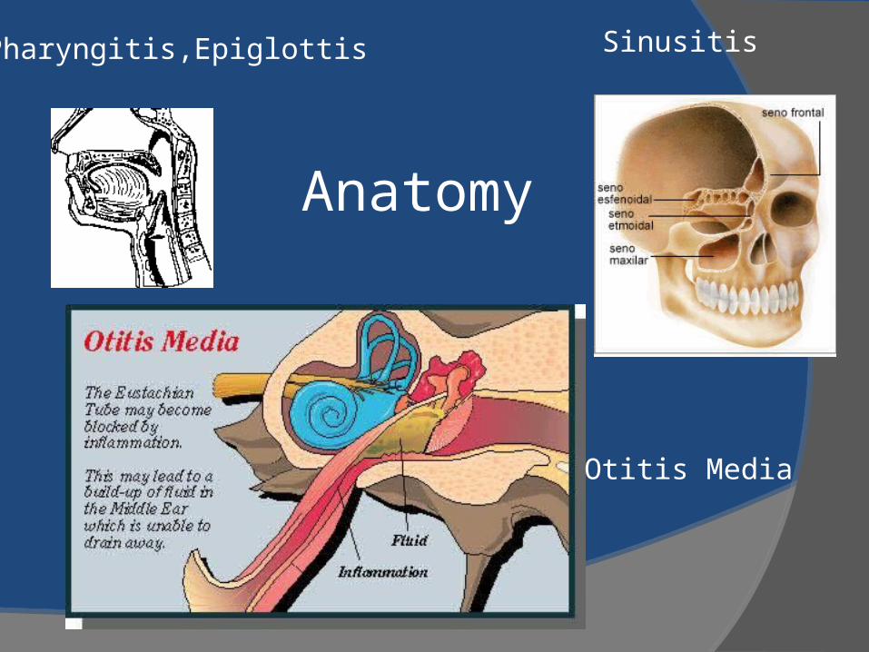
Anatomy
Otitis Media
SinusitisPharyngitis,Epiglottis
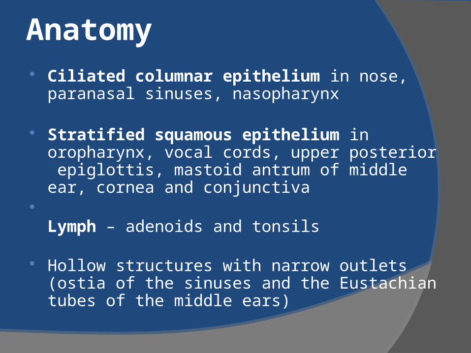
Anatomy Ciliated columnar epithelium in nose,
paranasal sinuses, nasopharynx
Stratified squamous epithelium in oropharynx, vocal cords, upper posterior epiglottis, mastoid antrum of middle ear, cornea and conjunctiva
Lymph – adenoids and tonsils
Hollow structures with narrow outlets (ostia of the sinuses and the Eustachian tubes of the middle ears)
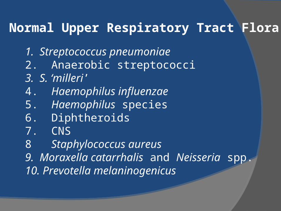
Normal Upper Respiratory Tract Flora
1. Streptococcus pneumoniae2. Anaerobic streptococci3. S. ‘milleri’4. Haemophilus influenzae5. Haemophilus species6. Diphtheroids7. CNS8 Staphylococcus aureus9. Moraxella catarrhalis and Neisseria spp.10. Prevotella melaninogenicus

The Common Cold
Causative agents: Coronaviruses, etc Epidemiology: usually common in the winter
months Presentation: rhinitis, headache, conjunctival
suffusion Management: Antimicrobial agents not to
be given. Symptomatic relief may be accompanied by mucopurulent rhinitis (thick,opaque or discolored nasal discharge), this is not an indication for antimicrobial treatment unless it persists without signs of improvement 10-14 days suggesting possible sinusitis.
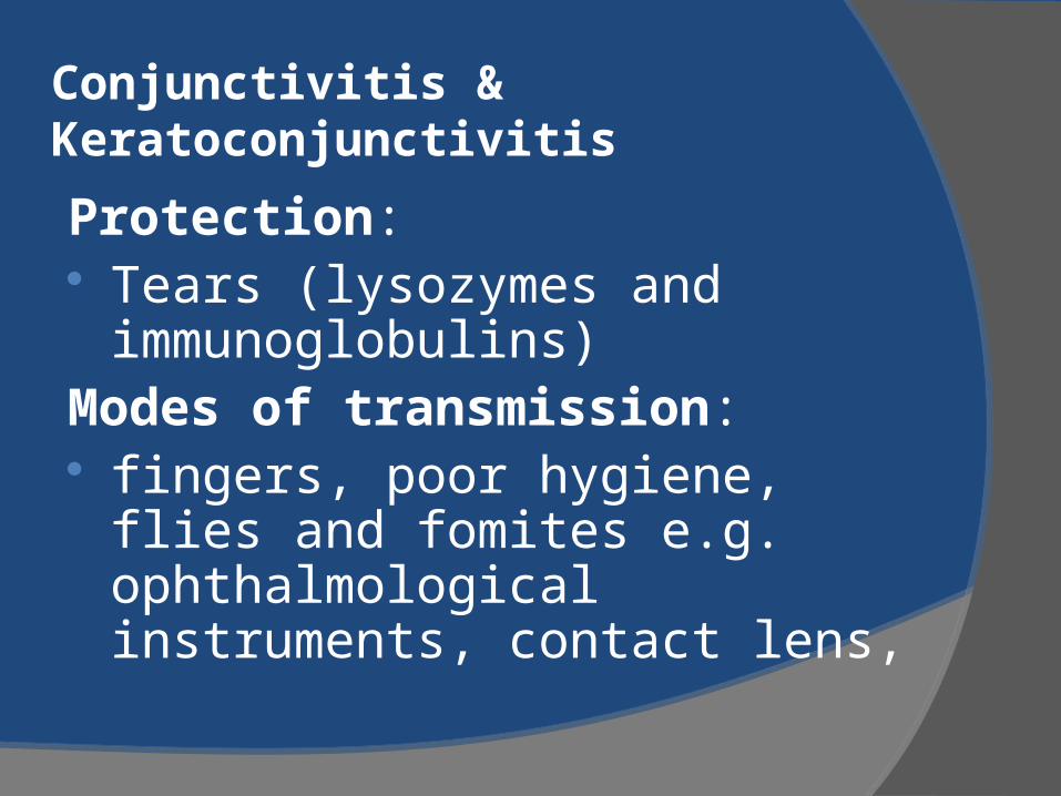
Conjunctivitis & Keratoconjunctivitis
Protection: Tears (lysozymes and
immunoglobulins)Modes of transmission: fingers, poor hygiene, flies and
fomites e.g. ophthalmological instruments, contact lens,
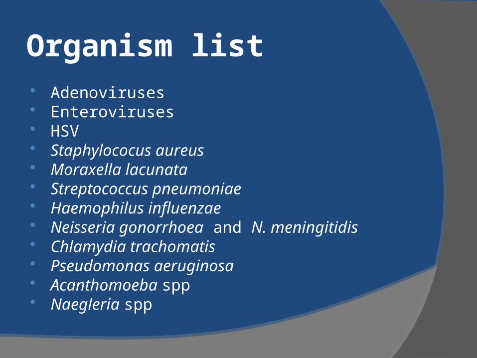
Organism list Adenoviruses Enteroviruses HSV Staphylococus aureus Moraxella lacunata Streptococcus pneumoniae Haemophilus influenzae Neisseria gonorrhoea and N. meningitidis Chlamydia trachomatis Pseudomonas aeruginosa Acanthomoeba spp Naegleria spp
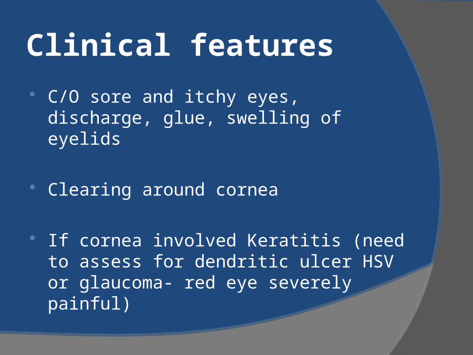
Clinical features
C/O sore and itchy eyes, discharge, glue, swelling of eyelids
Clearing around cornea
If cornea involved Keratitis (need to assess for dendritic ulcer HSV or glaucoma- red eye severely painful)
Pharyngitis Definition: Inflammatory Syndrome of
the pharynx caused by several microorganisms
Causes: most commonly viral also occur as part of common cold or influenza syndrome
The most common bacterial cause is Group A Streptococcus (Streptococcus pyogenes)-5-20%
Review: NEJM 344:205 2001
Pharyngitis Presentation

ETIOLOGYPathogen Syndrome/Disease Estimated
Importance
ViralRhinovirus (100 types and 1 subtype)Coronavirus (3 or more types)Adenovirus (types 3, 4, 7, 14, 21)Herpes simplex virus (types 1 and 2)Parainfluenza virus (types 1-4)Influenza virus (types A and B)Cocksackievirus A (types 2, 4-6, 8, 10)Epstein-Barr virusCytomegalovirusHIV-1
Common coldCommon coldPhayrngoconjunctival fever, ARDGingivitis, stomatitis, PharyngitisCommon cold, croupInfluenzaHerpanginaInfectious mononucleosisInfectious mononucleosisPrimary HIV infection
205
5422
<1<1<1<1
BacterialStreptococcus pyogenes (group A -hemolytic streptococci)Group C -hemolytic streptococci
Mixed anaerobic infectionNeisseria gonorrhoeaeCorynebacterium diphtheriaeCorynebacterium ulceransArcanobacterium haemolyticum (Corynebacterium haemolyticum)Yersinia enterocoliticaTreponema pallidumChlamydialChlamydia pneumoniaeMycoplasmalMycoplasma pneumoniaeMycoplasma hominis (type 1)Unknown
Pharyngitis/tonsillitis, scarlet feverGingivitis, Pharyngitis (Vincent’s angina)Peritonsillitis/peritonsillar abscess (quinsy)
PharyngitisDiphtheriaPharyngitis, diphtheriaPharyngitis, scarlatiniform rash
Pharyngitis, enterocolitisSecondary syphilis
Pneumonia/bronchitis/Pharyngitis
Pneumonia/bronchitis/PharyngitisPharyngitis in volunteers
15-305-10
<1<1<11<1<1<1<1
Unknown<1
Unknown
Approximately 15% of all cases of Pharyngitis are due to S. pyogenes. Strep. Group C and B have also been implicated in some cases.
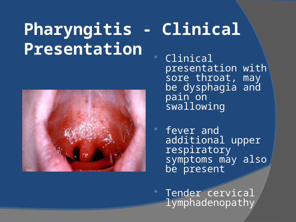
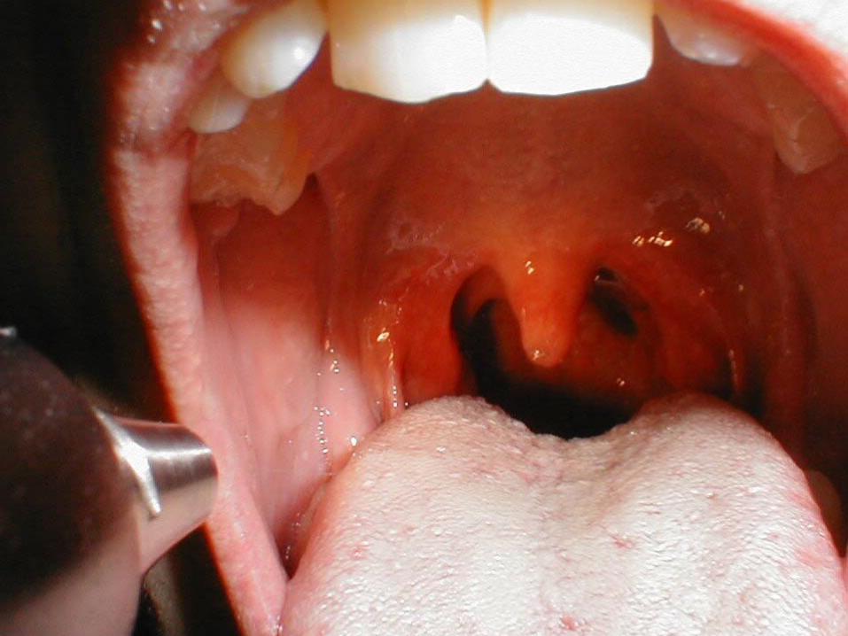
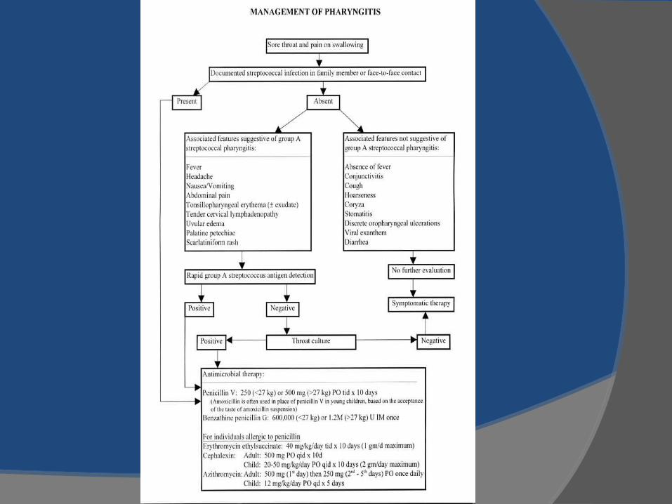
Pharyngitis - Clinical Presentation
Clinical presentation with sore throat, may be dysphagia and pain on swallowing
fever and additional upper respiratory symptoms may also be present
Tender cervical lymphadenopathy
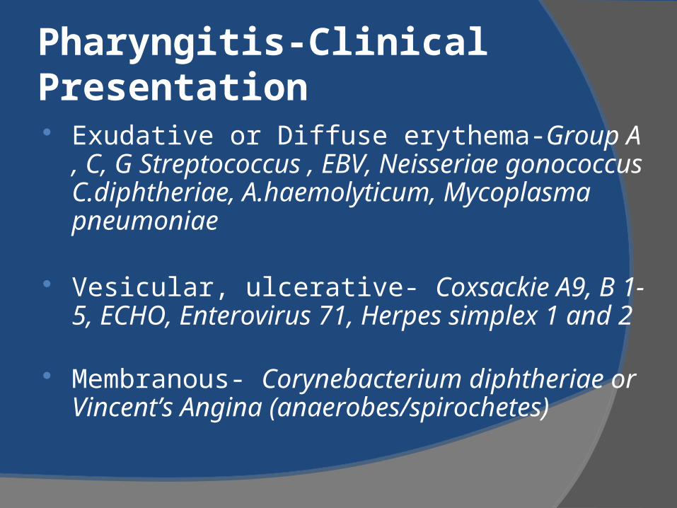
Pharyngitis-Clinical Presentation Exudative or Diffuse erythema-Group A , C, G
Streptococcus , EBV, Neisseriae gonococcus C.diphtheriae, A.haemolyticum, Mycoplasma pneumoniae
Vesicular, ulcerative- Coxsackie A9, B 1-5, ECHO, Enterovirus 71, Herpes simplex 1 and 2
Membranous- Corynebacterium diphtheriae or Vincent’s Angina (anaerobes/spirochetes)
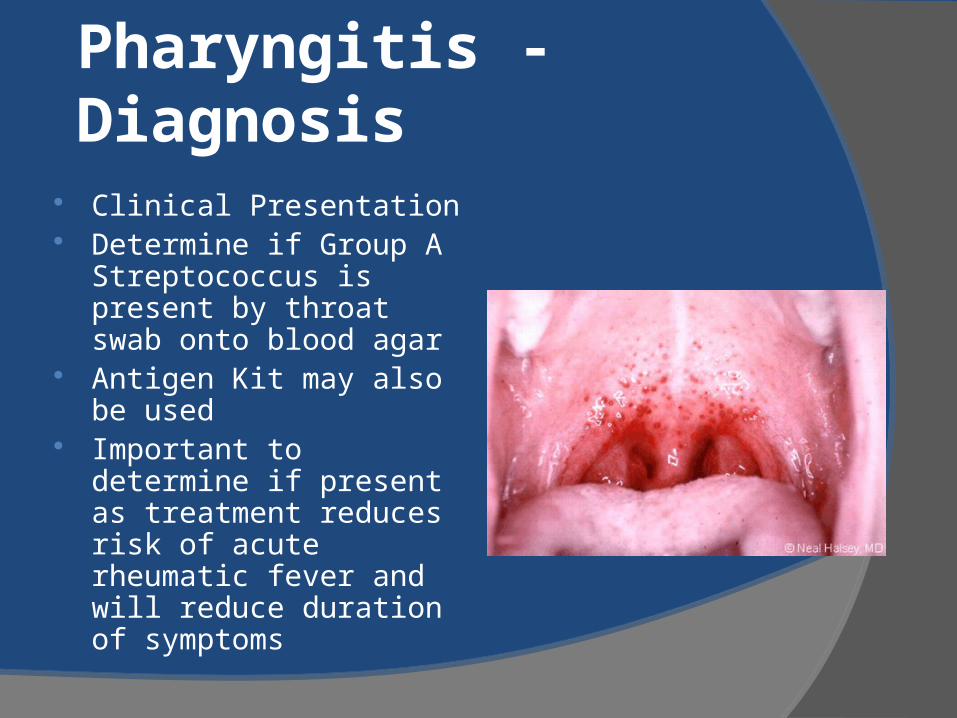
Pharyngitis - Diagnosis
Clinical Presentation Determine if Group A
Streptococcus is present by throat swab onto blood agar
Antigen Kit may also be used
Important to determine if present as treatment reduces risk of acute rheumatic fever and will reduce duration of symptoms
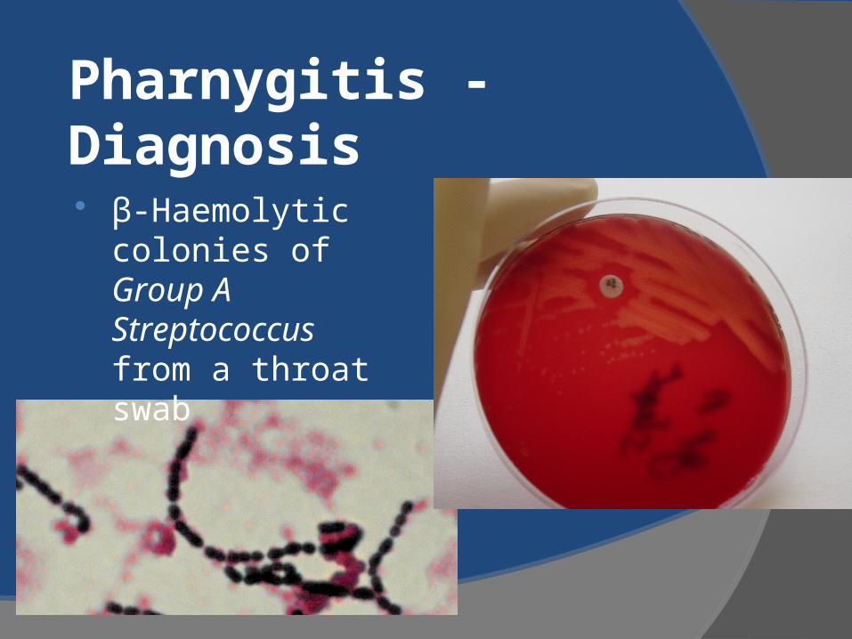
Pharnygitis - Diagnosis
β-Haemolytic colonies of Group A Streptococcus from a throat swab
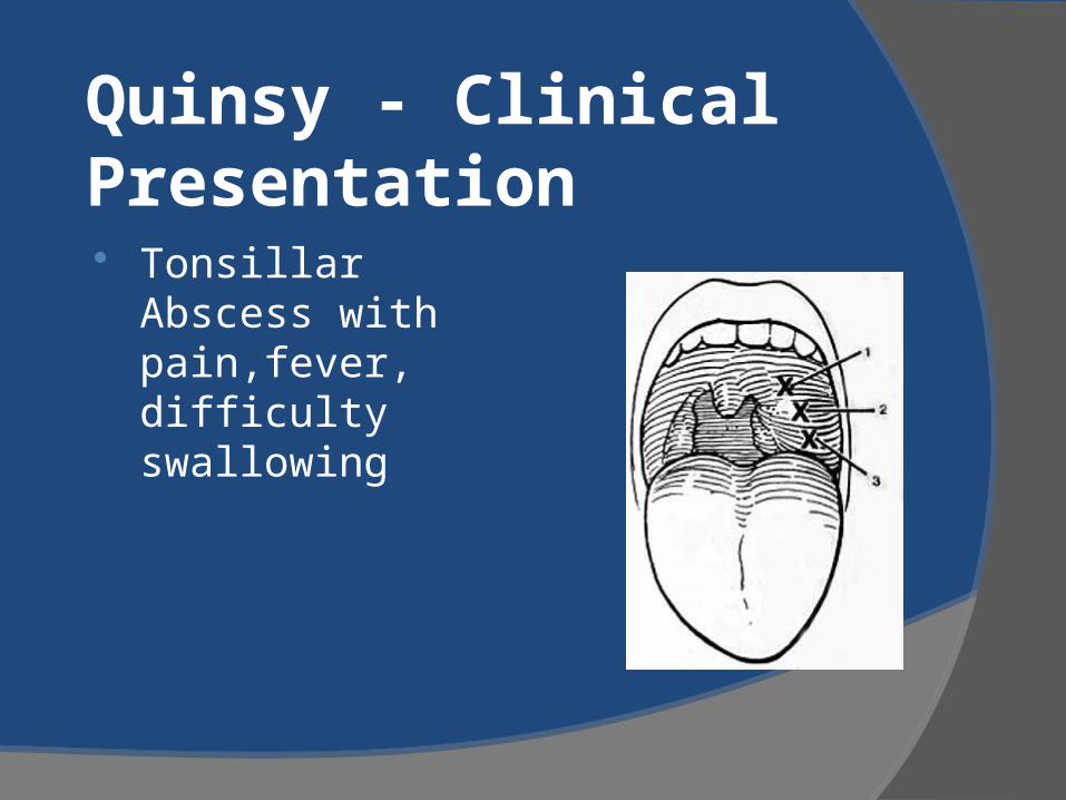
Quinsy - Clinical Presentation Tonsillar Abscess
with pain,fever, difficulty swallowing
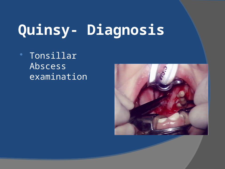
Quinsy- Diagnosis
Tonsillar Abscess examination

Quinsy - Clinical Management
Drainage of Abscess and antimicrobial therapy
Retropharyngeal Abscess Abscess in tissues behind the pharynx With oedema and pus, may get
compression and airway obstruction Potential medical emergency X-rays show wide soft tissue space Emergency tracheostomy
Epiglottitis
Definition: Inflammation of the epiglottis due to infection
Epidemiology: usually occurs in the winter months
Causative Bacterial Organisms: H.influenzae (now rare), S.pyogenes, Pneumococcus, Staphylococcus aureus
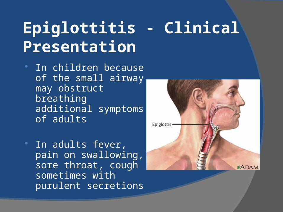
Epiglottitis - Clinical Presentation In children because of
the small airway may obstruct breathing additional symptoms of adults
In adults fever, pain on swallowing, sore throat, cough sometimes with purulent secretions
Epiglottitis - Diagnosis
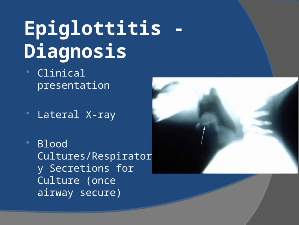
Clinical presentation
Lateral X-ray
Blood Cultures/Respiratory Secretions for Culture (once airway secure)
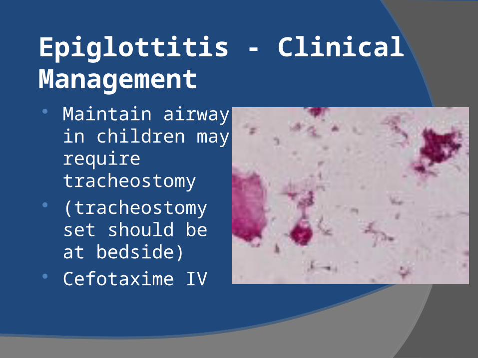
Epiglottitis - Clinical Management Maintain airway in
children may require tracheostomy
(tracheostomy set should be at bedside)
Cefotaxime IV

Haemophilus influenzae on Culture
OTITIS MEDIAAmerican Academy of Pediatrics and American Academy of Family PhysiciansClinical Practice GuidelinesPediatrics Vol. 113 No.5 May 2004
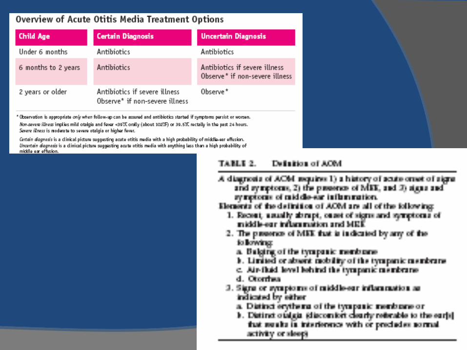
Otitis Media Definition: for diagnosis requires 3 things Confirmation of acute onset Signs of Middle Ear Effusion (Pneumatic
otoscopy): Bulging of TM, limited mobility, air-fluid level, otorrhoea
Evaluation of Signs and Symptoms of Middle Ear Inflammation: Erythema of TM or Distinct otalgia ( interferes with sleep)
Epidemiology : AOM must common cause of antibiotic prescribing in paediatric population, cost $1.96 billion in U.S, more common in some conditions such as cleft palate, Down's syndrome, genetic influences, occurs in the winter months but may be recurrent
Otitis Media
Causative Organisms: Streptococcus pneumoniae-25-50% Haemphilus Influenzae-15-30% Moraxella catarrhalis-3-30% Rhinovirus/RSV/Coronaviruses/
Adenoviruses/Enteroviruses –40-75%
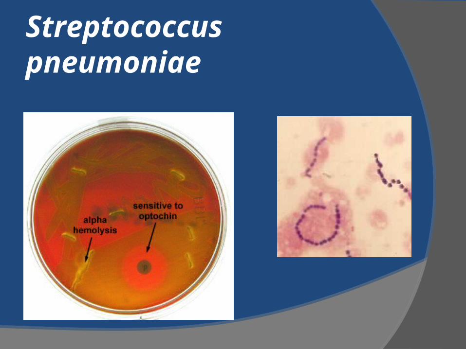
Streptococcus pneumoniae

Otitis Media Clinical Presentation
Symptoms: Infant excessive crying, pulling ear
Toddler: irritability , earache
Both may have otorrhoea Signs: Fever , bulging
eardrum, fullness and erythema of tympanic membrane
May also be additional upper respiratory symptoms
Recommendation 2
The management of Acute Otitis Media should include an assessment of Pain
and treatment accordingly
Recommendation 3a
Observation without use of antimicrobial agents in a child with uncomplicated AOM is an option for selected children based on diagnostic certainty, age, illness severity and assurance of follow-up
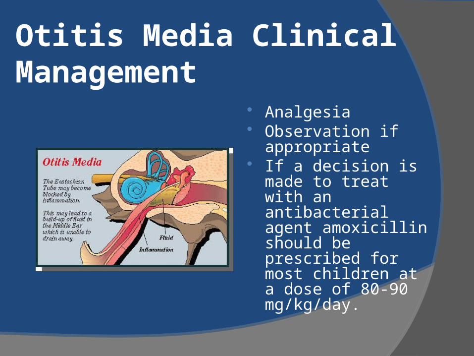
Otitis Media Clinical Management
Analgesia Observation if
appropriate If a decision is
made to treat with an antibacterial agent amoxicillin should be prescribed for most children at a dose of 80-90 mg/kg/day.
Recommendation 4 If there is no clinical improvement in 48-
72 hours Reassess and confirm or exclude
diagnosis of AOM If Observation arm: treat If Treatment arm: Change therapy Duration of therapy: 10 days if 2 or less
or severe 10 days , if > 2 years 5-7 days

Recurrent Otitis Media
Sinusitis
Definition:Acute Bacterial Sinusitis, subacute Bacterial Sinusitis, Recurrent acute, Chronic sinusitis , Superimposed
Epidemiology:children has 6-8 viral URTI per year and 5-13% may be complicated by sinusitis
Definitions of Sinusitis Acute Bacterial : Bacterial Infection of the paranasal
sinuses lasting less than 30days in which symptoms resolve completely
Subacute Bacterial Sinusitis: Lasting between 30 and 90 days in which symptoms resolve completely
Recurrent acute bacterial sinusitis: Each episode lasting less than 30 days and separated by intervals of at least 10days during which the patient is asymptomatic
Chronic Sinusitis: Episode lasting longer than 90 days Patients have persistent residual respiratory symptoms such as cough, rhinnorrhoea or nasal obstruction
Chronic Sinusitis: New symptoms resolve but underlying residue symptoms do not.
Sinusitis
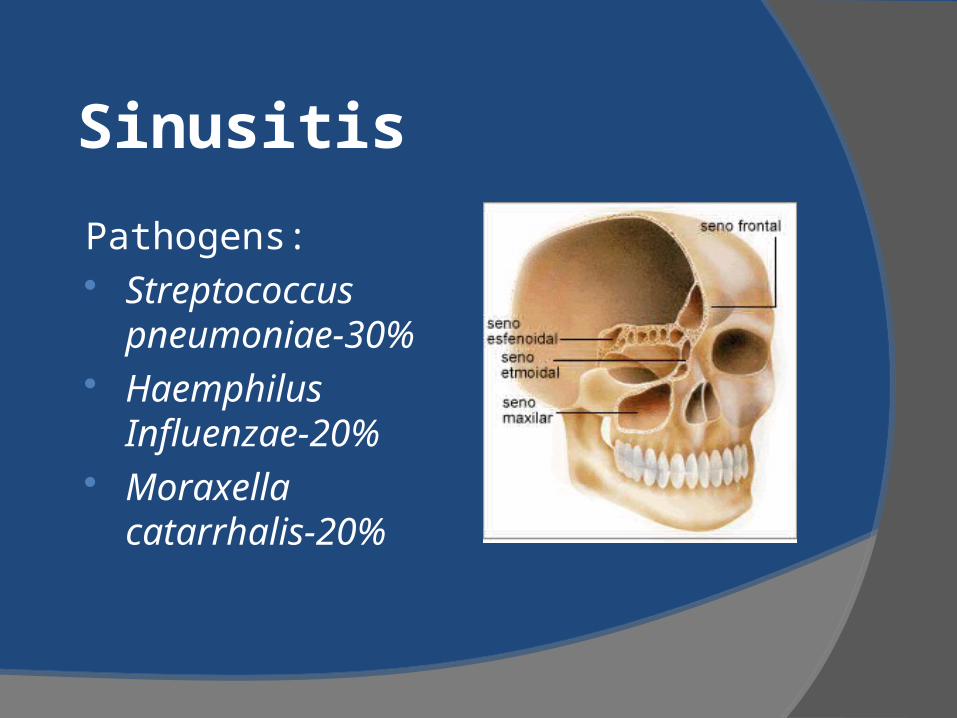
Pathogens: Streptococcus
pneumoniae-30% Haemphilus
Influenzae-20% Moraxella
catarrhalis-20%
Sinusitis
Diagnosis: > or = 10,000 cfu/ml from the cavity of paranasal sinus- but this is invasive
Recommendation 1 Diagnosis is based on clinical criteria who
have upper RT symptoms that are persistent or severe
Acute bacterial Persistent symptoms: nasal or postnasal D/C ,
daytime cough(worse at night) or both Severe Symptoms: Temp(>39 C) and purulent
nasal D/C present concurrently for at least 3-4 days in a child who seems ill
Recommendation 2a
Imaging studies are not necessary to confirm a diagnosis of clinical sinusitis in children less than 6 year of age
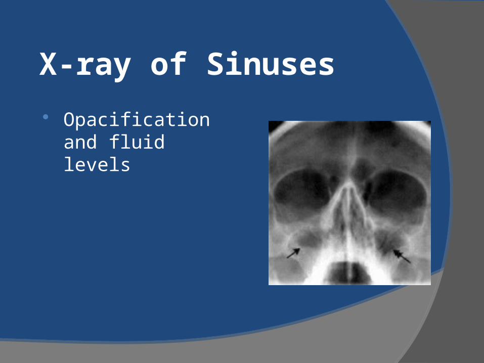
X-ray of Sinuses
Opacification and fluid levels
Recommendation 2b
CT scans should be preserved for those who may require surgery as part of management
Recommendation
Antibiotics are recommended for Acute Bacterial Sinusitis to achieve a more rapid clinical cure
Amoxicillin at 45 or 90 mg/kg.day recommendedMost response in 48-72 hoursDuration :until symptom free plus 7 days
Recommendation
Children with complications or suspected should be treated promptly and aggressively
Referral to ENT specialist, Ophthalmologist, ID physicians and neurosurgeon
Complications involve orbit and Central Nervous System

Thank you