JBC Papers in Press. Published on January 18, 2012 as ... · 3D structure of an α-(1 ... dextran...
22
3D structure of an α-(1→2) branching sucrase 1 Functional and structural characterization of an α-(1→2) branching sucrase derived from DSR-E glucansucrase* Yoann Brison 1 , Tjaard Pijning 2 , Yannick Malbert 1 , Émeline Fabre 3 , Lionel Mourey 4,5 , Sandrine Morel 1 , Gabrielle Potocki-Véronèse 1 , Pierre Monsan 1,6 , Samuel Tranier 4,5 , Magali Remaud- Siméon 1 , Bauke W. Dijkstra 2 1 From Université de Toulouse; INSA, UPS, INP, LISBP, 135 avenue de Rangueil, F-31077 Toulouse, France, CNRS UMR 5504, F-31400 Toulouse, France; INRA UMR 792 Ingénierie des Systèmes Biologiques et des Procédés, F-31400 Toulouse, France 2 Laboratory of Biophysical Chemistry, University of Groningen, Nijenborgh 7, 9747 AG Groningen, The Netherlands 3 Present address: Unité de Glycobiologie Structurale et Fonctionnelle, CNRS UMR 8576, IFR 147, Université Lille1, Sciences et Technologies, 59655 Villeneuve d’Ascq cedex, France 4 Institut de Pharmacologie et de Biologie Structurale, Centre National de la Recherche Scientifique, 205 route de Narbonne, 31077 Toulouse, France 5 Université de Toulouse, Université Paul Sabatier, Institut de Pharmacologie et de Biologie Structurale, 31077 Toulouse, France 6 Institut Universitaire de France, 103 Boulevard Saint-Michel, 75005 Paris, France *Running title: 3D structure of an α-(1→2) branching sucrase To whom correspondence should be addressed: Magali Remaud-Siméon, INSA, LISBP, 135 avenue de Rangueil, 31077 Toulouse, France, Tel: +33 561 559 446; Fax: +33 561 559 400; E-mail: [email protected] Or Samuel Tranier, Institut de Pharmacologie et de Biologie Structurale, 205 route de Narbonne, 31077 Toulouse, France. Tel.: +33 561 175 438; Fax: +33 561 175 994; E-mail: [email protected] Or Bauke W. Dijkstra, Laboratory of Biophysical Chemistry, University of Groningen, Nijenborgh 7, 9747 AG Groningen, The Netherlands. Tel.: +31 503 634 381; Fax: +31 503 634 800; E-mail: [email protected] Keywords: α-(1→2) linkage; dextran; glucansucrase; high resolution structure; DSR-E; prebiotic molecules Background: The transglucosidase GBD-CD2 shows a unique α-(1→2) branching specificity among GH70 family members when catalyzing dextran glucosylation from sucrose. Results: The truncated form ∆N 123 -GBD-CD2 was biochemically studied and structurally characterized at 1.90 Å resolution. Conclusion: Dextran recognition and regiospecificity clearly involves a residue in subsite +1. Significance: This is the first 3D structure of a GH70 enzyme that reveals determinants of α- (1→2) linkage specificity. SUMMARY ∆N 123 -GBD-CD2 is a truncated form of the bi-functional glucansucrase DSR-E from Leuconostoc mesenteroides NRRL B-1299. It was constructed by rational truncation of GBD-CD2, which harbours the second catalytic domain of DSR-E. Like GBD-CD2 this variant displays α-(1→2) branching activity when incubated with sucrose as glucosyl donor and (oligo-)dextran as acceptor, transferring glucosyl residues to the acceptor via a ping-pong bi-bi mechanism. This allows the formation of prebiotic molecules containing controlled amounts of α-(1→2) linkages. The crystal structure of the apo α-(1→2) branching sucrase ∆N 123 -GBD- CD2 was solved at 1.90 Å resolution. The protein adopts the unusual U-shape fold organized in five distinct domains, also found in GTF180-∆N and GTF-SI glucansucrases of http://www.jbc.org/cgi/doi/10.1074/jbc.M111.305078 The latest version is at JBC Papers in Press. Published on January 18, 2012 as Manuscript M111.305078 Copyright 2012 by The American Society for Biochemistry and Molecular Biology, Inc. by guest on July 5, 2018 http://www.jbc.org/ Downloaded from
Transcript of JBC Papers in Press. Published on January 18, 2012 as ... · 3D structure of an α-(1 ... dextran...

3D structure of an α-(1→2) branching sucrase
1
Functional and structural characterization of an α-(1→2) branching sucrase derived from DSR-E glucansucrase*
Yoann Brison1, Tjaard Pijning 2, Yannick Malbert 1, Émeline Fabre3, Lionel Mourey4,5, Sandrine
Morel1, Gabrielle Potocki-Véronèse1, Pierre Monsan1,6, Samuel Tranier4,5, Magali Remaud-
Siméon1, Bauke W. Dijkstra2
1From Université de Toulouse; INSA, UPS, INP, LISBP, 135 avenue de Rangueil, F-31077 Toulouse,
France, CNRS UMR 5504, F-31400 Toulouse, France; INRA UMR 792 Ingénierie des Systèmes Biologiques et des Procédés, F-31400 Toulouse, France
2Laboratory of Biophysical Chemistry, University of Groningen, Nijenborgh 7, 9747 AG Groningen, The Netherlands
3Present address: Unité de Glycobiologie Structurale et Fonctionnelle, CNRS UMR 8576, IFR 147, Université Lille1, Sciences et Technologies, 59655 Villeneuve d’Ascq cedex, France
4Institut de Pharmacologie et de Biologie Structurale, Centre National de la Recherche Scientifique, 205 route de Narbonne, 31077 Toulouse, France
5Université de Toulouse, Université Paul Sabatier, Institut de Pharmacologie et de Biologie Structurale, 31077 Toulouse, France
6Institut Universitaire de France, 103 Boulevard Saint-Michel, 75005 Paris, France
*Running title: 3D structure of an α-(1→2) branching sucrase
To whom correspondence should be addressed: Magali Remaud-Siméon, INSA, LISBP, 135 avenue de Rangueil, 31077 Toulouse, France, Tel: +33 561 559 446; Fax: +33 561 559 400; E-mail: [email protected] Or Samuel Tranier, Institut de Pharmacologie et de Biologie Structurale, 205 route de Narbonne, 31077 Toulouse, France. Tel.: +33 561 175 438; Fax: +33 561 175 994; E-mail: [email protected] Or Bauke W. Dijkstra, Laboratory of Biophysical Chemistry, University of Groningen, Nijenborgh 7, 9747 AG Groningen, The Netherlands. Tel.: +31 503 634 381; Fax: +31 503 634 800; E-mail: [email protected] Keywords: α-(1→2) linkage; dextran; glucansucrase; high resolution structure; DSR-E; prebiotic molecules
Background: The transglucosidase GBD-CD2 shows a unique α-(1→2) branching specificity among GH70 family members when catalyzing dextran glucosylation from sucrose. Results: The truncated form ∆N123-GBD-CD2 was biochemically studied and structurally characterized at 1.90 Å resolution. Conclusion: Dextran recognition and regiospecificity clearly involves a residue in subsite +1. Significance: This is the first 3D structure of a GH70 enzyme that reveals determinants of α-(1→2) linkage specificity. SUMMARY
∆∆∆∆N123-GBD-CD2 is a truncated form of the bi-functional glucansucrase DSR-E from
Leuconostoc mesenteroides NRRL B-1299. It was constructed by rational truncation of GBD-CD2, which harbours the second catalytic domain of DSR-E. Like GBD-CD2 this variant displays αααα-(1→2) branching activity when incubated with sucrose as glucosyl donor and (oligo-)dextran as acceptor, transferring glucosyl residues to the acceptor via a ping-pong bi-bi mechanism. This allows the formation of prebiotic molecules containing controlled amounts of αααα-(1→2) linkages. The crystal structure of the apo αααα-(1→2) branching sucrase ∆∆∆∆N123-GBD-CD2 was solved at 1.90 Å resolution. The protein adopts the unusual U-shape fold organized in five distinct domains, also found in GTF180-∆∆∆∆N and GTF-SI glucansucrases of
http://www.jbc.org/cgi/doi/10.1074/jbc.M111.305078The latest version is at JBC Papers in Press. Published on January 18, 2012 as Manuscript M111.305078
Copyright 2012 by The American Society for Biochemistry and Molecular Biology, Inc.
by guest on July 5, 2018http://w
ww
.jbc.org/D
ownloaded from

3D structure of an α-(1→2) branching sucrase
2
glycoside hydrolase family 70 (GH70). Residues forming subsite -1, involved in binding the glucosyl residue of sucrose and catalysis, are strictly conserved in both GTF180-∆∆∆∆N and ∆∆∆∆N123-GBD-CD2. Subsite +1 analysis revealed three residues (A2249, G2250 and F2214) that are specific to ∆∆∆∆N123-GBD-CD2. Mutation of these residues to the corresponding residues found in GTF180-∆∆∆∆N showed that A2249 and G2250 are not directly involved in substrate binding and regiospecificity. In constrast, mutant F2214N had lost its ability to branch dextran, although it was still active on sucrose alone. Furthermore, three loops belonging to domains A and B at the upper part of the catalytic gorge are also specific to ∆∆∆∆N123-GBD-CD2. These distinguishing features are also proposed to be involved in the correct positioning of dextran acceptor molecules allowing the formation of αααα-(1→2) branches.
Glucansucrases from glycoside hydrolase
family 70 (GH70) are transglucosidases produced by lactic acid bacteria from the genera Leuconostoc, Lactobacillus, Streptococcus, Weissella and Oenococcus (1). They naturally catalyze the polymerization of glucosyl residues with concomitant fructose release from sucrose, a cheap agroresource. Depending on the enzyme specificity, a large variety of glucans containing all types of glucosidic bonds, namely α-(1→2), α-(1→3), α-(1→4) or α-(1→6) and varying in terms of size, structure, degree of branching, and spatial arrangement are synthesized. These enzymes are also able to transfer the glucosyl unit from sucrose onto hydroxylated acceptor molecules added in the reaction medium and stand as very attractive biocatalysts for the production of novel biopolymers, prebiotic oligosaccharides and new glucoderivatives (2).
Sequence analysis, functional characterization and protein engineering showed that glucansucrases are structurally and mechanistically related to GH family 13 (3). They are α-retaining enzymes and sucrose cleavage is predicted to occur through the formation of a β-D-glucosyl covalent intermediate. This reaction involves one unique active site and requires the concerted action of an aspartate and a glutamic acid, which act as the nucleophile and the acid-base catalyst, respectively (4). Secondary structure predictions
suggested that the catalytic domain of GH70 glucansucrases consists of a circularly permuted (β/α)8 barrel compared with that of GH13 family enzymes (5). These predictions have been recently confirmed by the elucidation of the 3D-structures of the GH70 glucansucrases GTF180-∆N and GTF-SI (6-9). GTF180-∆N is a dextransucrase synthesizing mainly α-(1→6) glucosidic linkages, whereas GTF-SI mutansucrase is specific for α-(1→3) bond formation.
In the GH70 family, the enzyme DSR-E from L. mesenteroides NRRL B-1299 drew our attention as it was one of the rare enzymes able to synthesize dextrans with high amounts of α-(1→2) branch linkages. Sequence analysis of this very large enzyme (313 kDa) revealed the presence of two catalytic domains, CD1 and CD2, separated by a glucan-binding domain (GBD). CD1 and CD2, which share 45% identity and 65% similarity, were both classified in family GH70 (10). Both contain the highly conserved amino acids proposed to be involved in the formation of the glucosyl enzyme intermediate. Biochemical characterization of two recombinant truncated forms (CD1-GBD and GBD-CD2) showed that CD1-GBD acts as a polymerase, producing a glucan containing 86% α-(1→6), 11% α-(1→3) and 3% α-(1→4) glucosidic bonds. The second form (GBD-CD2) was found to be exclusively responsible for the synthesis of α-(1→2) linkages (11). Indeed, this enzyme acts as a very efficient transglucosidase in the presence of sucrose and either linear α-(1→6) glucans (dextrans) or linear gluco-oligosaccharides, which are used as acceptors. Steady-state kinetic analysis of α-(1→2) branch formation revealed that the enzyme displays a ping-pong bi-bi mechanism (12). In addition, experimental conditions have been established that enable the production of new dextrans with controlled sizes and α-(1→2) linkage contents (12). The presence of α-(1→2) linkages renders these products resistant to the action of mammalian digestive enzymes and promotes the growth of beneficial bacteria of the gut microbiome (13-17).
Due to this unique specificity, GBD-CD2 holds a great potential for the production of novel functional foods. To further investigate structure-function relationships of this α-(1→2) branching sucrase, we performed rational
by guest on July 5, 2018http://w
ww
.jbc.org/D
ownloaded from

3D structure of an α-(1→2) branching sucrase
3
truncations of GBD-CD2 in order to obtain a pure and crystallizable enzyme form. The specificity and kinetic properties of the variant ∆N123-GBD-CD2 were investigated and the apo X-ray structure was solved at 1.90 Å resolution. Additionally, the X-ray structure of this enzyme was solved at 3.3 Å resolution in a different crystal form. These are the first 3D-structures of an α-(1→2) branching sucrase. When compared to the GTF180-∆N glucansucrase and GTF-SI mutansucrase structures, the GBD-CD2 structure revealed common but also very distinctive features which are discussed with regard to the α-(1→2) branching properties.
EXPERIMENTAL PROCEDURES
Production of ∆N123-GBD-CD2 - The gbd-cd2 gene inserted into pBAD TOPO TA vector (Invitrogen) was amplified by PCR using the forward primer CACCATGGCACAAGCAGGTCACTATATCACGAAAA and reverse primer AGCTTGAGGTAATGTTGATTTATC for ∆N123-gbd-cd2. The primers used to generate other truncated mutants are listed in supplemental Table S1. The purified PCR products were cloned into a pBAD TOPO Directional 102 vector (Invitrogen). After ligation, the N-terminal thioredoxin tag was removed by NcoI restriction endonuclease digestion (supplemental Table S1). The constructs resulted in proteins with a C-terminal V5 epitope – His6 tag. The Pfu Turbo polymerase (Stratagene) and all restriction enzymes (New England Biolabs) were used according to manufacturer’s instructions. DNA sequencing of the ∆N123-gbd-cd2 gene did not reveal any mutation (MilleGen, Labège, France). Transformed E. coli strain TOP10 One shot (Invitrogen) was grown in Luria-Bertani medium supplemented with 100 µg/mL ampicillin. Induction with 0.02% (w/v) L-arabinose was performed at an OD (600 nm) of 0.5. Cells were grown for 8 additional hours, harvested by centrifugation, resuspended in PBS buffer (pH 7.0) supplemented with EDTA free anti-protease tablets (Roche) and disrupted by sonication. The ∆N123-GBD-CD2 enzyme was recovered as inclusion bodies in the crude cell extract. Purification of ∆N123-GBD-CD2 - Inclusion
bodies were recovered by three cycles of washing, with PBS buffer supplemented with 1
mM EDTA and 1% (v/v) Triton X-100, followed by centrifugation at 20,000 g during 20 min at 4°C. They were denaturated using 8 M urea buffer (pH 8.0). The denatured protein preparation (120 mL) was supplemented with 500 mM NaCl, 0.5% (v/v) glycerol and 25 mM imidazole, adjusted to pH 7.4, and centrifuged at 20,000g for 20 min. The supernatant was injected at a flow rate of 3 mL/min onto a 20 mL Ni-NTA Sepharose column- (GE Healthcare Life Sciences), equilibrated with 8 M urea buffer (pH 8.0). Protein refolding was carried out on-column, with five column volumes of buffer (20 mM sodium phosphate, 500 mM NaCl, 1% (v/v) glycerol, pH 7.4). The protein was eluted with a gradient of imidazole and dialyzed overnight at 4°C against 20 mM sodium phosphate, 25 mM NaCl and 1% (v/v) glycerol, pH 7.4. The solution was then applied to Q Sepharose resin (GE Healthcare Life Sciences) and eluted using a gradient of 25-500 mM NaCl in 20 mM sodium phosphate, pH 7.4 supplemented with 1% (v/v) glycerol. The eluted protein was dialyzed overnight at 4°C against 20 mM sodium acetate, 150 mM NaCl, 2.5% (v/v) glycerol and 1 mM CaCl2, pH 5.75. Protein purity was checked by silver stained SDS-PAGE. Effect of calcium chloride on enzymatic
activities – See supplemental experimental method section. Standard activity determination - Standard
activities were determined as previously described (12) only in the presence of sucrose.
Acceptor reactions with ∆N123-GBD-CD2 - The reaction in the presence of sucrose and maltose was performed using standard conditions during 24 h using 146 mM of maltose and 1.0 U/mL of enzymatic activity.
Branching reactions of linear dextrans were carried out using standard conditions with 292 mM sucrose and 2,470, 1,237, 463, 311, 123, 92 or 62 mM of 70 kDa dextran acceptor (dextran concentrations expressed as anhydroglucosyl unit concentrations) with 1.5 U/mL of purified enzyme. The reaction medium was analyzed by HPLC as described in Brison et al. (12) to assay glucose, fructose, and leucrose production, sucrose depletion and by determining i) glucose production rates (hydrolysis activity), ii) fructose production rates which reflects both α-(1→2) glucosylation and hydrolysis activity, and iii) α-(1�2) linkage content (12). Approximately 10
by guest on July 5, 2018http://w
ww
.jbc.org/D
ownloaded from

3D structure of an α-(1→2) branching sucrase
4
mg of each purified branched dextran was purified and analysed by 1H NMR to determine its α-(1�2) linkage content (12).
Steady-state kinetics - The ∆Ν123-GBD-CD2 kinetic mechanism was investigated by steady-state kinetics following the methodology described by Brison et al. (12). Crystallization and data collection - Freshly
purified enzyme was concentrated using a centrifugal filter device (Amicon Ultra – 4 Ultracel – 50 kDa, Millipore) to 3 - 4 mg/mL estimated by spectroscopy at 280 nm with theoretical molar extinction coefficient and molecular weight calculated using the ExPASy ProtParam tool. The protein to reservoir volume ratio in the 2 µL hanging-drop was 1:1. Crystals were obtained from two crystallization conditions (I and II). In condition I, ∆Ν123-GBD-CD2 enzyme crystallized either as needles or plate clusters over several weeks at 285 K using 17% (w/v) PEG 3350, 0.2 M NH4I, 80 mM ammonium citrate, 2% (v/v) glycerol, pH 5.0, as precipitant. Streak seeding resulted in single or clustered plate crystals with a thickness of 10-20 µm. Crystals were cryo-protected in reservoir solution supplemented with 15% glycerol (w/v) and then cryo-cooled in a gaseous nitrogen flux at 100 K. The majority of the plate crystals usually diffracted to 5 – 8 Å resolution; a crystal (100 × 100 × 10 µm3), obtained by co-crystallization with the glucohexaose α-D-Glcp-(1�6)-α-D-Glcp-(1�6)-α-D-Glcp-(1�6)-α-D-Glcp-(1�6)-α-D-Glcp-(1�4)-D-Glcp, diffracted anisotropically to 3.2 Å resolution in one direction and to 3.4 Å in the other direction. This crystal was used for data collection at the European Synchrotron Radiation Facility (Grenoble, France) at 100 K on beam line ID14-2. The data were processed using iMOSFLM and SCALA (18-20). According to the methodology for structure determination at low-resolution described by Brünger et al. (21), we fixed the resolution limit at an I/σ(I) cutoff of 1.6 (at 3.3 Å resolution). In condition II, pyramidal crystals grew over
several weeks at 285 K in 15% PEG 3350 and 0.1 M NH4NO3. They were cryo-protected in 20% PEG 3350, 0.1 M NH4NO3 and 11% (v/v) glycerol. One of these crystals was used for data collection at the Soleil synchrotron (Gif-sur-Yvette, France) at 100K on the PROXIMA1 beam line. The resulting 1.90 Å resolution data
set was indexed, integrated and scaled using XDS (22). Structure determination and refinement –
Details of data collection, cell parameters and processing statistics are presented in Table 1. The dataset at 3.3 Å resolution was the first to
be collected. CHAINSAW (23) was used to obtain a mixed homology model for domains A, B and C using the structure of GTF180-∆N (PDB entry: 3KLK (8)) and the ∆Ν123-GBD-CD2 sequence. This model was used as a template for molecular replacement with PHASER (24). Four loosely packed molecules were found in the asymmetric unit. Model building and refinement were done using COOT (25) and REFMAC5 (26), respectively, applying NCS restraints and resulted in an incomplete model. The high-resolution structure of ∆Ν123-GBD-
CD2 was obtained by molecular replacement using PHASER with the incomplete 3.3 Å resolution structure. Then, ARP/wARP (27) was used to entirely rebuild the structure and place water molecules. The final Rwork and Rfree for the high-resolution structure were 0.157 and 0.198, respectively. The high-resolution structure was in turn used to re-examine and complete the low-resolution structure leading to final Rwork and Rfree values of 0.224 and 0.291, respectively. Residue numbering refers to the protein sequence of the full DSR-E dextransucrase sequence available in UniProt (accession number Q8G9Q2). The coordinates and structure factors have been deposited in the PBD (entries 3TTO and 3TTQ). Mutagenesis studies: Mutants A2249W, G2250W, A2249D-G2250W and F2214N were constructed by inverse PCR using ∆N123-gbd-cd2 as template and the primers described in supplemental Table S2. E. coli TOP10 cells (Invitrogen) were used as host for gene expression and mutant production. The mutants were tested in the presence of sucrose alone or with additional maltose acceptor or 1 kDa dextran acceptor. The reaction products were analyzed by HPAEC-PAD and compared with those obtained with the wt ∆N123-GBD-CD2 enzyme. Docking study – Docking of isomaltotriose in a model of the ∆N123-GBD-CD2 glucosyl enzyme intermediate is described in the supplemental experimental procedure section.
by guest on July 5, 2018http://w
ww
.jbc.org/D
ownloaded from

3D structure of an α-(1→2) branching sucrase
5
RESULTS AND DISCUSSION Construction and production of a truncated
GBD-CD2 enzyme - Attempts to crystallize the full length GBD-CD2 failed. We assumed that the length and the quite hydrophobic nature of GBD (849 amino acids) did not favour crystallization. Indeed, the N-terminal part of the protein contains 41 consecutive repeat units rich in aromatic residues and homologous to the Cell Wall binding units (CW, Pfam family PF01473) of the C-terminal choline binding domain of Streptococcus pneumoniae autolysin A (C-LytA) (28,29). Therefore, the C-LytA three-dimensional structure was used to find twelve truncation sites in GBD (supplemental Fig. S1). Out of the twelve constructs, the shortest active truncated form, ∆N123-GBD-CD2 (123 kDa; 1108 residues) with a glucan-binding domain reduced by 76%, was retained for further characterization. The purification procedure, which included isolation of inclusion bodies, immobilized metal ion affinity chromatography and ion exchange chromatography yielded 14 mg of pure ∆N123-GBD-CD2 enzyme per liter of culture. Functional characterization of ∆N123-GBD-
CD2 - Using 292 mM sucrose, ∆N123-GBD-CD2 mainly catalyzed sucrose hydrolysis. Several by-products, including leucrose (α-D-Glcp-(1�5)-D-Frup), kojibiose (α-D-Glcp-(1�2)-D-Glcp) and traces of maltulose (α-D-Glcp-(1�4)-D-Fruf), were also obtained. They result from glucosyl transfer onto the fructose and glucose units released by hydrolysis (supplemental Fig. S2A). In the presence of sucrose and maltose, the enzyme yielded the same products as with sucrose alone, indicating that maltose is not an acceptor of ∆N123-GBD-CD2 (supplemental Fig. S2B).
∆N123-GBD-CD2 was also tested for its ability to branch dextran. 1H NMR spectra confirmed that ∆N123-GBD-CD2 catalyzes the formation of branched dextrans containing α-(1�2) glucosidic bonds (Fig. 1A). Branching amounts could be controlled by modulating the [sucrose]/[dextran] molar ratio (Fig. 1B). As shown in Fig. 1C, when the dextran concentration was increased, glucose and leucrose production was reduced. Indeed, at low molar [sucrose]/[dextran] ratios (0.12 and 0.24), the glucosyl units from sucrose are almost
exclusively transferred onto dextran, similar to what has been observed for GBD-CD2 (11,12). ∆N123-GBD-CD2 displays Michaelis-Menten kinetics for sucrose hydrolysis. KM,SucH and kcat,SucH values are 7.5±1.0 mM and 76 s-1, respectively (Table 2). In the presence of dextran as acceptor, the initial velocities of the α-(1→2) glucosylation obey a ping-pong bi-bi mechanism. The catalytic constants kcat,T for ∆N123-GBD-CD2 and GDB-CD2 are not significantly different. In contrast, the apparent KM,DexT and KM,SucT are 1.6- and 4.9-fold higher than those observed for GBD-CD2 indicating that the deleted part of the GBD may favor binding of both sucrose and high molecular weight dextran. Deletion of the complete GBD (from Q1141 to L1980) resulted in an almost inactive variant (11). The remaining part of GBD found in ∆N123-GBD-CD2 is thus necessary to maintain enzymatic activity. Crystal structures of ∆N123-GBD-CD2 - The
structure of ∆N123-GBD-CD2 was solved at 1.90 Å resolution in its apo form. Refinement statistics are summarized in Table 1. The polypeptide chain (1043 residues) is organized in five distinct domains named C, A, B, IV and V (Fig. 2A), which are not consecutively arranged along the peptide chain, similarly to the fold of other GH70 enzymes (Fig. 2B). A structure was also solved at 3.3 Å resolution in another space group. The superimposition of the two structures of ∆N123-GBD-CD2 showed that the positions of backbone atoms of domains IV and V are shifted by 1.8 to 7.0 Å (supplemental Fig. S3). In addition, no glucohexaose molecule was identified in the enzyme active site although the protein was co-crystallized with this substrate. At the interface of domains A and B, a heptacoordinated metal ion is bound, which is probably a calcium ion. Indeed, the binding site is homologous to the Ca2+-binding site of GTF180-∆N in which D2164 of ∆N123-GBD-CD2 replaces E979 of GTF180-∆N (8). Distances between ligands (i.e. carbonyl group of D2164, Oδ1 and Oδ2 of D2170, carbonyl group of F2214, Oδ1 of N2693 and two water molecules) and the metal ion are ranging from 2.32 to 2.61 Å (supplemental Fig. S4). In addition, activity measurements showed that the activity of ∆N123-GBD-CD2 decreased by 14% in the presence of the Ca2+-chelating agent
by guest on July 5, 2018http://w
ww
.jbc.org/D
ownloaded from

3D structure of an α-(1→2) branching sucrase
6
EDTA. This is in agreement with calcium dependency (supplemental Table S3) (30). Individual domain description - Domain C
consists of eight anti-parallel β strands and includes a modified Greek-key motif. The function of this domain remains unknown. Domain A is the largest domain; it forms the
catalytic core together with elements from domain B. It comprises a (β/α)8 barrel similar to that of GTF180-∆N and GFT-SI glucansucrases, which is circularly permuted compared to that of GH13 enzymes (Fig. 3) (8,9) (supplemental Fig. S5). Domain C is inserted between helix α8 and strand β1, and domain B connects strand β3 to helix α3. In domain A, many structural features distinguish ∆N123-GBD-CD2 from GTF180-∆N and GTF-SI. First, helix α5, which is downstream the putative catalytic E2248, is three residues shorter than that of GTF180-∆N and GTF-SI and adopts a different position (supplemental Fig. S5). Conversely, the loop from G2731 to S2796 is 25 residues longer than the equivalent loop of GTF180-∆N (supplemental Fig. S5). Starting with two contiguous α-helices (residues Q2734 to Y2739 and Q2741 to K2750) followed by a β-hairpin covering helices α3, α4 and α5, it protrudes from domain B and contributes to domain A (Fig. 3). Loop D2292 to I2299, which connects helix α6 to strand β7 of the (β/α)8 barrel, is also four residues longer than its equivalent in GTF180-∆N (supplemental Fig. S5). Loops G2731 to S2796 and D2292 to I2299 are both located at the upper part of the catalytic gorge, which is also delineated by a small sub-domain inserted between strand β6 and helix α7 (from G2324 to N2368). This small subdomain comprises two helices, H1 and H2, also found in GTF-180-∆N and GTF-SI (Fig. 3). Domain B is folded into a five-stranded β-
sheet. A comparison with domain B of GTF180−∆Ν shows that 94 Cα out of 99 Cα superimpose well (rmsd 1.2 Å). However, the loop inserted between R2157 and F2163, also located at the upper part of the catalytic gorge is eleven residues shorter than the equivalent one of GTF180-∆N (supplemental Fig. S5 and S6). Domain IV can be superimposed with that of
GTF180-∆N with secondary structure elements conserved but slightly shifted. A DALI analysis showed no significant structural similarities of this domain to other proteins in the PDB (31).
With a global V shape, domain V adopts an original fold that is only similar to domain V of GTF180-∆N, but not to any other protein domain in the PDB (DALI analysis) (Fig. 4). While in GTF180-∆N both the N- and C-terminal parts of the peptide chain contribute to domain V, in ∆N123-GBD-CD2 it is only made up from residues of the N-terminal part. It comprises three subdomains exhibiting an organization in which a three-stranded β-sheet is connected to two consecutive β-hairpins. Each β-hairpin is constituted of two β-strands of 3 to 6 residues separated by short loops of 2 to 11 amino acids. The two β-hairpins and the three-stranded β-sheet of each sub-domain are rotated by approximately 120° with respect to each other. Sucrose specificity at subsites -1 and +1 - The
∆N123-GBD-CD2 active site forms a pocket into a large gorge (supplemental Fig. S7). Since ∆Ν123-GBD-CD2 and GTF180-∆N both use sucrose as substrate, we superimposed the structure of ∆Ν123-GBD-CD2 with that of the GTF180-∆N:sucrose complex (PDB entry 3HZ3) to investigate the functional role of specific residues lining the active site of ∆Ν123-GBD-CD2. As shown in Fig. 5, seventeen residues defining subsites -1 and +1 (32) in ∆N123-GBD-CD2 align well with the corresponding residues of GTF180-∆N (rmsd 0.53 Å on Cα). At subsite -1, ten conserved residues (R2208, D2210, E2248, H2321, D2322, N2596, D2643, Y2650, D2689 and Q2694) correspond to residues that interact with the glucosyl moiety of sucrose in GTF180-∆N (Fig. 5). In particular, residues D2210 and E2248 coincide with the nucleophile and the general acid/base catalyst of GTF180-∆N and GH13 enzymes (8,33,34) and are in a position to play the same role in ∆Ν123-GBD-CD2. Likewise, D2322 is equivalent to the transition state stabilizing residue D1136 of GTF180-∆N. At subsite +1, seven residues of GTF180-∆N
have interactions with the fructosyl ring of sucrose (8). Three of them (L2166, L2167 and Q2326) are conserved between ∆Ν123-GBD-CD2 and GTF180-∆N. The two leucine residues are involved in van der Waals interactions with the fructosyl moiety of sucrose. Residue Q2326 is well positioned to make a hydrogen bond with the C6 hydroxyl of the fructosyl moiety. In
by guest on July 5, 2018http://w
ww
.jbc.org/D
ownloaded from

3D structure of an α-(1→2) branching sucrase
7
contrast, two residues of GTF180-∆N subsite -1 (N1029 and W1065), which are H-bonded to the fructosyl ring of sucrose, have no equivalent in ∆N123-GBD-CD2. Noteworthy, these residues are usually conserved in the GH70 family except for GBD-CD2. As shown in Fig 5, N1029 is substituted by F2214 and the position corresponding to W1065 is occupied by A2249 and G2250. None of these residues can make H-bonds with the fructosyl residue. Only K2323 may establish a hydrogen bond with the O4 of the fructosyl unit, suggesting a weaker sucrose binding in ∆N123-GBD-CD2. This may explain why ∆N123-GBD-CD2 has a 32-fold higher apparent Km for sucrose than GTF180-∆N (6). Acceptor Recognition - In GH70 enzymes, subsite +1 not only has to accommodate the fructosyl unit of the donor substrate, but must also bind acceptors. We have shown that ∆N123-GBD-CD2 does not glucosylate maltose. This disaccharide is known to be the most efficient acceptor for glucansucrases (35). Analysis of the GTF180-∆N:maltose complex structure (PDB entry: 3KLL) showed that the interaction between the maltose residue and W1065 is essential for maltose glucosylation, which results in the formation of panose (α-D-Glcp-(1→6)-α-D-Glcp-(1→4)-D-Glcp) (8). W1065 is not conserved in ∆N123-GBD-CD2 and there is no stacking platform at this position to keep the non-reducing end residue of maltose in a correct position for glucosylation. Mutagenesis studies - To further investigate the possible involvement in acceptor binding of the non-conserved residues A2249, G2250 and F2214, we constructed mutants A2249W, G2250W, A2249D-G2250W, and F2214N, in which the amino acids were replaced by the corresponding residues of GTF180-∆N. Since the structural alignment revealed that W1065 of GTF180-∆N occupies a position in between A2249 and G2250, both A2249 and G2250 were individually replaced by a tryptophan residue. The mutants were tested in the presence of sucrose alone or with additional acceptors (maltose or 1kDa dextran). When using only sucrose as a substrate, all mutants were unable to form polymer, and showed reduced hydrolytic activity (Table 3). Apparently, subsite +1 is tolerant to mutations with regard to sucrose utilization.
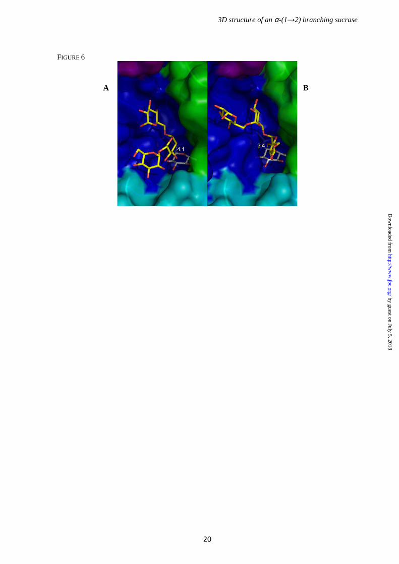
With maltose, none of the mutants showed transferase activity. In particular, the introduction of a tryptophan residue at position 2249 and/or 2250 did not promote the transfer reaction. Likely, other changes at the acceptor binding subsite +1 are necessary to generate such activity. However, when 1 kDa dextran was added as an acceptor, mutants at positions 2249 and/or 2250 were still able to catalyze the formation of α-(1→2) branches like the wt enzyme (supplemental Fig. S8). Apparently these positions are not directly involved in regiospecificity. In contrast, mutant F2214N was unable to use 1 kDa dextran as an acceptor (Table 3 and supplemental Fig. S9). Thus, this residue is critical for dextran binding and branching. To catalyze glucosyl transfer onto dextrans, ∆N123-GBD-CD2 must first bind sucrose, and then catalyze the formation of the covalent β-D-glucosyl enzyme complex. Fructose must be released from subsite +1 to allow the acceptor to enter. The dextran glucosyl residue that will be branched must bind in subsite +1 with its C2 hydroxyl group properly oriented towards the C1 of the β-D-glucosyl-enzyme intermediate. In accordance with the ping-pong bi-bi mechanism of ∆N123-GBD-CD2, the product has to leave the active site after branching, to allow the binding of a new sucrose molecule. How acceptor molecules are accommodated in the catalytic gorge in a position to be α-(1→2) glucosylated remains to be elucidated. Despite numerous attempts to crystallize complexes of ∆N123-GBD-CD2 with isomaltotriose, linear, or α-(1→2) branched gluco-oligosaccharides, we were not successful. Docking of isomaltotriose was thus attempted in a model of the glucosyl-enzyme intermediate of ∆Ν123-GBD-CD2. Two main groups of docked isomaltotriose were obtained with a glucosyl unit of the acceptor chain appropriately oriented to allow the formation of an α-(1→2) linkage. In the first group, the central glucosyl unit of isomaltotriose is in a position to be α-(1→2) glucosylated (Fig. 6A), with a shortest distance of 4.10 Å between the O2 atom of the acceptor glucosyl unit and the C1 atom of the glucosyl enzyme intermediate. In the second group, the glucosyl unit at the non-reducing extremity would be glucosylated with a distance between O2 and C1 of 3.45 Å (Fig. 6B). These models reveal that α-
by guest on July 5, 2018http://w
ww
.jbc.org/D
ownloaded from

3D structure of an α-(1→2) branching sucrase
8
(1→2) branching occurring at either the non-reducing end or at a residue of the dextran backbone is feasible. Moreover, structural analysis of ∆N123-GBD-CD2 has identified residues that line its catalytic gorge and that could be involved in dextran binding or enzyme regiospecificity (Fig. 7). Indeed, one of these residues, F2214, was shown by site-directed
mutagenesis to be crucial for dextran accommodation. The roles of the other residues, as well as residues in the three loops specific for GBD-CD2, will now be explored to further deepen our understanding of the enzyme’s mechanism and specificity.
by guest on July 5, 2018http://w
ww
.jbc.org/D
ownloaded from

3D structure of an α-(1→2) branching sucrase
9
REFERENCES 1. Henrissat, B., and Bairoch, A. (1996) Biochem. J. 316, 695-696 2. Monsan, P., Remaud-Siméon, M., and André, I. (2010) Curr. Opin. Microbiol. 13, 293-300 3. MacGregor, E. A., Janeček, Š., and Svensson, B. (2001) Biochim. Biophys. Acta 1546, 1-20 4. Moulis, C., Joucla, G., Harrison, D., Fabre, E., Potocki-Veronese, G., Monsan, P., and
Remaud-Siméon, M. (2006) J. Biol. Chem. 281, 31254-31267 5. MacGregor, E. A., Jespersen, H. M., and Svensson, B. (1996) FEBS Lett. 378, 263-266 6. Pijning, T., Vujičić-Žagar, A., Kralj, S., Eeuwema, W., Dijkhuizen, L., and Dijkstra, B.W.
(2008) Biocatal. Biotransfor. 26, 12-17 7. Ito, K., Ito, S., Shimamura, T., Kawarasaki, Y., Abe, K., Misaka, T., Kobayashi, T., and Iwata,
S. (2010) Acta Crystallogr. F. Struct. Biol. Cryst. Commun.66, 1086-1088 8. Vujičić-Žagar, A., Pijning, T., Kralj, S., López, C.A., Eeuwema, W., Dijkhuizen, L., and
Dijkstra, B.W. (2010) Proc. Natl. Acad. Sci. U S A 107, 21406-21411 9. Ito, K., Ito, S., Shimamura, T., Weyand, S., Kawarasaki, Y., Misaka, T., Abe, K., Kobayashi,
T., Cameron, A.D., and Iwata, S. (2011) J. Mol. Biol. 408, 177-186 10. Bozonnet, S., Dols-Laffargue, M., Fabre, E., Pizzut, S., Remaud-Siméon, M., Monsan, P., and
Willemot, R.M. (2002) J. Bacteriol. 184, 5753-5761 11. Fabre, E., Bozonnet, S., Arcache, A., Willemot, R.M., Vignon, M., Monsan, P., and Remaud-
Siméon, M. (2005) J. Bacteriol. 187, 296-303 12. Brison, Y., Fabre, E., Moulis, C., Portais, J.-C., Monsan, P., and Remaud-Siméon, M. (2010)
Appl. Microbiol. Biotechnol. 86, 545-554 13. Valette, P., Pelenc, V., Djouzi, Z., Andrieux, C., Paul, F., Monsan, P., and Szylit, O. (1993) J.
Sci. Food Agric. 62, 121-127 14. Djouzi, Z., Andrieux, C., Pelenc, V., Somarriba, S., Popot, F., Paul, F., Monsan, P., and Szylit,
O. (1995) J. Appl. Microbiol. 79, 117-127 15. Djouzi, Z., and Andrieux, C. (1997) Br. J. Nutr. 78, 313-324 16. Flickinger, E.A., Wolf, B.W., Garleb, K.A., Chow, J., Leyer, G.J., Johns, P.W., and Fahey,
G.C., Jr. (2000) J. Nutr. 130, 1267-1273 17. Sarbini, R. S., Kolida, S., Naeye, T., Einerhand, A., Brison, Y., Remaud-Siméon, M., Monsan,
P., Gibson, G. R., and Rastall, R. A. (2011) Appl. Environ. Microbiol. 77, 5307-5315 18. Battye, T. G., Kontogiannis, L., Johnson, O., Powell, H. R., and Leslie, A. G. (2011)
Acta Crystallogr. D. Biol. Crystallogr. 67, 271-281 19. Evans, P. (2006) Acta Crystallogr. D. Biol. Crystallogr. 62, 72-82 20. Winn, M. D., Ballard, C. C., Cowtan, K. D., Dodson, E. J., Emsley, P., Evans, P. R., Keegan,
R. M., Krissinel, E. B., Leslie, A. G., McCoy, A., McNicholas, S. J., Murshudov, G. N., Pannu, N. S., Potterton, E. A., Powell, H. R., Read, R. J., Vagin, A., and Wilson, K. S. (2011) Acta Crystallogr. D. Biol. Crystallogr. 67, 235-242
21. Brünger, A. T., DeLaBarre, B., Davies, J. M., and Weis, W. I. (2009) Acta Crystallogr. D. Biol. Crystallogr. 65, 128-133
22. Kabsch, W. (2010) Acta Crystallogr. D. Biol. Crystallogr. 66, 125-132 23. Stein, N. (2008) J. Appl. Crystallogr. 41, 641-643 24. McCoy, A.J., Grosse-Kunstleve, R.W., Adams, P.D., Winn, M.D., Storoni, L.C., and Read,
R.J. (2007) J. Appl. Crystallogr. 40, 658-674 25. Emsley, P., Lohkamp, B., Scott, W.G., and Cowtan, K. (2010) Acta Crystallogr. D. Biol.
Crystallogr. 66, 486-501 26. Murshudov, G.N., Vagin, A.A., and Dodson, E.J. (1997) Acta Crystallogr. D. Biol.
Crystallogr. 53, 240-255 27. Langer, G., Cohen, S. X., Lamzin, V. S., and Perrakis, A. (2008) Nat. Protoc. 3, 1171-1179 28. Fernandez-Tornero, C., Lopez, R., Garcia, E., Gimenez-Gallego, G., and Romero, A. (2001)
Nat. Struct. Biol. 8, 1020-1024 29. Shah, D.S.H., Joucla, G., Remaud-Simeon, M., and Russell, R.R. (2004) J. Bacteriol. 186,
8301-8308
by guest on July 5, 2018http://w
ww
.jbc.org/D
ownloaded from

3D structure of an α-(1→2) branching sucrase
10
30. Robyt, J.F., and Walseth, T.F. (1979) Carbohydr. Res. 68, 95-111 31. Holm, L., Kaariainen, S., Rosenstrom, P., and Schenkel, A. (2008) Bioinformatics 24, 2780-
2781 32. Davies, G. J., Wilson, K. S., and Henrissat, B. (1997) Biochem. J., 321, 557-559 33. Jensen, M.H., Mirza, O., Albenne, C., Remaud-Siméon, M., Monsan, P., Gajhede, M., and
Skov, L.K. (2004) Biochemistry 43, 3104-3110 34. Uitdehaag, J.C.M., Mosi, R., Kalk, K.H., van der Veen, B.A., Dijkhuizen, L., Withers, S.G.,
and Dijkstra, B.W. (1999) Nat. Struct. Biol. 6, 432-436 35. Remaud-Siméon, M., Willemot, R.-M., Sarçabal, P., Potocki de Montalk, G., and Monsan, P.
(2000) J. Mol. Catal. B: Enzym. 10, 117-128 Acknowledgments-We thank the region Midi-Pyrénées (France) and The European Molecular Biology Organization (EMBO) for financial support; the staff of beam line ID14-2 at the ESRF (Grenoble, France) and Pierre Legrand from the PROXIMA1 beam line at the SOLEIL synchrotron (Gif-sur-Yvette, France) for data collection facilities and assistance. We greatly thank Nelly Monties, Pierre Escalier, Sandra Pizzut-Serin and Virginie Rivière for technical assistance. FOOTNOTES The abbreviations used are: C-LytA: C-terminal choline binding domain of Streptococcus pneumoniae autolysin A; CW: Cell Wall; Fruf: fructofuranose; Frup: fructopyranose; GBD: Glucan Binding Domain; GBD-CD2: Glucan Binding Domain - Catalytic Domain 2; GH: Glycoside Hydrolase; Glcp: glucopyranose FIGURE LEGENDS FIGURE 1. A: Anomeric region of 1H NMR spectra obtained at 298 K for purified α-(1→2) branched dextrans. Black, dextran 70 kDa standard; blue, dextran α-(1→2) branched at 11%; pink, dextran α-(1→2) branched at 19%; orange, dextran α-(1→2) branched at 33%; green, dextrans α-(1→2) branched between 35 and 37%. B: Percentage of α-(1→2) linkage as a function of [sucrose]/[dextran] molar ratios used for the acceptor reactions. Empty and filled circles correspond to values obtained after 1H NMR and HPLC measurement, respectively, of the α-(1→2) linkage content in dextrans synthesized by ∆N123-GBD-CD2. Red crosses, 1H NMR results for dextrans branched by GBD-CD2 (12). C: Effects of the [sucrose]/[dextran] molar ratio on α-(1→2) branched dextran yields. Reactions were carried out at 292 mM sucrose and various dextran concentrations. Main final reaction products are residual sucrose, glucose (from sucrose hydrolysis), leucrose (from fructose glucosylation) and α-(1→2) branched dextran.
FIGURE 2. A: Stereo view of ∆N123-GBD-CD2 domain organization. Magenta, domain C; blue, domain A which includes the (β/α)8 barrel; green, domain B; yellow, domain IV; red, domain V. B: Schematic representation of the domain arrangement along the polypeptide chains of crystallized GH70 glucansucrases, from left to right, GTF180-∆N, GTF-SI and ∆N123-GBD-CD2. The colour code is identical to Fig. 2A; striped red, parts of domains V of GTF-SI and ∆N123-GBD-CD2, which are not visible in the electron density map; white, purification tag.
FIGURE 3. Stereo view of the secondary structure elements of ∆N123-GBD-CD2 domains A and B. Domain A: blue, (β/α)8 barrel; cyan, subdomain H1- H2; purple, loop G2731 to S2796 protruding from domain B and contributing to domain A. Domain B: green. FIGURE 4: Upper part, stereo view of the secondary structure elements of domain V (truncated Glucan Binding Domain) of ∆N123-GBD-CD2. The three-stranded β-sheets and β-hairpins of the
by guest on July 5, 2018http://w
ww
.jbc.org/D
ownloaded from

3D structure of an α-(1→2) branching sucrase
11
three sub-domains are represented in red and salmon, respectively. Lower part, sequence alignment of the domain V, underlined and colored residues are β-strands, N-terminal residues in italics are not visible in the electron density map. FIGURE 5. Stereo view of subsites -1 and +1 of ∆N123-GBD-CD2, with sucrose from the GTF180-∆N:sucrose complex superimposed. The catalytic residues are D2210 (nucleophile), E2248 (acid/base) and D2322 (transition state stabilizer). Sucrose is shown with yellow carbons. Residues of the inactive GTF180-∆N mutant (D1025N) that interact with sucrose (8) are represented in grey. The carbon atoms of their structural equivalents in ∆N123-GBD-CD2 are shown in blue (domain A), cyan (subdomain H1 - H2) and green (domain B). FIGURE 6. Isomaltotriose docking in the catalytic groove of the modeled glucosyl-enyme intermediate of ∆N123-GBD-CD2. Two binding modes were found that allow glucosylation through α-(1→2) linkage formation onto A) the central glucosyl unit or B) the non-reducing end extremity. The covalently linked glucosyl unit in subsite -1 is represented in grey and isomaltotriose is in yellow. Distance between O2 atom of isomaltotriose and C1 atom of the glucosyl enzyme intermediate (in Å) are shown in yellow. Domain coloring is as in Figure 3. FIGURE 7. Stereo view of the catalytic gorges of GTF180-∆N and ∆N123-GBD-CD2. For clarity, only different residues or residues adopting different conformations are shown. Backbone atoms of ∆N123-GBD-CD2 are depicted in lightblue. Residues from domain A of ∆N123-GBD-CD2 are depicted in blue, cyan and purple (see Fig. 3). Residues from domain B of ∆N123-GBD-CD2 are shown in green. Grey residues belong to GTF180-∆N. Sucrose from GTF180-∆N:sucrose complex (PDB entry 3HZ3) in subsites -1 and +1 is represented as yellow carbons.
by guest on July 5, 2018http://w
ww
.jbc.org/D
ownloaded from

3D structure of an α-(1→2) branching sucrase
12
TABLE 1: Data collection and refinement statistics. Numbers in brackets refer to statistics for the outer resolution shell.
Data collection
Triclinic crystals Orthorhombic crystals
Wavelength (Å) 0.933 0.954
Cell dimensions, a b c (Å), α β γ (°) 66.8, 140.0, 155.5,
85.4, 90.9, 76.9
68.2, 100.2, 187.2,
90.0, 90.0, 90.0
Space group P1 P212121
Molecules per asymmetric unit 4 1
Resolution limit (Å) 51.6 - 3.3 (3.48 – 3.30) 46.0 – 1.9 (1.95 – 1.90)
Reflections (total/unique) 153,233 / 80,648 610,291 / 98,989
Completeness (%) 97.5 (97.0) 97.2 (97.0)
Rmerge (%) 20.3 (53.3) 7.1 (45.4)$
I/σ(I) 4.1 (1.6)* 18.0 (4.4)
Wilson B-factor (Ų) 42.3 29.7
Refinement statistics
Reflections (working/test) 76,146 / 4,029 94,038 / 4,950
Rcryst/Rfree 0.224 / 0.291 0. 157 / 0.198
Number of atoms 32,344 9,331
protein 8123 / 8106 / 8072 / 7952 8128
ligand# 54 152
ion/water 4 / 33 2 / 1049
B factor (Ų)
main-chain 25.6 25.7
side-chain 26.5 27.7
ion/water/ligand 30.0 / 20.0 / 30.0 30.6 / 34.6 / 54.7
Stereochemical quality of the model
rmsd from bond lenghts (Å) 0.009 0.020
rmsd from angles (°) 1.107 1.835
Ramachandran favoured (%) 93.1 97.2
Ramachandran allowed (%) 98.9 99.9
Ramachandran disallowed (%) 1.1 0.1
* Mean I/σ(I); $ Rmeas; # glycerol or polyethylene glycol molecules
by guest on July 5, 2018http://w
ww
.jbc.org/D
ownloaded from

3D structure of an α-(1→2) branching sucrase
13
TABLE 2: Comparison of the apparent kinetic parameters determined for sucrose hydrolysis (subscript H) and α-(1→2) dextran branching activities (subscript T) for GBD-CD2 and ∆N123-GBD-CD2.
Apparent kinetic parameters GBD-CD2a ∆∆∆∆N123-GBD-CD2
In the presence of sucrose
VmaxH (µmol.min-1.mg-1 of purified enzyme) 34.6 ± 0.5 36.3 ± 0.6
KM,SucH (mM) 10.8 ± 0.8 7.5 ± 1.0
kcat,SucH (s-1) 109 76
In the presence of sucrose and dextran 70 kDa
VmaxT (µmol.min-1.mg-1 of purified enzyme) 303 ± 5 462 ± 45
KM,SucT (mM) 42 ± 2 206 ± 34
KM,DexT (mM of anhydroglucosyl units) 75 ± 3 125 ± 21
KM,DexT (mM) 0.174 ± 0.008 0.30 ± 0.05
kcat T (s-1) 970 947
kcat T/KM,SucT (s-1.mM-1) 23 4.6
kcat T/KM,DexT (s-1.mM-1) 13 7.6
a from (12).
by guest on July 5, 2018http://w
ww
.jbc.org/D
ownloaded from

3D structure of an α-(1→2) branching sucrase
14
TABLE 3: Characterization of single and double mutants targeting non-conserved residues of subsite +1.
Relative activitya
(%)
Sucrose (mM)
Acceptor, concentration (mM)
Hydrolysisb yield (%)
Sucrose isomer
yieldc (%)
Transfer onto
acceptord
(%)
146 - 87 13 -
146 1 kDa Dextran, 146 20 1 79 ∆N123-GBD-CD2 WT
100
146 Maltose, 146 88 12 nd
146 - 89 11 -
146 1 kDa dextran, 146 24 2 74 ∆N123-GBD-CD2 A2249W
21.7
146 Maltose, 146 90 10 nd
146 - 89 11 -
146 1kDa dextran, 146 45 4 51 ∆N123-GBD-CD2 G2250W
1.1
146 Maltose, 146 90 10 nd
146 - 90 10 -
146 1 kDa dextran, 146 36 4 60 ∆N123-GBD-CD2 A2249D-G2250W
89.3
146 Maltose, 146 90 10 nd
146 - 100 nde -
146 1 kDa dextran, 146 100 nd nd ∆N123-GBD-CD2 F2214N
18.6
146 Maltose, 146 100 nd nd aThe relative activity was determined as the ratio of sucrose consumption (intial rate) of each mutant versus sucrose consumption (initial rate) of ∆N123-GBD-CD2. bHydrolysis yield= Glc (mol)/ Sucrose consumed (mol) *100 cSucrose isomer = Glc incorporated into leucrose (mol)/ Sucrose consumed (mol) *100 dTransfer onto acceptor = 100 – Hydrolysis ratio % - Sucrose isomer yield % e Not detectable
by guest on July 5, 2018http://w
ww
.jbc.org/D
ownloaded from

3D structure of an α-(1→2) branching sucrase
15
FIGURE 1
C
A B
by guest on July 5, 2018http://w
ww
.jbc.org/D
ownloaded from

3D structure of an α-(1→2) branching sucrase
16
FIGURE 2
A B
by guest on July 5, 2018http://w
ww
.jbc.org/D
ownloaded from

3D structure of an α-(1→2) branching sucrase
17
FIGURE 3
by guest on July 5, 2018http://w
ww
.jbc.org/D
ownloaded from

3D structure of an α-(1→2) branching sucrase
18
FIGURE 4
by guest on July 5, 2018http://w
ww
.jbc.org/D
ownloaded from

3D structure of an α-(1→2) branching sucrase
19
FIGURE 5
by guest on July 5, 2018http://w
ww
.jbc.org/D
ownloaded from
3D structure of an α-(1→2) branching sucrase
20
FIGURE 6
A B
by guest on July 5, 2018http://w
ww
.jbc.org/D
ownloaded from

3D structure of an α-(1→2) branching sucrase
21
FIGURE 7
by guest on July 5, 2018http://w
ww
.jbc.org/D
ownloaded from

Remaud-Siméon and Bauke W. DijkstraSandrine Morel, Gabrielle Potocki-Véronèse, Pierre Monsan, Samuel Tranier, Magali
Yoann Brison, Tjaard Pijning, Yannick Malbert, Émeline Fabre, Lionel Mourey,derived from DSR-E glucansucrase
2) branching sucrase→-(1αFunctional and structural characterization of an
published online January 18, 2012J. Biol. Chem.
10.1074/jbc.M111.305078Access the most updated version of this article at doi:
Alerts:
When a correction for this article is posted•
When this article is cited•
to choose from all of JBC's e-mail alertsClick here
Supplemental material:
http://www.jbc.org/content/suppl/2012/01/18/M111.305078.DC1
by guest on July 5, 2018http://w
ww
.jbc.org/D
ownloaded from