JB Accepts, published online ahead of print on 2 December...
30
1 The synthetic lethality of lytE cwlO in Bacillus subtilis is caused by lack of 1 D,L-endopeptidase activity at the lateral cell wall 2 3 Masayuki Hashimoto, 1 Seika Ooiwa, 2 and Junichi Sekiguchi 2,* 4 5 1 International Young Researchers Empowerment Center, 2 Department of Applied 6 Biology, Faculty of Textile Science and Technology, Shinshu University, Ueda, Japan 7 8 Running title: Synthetic lethality of lytE cwlO in B. subtilis 9 Section: Genetics and Molecular Biology 10 11 * Corresponding author. Department of Applied Biology, Faculty of Textile Science and 12 Technology, Shinshu University, 3-15-1 Tokida, Ueda-shi, Nagano 386-8567, Japan. 13 Tel: +81 268 21 5344; Fax: +81 268 21 5344; E-mail: [email protected] 14 15 16 17 18 Copyright © 2011, American Society for Microbiology and/or the Listed Authors/Institutions. All Rights Reserved. J. Bacteriol. doi:10.1128/JB.05569-11 JB Accepts, published online ahead of print on 2 December 2011 on July 18, 2018 by guest http://jb.asm.org/ Downloaded from
-
Upload
duongduong -
Category
Documents
-
view
217 -
download
0
Transcript of JB Accepts, published online ahead of print on 2 December...

1
The synthetic lethality of lytE cwlO in Bacillus subtilis is caused by lack of 1
D,L-endopeptidase activity at the lateral cell wall 2
3
Masayuki Hashimoto,1 Seika Ooiwa,2 and Junichi Sekiguchi2,* 4
5
1 International Young Researchers Empowerment Center, 2 Department of Applied 6
Biology, Faculty of Textile Science and Technology, Shinshu University, Ueda, Japan 7
8
Running title: Synthetic lethality of lytE cwlO in B. subtilis 9
Section: Genetics and Molecular Biology 10
11
* Corresponding author. Department of Applied Biology, Faculty of Textile Science and 12
Technology, Shinshu University, 3-15-1 Tokida, Ueda-shi, Nagano 386-8567, Japan. 13
Tel: +81 268 21 5344; Fax: +81 268 21 5344; E-mail: [email protected] 14
15
16
17
18
Copyright © 2011, American Society for Microbiology and/or the Listed Authors/Institutions. All Rights Reserved.J. Bacteriol. doi:10.1128/JB.05569-11 JB Accepts, published online ahead of print on 2 December 2011
on July 18, 2018 by guesthttp://jb.asm
.org/D
ownloaded from

2
(Abstract) 19
Bacterial peptidoglycan acts as an exoskeleton to protect the bacterial cell. 20
Although peptidoglycan biosynthesis by penicillin-binding proteins is well studied, 21
few studies have described peptidoglycan disassembly, which is necessary for a 22
dynamic structure that allows cell growth. In Bacillus subtilis, more than 35 genes 23
encoding cell wall lytic enzymes have been identified; however, only two 24
D,L-endopeptidases (lytE and cwlO) are involved in cell proliferation. In this study, 25
we demonstrated that the D,L-endopeptidase activity at the lateral cell wall is 26
essential for cell proliferation. Inactivation of LytE and CwlO by point mutation of 27
the catalytic residues caused cell growth defects. However, the forced expression of 28
LytF or CwlS, which are paralogs of LytE, did not suppress lytE cwlO synthetic 29
lethality. Subcellular localization studies of these D,L-endopeptidases showed LytF 30
and CwlS at the septa and poles, CwlO at the cylindrical part of the cell, and LytE 31
at the septa and poles as well as the cylindrical part. Furthermore, construction of 32
N-terminal and C-terminal domain-swapped enzymes of LytE, LytF, CwlS, and 33
CwlO revealed that localization was dependent on the N-terminal domains. Only 34
the chimeric proteins that were enzymatically active and localized to the sidewall 35
were able to suppress the synthetic lethality, suggesting that lack of 36
D,L-endopeptidase activity at the cylindrical part of the cell leads to a growth defect. 37
The functions of LytE and CwlO in cell morphogenesis were discussed. 38
on July 18, 2018 by guesthttp://jb.asm
.org/D
ownloaded from
3
(Introduction) 39
Autolysins are bacterial cell wall lytic enzymes found in all bacteria that possess 40
peptidoglycan. In the Bacillus subtilis genome, more than 35 definite or probable 41
autolysin genes have been identified and shown to be involved in cell morphogenesis, 42
cannibalism, sporulation, and germination. (22, 25). The bacterial peptidoglycan 43
sacculus requires a dynamic structure for cell elongation and separation; therefore, a 44
balance between peptidoglycan synthesis and disassembly is essential for cell 45
proliferation. Although a number of autolysins are thought to be involved in 46
peptidoglycan disassembly, none have been found to be essential for cell growth, 47
perhaps due to their functional redundancy. However, it was recently reported that 48
disruption of both lytE and cwlO in B. subtilis is lethal (4). To date this is the sole report 49
of an autolysin mutant of B. subtilis with a serious growth defect. Bisicchia et al. also 50
demonstrated that cwlO depletion in a lytE disrupted background strain impairs cell 51
elongation (4). 52
LytE and CwlO are D,L-endopeptidases that hydrolyze the linkage of 53
D-γ-glutamyl-meso-diaminopimelic acid in peptidoglycan (13, 27). The B. subtilis 54
genome contains seven D,L-endopeptidase genes. The mature forms of LytE, LytF, and 55
CwlS all contain N-terminal LysM repeats, although the number of LysM domains 56
differs, and C-terminal D,L-endopeptidase domains belonging to the NlpC/P60 family. 57
Although phenotypes of single-gene knockout mutants were indistinguishable from that 58
of wild type, multiple gene disruptions led to a chained-cell morphology (10, 13, 19), 59
suggesting that these proteins are involved in cell separation. In contrast, CwlO contains 60
a domain with unknown function at the N-terminus and a D,L-endopeptidase domain at 61
the C-terminus. The phenotype of the cwlO mutant was also indistinguishable from wild 62
on July 18, 2018 by guesthttp://jb.asm
.org/D
ownloaded from

4
type, but the lytE cwlO double disruption leads to synthetic lethality (4, 27). Two 63
D,L-endopeptidase genes (pgdS and cwlT) are not likely to be involved in cell 64
morphology, because the pgdS gene encodes a poly-γ-glutamic acid degradase, and the 65
cwlT gene is part of an integrative and conjugative element (11, 23). The other gene is a 66
function-unknown ykfC. Results of these previous studies indicate that LytE, LytF and 67
CwlS are cell separation enzymes, and LytE and CwlO are associated with cell growth. 68
Thus, although their catalytic domains show high amino acid sequence similarity, these 69
enzymes play different physiological roles in cell morphology. To elucidate the roles of 70
LytE and CwlO in cell morphogenesis, we investigated the main factors causing 71
synthetic lethality in B. subtilis. 72
73
MATERIALS AND METHODS 74
Bacterial strains and plasmids. The bacterial strains and plasmids used in this 75
study are listed in Table 1 and Table S1 in the Supplementary material, respectively. B. 76
subtilis 168 was used as the parent strain throughout this study. The details of the strains 77
and plasmids constructs used in this study are presented in the Supplementary material. 78
All constructed strains were confirmed by PCR. 79
General methods. The B. subtilis and Escherichia coli strains were grown at 37˚C 80
in Luria Broth (LB) (21). When required, antibiotics and chemical inducers were added 81
in the following concentrations: ampicillin, 100 μg/ml; tetracycline, 5 μg/ml; kanamycin, 82
25 μg/ml; spectinomycin, 50 μg/ml; erythromycin, 0.3 μg/ml chloramphenicol, 5 μg/ml; 83
isopropyl β-D-1-thiogalactopyranoside (IPTG), 1 mM; and xylose, 1%. 84
DNA manipulation and E. coli transformation were performed using standard 85
methods (21). B. subtilis transformation was performed by conventional transformation 86
on July 18, 2018 by guesthttp://jb.asm
.org/D
ownloaded from
5
procedures (1). 87
Sample preparation for immunofluorescence microscopy (IFM). Cells 88
harvested from an overnight culture in LB medium were diluted 50-fold in 5 ml fresh LB 89
medium. The cells were grown to the late exponential growth phase (optical density at 90
600 nm [OD600nm] = 2.0), and then the precultured cells were inoculated into fresh LB 91
medium to give an initial absorbance of OD600nm = 0.001. Cells corresponding to 0.3 of 92
the OD600nm unit for WECLytE6FL (LytE-6×FLAG), OH015 (CWBLytE-6×FLAG), 93
WECO6FL (CwlO-6×FLAG), OH013 (overexpressed CwlO-6×FLAG), or OH018 94
(overexpressed NTDCwlO-6×FLAG) were collected when each culture reached OD600nm 95
= 0.1. As described below, LytE-6×FLAG and CwlO-6×FLAG were functional for B. 96
subtilis cell proliferation. Likewise, 0.3 of the OD600nm unit cells were collected for 97
WECLytF6FL (LytF-6×FLAG) and OH014 (CWBLytF-6×FLAG) when the cultures 98
reached OD600nm = 0.6. Similarly, 0.3 of the OD600nm unit cells were collected for 99
WECS6FL (CwlS-6×FLAG) and OH016 (CWBCwlS-6×FLAG) when each culture 100
reached OD600nm = 2.0. To determine the subcellular localization of the domain-swapped 101
chimeric enzymes, cells were collected when the cultures reached OD600nm = 0.3 (for 102
chimeric proteins transcribed from the lytE promoter) or OD600nm = 0.1 (for those 103
transcribed from the cwlO promoter). Cell samples were prepared for IFM as described 104
previously (30). 105
Fluorescence microscopy. Fluorescence microscopy was performed as described 106
previously (29) with an Olympus BX61 microscope equipped with a BX-UCB control 107
unit, a UPPlan Apo Fluorite phase-contrast objective (×100 magnification; numerical 108
aperture, 1.3), and a standard rhodamine filter set for visualizing Cy3. Exposure times 109
on July 18, 2018 by guesthttp://jb.asm
.org/D
ownloaded from

6
were 0.1 s for phase-contrast microscopy and 0.1s (gain 2) for Cy3. The cells were 110
photographed with a charge-coupled device camera (CoolSNAP HQ; Nippon Roper) 111
driven by MetaMorph software (version 4.6; Universal Imaging). For Cy3 imaging, 112
out-of-focus light was removed using the two-dimensional deconvolution utility of the 113
AutoDeblur software. All images were processed with Adobe Photoshop software. 114
Western blot analysis and zymography. Sodium dodecyl sulfate-polyacrylamide 115
gel electrophoresis (SDS-PAGE) was performed with 14% (w/v) polyacrylamide gels as 116
described previously (15). For western blot analysis, the 6×FLAG-fused proteins were 117
separated by 14% SDS-PAGE gels. After electrophoresis, the proteins were transferred 118
to polyvinylidene fluoride membranes (Invitrogen) in a transfer buffer (25 mM Tris, 192 119
mM glycine, 20% [v/v] methanol, 0.1% SDS) using a semidry blotting system 120
(Bio-Rad). Immunoblot detection was carried out as described in the instruction manual 121
for the ECL Plus Western Blotting Detection System (Invitrogen) using a mouse 122
anti-FLAG M2 monoclonal antibody (Sigma) and horseradish peroxidase-labeled 123
anti-mouse IgG antibody. Zymography was performed as described previously using 124
14% SDS-PAGE gels containing 0.5 mg/ml B. subtilis cell wall extract (17). The cell 125
wall derived from B. subtilis 168 was prepared as described previously (8, 19). 126
Renaturation was performed at 37°C in a renaturation solution (25 mM Tris-HCl [pH 127
7.2], 1% [v/v] Triton X-100) as described previously (10). 128
129
RESULTS 130
D,L-Endopeptidase activity of LytE or CwlO is essential for cell proliferation. 131
The catalytic domains of LytE and CwlO belong to the NlpC/P60 family, which 132
on July 18, 2018 by guesthttp://jb.asm
.org/D
ownloaded from

7
hydrolyzes the γ- D -glutamyl-meso-diaminopimelic acid linkage or 133
N-acetylmuramoyl-L-alanine linkage. In this superfamily of papain-like enzymes, a 134
conserved cysteine residue was predicted to be a catalytic residue on amino acid 135
sequence alignment (2, 16). Recently, the three-dimensional structures of NlpC/P60 136
enzymes were reported (Spr from E. coli, ABA23003 from Anabaena variabilis, and 137
ACC79413 from Nostoc punctiforme) (3, 26). In these enzymes, the conserved cysteine 138
residues are located at a predicted active site and are structurally conserved. To 139
determine whether the conserved cysteine residues are involved in the catalytic activity 140
of D,L-endopeptidases, we constructed point mutations in LytE and CwlO, replacing the 141
conserved cysteine residue with a serine residue (LytEC247S and CwlOC377S). To evaluate 142
the lytic activities of these mutated enzymes, the intact or mutated catalytic domains of 143
LytE and CwlO were expressed in E. coli, and zymography was carried out with the cell 144
lysates using B. subtilis cell wall as a substrate (see Fig. S1B in the Supplementary 145
material). The intact catalytic domains of LytE and CwlO exhibited cell wall-degrading 146
activity, but mutants in which the cysteine residue had been replaced appeared to be 147
inactive. This finding suggests that the conserved cysteine residue is important for the 148
catalytic activity of NlpC/P60 enzymes. 149
Next, we examined whether the D,L-endopeptidase activities of LytE and CwlO are 150
involved in the synthetic lethality of the lytE cwlO double mutants (Fig. 1A and B). 151
OH004 (lytE-6×flag Pxyl-cwlO) grew normally without xylose induction of CwlO, 152
indicating that LytE-6×FLAG was functional. In contrast, the growth of OH005 153
(lytEC247S-6×flag Pxyl-cwlO) was normal in the presence of xylose, but was arrested in 154
the absence of xylose. Similarly, CwlO-6×FLAG was functional, but OH007 155
(cwlOC377S-6×flag Pspac-lytE) showed growth arrest without LytE induction by IPTG. 156
on July 18, 2018 by guesthttp://jb.asm
.org/D
ownloaded from

8
These results indicate that the D,L-endopeptidase activity of either LytE or CwlO is 157
essential for cell proliferation. 158
As described above, LytE, LytF, and CwlS exhibit similar domain structures. 159
However, lytE expression is regulated by σA and σH, cwlO expression is regulated by σA, 160
and lytF and cwlS are regulated by σD and σH, respectively (5, 13, 19, 27). The σD and σH 161
regulons are induced later than the σA regulon. Therefore, although LytF and CwlS can 162
suppress the synthetic lethality, the LytE CwlO double-depleted cells may be dead 163
before LytF or CwlS can be expressed. Consequently, OH009 (ΔlytE Pxyl-cwlO 164
Pspac-lytF) and OH012 (ΔlytE Pxyl-cwlO Pspac-cwlS) were constructed to determine 165
whether induction of LytF or CwlS could suppress the synthetic lethality. These strains 166
were cultured in the presence of 1 mM IPTG to induce LytF or CwlS and in the presence 167
or absence of 1% xylose to induce CwlO (Fig. 1C, D). Both strains grew normally when 168
CwlO was expressed; however, growth was arrested by CwlO depletion, even though 169
LytF or CwlS was expressed. The hydrolytic activities of induced LytF and CwlS were 170
confirmed by zymography with B. subtilis cell wall as a substrate (see Fig. S2 in the 171
Supplementary material). We found that LytF and CwlS are not able to suppress the LytE 172
CwlO-depleted synthetic lethality, even though their domain structures are similar to 173
that of LytE. 174
Subcellular localization of B. subtilis D,L-endopeptidases. The C-terminal 175
D,L-endopeptidase domains of LytE, LytF, CwlS, and CwlO show strong sequence 176
similarity. In contrast, the N-terminal domains of LytE, LytF, and CwlS contain different 177
numbers of the LysM repeats, and the N-terminus of CwlO contains a COG3883 domain. 178
Although the D,L-endopeptidase activity of either LytE or CwlO is essential for cell 179
proliferation, forced expression of LytF or CwlS did not suppress the lytE cwlO 180
on July 18, 2018 by guesthttp://jb.asm
.org/D
ownloaded from

9
synthetic lethality. These results suggest that the N-terminal domains are important for 181
the function of the D,L-endopeptidases. Previously, we reported that B. subtilis WE1, a 182
strain with defects in extracellular proteases WprE and Epr, accumulates 183
D,L-endopeptidases on the cell surface (29). Therefore, we evaluated the subcellular 184
localization of FLAG-tagged LytE, LytF, CwlS, and CwlO (full-length proteins and 185
N-terminal domains) by IFM with wprE epr-deleted WEC background strains. Because 186
these D,L-endopeptidases are regulated by different σ factors, we also evaluated the 187
localization of these enzymes during different growth phases. Full-length LytE and 188
CwlO and their N-terminal domains (CWBLytE and NTDCwlO, respectively) were 189
observed during early exponential growth phase (OD600nm = 0.1), full-length LytF and its 190
N-terminal domain (CWBLytF) were observed in mid-exponential growth phase 191
(OD600nm = 0.6), and full-length CwlS and its N-terminal domain (CWBCwlS) were 192
observed in early stationary phase (OD600nm = 2.0). The results showed that LytE is 193
localized at the cell septa, poles, and sidewall (Fig. 2A). LytF-6×FLAG and 194
CwlS-6×FLAG were localized at the cell septa and poles, but neither was detected at the 195
lateral cell wall (Fig. 2C and E). CwlO-6×FLAG expressed from the intact promoter was 196
weakly detected at the lateral cell wall but not at the septa or poles (Fig. 2G). To better 197
assess CwlO localization, we then used a CwlO-6×FLAG-overexpressing strain (Fig. 198
2H), which increased cell surface CwlO-6×FLAG expression to 2.4 times that of normal, 199
as determined by western blot analysis (data not shown). The overexpressed 200
CwlO-6×FLAG was more clearly visualized at the sidewall but not detected at the cell 201
septa or poles. To determine whether the localization of these D,L-endopeptidases 202
depends on the N-terminal domain, we investigated the subcellular localization of the 203
N-terminal domains under the same conditions used for the full-length proteins (Fig. 2B, 204
on July 18, 2018 by guesthttp://jb.asm
.org/D
ownloaded from

10
D, F, and I). The localization pattern of each N-terminal domain was identical to that of 205
the corresponding full-length protein, indicating that these D,L-endopeptidases localized 206
on the cell surface through their N-terminal domains. 207
Characterization of domain-swapped D,L-endopeptidases. IFM analysis 208
demonstrated that LytF and CwlS (involved in cell separation) localize to the septa and 209
poles, CwlO (involved in cell elongation) localizes to the lateral cell wall, and LytE 210
(involved both in cell separation and elongation) localizes to the septa, poles, and lateral 211
cell wall. These results suggest that the functions of these D,L-endopeptidases depend on 212
their subcellular localization. To test this hypothesis, we generated domain-swapped 213
D,L-endopeptidases and examined their ability to suppress the lytE cwlO synthetic 214
lethality. 215
Domain-swapped D,L-endopeptidases (other than NLytFCLytE) were generated by 216
C-terminal domain substitution at the original genetic loci of the N-terminal domains. 217
For example, NLytECCwlS was constructed by substituting the C-terminal domain of LytE 218
with that of CwlS at the lytE locus. Thus, the chimeric genes were transcribed from the 219
promoters of the gene encoding the N-terminal domain. However, NLytFCLytE was 220
constructed by substituting the N-terminal domain of LytE with that of LytF at the lytE 221
locus; the chimeric gene was transcribed from the lytE promoter. All chimeric proteins 222
were fused to a 6×FLAG tag at the C-terminus to evaluate their expression and 223
localization. Expression was confirmed by western blot analysis, and the chimeric 224
proteins were detected at positions corresponding to the predicted molecular sizes (Fig. 225
3A). Enzyme activity was assessed by zymography using the B. subtilis cell wall as a 226
substrate (Fig. 3B). The results show that the chimeric enzymes containing the CwlO 227
N-terminal domain did not retain cell wall-degrading activity. The C-terminal 228
on July 18, 2018 by guesthttp://jb.asm
.org/D
ownloaded from
11
D,L-endopeptidase regions of NCwlOCLytF and NCwlOCCwlS are the same as those of 229
NLytECLytF and NLytECCwlS, respectively. Since NLytECLytF and NLytECCwlS exhibited cell 230
wall-degrading activity, it was assumed that the C-terminal D,L-endopeptidase domains 231
of NCwlOCLytF and NCwlOCCwlS would exhibit enzyme activity as well; however, it is 232
possible that the N-terminal region of CwlO interfered with the C-terminal 233
D,L-endopeptidase domain activity in NCwlOCLytF and NCwlOCCwlS. Next, the subcellular 234
localization of these domain-swapped D,L-endopeptidases was visualized by IFM (Fig. 235
4). The chimeric proteins containing the LytE N-terminal domain (NLytECLytF and 236
NLytECCwlS) localized to the cell septa, poles, and lateral cell wall, similar to the 237
localization of LytE-6×FLAG and CWBLytE-6×FLAG. However, NLytFCLytE localized 238
only to the cell septa and poles, like LytF-6×FLAG and CWBLytF-6×FLAG. Only weak 239
fluorescence of the chimeric enzymes containing the N-terminal domain of CwlO 240
(NCwlOCLytF and NCwlOCCwlS) was detected. However, enhancing the signal intensity of 241
IFM images revealed that these chimeric enzymes were localized to the sidewall, similar 242
to full-length CwlO and its N-terminal domain. These results demonstrate that the 243
N-terminal domains of D,L-endopeptidases determine their subcellular localization. 244
Finally, we assessed whether these domain-swapped D,L-endopeptidases were able to 245
suppress the lytE cwlO synthetic lethality (Fig. 4). The transcription of cwlO was 246
induced by xylose in strains expressing LytE or LytF N-terminal domain-containing 247
chimeric enzymes (NLytECLytF, NLytECCwlS, or NLytFCLytE), whereas lytE gene transcription 248
was induced by IPTG in strains expressing the CwlO N-terminal domain-containing 249
chimeric enzymes (NCwlOCLytF and NCwlOCCwlS). After exposure to the appropriate 250
inducer, an aliquot of each culture was washed to remove the inducer, and the cells were 251
inoculated into fresh medium with or without the inducer. OH019 (lytE::NLytECLytF 252
on July 18, 2018 by guesthttp://jb.asm
.org/D
ownloaded from
12
Pxyl-cwlO) and OH020 (lytE::NLytECCwlS Pxyl-cwlO) were found to partially suppress the 253
lytE cwlO synthetic lethality without xylose induction of cwlO. As described above, 254
these chimeric proteins were enzymatically active and detected at the cell septa, poles, 255
and sidewall. However, strains expressing chimeric proteins containing the CwlO 256
N-terminal domain (OH023 [cwlO::NCwlOCLytF Pspac-lytE] and OH024 [cwlO::NCwlOCCwlS 257
Pspac-lytE]), which were not enzymatically active, were localized at the lateral cell wall, 258
but not able to grow without IPTG induction of lytE. Furthermore, lack of xylose caused 259
the growth arrest of OH022 (lytE::NLytFCLytE Pxyl-cwlO). This strain expressed NLytFCLytE, 260
which retained enzymatic activity but was not localized at the cellular sidewall. 261
Taken together, our findings show that only strains expressing at least one active 262
D,L-endopeptidase localized at the lateral cell wall were able to proliferate. Therefore, 263
we conclude that localization of D,L-endopeptidase activity at the lateral cell wall is 264
essential for cell proliferation. 265
266
DISCUSSION 267
Peptidoglycan forms a network on the outer surface of bacterial cells. The dynamic 268
structure of the peptidoglycan sacculus allows cell growth; therefore, maintaining the 269
balance of peptidoglycan synthesis and disassembly is important. To the best of our 270
knowledge, the synthetic lethality of lytE cwlO in B. subtilis is the only report of an 271
autolysin mutant with a serious growth defect (4). In this study, we found that 272
subcellular localization of these enzymes is determined by their N-terminal domains, 273
and synthetic lethality is caused by the lack of D,L-endopeptidase activity at the lateral 274
cell wall. The D,L-endopeptidases required for cell separation (LytE, LytF, and CwlS) 275
were detected at the septa and poles, and the enzymes involved in cell elongation (LytE 276
on July 18, 2018 by guesthttp://jb.asm
.org/D
ownloaded from

13
and CwlO) were detected at the cylindrical part of the cell. These results strongly 277
suggest that the function of these autolysins depends on their subcellular localization. 278
Our findings are consistent with a previous study reporting that a lytF cwlO double 279
mutant and a lytE lytF cwlS triple mutant were not defective in cell growth (10, 27). 280
LytE and CwlO may participate in loosening the peptidoglycan sacculus of B. 281
subtilis during growth. The cell wall of B. subtilis is comprised of multi-layered thick 282
peptidoglycan. Electron microscopy images show that the thick peptidoglycan consists 283
of three distinct parts (18). Results of pulse-labeling studies revealed a delay between 284
the incorporation of new material into the cell wall and its eventual appearance in the 285
culture (12, 20). These results suggest that the inner zone of the thick peptidoglycan 286
contains the newly synthesized layers, and the outer zone consists of old peptidoglycan 287
(i.e., inside-to-outside peptidoglycan sacculus formation) (12, 18, 20). 288
Peptidoglycan-synthesizing enzymes are anchored to cytoskeleton proteins (MreB 289
homologs and FtsZ), and localize to the outside surface of the cytoplasmic membrane 290
(6). Thus, the peptidoglycan-synthesizing enzymes are accessible to the inner zone of 291
peptidoglycan. Degradation of the outer zone loosens the cell wall, enabling 292
construction of a new peptidoglycan layer inside the preexisting peptidoglycan sacculus 293
(22). Since lytE cwlO double disruption leads to synthetic lethality and impaired cell 294
elongation, these autolysins are strong candidates for participation in the peptidoglycan 295
dynamics. Consistent with this hypothesis, our results show that the cell elongation 296
defect due to the lytE cwlO disruption is caused by the absence of D,L-endopeptidase 297
activity at the lateral cell wall. However, results of a pulse-labeling experiment show that 298
the rate of N-acetylglucosamine incorporation is not the same for lytE and cwlO mutants, 299
demonstrating that LytE behavior differs from that of CwlO (4). LytE and CwlO differ in 300
on July 18, 2018 by guesthttp://jb.asm
.org/D
ownloaded from
14
their subcellular localizations and specific activities (28). In addition, CwlO was rapidly 301
degraded and released into culture medium, whereas most of LytE adsorbed to cell 302
surface (27). Taken together, these findings demonstrate that although these two 303
enzymes possess similar D,L-endopeptidase domains, they appear to have different 304
functions in cell growth. 305
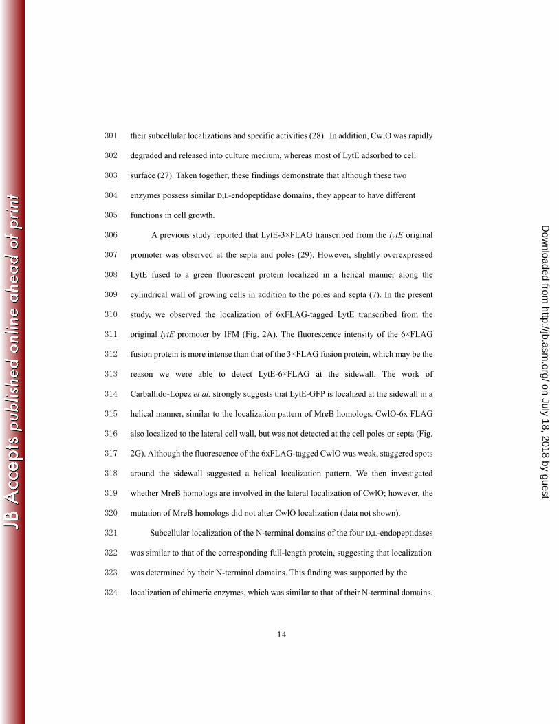
A previous study reported that LytE-3×FLAG transcribed from the lytE original 306
promoter was observed at the septa and poles (29). However, slightly overexpressed 307
LytE fused to a green fluorescent protein localized in a helical manner along the 308
cylindrical wall of growing cells in addition to the poles and septa (7). In the present 309
study, we observed the localization of 6xFLAG-tagged LytE transcribed from the 310
original lytE promoter by IFM (Fig. 2A). The fluorescence intensity of the 6×FLAG 311
fusion protein is more intense than that of the 3×FLAG fusion protein, which may be the 312
reason we were able to detect LytE-6×FLAG at the sidewall. The work of 313
Carballido-López et al. strongly suggests that LytE-GFP is localized at the sidewall in a 314
helical manner, similar to the localization pattern of MreB homologs. CwlO-6x FLAG 315
also localized to the lateral cell wall, but was not detected at the cell poles or septa (Fig. 316
2G). Although the fluorescence of the 6xFLAG-tagged CwlO was weak, staggered spots 317
around the sidewall suggested a helical localization pattern. We then investigated 318
whether MreB homologs are involved in the lateral localization of CwlO; however, the 319
mutation of MreB homologs did not alter CwlO localization (data not shown). 320
Subcellular localization of the N-terminal domains of the four D,L-endopeptidases 321
was similar to that of the corresponding full-length protein, suggesting that localization 322
was determined by their N-terminal domains. This finding was supported by the 323
localization of chimeric enzymes, which was similar to that of their N-terminal domains. 324
on July 18, 2018 by guesthttp://jb.asm
.org/D
ownloaded from

15
The localization of the LytF N-terminal domain at the cell poles and septa was 325
previously reported (30). As expected, the localization of LytE and CwlS was dependent 326
on their N-terminal domains, which contained LysM repeats like that of LytF. Yamamoto 327
et al. also reported a helical localization of LytF-6×FLAG at the sidewall after partial 328
removal of wall teichoic acid (30), suggesting that the cylindrical localization of 329
N-terminal domains of LytE and CwlS are regulated by wall teichoic acid. 330
Carballido-López et al. reported that LytE localization at the sidewall is dependent on 331
MreBH, indicating that MreBH may regulate wall teichoic acid localization (7). It was 332
reported that, the helical localization of the major wall teichoic acid synthesis proteins 333
was not altered in three mreB homolog single mutants (9). However, we note that these 334
cells were cultured with 20 mM MgCl2, which suppresses mreB homolog deficiency 335
(14). 336
The CwlO N-terminus contains a COG3883 domain, which is an uncharacterized 337
conserved domain in bacteria. According to Teng et al., a secreted antigen (SagA) from 338
Enterococcus faecium containing a COG3883 domain showed broad-spectrum binding 339
to extracellular matrix proteins such as fibrinogen, collagen type I, collagen type IV, 340
fibronectin, and laminin (24). However, full-length CwlO and its N-terminal domain did 341
not bind some of the matrix proteins evaluated in this study (data not shown). The SagA 342
protein migrated more slowly on cell wall-containing PAGE than on SDS-PAGE, 343
suggesting an interaction between SagA and the cell wall (24); however, the purified 344
CwlO protein did not bind to the cell wall in vitro (27). In the present study, we 345
demonstrated the involvement of the CwlO N-terminal domain in cell surface 346
localization. Taken together, these results suggest that CwlO interacts directly, but 347
weakly, with the cell wall or a cell surface protein. 348
on July 18, 2018 by guesthttp://jb.asm
.org/D
ownloaded from

16
In this study, we found that the subcellular localization of LytE, LytF, CwlS, and 349
CwlO is dependent on their N-terminal domains, and that D,L-endopeptidase activity at 350
the lateral cell wall is essential for cell proliferation. These results strongly suggest that 351
LytE and CwlO are involved in cell elongation and support the inside-to-outside model 352
for peptidoglycan sacculus formation. A more detailed study is necessary to clarify the 353
role of D,L-endopeptidases in peptidoglycan dynamics and characterize the localization 354
mechanisms of these proteins. 355
356
ACKNOWLEDGMENTS 357
We would like to thank the members of our group, particularly Hiroki Yamamoto 358
and Tatsuya Fukushima, for the helpful advice and discussion. We also thank N. 359
Hariyama and Y. Miyake for technical assistance with strain construction and 360
microscopy analysis. This work was supported by Grants-in-Aid for Scientific Research 361
(B) (19380047) and (A) (22248008), the New Energy and Industrial Department 362
Organization (NEDO), the Global COE programs (JS), and the Program for 363
Dissemination of Tenure-Track System funded by the Ministry of Education and 364
Science, Japan (MH). 365
REFERENCES 366
1. Anagnostopoulos, C., and J. Spizizen. 1961. Requirements for transformation 367
in Bacillus subtilis. J. Bacteriol. 81:741-746. 368
2. Anantharaman, V., and L. Aravind. 2003. Evolutionary history, structural 369
features and biochemical diversity of the NlpC/P60 superfamily of enzymes. 370
Genome Biol. 4: R11. 371
on July 18, 2018 by guesthttp://jb.asm
.org/D
ownloaded from

17
3. Aramini, J. M., P. Rossi, Y. J. Huang, L. Zhao, M. Jiang, M. Maglaqui, R. 372
Xiao, J. Locke, R. Nair, B. Rost, T. B. Acton, M. Inouye, and G. T. 373
Montelione. 2008. Solution NMR structure of the NlpC/P60 domain of 374
lipoprotein Spr from Escherichia coli: Structural evidence for a novel cysteine 375
peptidase catalytic triad. Biochemistry. 47:9715-9717. 376
4. Bisicchia, P., D. Noone, E. Lioliou, A. Howell, S. Quigley, T. Jensen, H. 377
Jarmer, and K. M. Devine. 2007. The essential YycFG two-component 378
system controls cell wall metabolism in Bacillus subtilis. Mol. Microbiol. 379
65:180-200. 380
5. Britton, R. A., P. Eichenberger, J. E. Gonzalez-Pastor, P. Fawcett, R. 381
Monson, R. Losick, and A. D. Grossman. 2002. Genome wide analysis of the 382
stationary phase sigma factor (sigma-H) regulon of Bacillus subtilis. J. 383
Bacteriol. 184:4881-4890. 384
6. Cabeen, M. T., and C. Jacobs-Wagner. 2005. Bacterial cell shape. Nat. Rev. 385
Microbiol. 3:601-610. 386
7. Carballido-Lopez, R., A. Formstone, Y. Li, S. D. Ehrlich, P. Noirot, and J. 387
Errington. 2006. Actin homolog MreBH governs cell morphogenesis by 388
localization of the cell wall hydrolase LytE. Dev. Cell. 11:399-409. 389
8. Fein, J. E., and H. J. Rogers. 1976. Autolytic enzyme deficient mutants of 390
Bacillus subtilis 168. J. Bacteriol. 127:1427-1442. 391
9. Formstone, A., R. Carballido-Lopez, P. Noirot, J. Errington, and D. J. 392
Scheffers. 2008. Localization and interactions of teichoic acid synthetic 393
enzymes in Bacillus subtilis. J. Bacteriol. 190:1812-1821. 394
on July 18, 2018 by guesthttp://jb.asm
.org/D
ownloaded from

18
10. Fukushima, T., A. Afkham, S. Kurosawa, T. Tanabe, H. Yamamoto, and J. 395
Sekiguchi. 2006. A new D,L-endopeptidase gene product, YojL (renamed 396
CwlS), plays a role in cell separation with LytE and LytF in Bacillus subtilis. J. 397
Bacteriol. 188:5541-5550. 398
11. Fukushima, T., T. Kitajima, H. Yamaguchi, Q. Ouyang, K. Furuhata, H. 399
Yamamoto, T. Shida, and J. Sekiguchi. 2008. Identification and 400
characterization of novel cell wall hydrolase CwIT - A two-domain autolysin 401
exhibiting N-acetylmuramidase and dl-endopeptidase activities. J. Biol. Chem. 402
283:11117-11125. 403
12. Graham, L. L., and T. J. Beveridge. 1994. Structural differentiation of the 404
Bacillus subtilis 168 cell wall. J. Bacteriol. 176:1413-1421. 405
13. Ishikawa, S., Y. Hara, R. Ohnishi, and J. Sekiguchi. 1998. Regulation of a 406
new cell wall hydrolase gene, cwlF, which affects cell separation in Bacillus 407
subtilis. J. Bacteriol. 180:2549-2555. 408
14. Kawai, Y., K. Asai, and J. Errington. 2009. Partial functional redundancy of 409
MreB isoforms, MreB, Mbl and MreBH, in cell morphogenesis of Bacillus 410
subtilis. Mol. Microbiol. 73:719-731. 411
15. Laemmli, U. K. 1970. Cleavage of structural proteins during assembly of head 412
of bacteriophage T4. Nature. 227:680-685. 413
16. Layec, S., B. Decaris, and N. Leblond-Bourget. 2008. Characterization of 414
proteins belonging to the CHAP related superfamily within the firmicutes. J. 415
Mol. Microbiol. Biotechnol. 14:31-40. 416
on July 18, 2018 by guesthttp://jb.asm
.org/D
ownloaded from

19
17. Leclerc, D., and A. Asselin. 1989. Detection of bacterial cell wall hydrolases 417
after denaturing polyacrylamide gel electrophoresis. Can. J. Microbiol. 418
35:749-753. 419
18. Merad, T., A. R. Archibald, I. C. Hancock, C. R. Harwood, and J. A. 420
Hobot. 1989. Cell wall assembly in Bacillus subtilis visualization of old and 421
new wall material by electron microscopic examination of samples stained 422
selectively for teichoic acid and teichuronic acid. J. Gen. Microbiol. 423
135:645-655. 424
19. Ohnishi, R., S. Ishikawa, and J. Sekiguchi. 1999. Peptidoglycan hydrolase 425
LytF plays a role in cell separation with Cw1F during vegetative growth of 426
Bacillus subtilis. J. Bacteriol. 181:3178-3184. 427
20. Pooley, H. M. 1976. Layered distribution, according to age, within cell wall of 428
Bacillus subtilis. J. Bacteriol. 125:1139-1147. 429
21. Sambrook, J., E. F. Fritch, and T. Maniatis. 1989. Molecular cloning: a 430
laboratory manual, 2nd ed. Cold Spring Harbor Laboratory, Cold Spring Harbor, 431
NY. 432
22. Smith, T. J., S. A. Blackman, and S. J. Foster. 2000. Autolysins of Bacillus 433
subtilis: multiple enzymes with multiple functions. Microbiology. 146:249-262. 434
23. Suzuki, T., and Y. Tahara. 2003. Characterization of the Bacillus subtilis 435
ywtD gene, whose product is involved in gamma-polyglutamic acid degradation. 436
J. Bacteriol. 185:2379-2382. 437
24. Teng, F., M. Kawalec, G. M. Weinstock, W. Hryniewicz, and B. E. Murray. 438
2003. An Enterococcus faecium secreted antigen, SagA, exhibits broad 439
on July 18, 2018 by guesthttp://jb.asm
.org/D
ownloaded from

20
spectrum binding to extracellular matrix proteins and a appears essential for E. 440
faecium growth. Infect. Immun. 71:5033-5041. 441
25. Vollmer, W., B. Joris, P. Charlier, and S. Foster. 2008. Bacterial 442
peptidoglycan (murein) hydrolases. FEMS Microbiol. Rev. 32:259-286. 443
26. Xu, Q. P., S. Sudek, D. McMullan, M. D. Miller, B. Geierstanger, D. H. 444
Jones, S. S. Krishna, G. Spraggon, B. Bursalay, P. Abdubek, C. Acosta, E. 445
Ambing, T. Astakhova, H. L. Axelrod, D. Carlton, J. Caruthers, H. J. Chiu, 446
T. Clayton, M. C. Deller, L. Duan, Y. Elias, M. A. Elsliger, J. Feuerhelm, S. 447
K. Grzechnik, J. Hale, G. W. Han, J. Haugen, L. Jaroszewski, K. K. Jin, H. 448
E. Klock, M. W. Knuth, P. Kozbial, A. Kumar, D. Marciano, A. T. Morse, E. 449
Nigoghossian, L. Okach, S. Oommachen, J. Paulsen, R. Reyes, C. L. Rife, 450
C. V. Trout, H. van den Bedem, D. Weekes, A. White, G. Wolf, C. Zubieta, 451
K. O. Hodgson, J. Wooley, A. M. Deacon, A. Godzik, S. A. Lesley, and I. A. 452
Wilson. 2009. Structural basis of murein peptide specificity of a 453
gamma-D-glutamyl-L-diamino acid endopeptidase. Structure. 17:303-313. 454
27. Yamaguchi, H., K. Furuhata, T. Fukushima, H. Yamamoto, and J. 455
Sekiguchi. 2004. Characterization of a new Bacillus subtilis peptidoglycan 456
hydrolase gene, yvcE (named cwlO), and the enzymatic properties of its 457
encoded protein. J. Biosci. Bioeng. 98:174-181. 458
28. Yamamoto, H., M. Hashimoto, Y. Higashitsuji, H. Harada, N. Hariyama, 459
L. Takahashi, T. Iwashita, S. Ooiwa, and J. Sekiguchi. 2008. 460
Post-translational control of vegetative cell separation enzymes through a direct 461
interaction with specific inhibitor IseA in Bacillus subtilis. Mol. Microbiol. 462
70:168-182. 463
on July 18, 2018 by guesthttp://jb.asm
.org/D
ownloaded from

21
29. Yamamoto, H., S. Kurosawa, and J. Sekiguchi. 2003. Localization of the 464
vegetative cell wall hydrolases LytC, LytE, and LytF on the Bacillus subtilis cell 465
surface and stability of these enzymes to cell wall bound or extracellular 466
proteases. J. Bacteriol. 185:6666-6677. 467
30. Yamamoto, H., Y. Miyake, M. Hisaoka, S. I. Kurosawa, and J. Sekiguchi. 468
2008. The major and minor wall teichoic acids prevent the sidewall localization 469
of vegetative DL-endopeptidase LytF in Bacillus subtilis. Mol. Microbiol. 470
70:297-310.471
on July 18, 2018 by guesthttp://jb.asm
.org/D
ownloaded from
22
Figure legends 472
473
FIG. 1. D,L-endopeptidase activity of LytE and CwlO is important for cell proliferation, 474
and LytF or CwlS induction could not suppress lytE cwlO synthetic lethality. Strains 475
were precultured with the appropriate inducer until late exponential phase 476
(OD600nm=2.0). An aliquot of each culture was washed and inoculated into fresh medium 477
with or without the inducer to OD600nm=0.01. The × symbol in panels A to D indicates 478
the wild type 168 strain. (A) Growth of OH005 (lytEC247S-6×flag Pxyl-cwlO; open circles) 479
and OH004 (lytE-6×flag Pxyl-cwlO; closed circles). Xylose (1%) was added to the 480
preculture, but CwlO expression was not induced by xylose in the main culture. (B) 481
Growth of OH007 (cwlOC377S-6×flag Pspac-lytE; open circles) and OH006 (cwlO-6×flag 482
Pspac-lytE; closed circles). IPTG (1 mM) was added to the preculture, but LytE 483
expression was not induced by IPTG in the main culture. (C) Growth of OH009 (ΔlytE 484
Pxyl-cwlO Pspac-lytF). The strain was cultured with 1 mM IPTG to induce LytF 485
expression, and with 1% xylose to induce CwlO induction (closed circles) or without 486
xylose (open circles). (D) Growth of OH012 (ΔlytE Pxyl-cwlO Pspac-cwlS). The strain was 487
cultured with 1 mM IPTG to induce CwlS expression, and with 1% xylose to induce 488
CwlO expression (closed circles) or without xylose (open circles). 489
490
FIG. 2. Subcellular localization of full-length D,L-endopeptidases and their N-terminal 491
domains. Phase-contrast and immunofluorescence microscopy analysis of 492
FLAG-tagged proteins. The OD600nm values at the sampling times were 0.1 for LytE and 493
CwlO and their N-terminal domains (CWBLytE and NTDCwlO, respectively), 0.6 for LytF 494
and its N-terminal domain (CWBLytF), and 2.0 for CwlS and its N-terminal domain 495
on July 18, 2018 by guesthttp://jb.asm
.org/D
ownloaded from

23
(CWBCwlS). A, WECLytE6FL (LytE-6×FLAG); B, OH015 (CWBLytE-6×FLAG); C, 496
WECLytF6FL (LytF-6×FLAG); D, OH014 (CWBLytF-6×FLAG); E, WECS6FL 497
(CwlS-6×FLAG); F, OH016 (CWBCwlS-6×FLAG); G, WECO6FL (CwlO-6×FLAG); H, 498
OH013 (overexpressed CwlO-6×FLAG); and I, OH018 (overexpressed 499
NTDCwlO-6×FLAG). Scale bars = 5 μm. 500
501
FIG. 3. Expression and activity of domain-swapped D,L-endopeptidases. Strains were 502
exposed to 1% xylose or 1 mM IPTG for 2 hours to induce Pxyl-cwlO and Pspac-lytE 503
expression, respectively. 1, OH019 (NLytECLytF Pxyl-cwlO, 41 kDa); 2, OH020 (NLytECCwlS 504
Pxyl-cwlO, 40 kDa); 3, OH022 (NLytFCLytE Pxyl-cwlO, 53 kDa); 4, OH023 (NCwlOCLytF 505
Pspac-lytE, 55 kDa); and 5, OH024 (NCwlOCCwlS Pspac-lytE, 56 kDa). (A) 506
Domain-swapped D,L-endopeptidases were evaluated by western blot analysis with an 507
anti-FLAG antibody. Degraded products of the chimeric enzymes appear on lanes 4 and 508
5. (B) Zymography of the chimeric enzymes using B. subtilis cell wall as a substrate. 509
Asterisks indicate clear zones produced by the chimeric enzymes. 510
511
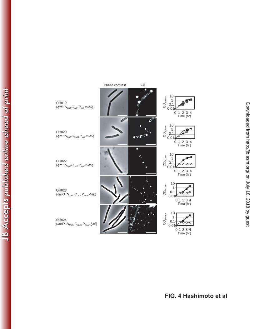
FIG. 4. Subcellular localization of domain-swapped D, L-endopeptidases and 512
suppression of the lytE cwlO synthetic lethality by these proteins. For microscopic 513
imaging, OH019 (lytE ::NLytECLytF Pxyl-cwlO), OH020 (lytE ::NLytECCwlS Pxyl-cwlO), and 514
OH022 (lytE ::NLytFCLytE Pxyl-cwlO) were cultured with 1% xylose to induce CwlO, and 515
OH023 (cwlO ::NCwlOCLytF Pspac-lytE) and OH024 (cwlO ::NCwlOCCwlS Pspac-lytE) were 516
cultured with 1 mM IPTG to induce LytE. For suppression assays, the strains were 517
grown under the same conditions as those described in Fig. 1. They were cultured with 518
xylose (closed circles) or without xylose (open circles) for Pxyl-cwlO and IPTG for 519
on July 18, 2018 by guesthttp://jb.asm
.org/D
ownloaded from

24
Pspac-lytE. The × symbol indicates the wild type 168 strain. Scale bars = 5 μm. 520
521
on July 18, 2018 by guesthttp://jb.asm
.org/D
ownloaded from

25
TABLE 1. Bacterial strains used in this study. 522
________________________________________________________________________________________________________ 523 Strains Relevant genotype Source or reference a 524 ________________________________________________________________________________________________________ 525 E. coli strains 526 JM109 recA1 endA1 gyrA96 thi-1 hsdR17 relA1 supE44 Δ (lac-proAB) 527 /F’ [traD36 proAB lacIq lacZ ΔM15] Takara 528 C600 supE44 hsdR17 thi-1 thr-1 IeuB6 lacY1 tonA21 Laboratory stock 529 M15/pREP4 lac ara gal mtl F- recA+ uvr+ / lacI kan Qiagen 530 B. subtilis 531 168 trpC2 S. D. Ehrlich 532 FTD trpC2 lytE::tet 30 533 OH001 trpC2 cwlO:: pXyl-cwlO (Pxyl -cwlO) pXyl-cwlO -> 168 534 OH002 trpC2 lytE::tet cwlO::pXyl-cwlO (Pxyl -cwlO) OH001 -> 168FTD 535 OH003 trpC2 lytE::pM4LYTE pM4LYTE -> 168 536 OH004 trpC2 lytE::lytE-6×flag cwlO::pXyl-cwlO (Pxyl -cwlO) pCA6FLCF -> OH001 537 OH005 trpC2 lytE::lytEC247S-6×flag cwlO::pXyl-cwlO (Pxyl -cwlO) pCALEC247S -> OH001 538 OH006 trpC2 cwlO::cwlO-6×flag lytE::pM4LYTE (Pspac -lytE) Supplementary data 539 OH007 trpC2 cwlO::cwlOC377S-6×flag lytE::pM4LYTE (Pspac -lytE) Supplementary data 540 OH008 trpC2 lytF::pM4LYTF pM4LYTF -> 168 541 OH009 trpC2 lytE::tet cwlO::pXyl-cwlO (Pxyl -cwlO) lytF::pM4LYTF (Pspac -lytF) OH008 -> OH002 542 BKD trpC2 lytC::kan 27 543 OH010 trpC2 lytE::tet cwlO::pXyl-cwlO lytF::pM4LYTF lytC::kan 168BKD -> OH009 544 OH011 trpC2 cwlS::pM4SDΔojL pM4SD∆ojL -> 168 545 OH012 trpC2 lytE::tet cwlO::pXyl-cwlO (Pxyl -cwlO) cwlS::pM4SDΔojL (Pspac -cwlS) OH011 -> OH002 546 WEC trpC2 ΔwprA Δepr 30 547 WECLytF6FLb trpC2 ΔwprA Δepr lytF::pCA6FLCE 30 548 WECLytE6FLb trpC2 ΔwprA Δepr lytE::pCA6FLCF 30 549 WECS6FL trpC2 ΔwprA Δepr cwlS::pCA6FLCS 30 550 WECO6FL trpC2 ΔwprA Δepr cwlO::pCA6FLCO pCA6FLCO -> WEC 551 OH013 trpC2 ΔwprA Δepr / pDG-O6FL pDGO6FL -> WEC 552 OH014 trpC2 ΔwprA Δepr lytF::pCA6FLCWBE pCA6FLCWBE -> WEC 553 OH015 trpC2 ΔwprA Δepr lytE::pCA6FLCWBF pCA6FLCWBF -> WEC 554 OH016 trpC2 ΔwprA Δepr cwlS::pCA6FLCWBS pCA6FLCWBS -> WEC 555 OH017 trpC2 ΔwprA Δepr cwlO::pCA6FLNTDO pCA6FLNTDO -> WEC 556 OH018 trpC2 ΔwprA Δepr / pDGNO6FL pDGNO6FL -> WEC 557 OH019 trpC2 lytE::pCA-FbEcII (NLytECLytF) cwlO::pXyl-cwlO (Pxyl -cwlO) pCA-FbEcII -> OH002 558 OH020 trpC2 lytE::pCA-FbSc (NLytECCwlS) cwlO::pXyl-cwlO (Pxyl -cwlO) pCA-FbSc -> OH002 559 OH021 trpC2 lytE::pBlue-FtEbkan (5’-lytF kan) cwlO::pXyl-cwlO (Pxyl -cwlO) pBlue-FtEbkan -> OH002 560 OH022 trpC2 lytE::NLytFCLytE cwlO::pXyl-cwlO (Pxyl -cwlO) Supplementary material 561 OH023 trpC2 cwlO::NCwlOCLytF lytE::pM4LYTE (Pspac -lytE) Supplementary material 562 OH024 trpC2 cwlO::NCwlOCCwlS lytE::pM4LYTE (Pspac -lytE) Supplementary material 563 ________________________________________________________________________________________________________ 564 aSources shown before and after the arrows indicate donor DNA and recipient cells of 565
transformation, respectively. 566
bThe previous strain names, WECE6FL and WECF6FL (30), are changed to 567
on July 18, 2018 by guesthttp://jb.asm
.org/D
ownloaded from

26
WECLytF6FL and WECLytE6FL, respectively, to avoid the confusion of gene names. 568
on July 18, 2018 by guesthttp://jb.asm
.org/D
ownloaded from

FIG. 1 Hashimoto et al
0.01
0.1
1
10
0 2 4O
D60
0nm
Time (hr)1 3
A
0.01
0.1
1
10
0 2 4
OD
600n
m
Time (hr)1 3
B
0.01
0.1
1
10
0 2 4
OD
600n
m
Time (hr)1 3
C
0.01
0.1
1
10
0 2 4
OD
600n
m
Time (hr)1 3
D
on July 18, 2018 by guesthttp://jb.asm
.org/D
ownloaded from

FIG. 2 Hashimoto et al
Phase contrast IFM Phase contrast IFM
A
LytE-6xFLAG
B
CWBLytE-6xFLAG
C
LytF-6xFLAG
D
CWBLytF-6xFLAG
E
CwlS-6xFLAG
F
CWBCwlS-6xFLAG
G
CwlO-6xFLAG
H
Over expressedCwlO-6xFLAG
I
Over expressedNTDCwlO-6 xFLAG
Full-length enzymes N-terminal domains
on July 18, 2018 by guesthttp://jb.asm
.org/D
ownloaded from
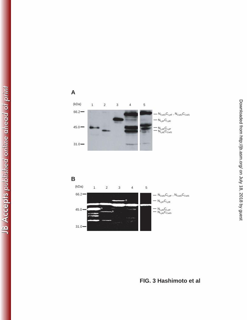
FIG. 3 Hashimoto et al
A
66.2
45.0
31.0
(kDa)
NLytFCLytE
NLytECLytFNLytECCwlS
NCwlOC CwlS, NCwlOCLytF
66.2
45.0
31.0
(kDa) 1 2 3 4 5
NLytFCLytE
B
NLytECLytFNLytECCwlS
NCwlOC CwlS, NCwlOCLytF
1 2 3 4 5
on July 18, 2018 by guesthttp://jb.asm
.org/D
ownloaded from
OH020(lytE::NLytECCwlS Pxyl-cwlO)
OH022(lytE::NLytFCLytE Pxyl-cwlO)
OH019(lytE::NLytECLytF Pxyl-cwlO)
OH024(cwlO::NCwlOCCwlS Pspac-lytE)
OH023(cwlO::NCwlOCLytF Pspac-lytE)
0.010.1
110
0 1 2 3 4
OD
600n
m
Time (hr)
0.010.1
110
0 1 2 3 4
OD
600n
m
Time (hr)
0.010.1
110
0 1 2 3 4
OD
600n
m
Time (hr)
0.010.1
110
0 1 2 3 4
OD
600n
m
Time (hr)
0.010.1
110
0 1 2 3 4
OD
600n
m
Time (hr)
Phase contrast IFM
FIG. 4 Hashimoto et al
on July 18, 2018 by guesthttp://jb.asm
.org/D
ownloaded from