Jaw Anomalies
12
JAW ANOMALIES JAW ANOMALIES DRG. SHANTY CHAIRANI DRG. SHANTY CHAIRANI
description
aaa
Transcript of Jaw Anomalies

JAW ANOMALIESJAW ANOMALIES
DRG. SHANTY CHAIRANIDRG. SHANTY CHAIRANI

A developmental defect of the palate characterized A developmental defect of the palate characterized by a lack of complete fusion of the two lateral by a lack of complete fusion of the two lateral portions of the palate, resulting in a communication portions of the palate, resulting in a communication with the nasal cavity. with the nasal cavity. Clinical features : Clinical features : – May be unilateral or bilateralMay be unilateral or bilateral– The cleft may vary in severity : involving only the uvula or The cleft may vary in severity : involving only the uvula or
soft palate, or extending all the way through the palate and soft palate, or extending all the way through the palate and including the alveolar ridge. including the alveolar ridge.
Radigraphic features : the alveolar bone in the Radigraphic features : the alveolar bone in the region of the cleft may reveal numerous dental region of the cleft may reveal numerous dental anomalies. These may include the absence of the anomalies. These may include the absence of the maxillary lateral incisor dan the presence of maxillary lateral incisor dan the presence of supernumerary teeth in this region. Often the teeth supernumerary teeth in this region. Often the teeth in this region are malformed and poorly positioned.in this region are malformed and poorly positioned.
CLEFT PALATECLEFT PALATE


CLEIDOCRANIAL DYSPLASIACLEIDOCRANIAL DYSPLASIA
It is a developmental anomaly of the skeleton It is a developmental anomaly of the skeleton and teethand teethClinical features : Clinical features : – Failure of formation of claviclesFailure of formation of clavicles– Maxillary micrognathiaMaxillary micrognathia– Prolonged retention of primary dentition and Prolonged retention of primary dentition and
delayed eruption of the permanent dentition.delayed eruption of the permanent dentition.– A large number of unerupted supernumerary A large number of unerupted supernumerary
teeth.teeth.
Radiographic features :Radiographic features :– Typically multiple supernumeray unerupted teeth.Typically multiple supernumeray unerupted teeth.– Prolonged retention of primary dentition.Prolonged retention of primary dentition.

Maxillary retrognathism in cleidocranial dysplasiaMaxillary retrognathism in cleidocranial dysplasia

In this 28 years old, there are many of the permanent In this 28 years old, there are many of the permanent teeth fail to eruptteeth fail to erupt

Also known as Treacher Collins syndrome Also known as Treacher Collins syndrome It is a developmental anomaly.It is a developmental anomaly.Clinical features :Clinical features :– Underdevelopment of the zygomatic bones and Underdevelopment of the zygomatic bones and
mandible. mandible. – MacrostomiaMacrostomia– Malformation of the external ears and absence of Malformation of the external ears and absence of
external auditory canal.external auditory canal.– Occasional facial cleft.Occasional facial cleft.
Radiographic features :Radiographic features :– Consistent with the clinical observationConsistent with the clinical observation
MANDIBULOFACIAL DYSOSTOSISMANDIBULOFACIAL DYSOSTOSIS
In this patient with mandibulofacial dysostosis, the maxilla and In this patient with mandibulofacial dysostosis, the maxilla and mandible are both underdeveloped, the latter markedly so, mandible are both underdeveloped, the latter markedly so, producing a receding chin.producing a receding chin.

There is malocclusion of the maxillary and mandibular incisors There is malocclusion of the maxillary and mandibular incisors and crowding of the teeth. The inferior border of the mandible and crowding of the teeth. The inferior border of the mandible is concave with pronounced antegonial notching and ,in this is concave with pronounced antegonial notching and ,in this patients, the right maxillary antrum is less well developed.patients, the right maxillary antrum is less well developed.

HEMIFACIAL HYPERTOPHYHEMIFACIAL HYPERTOPHYIs a condition in which half of the face and jaws, Is a condition in which half of the face and jaws, alone or in concert with other parts of the body, alone or in concert with other parts of the body, grow to unusual proportions.grow to unusual proportions.The cause of this condition is unknown, but heredity The cause of this condition is unknown, but heredity is suggested play a rule.is suggested play a rule.Clinical features :Clinical features :– Begins during youth, sometimes at birth.Begins during youth, sometimes at birth.– The dentition of affected individual may show enlargement The dentition of affected individual may show enlargement
of the canine, premolar and first molar crown and roots and of the canine, premolar and first molar crown and roots and accelerated development.accelerated development.
– Typically primary teeth shed prematurely.Typically primary teeth shed prematurely.– The tongue and alveolar bone are enlarged on the involved The tongue and alveolar bone are enlarged on the involved
sideside
Radiographic features :Radiographic features :– Enlargement of the bones on the affected side, it may Enlargement of the bones on the affected side, it may
include enlargement of the mandible, maxilla and zygoma.include enlargement of the mandible, maxilla and zygoma.

HEMIFACIAL HYPOPLASIAHEMIFACIAL HYPOPLASIA
There is reduced growth of half of the faceThere is reduced growth of half of the faceClinical features :Clinical features :– Usually begins early in life.Usually begins early in life.– A striking appearance as a result of progressive failure A striking appearance as a result of progressive failure
of growth of the affected side, with the result that there of growth of the affected side, with the result that there is a reduced dimension of the involved side of the face.is a reduced dimension of the involved side of the face.
Radiographic features :Radiographic features :– A reduction in the size of the bones on the affected side.A reduction in the size of the bones on the affected side.– Mostly determined in the mandible, which may show a Mostly determined in the mandible, which may show a
reduction in the size of the condyle, coronoid process or reduction in the size of the condyle, coronoid process or overal dimensions of the body and ramus of the overal dimensions of the body and ramus of the mandible.mandible.
– The dentition of the affected side show a reduction in The dentition of the affected side show a reduction in the number or size of the teeth.the number or size of the teeth.
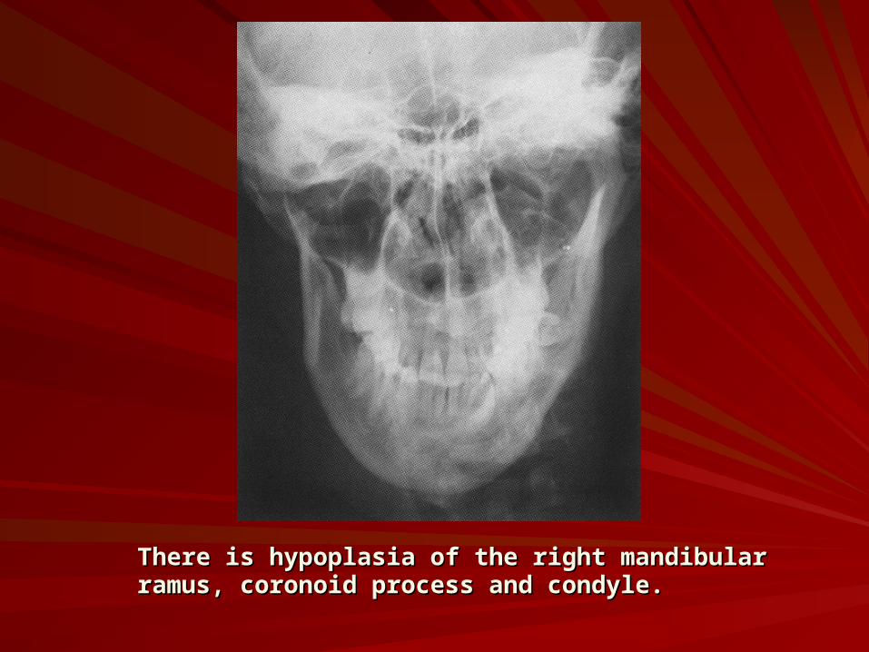
There is hypoplasia of the right mandibular There is hypoplasia of the right mandibular ramus, coronoid process and condyle.ramus, coronoid process and condyle.