Jan-March 2016 Jan - Mar 2017 · 7 8 IDA Jan-Mar 2017, Volume 2, Issue 1 It is important to...
29
BDJ BDJ BANGALORE DENTAL JOURNAL Official Publication of IDA Bangalore Branch ISSN : 2278-6686 Issue 1 Volume 2 Jan - Mar 2017 A comparative evaluation of different polishing aids after scaling and root planing-a split mouth clinical trial. 6 A comparitive study of lipid profile on healthy smokers and non smokers 15 Pills of the future: Nanoparticles in Oral Medicine 17 Angular Cheilitis - A Review 21
-
Upload
truonglien -
Category
Documents
-
view
218 -
download
0
Transcript of Jan-March 2016 Jan - Mar 2017 · 7 8 IDA Jan-Mar 2017, Volume 2, Issue 1 It is important to...

ISSN : 2278-6686
Issue 1
Volume 1
Jan-March 2016
BDJBDJ
BANGALORE DENTAL JOURNAL
Official Publication of IDA Bangalore Branch
ISSN : 2278-6686
Issue 1
Volume 2
Jan - Mar 2017
A comparative evaluation of different polishing aids after scaling and root planing-a split mouth clinical trial. 6
A comparitive study of lipid profile on healthy smokers and non smokers 15
Pills of the future: Nanoparticles in Oral Medicine 17
Angular Cheilitis - A Review 21


3
EDIT
OR
IAL
Review Board
Editorial Board
ORAL MEDICINE AND RADIOLOGY :Dr. JAIKRISHNADr. SHILPA PATIL ORAL PATHOLOGY :Dr. GIRISH H.CDr. SANJAY MURUGOD ORAL SURGERY :Dr. MADANDr. PREETHAM SHETTY
PEDODONTIA :Dr. PAVANDr. VENKATESH BABU
PERIODONTIA :Dr. SHYAM PADMANABHANDr. ANIERBAN CHATERJE
Associate Editor :Dr. CHETHAN.R
PROSTHODONTIA :Dr. PURUSHOTAM MANVIDr. SHILPA SHETTY
COMMUNITY DENTISTRY :Dr. MURALI IYERDr. PADMA BHAT
CONSERVATIVE :Dr. NAVEENDr. KAVITHA RANGANATH
ORTHODONTIA :Dr. HEMANTHDr. VINAY
I have been pleased and honored to serve you, the members and the Indian Dental Association Karnataka State Branch as President this year. The mission of IDA is
to advance increase knowledge for the improvement of oral health across the region ; to support and represent the oral health community; and tofacilitate the communication and application by Awarenesss among the public.
In support of the mission, the Association continued to provide professional
development and publication opportunities for members.Large number of attendees from all the branches allow delegatesopportunities to network with the community of clinicians and researchers while exploring the latest scientific discoveries in the field.
The IDA Karnataka StateJournal continued to serve this year, thanks to
the high quality of research that scientists and clinicians submitted for publication in the Journal. The high caliber of science of the Journal has had high impact and helped it achieve appreciationproviding increased opportunities for publication encompassedthe complete spectrum of oral, dental and craniofacial investigation with a focus onclinical and translational research.
The increasing importance of translating findings into clinical practice provided impetus for Research. Under the editorship of Dr Rajkumar. This groundbreaking new softcopy version of the print journal will be dedicated to publishing more original dental, oral and craniofacial research at the interface between scientific discovery and clinical application with the translation of research into healthcare delivery at the individual patient, clinical practice and community levels.
As scientists, we know that research discovery does not occur in isolation. Science is a continuum of knowledge that builds on the previous work of others, and today's discoveries will provide the foundation for further efforts. We must support our research community and extend our reach if we wish to further our science.
Also IDA continues to encourage members to collaborate with their fellow member colleagues and be active in the Association by participating in at least at the CDE for clinical and research updaesboth at the State and local branch meetings.
I encourage all Ida members to remainengaged so that together we can support the IDA mission and improve oral health Nationwide.
Sincerely,
B Nandlal
With Warm Regards,Dr. SATHEESHA REDDY B H
Editor-in-Chief, IDA BANGALORE

4
IDA BANGALORE BRANCHLIST OF OFFICE BEARERS FOR THE YEAR 2017-18
Hon. Treasurer:
Dr B K SrivastavaDr
Dr Tilak Raj T NDr Manjunath BDr Kumar NCDr Madhu KDr DhayakarDr Kishore HCDr Prashanth BRDr Manjunath VK
President Elect:
Imm. Past President:
Vice Presidents:
Dr. Girish Sharma
Dr. Ashwatharaju .P
Dr Annaji
Dr Sanjay Kumar
Dr Utkarsh .L
DrMurali .R
Hon. Joint Secretary:
Hon. Asst. Secretary:
Hon. Editor:
Chairman CDE:
Chairman CDH:
Dr Smitha T
Dr Sudarshan
Dr Satheesha Reddy B H
Dr Suresh .T
Dr Ramesh L
Executive Committee Members:
Dr Vidhya SagarDr Ramamurthy TKDr Sai RameshDr PremnathDr Chaithnya Babu NDr Raghu TNDr Rohith SDr Charan Shetty
Representatives to State:
Dr Veerendra KumarDr Hegde BTDr Sadanand MPDr Mohammed NoomanDr Madhusudhan ReddyDr Sudharshan KumarDr Ullhas AmasiDr Shivu ME
Dr Uma SRDr Prasad MGSDr Raghunatha KDr Akshay ShettyDr Chethan RDr Sandeep JNDr Dhanu
President
Dr. Nanda Kishore BHon. State Secretary
Mahesh Chandra

Dr. Nanda Kishore B
PR
ESID
ENT’
S M
ESSA
GE
President, IDA Bangalore Branch
SEC
RET
AR
Y’S
MES
SAG
E
Dr Mahesh ChandraHon. Secretary
IDA Bangalore Branch
5
It is with great pride, enthusiasm, and anticipation that I invite you to read the inaugural issue of the IDA BANGALORE DENTAL JOURNAL, a new kind of research journal.
An enormous amount of work has gone into the development of this journal and I believe you will see that effort reflected in this journal and in the impact it will have on the field. It has been an interesting journey, the journey has not been one with a completely charted course. It could not have been, given our time constraints.
As we look at Journal, it is important to keep in mind that it represents the collective thinking of a group of innovative individuals with whom I am privileged to work. First, we want Journal to be the premiere scientific journal in Dental Sciences. We want it to look different, to be different, to be one journal that, with its related website, will be as dynamic as the work going on in our disciplines, a rarity in academic publishing. Second, we want it to be a vehicle for a new type of conversation about dental practice and its place in the academic review, tenure, promotion, and reward process. That’s a tall order, but with your help we will make it happen.
Over the past six years, having acquired considerable new experience in Indian Dental Association with such experienced and well informed colleagues from all the Dental Colleges , and papers of various qualities covering all fields of dental medicine, I believe this is the proper time to initiate some new activities. Setting a web site is such an activity; I believe quite an important activity, which will add to the Journals wider recognition and, consequently, better and more efficient communication and exchange of scientific ideas. Now, on the web site, the BDJ will be easily found, and I hope that this will enable the BDJ to become a well-known international scientific journal, covering all aspects of Dental Medicine.
Dear members,
It gives me immense pleasure to present to you the first issue of the current edition of BDJ for the year 2017.
It's been a very enriching and memorable journey as President, IDA Bangalore branch, which has given me an opportunity to evolve as a person and to serve our fraternity in my capacity.
I would like to thank all the office bearers of the IDA Bangalore branch and all the people who have supported me through this journey.
I would like to express my heartfelt thanks to Dr. Satheesha Reddy B H, our editor for his enduring efforts in ensuring the publication of this journal.
Dear respected IDA member,

6
ABSTRACT:
Aim-To evaluate and compare the efficacy of different polishing aids after scaling and root planing.
Objective-The primary objective of the study was to evaluate and compare the efficacy of different polishing aids (rubber cup, bristle brush and air polisher) after scaling and root planing.
Materials and methods- A clinical split-mouth study was carried out with a total sample size of 100 individuals within the age range of 18–65 years, having all teeth except third molars and a probing depth of not more than 5 mm, and suffering from chronic marginal/papillary gingivitis with localized periodontitis. Each quadrant was assigned randomly with a polishing aid except the last quadrant after the scaling and root planing .Three clinical parameters were assessed-plaque index, gingival bleeding index and extrinsic stain index at baseline ,7 and 21 days.
Results-As per mean score change from baseline, air polisher showed better improvement than other quadrants in Plaque score. All Quadrants showed early and progressive improvement in Lobene's stain index and gingival bleeding index at day 7 and day 21 over baseline. The noted improvement was statistically insignificant at day 7 and day 21 in comparison to baseline.
Conclusion.- All the quadrants showed improvement in all the clinical parameters but air polisher showed significant improvement in all the parameters compared to other quadrants.
Key words : Plaque ,calculus ,stains, polishing aids, scaling and root planing
AUTHORS: 1 1 2 3 4 4Sircar Trisha , Debnath koel , chatterjee Anirban , Raghunathan Vinayak , Jayaram Praveen , RM Rosh
A comparative evaluation of different polishing aids after scaling and root planing-a split mouth clinical trial.
Department of Periodontology,
The Oxford Dental College and Hospital,
Karnataka, Bangalore
1. Dr. Sircar Trisha, 1. Dr. Debnath Koel,
3. Dr. Chatterjee Anirban, HOD and Professor3. Dr. Raghunathan Vinayak, Senior Lecturer4. Dr. Jayaram Praveen, Reader4. Dr. RM Rosh, Reader
Post Graduate StudentPost Graduate Student
INTRODUCTION
Periodontal disease is a multi factorial disease in which plaque being one of the major contributor factor in the progression of a disease. It is defined clinically as a structured resilient yellowish grayish substance that adheres tenaciously to the intraoral hard surface including removable and fixed
[1]restorations .Dental biofilm and plaque calcifies and forms calculus. Therefore it is important to control the accumulation of biofilm and plaque.
Many physical and chemical agents are capable of producing discoloration of the dentition. There are two types of stains -Extrinsic and Intrinsic stains. Extrinsic stains result from the deposition of a film, pigment or calculus on the surface of enamel,
[2]. exposed dentin or cementum Intrinsic discoloration occurs following a change to the structural composition or thickness of the dental hard tissues.
The normal colour of teeth is determined by the blue, green and pink tints of the enamel and is reinforced by the yellow to brown shades of dentine beneath. A number of metabolic diseases and systemic factors are known to affect the developing dentition and
[3]cause discolouration as a consequence
In a periodontal disease or a routine cleaning of the oral cavity, it consists of full mouth scaling either by manual or ultra sonic scalers as primary treatment. Tooth polishing is a procedure carried out as a part of oral prophylaxis in most dental practices. It is an act of smoothening the tooth surfaces to make it glossy and lustrous. Although the term polishing has been used to describe the professional removal of soft deposits and stains from the tooth surfaces, in reality, this
[4] includes both cleaning and polishing .During polishing, plaque, bio film, stains and acquired pellicle are removed.

7
8
IDA Jan-Mar 2017, Volume 2, Issue 1
It is important to understand the patient's expectations when considering tooth polishing. They simply like the look and feel of polished teeth. An important factor is that patients respond positively to the smooth and clean feel that polishing produces. Over the years many polishing aids have been used. There are many studies showing the efficacy of Rubber cups and Bristle brush but very few studies have been done on air polisher. It is an excellent aid to remove plaque, calculus and extrinsic stains but very less in vivo studies have been done. So the present study aims to evaluate the efficacy of different polishing aids that is the rubber cup, bristle brush and air polisher (APP) after scaling and root planing.
AIM
To evaluate and compare the efficacy of different polishing aids after scaling and root planing.
OBJECTIVE
Over the years many polishing aids have been used. There are many studies showing the efficacy of Rubber cups and Bristle brush but very few studies have been done on air polisher. It is an excellent aid to remove plaque, calculus and extrinsic stains but very less in vivo studies have been done. So the present study aims to evaluate the efficacy of different polishing aids that is the rubber cup, bristle brush and air polisher (APP) after Scaling and root planing.
MATERIALS AND METHOD
A split-mouth randomized clinical study was carried out at the Department of Periodontics in The Oxford Dental College and Hospital,Bangalore,Karnataka to evaluate the comparative effectiveness of air polisher, rubber-cup and bristle brush with abrasive paste after scaling and root planing.
The study population consisted of a total of 100 individuals (69 males and 31 females) within the age range of 18–65 years, having > 20 teeth except third molars and a probing depth < 5 mm, smokers as well as non smokers with the presence of plaque or calculus and suffering from chronic marginal/ papillary gingivitis with localized periodontitis were included in the study . Individuals with history or signs of periodontitis, systemic disorder, or contagious disease, pregnant/lactating women, chronic illness/condition (hypertension, diabetes, respiratory diseases, etc.) and those who had undergone
radiotherapy or chemotherapy, patients undergoing orthodontic treatment, fixed prosthesis or faulty restorations, immunosuppressant and xerostomia patients were excluded . Among which14 patients dropped out during the study. This study was explained to each patient and informed consent was recorded. Those who met the selection criteria were enrolled in the study.
The relevant data pertaining to fulfilling all the requisites the case history was recorded in a special Performa. Before commencement of the study, all subjects underwent scaling for removal of deposits, immediately following which each quadrant of the patient's mouth was randomly assigned and polished as test side and the other quadrants as control side by coin toss method. In the present study, split-mouth design was used . plaque index, gingival bleeding index and extrinsic stains index were the three parameters that were recorded.
Figure 1- Distribution of quadrants and random use of different polishing aids in each quadrant.
1st quadrant SRP + Bristle
polishing
2nd quadrant SRP+APP
3 rdquadrant
RSRP + ubber cup
4th quadrant SRP

8
STUDY DESIGN®In the test group, APP system (Air Prophy unit ;
Compass international, Guangdong, China) with sodium bicarbonate powder (cleaning powder for Prophy unit; Greeloy, Shanghai, China) was used (particle size standardized up to 250 µm). The technique used for APP involved positioning the nozzle 5–6 mm away from the tooth surface [5] with the spray directed toward the middle third of the crowns of two to three teeth at one time, cleansed
[6,7]with a constant circular motion. Since the APP device generates aerosol, a mask and protective
[8]eyewear were used . In the control group, the bristle brush followed by rubber-cup with prophylaxis paste was used in circular motion for polishing. Time employed for both procedures was held constant at 5
[9] min for each technique.
® Two-tone plaque disclosing agent (AlphaPlac ; Dental Products of India, Mumbai, India) was used on the facial and lingual surfaces of all teeth . To assess the Plaque Score ,Quigley Hein Index (1962), Extrinsic Stain score by Lobene's Stain index and Gingival Bleeding score by Modified Sulcular Bleeding Index by Mombelli (1987), were used respectively. The measurements were assessed thrice for each subject, i.e. at baseline, after 7 and 21 days postoperatively. All individuals were demonstrated the oral hygiene instructions.
All the steps in this descriptive statistical study were carried out by a single operator. The power of the study was calculated by considering 95% confidence interval. The entire data obtained from the study population at all the three time intervals were put to statistical analysis(SPSS software package 16), and the mean, standard deviation, standard error and paired t-test were calculated to derive an evidence based scientific interpretation.
st Table 1-Comparison of plaque index at baseline,7 and 21 day.
At baseline plaque index(PI), bleeding index (SBI) and extrinsic stain index were same in all the groups.But at
st21 follow up quadrant I showed statistically significant improvement in plaque index comparison to baseline. However, Quadrants II, III and IV showed statistically significant improvement in plaque index at day 7 and at day 21 in comparison to baseline with a P value of 0.0008.
· As per mean score change from baseline, Quadrant II showed better improvement than Quadrant I, III, and IV.
After 21 days ,the observed improvement noted in
descending order as per the %mean change over
baseline was ,
Quadrant II Quadrant III Quadrant IV
Quadrant I
Figure 2- Participation of patients in the study.
IDA Jan-Mar 2017, Volume 2, Issue 1

9
stFigure 3-Comparison of plaque index at baseline,7 and 21 day among the quadrants.
th st Table 2 - Turkey grouping among the quadrants at baseline,7 and 21 day.
IDA Jan-Mar 2017, Volume 2, Issue 1

10
· At baseline, no significant difference was observed between the quadrants with tukey test.
· At day 7, Quadrant I and IV are significantly different from quadrant III and II.The same perception was maintained till day 21.
LOBENE'S STAIN INDEX
All Quadrants showed early and progressive improvement in Lobene stain index at day 7 and day 21 over baseline. The noted improvement was statistically insignificant at day 7 and day 21 in comparison to baseline.
th stTable 3 - Shows the comparison of extrinsic stain index at baseline,7 and 21 day
Figure 4- Pictorial presentation of comparison of extrinsic stains index among quadrants atdifferent recall appointments.
IDA Jan-Mar 2017, Volume 2, Issue 1
11
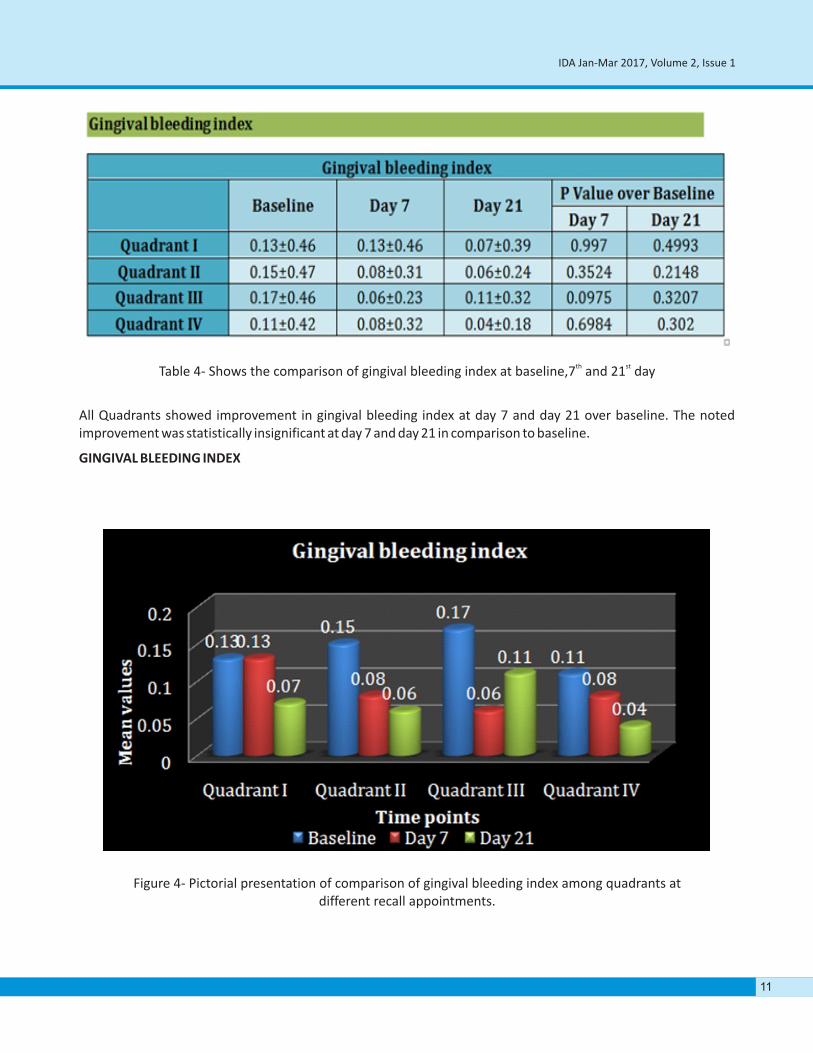
All Quadrants showed improvement in gingival bleeding index at day 7 and day 21 over baseline. The noted improvement was statistically insignificant at day 7 and day 21 in comparison to baseline.
GINGIVAL BLEEDING INDEX
th stTable 4- Shows the comparison of gingival bleeding index at baseline,7 and 21 day
Figure 4- Pictorial presentation of comparison of gingival bleeding index among quadrants at different recall appointments.
IDA Jan-Mar 2017, Volume 2, Issue 1

12
th stTable 5-Comparison of all the indices at baseline, 7 and 21 day.
Plaque index: There was early statistically significant improvement was observed in plaque index at day 7 over baseline. The improvement was further progressed and showed highly statistically significant improvement at day 21 over baseline. As per the mean score change, after 21 days of treatment, 65.83% of the population improved in plaque index over baseline.
GBI: Gingival bleeding index showed statistically significant improvement at day 7 and day 21 over baseline. The improvement was progressive with time. As per the mean score change, after 21 days of treatment, 82.09% of the population improved in gingival bleeding index over baseline.
LI: Lobene's index alsoshowed statistically significant improvement at day 7 and day 21 over baseline. The improvement was progressive with time. As per the mean score change, after 21 days of treatment, 87.69% of the population improved in lobene's index over baseline.
th st Figure 6- Pictorial presentation of comparison of all the indices at baseline, 7 and 21 day.
IDA Jan-Mar 2017, Volume 2, Issue 1
13
DISCUSSION
The primary objective of the study was to evaluate and compare the efficacy of different polishing aids (rubber cup, bristle brush and air polisher) after scaling and root planing.
The present study compared the clinical efficiency of air polisher and rubber cup and bristle brush in removing plaque and calculus, and also aimed to study their effects on gingival status in a split-mouth design. Also, all the treatment modalities showed a significant change in plaque accumulation and gingival status from baseline to 7 and 21 days post-op.
The design of the APP system uses a mixture of air, water, and sodium bicarbonate powder to deliver a controlled stream of sodium bicarbonate particles onto the tooth surface. This slurry of powder and water polishes the surface by removing deposits attached to it or smoothing its texture. The APP powder used in this study was the sodium bicarbonate powder, which is readily available, biocompatible, and is relatively soft and only mildly
[10]abrasive by De Spain.
In many study, it was shown that air-polishing devices became time-saving and effective in the application
[11,16]on normal enamel surface .However, it does not generally lead to surface modification and loss of
[16,17]materials to be able to be detected clinically . In contrast, spray may occur a significant amount of loss of material, if applied directly on root surface or dentin. As a rule, it is known that it should be certainly
[17]avoided to use these devices on dentin and cement . Tissue loss caused by the technique is depends on application time, powder and water application as much as the probe distance and the application
[17,18]surface . While we used air powder instrument in our study, the application was done by the same researcher from 1-1.5 cm by approaching at a right angle to the tooth surface. Likewise, the polishing application that was done by using rotary rubber cup was performed by the same researcher only by the weight of rotary instrument without extra pressure.
The one of the most commonly used polishing method is prophylaxis paste used with rotary rubber cup/brush. The abrasive properties of paste vary by content and size of paste. However, fine-grained paste can be more abrasive than a thick grained paste, because there is no standard in abrasiveness of paste among manufacturers.
In this study, it was studied that prophylaxis paste and air-flow powder were provided to be completely the same properties in order to be able to eliminate the effects of abrasive powder used in air-polishing techniques on the amount of abrasion. Therefore, the same paste and powder products having the same contents and produced by the same manufacturer were used for testing. In this way, it was evaluated if the application of the products having the same abrasive properties with the rotary instruments and aerator devices affected on surface roughness. According to the statistical analysis of data, it was determined that reduction observed in roughness values of prophylaxis paste group has been
[19]significant.
[20] A study was done by Patil in 2015 in which they compared the clinical efficiency of APP and RCP in removing supragingival plaque, and also aimed to study their effects on gingival status in a split-mouth design. The results indicated that when comparing the effectiveness of polishing treatments, there was no statistically significant difference in plaque removal or gingival status within the established time interval. Also, both the treatment modalities showed a significant change in plaque accumulation and gingival status from baseline, immediate post-op to 15 days post-op.
There was a significant reduction in plaque scores from baseline to immediate post-op in both the treatments (APP and RCP), but there was a considerable increase in GI and SBI scores from baseline to immediate post-op period. This finding could be attributed to the increase in gingival bleeding in the immediate post-op period as compared to the baseline scores, due to the therapy performed. At 15 days of follow-up, there was a substantial increase in plaque as compared to the immediate post-op findings, but there was a remarkable reduction in gingival and bleeding scores. This observation could not be attributed to either of the treatment modalities (APP or RCP), as this could entirely be credited to supra- and sub gingival scaling alone. But the improvement in the gingival status from immediate post-op period to 15 days postoperatively indicated that although both the polishing methods were traumatic, their effects on the soft tissues were temporary.Therfore this present study was not in accordance with the study done by Patil et al in 2015.
Almost all the patients in this clinical trial were
IDA Jan-Mar 2017, Volume 2, Issue 1

14
comfortable with all the treatment modalities and no patient suffered from any adverse effect of any sort. There was no complaint of any discomfort/sensitivity from any patient after APP or bristle brush or rubber cup at 7 and 21 days post-treatment. Overall, APP has proven to be causing less operator fatigue, compared to rubber cup and bristle brush.
CONCLUSION
Hence, it can be concluded that air polisher ( APP) device has shown better result compared to bristle brush and rubber cup. Further studies still need to be carried out to test its efficacy.
Financial support and sponsorship- Nil.
Conflicts of interest
There are no conflicts of interest
REFERENCES
1 Nature of plaque. Bowen W.H. Oral Sci Rev. 1976;9:3-21.
2. Intrinsic and extrinsic discoloration of the dentition. (A literature review) R. I. Vogel.Periodontology and Implant Dentistry Journal of Oral Medicine.1975, 4,99-104.
3. Tooth discoloration and staining Tooth discoloration and staining: a review of the literature. A Watts, Addy. British dental journal .2001,190(6):309-316.
4. Francis B, Barnes CM. Cosmetic and therapeutic polishing. Daniel SJ, Harfst SA, Wilder R, editors. Mosby's Dental Hygiene: Concepts, Cases and Competencies. Missouri: Elsevier; 2008. 599–622.
5. Rethman J. Polishing angles, cups and pastes. Pract Hyg. 1997; 1:32
6. Madan C, Bains R, Bains VK. Tooth polishing: Relevance in present day periodontal practice. Madan C, Bains R, Bains VK.. J Indian Soc Periodontol 2009;13:58-9.
7.Petersilka GJ, Ehmke B, Flemmig TF. Antimicrobial effects of mechanical debridement. Periodontol 2000. 2002;28:56–71
8.Boyde A. Air polishing effects on enamel, dentine, cement and bone. Br Dent J. 1984; 156:287–91
9.Trade news – New toothpaste. Br Dent J. 2003; 195:112.
10.De Spain B, Nobis R. Comparison of rubber cup polishing and air polishing on stain, plaque, calculus and gingival. Dent Hyg. 1988; 62-55.
11.Weaks LM, Lescher NB, Barnes CM, Holroyd SV (1984)Clinical evaluation of the Prophy-Jet as an instrument for routine removal of tooth stain and plaque. J Periodontol55: 486-488
12.Berkstein S, Reiff RL, McKinney JF, Killoy WJ (1987) Supragingival root surface removal during maintenance procedures utilizing an air-powder abrasive system or hand scaling. J Periodontol 58: 327-329.
13.Horning GM, Cobb CM, Killoy WJ (1987)Effect of an air-powder abrasive system on root surfaces in periodontal surgery. J ClinPeriodontol 14: 213-220.
14.Barnes CM, Russel CM, Gerbo LR, Wells BR, Barnes DW (1990) Effects of an air-powder polishing system on orthodontically bracketed and banded teeth. Am J of OrthodDento-facial Orthop 97: 74-81.
15.Kontturi-Närki V,Markkanen S, MarkkanenH (1990) Effects of air polishing on dental plaque removal and hard tissue as evaluated by scanning electron microscopy. J Periodontol 61: 334-338.
16.Mahledorff M (1989)Evaluation of the relationships between abrasion and surface alterations after professional cleaning. Mahledorff M.1989, 44: 203-204.
17.Petersilka GJ, Bell M, Häberlein I, Ehl A, Hickel R, et al. (2003) In vitro evaluation of novel low abrasive air polishing powders. J ClinPeriodontol 30: 9-13.
18. Jost-Brinkmann PG (1998)The influence of air polishers on tooth enamel. An in vitro study. J Orofacial Orthopedics59: 1-16.
19. * S Limaye Niraj P Chaudhari
A Comperative Evaluation of 3 Different Polishing Methods on Tooth Surface Roughness Fatma Karacaoglu, Neyran Yılmaz Tuzcel and Murat Akkaya J Biomedical Sci. 2016, 6:1. doi:10.4172/2254-609X.100046.20.A comparative evaluation of plaque-removing efficacy of air polishing and rubber-cup, bristle brush with paste polishing on oral hygiene status: A clinical study.,,, . Prevent Communit Dent 2015;5:457-62
Saurabh S Patil Purshottam S Rakhewar Priyanka
Corresponding author: trisha sircaremail: [email protected]
IDA Jan-Mar 2017, Volume 2, Issue 1

15
AUTHORS: 1 2 3 4 5Dr Roshan .P. Verghese , Dr Anjali .P , , Dr Ramamurthy T K , Dr Satheesha Reddy Dr. Chethan
A comparitive study of lipid profile on healthy smokers and non smokers
Introduction
Smoking is considered to cause heart disease, cancer, stroke and also have close relationship with gastric ulcer, periodontal disease, sudden infant death
(1-5)syndrome, and metabolic syndrome.
It has significant detrimental effect on various systems of body especially on cardiovascular system.
Smoking in different forms is a major risk factor for (6-9)
atherosclerosis and coronary heart disease.
Smoking cigarette or bidi leads to increase in concentration of serum total cholesterol, triglyceride, low density lipoprotein and very low density lipoprotein and fall in levels of anti-atherogenic high density lipoprotein as reported by various workers.
There is a dose response relationship between number of cigarettes/bidis smoked and cardio-vascular morbidity and mortality.
Thus lipid profile is a simple investigation which helps estimate future cardiovascular morbidity and mortality among smokers. The present study was conducted to demonstrate the effect of smoking on lipid profile and thus on cardiovascular system.
Aims and objectives-
To Study the effect of smoking on lipid profile of healthy smokers.
To compare the lipid profile of both smokers and non-smokers .
Materials and methods-
The present study was undertaken in the department of Oral medicine & Radiology.
The study was conducted on 10 healthy male smokers and 10 healthy non-smokers selected from volunteers and patients attending the hospital OPD.
Procedure for selection-
Inclusion Criteria:
Age: 25 – 45 years, Control: Who never smoked
Subjects: Who smoked at least once every week for a year or more and are non-symptomatic.
Exclusion Criteria:
Diabetes and endocrine Disorder
Hypertension, Renal Disorder, Coronary Artery Disease, History of Drug intake, History of Alcohol Intake/Drug abuse
Method of sample collection-
A detailed history was taken & subjects were explained about the study and written informed consent was taken.
Prior approval of Institute's Ethical Committee was taken.
Blood Sample was collected after overnight fasting under all aseptic precautions and sample was centrifuged at 2000rpm for one minute.
Lipid profile estimation which includes serum cholesterol, serum triglyceride, High density lipoprotein, low density lipoprotein and very low density lipoprotein was done.
Stastical analysis-
The statistical analysis was done by computer programs using microsoft excel & SPSS. 11.3
Results-
Data was collected in pre-designed validated Proforma and results were tabulated .
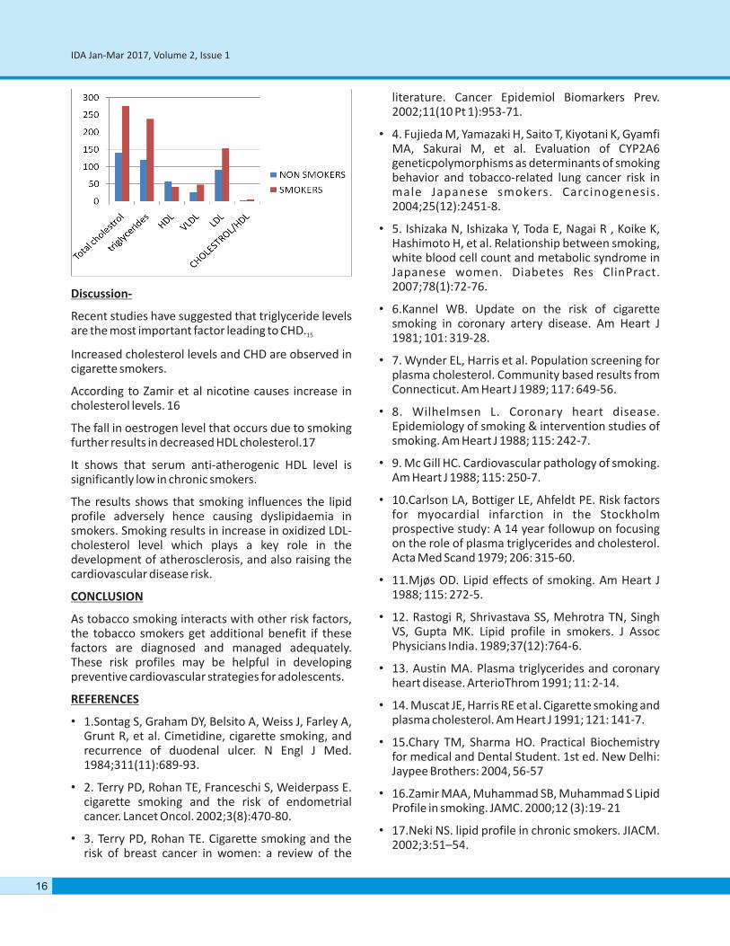
The total serum cholesterol, LDL, VLDL and Triglyceride values were higher in smokers as compared to Non-smokers.
Serum levels of HDL are lower in smokers than the same in non-smokers.
LIPID PROFILE IN SMOKERS & NON SMOKERS
Department of Oral Medicine and Radiology,A.E.C.S. Maaruti College of Dental Sciences and Research Centre.Bangalore, Karnataka
1. Dr Roshan .P. Verghese, Post Graduate Student2. Dr Anjali .P, Post Graduate Student3. Dr Satheesha Reddy, H.O.D. and Professor4. Dr Ramamurthy T K, Professor5. Dr. Chethan, Senior Lecturer
16
Discussion-
Recent studies have suggested that triglyceride levels are the most important factor leading to CHD.15
Increased cholesterol levels and CHD are observed in cigarette smokers.
According to Zamir et al nicotine causes increase in cholesterol levels. 16
The fall in oestrogen level that occurs due to smoking further results in decreased HDL cholesterol.17
It shows that serum anti-atherogenic HDL level is significantly low in chronic smokers.
The results shows that smoking influences the lipid profile adversely hence causing dyslipidaemia in smokers. Smoking results in increase in oxidized LDL-cholesterol level which plays a key role in the development of atherosclerosis, and also raising the cardiovascular disease risk.
CONCLUSION
As tobacco smoking interacts with other risk factors, the tobacco smokers get additional benefit if these factors are diagnosed and managed adequately. These risk profiles may be helpful in developing preventive cardiovascular strategies for adolescents.
REFERENCES
• 1.Sontag S, Graham DY, Belsito A, Weiss J, Farley A, Grunt R, et al. Cimetidine, cigarette smoking, and recurrence of duodenal ulcer. N Engl J Med. 1984;311(11):689-93.
• 2. Terry PD, Rohan TE, Franceschi S, Weiderpass E. cigarette smoking and the risk of endometrial cancer. Lancet Oncol. 2002;3(8):470-80.
• 3. Terry PD, Rohan TE. Cigarette smoking and the risk of breast cancer in women: a review of the
literature. Cancer Epidemiol Biomarkers Prev. 2002;11(10 Pt 1):953-71.
• 4. Fujieda M, Yamazaki H, Saito T, Kiyotani K, Gyamfi MA, Sakurai M, et al. Evaluation of CYP2A6 geneticpolymorphisms as determinants of smoking behavior and tobacco-related lung cancer risk in male Japanese smokers. Carcinogenesis. 2004;25(12):2451-8.
• 5. Ishizaka N, Ishizaka Y, Toda E, Nagai R , Koike K, Hashimoto H, et al. Relationship between smoking, white blood cell count and metabolic syndrome in Japanese women. Diabetes Res ClinPract. 2007;78(1):72-76.
• 6.Kannel WB. Update on the risk of cigarette smoking in coronary artery disease. Am Heart J 1981; 101: 319-28.
• 7. Wynder EL, Harris et al. Population screening for plasma cholesterol. Community based results from Connecticut. Am Heart J 1989; 117: 649-56.
• 8. Wilhelmsen L. Coronary heart disease. Epidemiology of smoking & intervention studies of smoking. Am Heart J 1988; 115: 242-7.
• 9. Mc Gill HC. Cardiovascular pathology of smoking. Am Heart J 1988; 115: 250-7.
• 10.Carlson LA, Bottiger LE, Ahfeldt PE. Risk factors for myocardial infarction in the Stockholm prospective study: A 14 year followup on focusing on the role of plasma triglycerides and cholesterol. Acta Med Scand 1979; 206: 315-60.
• 11.Mjøs OD. Lipid effects of smoking. Am Heart J 1988; 115: 272-5.
• 12. Rastogi R, Shrivastava SS, Mehrotra TN, Singh VS, Gupta MK. Lipid profile in smokers. J Assoc Physicians India. 1989;37(12):764-6.
• 13. Austin MA. Plasma triglycerides and coronary heart disease. ArterioThrom 1991; 11: 2-14.
• 14. Muscat JE, Harris RE et al. Cigarette smoking and plasma cholesterol. Am Heart J 1991; 121: 141-7.
• 15.Chary TM, Sharma HO. Practical Biochemistry for medical and Dental Student. 1st ed. New Delhi: Jaypee Brothers: 2004, 56-57
• 16.Zamir MAA, Muhammad SB, Muhammad S Lipid Profile in smoking. JAMC. 2000;12 (3):19- 21
• 17.Neki NS. lipid profile in chronic smokers. JIACM. 2002;3:51–54.
IDA Jan-Mar 2017, Volume 2, Issue 1

17
Pills of the future: Nanoparticles in Oral Medicine
1 1 2AUTHORS: Dr. N. Janani , Dr. Chethan .R , Dr. Satheesha Reddy B.H
ABSTRACT:Nanoparticles are defined as particles with a diameter smaller than 100 nm and are increasingly used in different applications. Nanoparticles research is currently the most studied branch of science with the number of uses of nanoparticles in various fields. The particles have wide variety of potential applications in biomedical, optical and electronic fields. Michael Faraday was the first to provide the description in scientific terms, of the optical properties of nanometer-scale metals in his 1857 paper.Keywords: Nanoparticles, cancer, stem cell therapy, applications
INTRODUCTION:
Nanoparticles are defined as particles with a diameter smaller than 100 nm, are increasingly used in different applications, including drug carrier systems and to pass organ barriers such as
[1]the blood-brain barrier.
It is classified according to size:
- In terms of diameter
a. Fine particles cover a range between 100 and 2500 nanometers
b. Ultrafine Particles are sized between 1 and 100 nanometers
Nanoparticles may or may not exhibit size-related properties that differ significantly from those
[4]observed in fine particles or bulk materials.
Nanoparticle research is currently an area of intense scientific interest due to a wide variety of potential applications in biomedical, optical and electronic
[4]fields.
Nanoparticles in general terms are defined as engineered structures with diameters of <100nm, are devices and systems produced by chemical and/or physical processes having specific properties. The reason why nanoparticles are attractive for such purposes is based on their important and unique features, such as their surface to mass ratio, which is much larger than that of other particles and materials, allowing for catalytic promotion of reactions, as well as their ability to adsorb and carry other compounds.
TYPES OF NANOPARTICLES:
a. Nanosphere b. Nanocapsulec. Dendrimer d. Polymeric micellese. Liposome f. SLN (Solid lipid Nanoparticles)
Types of Nanoparticles
1. Dr. N. Janani1. Dr. Chethan .R 2. Dr. Satheesha Reddy B.H
Department of Oral Medicine and Radiology,A.E.C.S. Maaruti College of Dental Sciences and Research Centre.Bangalore, Karnataka

18
APPLICATIONS OF NANOPARTICLES:
1. Targeted Drug Delivery2. Gold Nanoparticles detect cancer3. Nanoparticles target ovarian cancer4. Stem cell therapy5. To extend shelf life in containers6. In anthrax vaccine to produce immunity
APPLICATIONS:
1. TARGETED DRUG DELIVERY :
It is the accurate targeting of the drug to cells or tissues of choice. Today's delivery technologies are far away from the design of the so called “magic bullet”, proposed by Paul Ehrlich at the beginning
thof the 20 century, in which the drug is precisely
[1]targeted to the exact side of action.
Targeting is the ability to direct the drug-loaded system to the site of interest. Two major mechanisms can be distinguished for addressing the desired sites for drug release:
(i) Passive and
(ii)Active targeting
2 most important aspects of nanoparticle drug delivery must be:-
a. The specific targeting of the diseased tissue with
nanoparticles
b. The timed release of the drug
Advantages of using Nanoparticles in Drug Discovery:-
1. Particle size and surface characteristics of nanoparticles can be easily manipulated to achieve both passive and active drug targeting after parenteral administration
2. They control and sustain release of the drug during the transportation and at the site of localization, altering organ distribution of the drug so as to achieve increase in drug therapeutic efficacy and reduction in side effects
3. Site-specific targeting can be achieved by attaching targeting ligands to surface of particles or use of magnetic guidance
4. The system can be used for various routes of administration including oral, nasal, parenteral,
[2]intra-ocular etc
5. Nanoparticles can better deliver drugs to tiny areas within the body
6. Engineering on this scale enables researchers to exercise exquisite and previously unthinkable control over the physical attributes of polymers and other biomaterials
7. Nanoparticles overcome the resistance offered by the physiological barriers in the body because efficient delivery of drug to various parts of the body is directly affected by particle size
8. Nanoparticles aid in efficient drug delivery to improve aqueous solubility of poorly soluble drugs that enhance bioavailability for timed release of drug molecules, and precise drug targeting
9. The surface properties of nanoparticles can be modified for targeted drug delivery
10. Targeted Nano drug carriers reduce drug toxicity and provide more efficient drug distribution
11. Nanocarriers holds promise to deliver biotech drugs over various anatomic extremities of body
[3]such as blood brain barrier
2. GOLD NANOPARTICLES DETECT CANCER:
Gold nanoparticles have been used as
ultrasensitive fluorescent probes to detect cancer
biomarkers in human blood. The approach is so
sensitive that it outstrips current methods by
several orders of magnitude and could also be
IDA Jan-Mar 2017, Volume 2, Issue 1

19
employed in direct detection of viral or bacterial
DNA.
Gold nanoparticles are promising probes for
biomedical applications because they can be
easily prepared and, unlike other fluorescent
probes such as quantum dots or organic dyes,
don't burn out after long exposure to light. In a
newer study, the application of nanoparticles to
detect carcinoembryonic antigen (CEA) and alpha
foetal protein (AFP)-2 biomarkers in the diagnosis
of various cancers, including liver, lung and breast [1]
cancer have been tried.
3. NANOPARTICLES TARGET OVARIAN CANCER:
Tiny particles carrying a killer gene can effectively
suppress ovarian tumor growth in mice, according
to a team of researchers from MIT and the
Lankenau Institute, ovarian cancer is one of the
most deadly forms of the disease as it is usually
diagnosed at a relatively late stage.
The treatment delivers a gene that produces the
diphtheria toxin, which kills cells by disrupting
their ability to manufacture proteins. The toxin is
normal ly produced by the bacter ium
Corynebacterium diphtheria.
To further ensure tumor-focused effects, the
nanoparticles were administered by injection into
the peritoneal cavity, which encases abdominal
organs such as the stomach, liver, spleen, ovaries
4. STEM CELL THERAPY:
Nanoparticles prove effective tools for improving stem cell therapy. It has been successfully used to enhance stem cell's ability to stimulate regeneration of damaged vascular tissue and reduce muscle degeneration in mice.
Researchers also suggested that after implantation into a living organism, cells may not continue to renew tissue effectively enough to keep the tissue alive long-term. The cells can therefore benefit from help with performance-enhancing genes,
[1]which promote growth in the target tissue.
and uterus. The new nanoparticles are made with
positively charged, biodegradable polymers
known as poly (beta-amino esters). In addition,
these nanoparticles have demonstrated potential
for treatment of a variety of diseases, including [1]prostate cancer and viral infection.
IDA Jan-Mar 2017, Volume 2, Issue 1

20
5. NANOPARTICLES IN CONTAINERS TO EXTEND SHELF LIFE:
The use of silver nanoparticles in plastic containers to keep foods fresher longer, pointed the way forward for processors looking to incorporate the technology into their packaging.
In the food-packaging arena, nanomaterials are being developed with enhanced mechanical and thermal properties to ensure better protection of foods from exterior mechanical, thermal, chemical or
[1]microbiological effects.
6. USE OF NANOPARTICLES IN ANTHRAX VACCINE TO PRODUCE IMMUNITY:
The use of nanoparticles in a vaccine against anthrax proved more effective and easier to administer in tests in mice and guinea pigs. It was able to trigger a strong immune response by treating the inside of the animals noses with a “nanoemulsion” a suspension of water, soybean oil, alcohol and surfactant emulsified to create droplets of only 200 to 300 nanometers in
[1]size.
Besides eliminating the need for needles, the nanoemulsion anthrax vaccine is easy to store and use in places where refrigeration is not available, also a nasal nanoemulsion based anthrax vaccine if proved effective in humans, could be given easily to people even after they are exposed in an anthrax attack,
along with antibiotics, as with some diseases, where vaccines given after exposure boost the speed of the immune response
CONCLUSION:
The application of nanoparticles in cancer diagnosis and treatment promises to have a profound impact on oral health care. Many of the technologies involving nanoparticles are in preclinical stages but have the potential to replace highly invasive conventional cancer detection and treatment, which include biopsies, irradiation and painful therapies.
The ability to diagnose malignant disease at the earliest also allows treatment options to be planned early thereby directly affecting the morbidity and mortality of head and neck cancer.
REFERENCES:
1. Potential applications of nanoparticles. International Journal of Pharma and BioSciencesV1(1)2010
2. Mohsen Jahanshahi and Zahra Babaei. (2008) Protein nanoparticle: A Unique system as drug delivery vehicles. African Journal of Biotechnology. 7(25):4926-4934
3. Manju Rawat, Deependra Singh, S.Saraf, and Swarnlata Saraf. (2006) Nanocarriers: Promising drug for Bioactive Drugs. Biol.Pharm.Bull.29 (9):1790-1798.
4. Ananya Mandal (2014) What are Nanoparticles?
5. James Chen Yong Kah et al. Early diagnosis of oral cancer based on the surface Plasmon resonance of gold nanoparticles. International journal of nanomedicine 2007:2(4) 785-798.
IDA Jan-Mar 2017, Volume 2, Issue 1

21
Angular Cheilitis - a Review
1 2AUTHORS: Torsha Ray , Dr. Satheesha Reddy .B.H
1. Post Graduate Student, Department of Oral Medicine and Radiology,A.E.C.S. Maaruti College of Dental Sciences and Research Centre.Bangalore, Karnataka
Torsha Ray 2. H.O.D. and professor, A.E.C.S. Maaruti College of Dental Sciences and Research Centre.Bangalore, Karnataka
Dr. Satheesha Reddy .B.HDepartment of Oral Medicine and Radiology,
ABSTRACT : Angular cheilitis is defined as symptomatic bilateral fissures of the corner of the mouth that are
common in patients with candida albicans infection in other parts of the mouth; characterized by cracking
,crusting and in severe cases bleeding. It is a multifactorial disease associated with several predisposing factors.
Several drugs, certain systemic disorders and also nutritional deficiencies can cause angular cheilitis. Candida
species can be detected in 93% of lesions; also staphylococcus aureus; ß-hemolytic streptococci or a combination
of the above species can cause angular cheilitis. Diagnosis is usually based on clinical finding of erythematous
fissures. Treatment modalities includes initial evaluation followed by prescribing a topical ointment or cream
(combination of antifungal and antibacterial).Follow-up is usually recommended after two weeks. Usually if
condition does not resolve, prescribing an appropriate systemic antifungal is considered. If further systemic issues
are suspected,patient is referred to their primary care physician for additional and management.
SYNONYMS: Rhagades, perleche, cheilosis,
angularcheilosis, commissuralcheilitis,angular 7stomatitis.
INTRODUCTION : Angular cheilitis is a multifactorial
disease affecting the commissure of the lips and is
commonly seen in denture wearers.A clinical
diagnosis of angular cheilitis is arrived at when other
specific lesions of the lip such as recurrent herpes
labialis, ulceration due to trauma ,environmental 1
exposure or syphilis are ruled out. This common
condition has a prevalence of 7 per 1000 and is most
often seen in older age group. The incidence is
increased about three fold in denture wearers and
two fold in men.Of HIV infected patients 10% may
have an opportunistic infection with candida albicans
,suspicious of angular cheilitis.Angularcheilitiswas
detected in 7.8% of patients with crohn's disease and 2
5% with ulcerative colitis.
DEFINITION :” Symptomatic bilateral fissures of the
corners of the mouth that are common in patients
with candida albicans infection in other parts of the
mouth and often intensified with mouth over closure 3;requires treatment with antifungal medication.”

20
CAUSATIVE AGENTS: The involved organisms are :
· Candida species alone(usually candida 4albicans)which accounts for about 20% of cases.
· Bacterial species :
- Staphyloccusaureus alone (acconts for 20% of cases).
- ß-hemolytic streptococci alone (detected in 8-15% of angular cheilitis, but less commonly
5present in isolation) .
- Or acombination of the above organisms(a polymicrobial infection)with about 60% of case
4,6involving both C. albicans and S.aureus.
Candida can be detected in 93% of angular cheilitislesions. This organism is found in the mouths of about 40% of healthy individuals, and it is considered by some to be normal commensal component of the oral microbiota. Potassium hydroxide preparation is recommended by some to help distinguish between the harmless and the pathogenic forms,and thereby highlight which cases
7of angular cheilitis are truly caused by candida.
CAUSE : Angular cheilitis is a multifactorial disease affecting the commissure of the lips,commonly seen in denture wearers .A clinical diagnosis of angular cheilitis is arrived at when other specific lesions of the lip such as recurrent herpes labialis, ulceration due to trauma, environmental exposure or syphilis are ruled out.This common condition of the lip has been associated with several predisposing factors .These factors includes infection, nutritional deficiencies and reduced vertical dimension of the mouth as seen in old age and in long term denture wearers. A majority of infections are candida associated, with nearly 20% of the cases arising due to candida albicans alone,60% due to a combined infection with candida albicans and 20% due to staphyloccusalone. Further,in nearly 80% of the cases of angular cheilitis, there is a co-existent
1 denture stomatitis. The condition is characterized by cracking,crusting and in severe cases bleeding. Probably, the most common cause is the recession of the bony support of the lower aspects of the mouth. This can result in an overbite with the upper lip protruding the lower. This situation can then be further aggravated by dentition in less than stellar condition,or dentures that have not been adjusted in
sometime.A set-up for the problem may even have been initiated by thumb sucking that continued long
8after the toddler years. In the past this lesion was considered as a sign of vitamin B deficiency ,treatment efforts were often erroneously directed towards
3correcting only that condition. Ultimately,mechanical trauma to the area is likely to be the primary culprit,but less common etiologies in practice better known by physicians in training are nutritional deficiency,particularly of riboflavin,iron,cobalamin or zinc.Nutritional deficiencies such as iron deficiency anaemia,vitamin B or folic acid deficiency have been strongly implicated in angular cheilitis,referred to as perleche.Ariboflavinosis induces circumoral lesions which are prone to become infected and when this happens,the lesions are indistinguishable from
1perleche of other causes. These deficiencies are often cited whether due to malabsorption from diseases such as celiac or malnutrition due to anorexia nervosa
9,as well as bulimia nervosa. Patients suffering from diabetes ,chronic renal failure, hepatitis, sjogren's syndrome, plummervinson or crohn's disease can
10-12present with angular cheilitis. Medications have also been shown to cause angular cheilitis. Antineoplastic agents such as sorafenile and selumetinile can cause cheilitis in patients being t reated with them for var ious types of
13,14malignancies. The condition is uncommonly caused or exacerbated by oral candidiasis or secondary
1 5bi lateral infect ions. In pat ients who are immunocompromised ,or have diabetes, malignancy or anaemia ,the likelihood of infection is
16increased. Immunodeficiency status such as human immunodeficiency virus infection are a few among a plethora of factors that predispose to the
17lesion. Angular cheilitis has been included in the classification and diagnostic criteria for oral lesions in
18HIV infection. Although angular cheilitis may not be very frequently observed during HIV disease, it is
19somewhat strongly associated.
NUTRITIONAL DEFICIENCIES: Several different nutritional deficiency status of vitamins or minerals have been linked to angular cheilitis. It is thought in about 25% of people with angular cheilitis, iron deficiency or deficiency of B vitamins are involved. Chronic iron deficiency may also cause koilonychia and glosssitis. It is not completely understood how iron deficiency causes angular cheilitis but it is known
IDA Jan-Mar 2017, Volume 2, Issue 1

21
that it causes a degree of immunocompromise which may in turn allow an opportunistic infection of candida.Vitamin B² deficiency (ariboflavinosis)may also cause angular cheilitis and other conditions such as redness of mucous membranes, magenta
5coloredglossitis. Vitamin B deficiency may also cause angular cheilitis along with glossitis, skin changes similar to seborrheic dermatitis around eyes, nose, mouth. Vitamin B12 deficiency is sometimes responsible for angular cheilitis and commonly occurs together with folate deficiency which causes glossitis and megaloblasticanaemia. Vitamin B3 deficiency is another possible cause in association with conditions such as dermatitis, diarrhoea, dementia and glossitis. Biotin (vitamin B 7) deficiency has also been reported to cause angular cheilitis along with alopecia and dry
20eyes. Zinc deficiency is also known to cause angular 2 1
cheilitis. Acrodermatitis enteropathicais an autosomal recessive genetic disorder causing impaired absorption of zinc and is associated with angular cheilitis. Ingeneral, these nutritional disorders may be caused by malnutrition such as may occur in alcoholism or in strict vegan diets or by malabsorption secondary to gastrointestinal disorders or surgeries and in turn lead to angular
20cheilitis.
SYSTEMIC DISORDERS : Some systemic disorders are involved in angular cheilitis by virtue of their association with malabsorption and the creation of nutritional deficiencies. Such examples include people with a anorexia nervosa. Other disorders may include lip enlargement (orofacialgranulomatosis) which alters the local anatomy and extenuates the skin fold at the corners of the mouth.More still may be involved because they effect the immune system, allowing normally harmless organisms like candida to become pathogenic and cause an infection. Xerostomia itself may have possible causes, but commonly the cause may be side effects of medications or conditions such as sjogren' ssyndrome. Xerostomia accounts for 5% of the cases of angular cheilitis. Conversely conditions which drooling or sialorrhea (excessive salivation)can cause angular cheilitis by creating a constant wet environment in the corners of the mouth. About 25% of people with downs syndrome appear to have
20angular cheilitis. This is due to relative macroglossia, an apparently large tongue in small mouth ,which may
constantly stick out of the mouth causing maceration of the corner of the mouth with saliva. Inflammatory bowel diseases (Crohn's disease or ulcerative coloitis)
22can be associated with angular cheilitis. In crohn's disease,it is likely the result of malabsorption and immunosuppressive therapy which gives rise to sores
23at corners of the mouth. Glucagonomas are rare pancreatic endocrine tumors, which secrete glucagon,and cause a syndrome of dermatitis, glucoseintolerance, weight loss and anaemia. Angularcheilitis is a common feature of glucagonoma
24 syndrome. Infrequently,angular cheilitis may be one of the manifestations of chronic mucocutaneous
4candidiasis and sometimes cases oropharyngeal and oesophageal candidiasis may accompany angular
7cheilitis. Angularcheilitis may be present in HIV 25 26 22
infection ,neutropenia or diabetes. Angular cheilitis is more common in people with eczema
7because their skin is more sensitive to irritants. Other conditions possibly associated include plasma cell
2 7gingivitis ,Melkersson Rosenthal syndrome, orsideropenic dysphagia (plummer–vinson syndrome
20or Paterson-brown-kelly syndrome).
DRUGS:Several drugs may cause angular cheilitis asa side effect, by various mechanisms, such as creating drug induced xerostomia. Various examples include
20isotretinoin, indinavir and sorafenile. Isotretinoin (Accutane),an analogue of vitamin A,is a medication which dries the skin. Lesscommonly, angularcheilitis is associated with primary hypervitaminosisA, which can occur when large amounts of liver oil(cod liver oils and other fish oils)are regularly consumed or as a result from man excess intake of vitamin A in the form of vitamin supplements. Recreationaldrug users may also develop angular cheilitis. Examples include c o c a i n e , m e t h a m p h e ta m i n e s , h e ro i n a n d
20hallucinogens.
CLINICAL FEATURES :
1· Occurs in both young children and adults.
· Population:
- Individuals with increased folding or wrinkling of skin at the corners of the mouth.
- Individuals wearing dentures with decreased vertical dimension of occlusion.
IDA Jan-Mar 2017, Volume 2, Issue 1

Ø Saliva pools in the fissures, creating a chronic moist environment for infection with candida a lb icans , staphy loccusaureus and /or streptococcus(rare).
Ø Individuals with underlying systemic conditions including endocrine disorders(eg. Diabetes), i m m u n o l o g i c a l d i s o r d e r s ( e g . H I V infection), nutritional deficiencies, hematologic malignancies or solid organ malignancies.
20
SIGNS:Red,scaly fissured lesions at the angles of the mouth,usually bilateral.
SYMPTOMS: Pain severity can range from asymptomatic to severe discomfort, burning,
28irritation,pruritis. Although,the lesions may occur alone, they are often associated with intraoral acute pseudomembranous or atrophic lesions in other parts of the mouth.The condition is uncommonly caused or exacerbated by oral candidiasis or secondary bacterial
3infections. Characterized symptomatically by a feeling of dryness and a burning sensation at the corners of the mouth. Clinically, the skin at the commisures appear wrinkled and somewhat macerated. Intime, the wrinkling becomes more pronounced to form one or more deep fissures or cracks which appear ulcerated, but which do not tend to bleed ,although a superficial exudate crust may form. These fissures do not involve the mucosal surface of the commissure inside the mouth,but stop at the mucocutaneous junction.The severity of the lesions waxes and wanes. If untreated, they often show a tendency for spontaneous remission. Subsequently exacerbation is common, however only rarely do the lesions completely disappears. 'Cheilocandidiasis' and 'juvenile juxtavermillion candidiasis' refer to more extensive and often desquamative lesions affecting the full width of the
lip, extending into adjacent lesions. They are associated with habitual lip sucking, prolonged dental appointments, sunlight and chronic candidal
1infection.
CLASSIFICATION:
· Angular cheilitis could be considered as a type of cheilitis or stomatitis.
· Where candida species are involved, angular cheilitis is classed as a type of oral candidiasis, specifically a primary (Group –I)candida
25 associated lesion. This form of angular cheilitis which is caused by candida is sometimes termed “Candida associate angular cheilitis” or less
7commonly “Monilialperleche”.
· Angular cheilitis can also be classified as acute(sudden, short lived appearance of the condition) or chronic (lasts a long time or keeps returning)or refractory(condition persists despite
7attempts to treat it).
INVESTIGATIONS:
1. If the patient wears dentures:
v Are the dentures stable and comfortable?
v Is the vertical dimension of occlusion appropriate?
[poorly fitting dentures could cause the wrinkling that creates a favourable environment for the condition to manifest]
2. Does the patient have a tendency to lick their lips/corners of the mouth?
3. Have there been any recent changes in saliva quantity /quality?
IDA Jan-Mar 2017, Volume 2, Issue 1

4. Does the patient report oral burning or taste alterations , which may be indicative of a generalized oral fungal infection? If present, it could be a source of re-infection.
5. Ask the patient about recently prescribed medications(including antibiotics) that may cause this condition.
6. Obtain a thorough dental and medical history:
v Recognize responses to review of systems that raise suspicion of underlying systemic disorders.
v Determine the stability of current medical conditions.
v Tobacco use may predispose to the development of this condition.
7. If a more thorough investigation is warranted, refer for cytology culture or laboratory assessment to rule out local or systemic
28predisposing factors.
DIAGNOSIS: Based on clinical finding of erythematous fissures at the angles of the mouth,a diagnosis of
28angular cheilitis is determined.
21
DIFFERENTIAL DIAGNOSIS :
· Contact dermatitis
28· Actinic cheilitis
TREATMENT :
Common initial treatments:
1.Initial evaluation of pre-disposing factors:
üEvaluation of prosthesis.
üMaintenance of oral hygiene.
üMaintenance of prosthesis hygiene.
üLocal salivary gland issues.
üIntraoral fungal infection.
2.Prescribe a topical ointment or cream:
üUsually a combination of topical antifungal and antibacterial (eg. Nystatin and mupirocin); consider the use of combination.
üAntifungal/antibacterial? glucocorticosteroid ointment (eg. Viaderm-KC ointment) as an alternative.
IDA Jan-Mar 2017, Volume 2, Issue 1

20
FOLLOW-UP:
Follow – up is recommended at two weeks.
¨ If the condition is resolved, continue monitoring.
¨ If the condition is not resolved, consider prescribing an appropriate systemic antifungal.
¨ If systemic issues are suspected as a cause, the patient should be referred to their primary care physician for additional evaluation and or
28management.
CONCLUSION :
Angular cheilitis remains a chronic problem and is
usually mechanical. While there are esoteric causes to
be considered, more mundane etiologies are more
likely. Identifying the underlying etiology is useful and
allows a more appropriate therapeutic approach in 8order to resolve the pathology.
REFERRENCES :
1. Rajendran R. Shafer's textbook of oral pathology.
Elsevier India; 2009.
2. Lisciandrano D, Sardella A, Campanini MC, Velio P,
Bianchi PA, Ranzi T, Carrassi A. Prevalence of oral
lesions in inflammatory bowel disease. American
Journal of Gastroenterology. 1996 Jan 1;91(1).
3. Sapp JP, Eversole LR, Wysocki G. Contemporary
oral and maxillofacial pathology. Implant
Dentistry. 1997 Oct 1;6(3):238-9.
4. Chelitis A. ANGULAR CHEILITIS: CASE REPORTS
AND LITERATURE REVIEW. Pakistan Oral & Dental
Journal. 2014 Dec;34(4).
5. Dacre IT, Kempson S, Dixon PM. Pathological
studies of cheek teeth apical infections in the
horse. 1: Normal endodontic anatomy and
dentinal structure of equine cheek teeth. The
Veterinary Journal. 2008 Dec 31;178(3):311-20.
6. Kerawala C, Newlands C (editors) (2010).Oral and
maxillofacial surgery. Oxford: Oxford University
Press. pp. 446, 447.ISBN9780199204830.
7. Park KK, Brodell RT, Helms SE. Angular cheilitis,
part 1: Local etiologies. Cutis. 2011 Jun;
87(6):289-95.
8. Angular cheilitis-A maligned condition,Volume
11,Issue 3,pp 198-200;July-aug 2013-Caren
Campbell, Lawrence Charles Parish.
9. Strumia R. Dermatologic signs in patients with
eating disorders. Am J Clin Dermatol.2005;6:165-
173.
10. UdayakumarP, BalasubramanianS, Ramalingam
KS, etal. Cutaneous manifestations in patients
with chronic renal failue on hemodialysis. Indian J
Dermatol Venerol Leprol.2006;72:119-125.
11. Soy M, Piskin S. Cutaneous findings in patients
with primary Sjogren's syndrome. Clinical
rheumatology. 2007 Aug 1;26(8):1350-2.
IDA Jan-Mar 2017, Volume 2, Issue 1
12. Novacek G. Plummer-Vinson syndrome.
Orphanet Journal of rare diseases. 2006 Sep
15;1(1):36.
13. Yang C-H,Lin W-C,Chuang C-k,et-al.Hand-foot skin
reaction in patients treated withsorafenib:
aclinicopathological study of cutaneous
manifestations due to multitargeted kinase
inhibitor therapy.Br.J.Dermatol .2008;158:592-
596.
14. BalagulaY, Barth Huston K, BusanKJ, et al.
Dermatologic side effects associated with the
MEK 1/2 inhibitor selumitinib ((AZD62,ARRY-
142886). Invest New Drugs. 2010.Available at
:https://www.ncbi.nlm.nih.gov/pubmed/209789
26 (accessed Apr-2017).
15. Sharon V,FazelN.Oral Candidiasis and angular
cheilitis. DermatolTher. 2010;23:230-242.
16. Rogers RS 3rd,BekicM.Diseases of tgelips.
SeminCutan Med surg.1997;16:328-336.
17. SamaranayakeLP, CheungKL, SamaranayakeYH.
Candidiasis and other fungal diseases of the
mouth.Dermatol Ther.2002;15:251-69.
18. Williams DM. Classification and diagnostic criteria
for oral lesions in HIV infection-EC clearing house
and WHO collaborating center in oral
manifestations of human immunodeficiency
virus(HIV) infection.Copenhagen Denmark. J Oral
Pathol Med.1993;22:289-91[Pubmed].
19. PindborgJJ.Classification of oral lesions
associated with HIV infection. OralSurg Oral Med
Oral Pathol 1989;67:292-5[Pubmed].
20. Park KK;BrodellRT;Helms SE(july 2011)."Angular
Cheilitis,Part 2:nutritional,systemic,and drug
related causes and treatment"(PDF).Cutis
.88(1):27-32.
21. GareauD,PietteF,CortotA,DumurV,Bergoend
H(1987)."[Cutaneous manifestations of zinc
d e f i c i e n c y i n e t hy l i c c i r r h o s i s ] " . A n n
DermatolVenerol 11(1)39-53.
22. Scully, crispian(2008). Oral and maxillifacial
medic ine: the basis of d iagnosis and ndtreatment(2 edition). Edinburgh: Churchill
Livingstone.pp 147-149.
23. Triester NS, Bruch JM (2010).Clinical oral
medicine and pathology. NewYork: Humana
Press.pp 92,93,144.
24. TadatakaYamada; DavidAlpers; AnthonyKalloo;
NeilKaplowitz; Chung Owyang; DonPowell, th
eds(2009). Textbook of gastroenterology (5
edition) Chichester, WestSussex: Blackwell
Pub.pp.1882-1883.
25. TyldesleyWR,FieldALongman L(2003). Tyldesley's thoral medicine (5 edition) Oxford : Oxford
University press. pp.37,40,46,63-67.
26. GreenbergMS,Glick M(2003).Burket's oral t h
medicine diagnosis and treatment(10
edition).Hamilton,Ont:BC Decker.pp.97,98,550.
27. Wood NK, Goaz PW. Differential diagnosis of oral
lesions. Mosby Elsevier Health Science; 1985.
28. Stoopler ET, Nadeau C, Sollecito TP. How do I
manage a patient with angular cheilitis?. Journal
(Canadian Dental Association). 2013;79:d68.
IDA Jan-Mar 2017, Volume 2, Issue 1