J. Fac. Fish. Anirn. Husb., Hiroshima Univ. (1975), 14 ... · J. Fac. Fish. Anirn. Husb., Hiroshima...
12
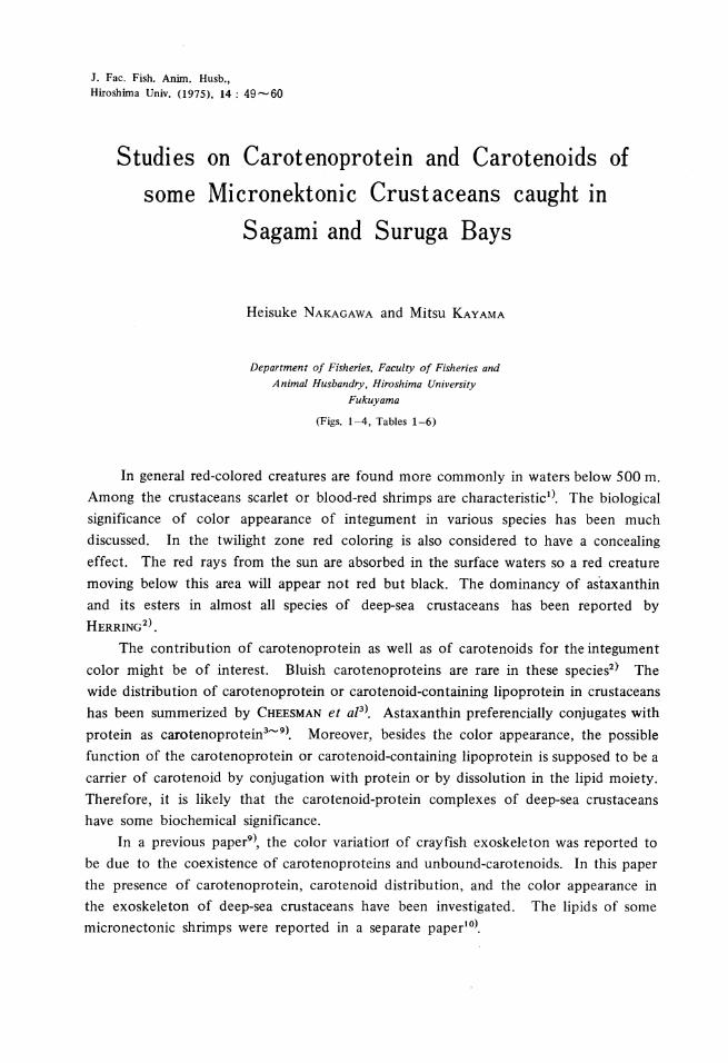
J. Fac. Fish. Anirn. Husb., Hiroshima Univ. (1975), 14 : 49-60 Studies on Carotenoprotein and Carotenoids of some Micronektonic Crustaceans caught S agami and S uruga Bays Heisuke NAKAGAWA and Mitsu KAYAMA Department of Fisheries. Faculty of Fisheries and Animal Husbandry, Hiroshima University Fukuyama (Figs. l-4, Tables 1-6) . ID In general red-colored creatures are found more commonly in waters below 500 m. Among the crustaceans scarlet or blood-red shrimps are characteristic 1 >. The biological significance of calor appearance of integument in various species has been much discussed. In the twilight zone red coloring is also considered to have a concealing effect. The red rays from the sun are absorbed in the surface waters so a red creature moving below this area will appear not red but black. The dominancy of astaxanthin and its esters in almost all species of deep-sea crustaceans has been reported by HERRING 2 ). The contribution of carotenoprotein as well as of carotenoids for the integument calor might be of interest. Bluish carotenoproteins are rare in these species 2 > The wide distribution of carotenoprotein or carotenoid-containing lipoprotein in crustaceans has been summerized by CHEESMAN et al 3 >. Astaxanthin preferencially conjugates with protein as carotenoprotein 3 - 9 >. Moreover, besides the calor appearance, the possible function of the carotenoprotein or carotenoid-containing lipoprotein is supposed to be a carrier of carotenoid by conjugation with protein or by dissolution in the lipid moiety. Therefore, it is likely that the carotenoid-protein complexes of deep-sea crustaceans have some biochemical significance. In a previous paper 9 >, the calor variation of crayfish exoskeleton was reported to be due to the coexistence of carotenoproteins and unbound-carotenoids. In this paper the presence of carotenoprotein, carotenoid distribution, and the calor appearance in the exoskeleton of deep-sea crustaceans have been investigated. The lipids of some micronectonic shrimps were reported in a separate paper 10 >.
Transcript of J. Fac. Fish. Anirn. Husb., Hiroshima Univ. (1975), 14 ... · J. Fac. Fish. Anirn. Husb., Hiroshima...
J. Fac. Fish. Anirn. Husb., Hiroshima Univ. (1975), 14 : 49-60
Studies on Carotenoprotein and Carotenoids of some Micronektonic Crustaceans caught
S agami and S uruga Bays
Heisuke NAKAGAWA and Mitsu KAYAMA
Department of Fisheries. Faculty of Fisheries and Animal Husbandry, Hiroshima University
Fukuyama
(Figs. l-4, Tables 1-6)
. ID
In general red-colored creatures are found more commonly in waters below 500 m. Among the crustaceans scarlet or blood-red shrimps are characteristic1>. The biological significance of calor appearance of integument in various species has been much discussed. In the twilight zone red coloring is also considered to have a concealing effect. The red rays from the sun are absorbed in the surface waters so a red creature moving below this area will appear not red but black. The dominancy of astaxanthin and its esters in almost all species of deep-sea crustaceans has been reported by HERRING2).
The contribution of carotenoprotein as well as of carotenoids for the integument calor might be of interest. Bluish carotenoproteins are rare in these species2> The wide distribution of carotenoprotein or carotenoid-containing lipoprotein in crustaceans has been summerized by CHEESMAN et al3>. Astaxanthin preferencially conjugates with
protein as carotenoprotein3- 9>. Moreover, besides the calor appearance, the possible function of the carotenoprotein or carotenoid-containing lipoprotein is supposed to be a carrier of carotenoid by conjugation with protein or by dissolution in the lipid moiety. Therefore, it is likely that the carotenoid-protein complexes of deep-sea crustaceans have some biochemical significance.
In a previous paper9 >, the calor variation of crayfish exoskeleton was reported to be due to the coexistence of carotenoproteins and unbound-carotenoids. In this paper the presence of carotenoprotein, carotenoid distribution, and the calor appearance in the exoskeleton of deep-sea crustaceans have been investigated. The lipids of some micronectonic shrimps were reported in a separate paper10>.

50 Heisuke NAKAGAWA and Mitsu KAYAMA
MATERIALS AND METHODS
Animals Four species belonging to the order of decapoda, Acanthephyra quadrispinosa,
Sergestes prehensilis, Sergestes lucens, Lucifer sp., and a species belonging to the order of euphausiacea, Euphausia similis, were collected at night from a surface circa 2000 m deep in Suruga and Sagami Bays by means of a large plankton net and Isaacs-Kidd
midwater frawl on the cruise of R/V Tansei Maru of the Ocean Research Institute,
Tokyo University.
Preparation of pigmented protein The exoskeletons of A. quadrispinosa and S. prehensilis were analyzed for caroteno
protein. They were homogenized by a Potter-Elvehem homogenizer with 0.6 M am
monium sulfate solution, according to the method used for the extraction of caroteno
protein by CECCARDI & ALLEMANn11>. The extract by 0.6 M ammonium sulfate solution
was obtained by centrifuging at 10,000 rpm for 10 minutes. The pigmented protein
was salted-out by addition of ammonium sulfate, collected by centrifugation, and redissolved in Tris-glycine buffer at pH 8.6. The thus obtained crude pigmented protein
was used for further analysis.
Identification of carotenoid
Carotenoid was repeatedly extracted with acetone. Adding water to the acetone
solution, carotenoids were transfered to a petroleum ether layer, then repeatedly washed
with water to remove acetone. The petroleum ether extract was dried over anhydrous
sodium sulfate and concentrated under nitrogen stream. Thus obtained carotenoids were separated by the thin-layer chromatography using a silicagel G (E. Merck,
Darmstadt) plate of thickness 0.25 mm. The solvent systems used are as follows; n
hexane, ethylacetate (85: 15 v/v) ; benzene, methanol (98:2 v/v). The Rf values were
compared with authentic standards on a plate. Alpha- and {3-carotenes, and canthaxanthin
were purchased. Echinenone was isolated from the gonad of sea urchin. Cryptoxanthin
and zeaxanthin were isolated from maize meal. The pigmented bands on a plate were
scratched and re-extracted with acetone. Carotenoids were identified by the following
methods ; partition test between 90 % methanol and petroleum ether, saponification
with 10% alcoholic potassium hydroxide, concentrated HC1 test, and reduction of
keto group with sodium borohydride. The absorption spectra of carotenoids were
recorded in a solution of petroleum ether, chloroform, carbon dioxide, and ethanol.
Quantitative determination of carotenoids
For the measurement of carotenoid composition, the carotenoids. were separated
on the silicagel plate, and the calor density of each band was measured on an Ozumor
Densitometer 82 with a filter No. 52 (520 nm). The relative amount of carotenoid was
given as percentage of the total. The carotenoid content was based on the specific
extinction coefficient at the wavelength of maximal absorption in a petroleum ether,

Carotenoprotein and carotenoid of micronektonic crustaceans 51
2,000 as the E~~-Disc electrophoresis
The disc electrophoresis of pigmented protein was carried out by the method of
ORNSTEIN & DAVIS 12 • 13>. The current intensity of 3 mA per gel of 6 cm was charged
for 60-90 minutes. After migration the protein fractions were stained with amidoblack
lOB.
Carotenoid pigments in all species were extracted from a whole animal with acetone.
The carotenoid content (mg%) in the whole animal weight are shown in Table I. The
schematic chromatograms of carotenoid composition on a silicagel plate are shown in
Table 1. Carotenoid contents of various crustaceans.
Content (mg%)
Muscle (mg%)
Exoskeleton (mg%)
front
origin
Acanthephyra Sergestes quadrispinosa prehensilis
Sergestes lucens
26.60 23.42
6.64 7.49
=
4.43
8.27
=
= = =
• •
~ -A.q.
=
= = - t.\\\\\\\\'1 ~-
S.p. S.l.
11.24
= =
= -= - = ~ -
Euphausia Wmilis
11.80
~----------------
: 7j
Lucifer rp.
1.56
Fig. 1. Thin-layer chromatograms of carotenoids obtained from various crustaceans. n- hexa •. e and ethylacetate (85:f5 v/v).
A.q. Acanthephyra quadrispinosa S.p. Sergestes prehensilis S.l. Sergestes lucens E.s. Euphausia similis L. Lucifer lip.
52 Heisuke NAKAGAWA and Mitsu KAYAMA
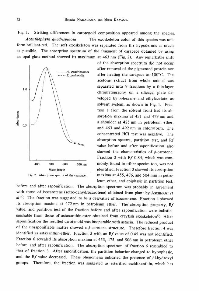
Fig. 1. Striking differences in carotenoid composition appeared among the species. Acanthephyra quadrispinosa The exoskeleton color of this species was uni-
form-brilliant-red. The soft exoskeleton was separated from the hypodermis as much as possible. The absorption spectrum of the fragment of carapace obtained by using an opal glass method showed its maximum at 463 nm (Fig. 2). Any remarkable shift
--A. quadrispinosa -- - - - S. preh en si lis
1.0
' '
'
\\. ......... -............ ..
400 500 600 700nm
Wave length Fig. 2. Absorption spectra of the carapace.
of the absorption spectrum did not occur after removal of the pigmented protein nor after heating the carapace at 100°C. The acetone extract from whole animal was separated into 9 fractions by a thin-layer chromatography on a silicagel plate de-veloped by n-hexane and ethylacetate as solvent system, as shown in Fig. I. Fraction 1 from the solvent front had its absorption maxima at 451 and 479 nm and a shoulder at 425 nm in petroleum ether, and 463 and 492 nm in chloroform. The concentrated HCI test was negative. The absorption spectra, partition test, and Rf
value before and after saponification also showed the characteristics of 13-carotene. Fraction 2 with Rf 0.84, which was commonly found in other species too, was not identified. Fraction 3 showed its absorption maxima at 455, 476, and 504 mm in petro-leum ether, and epiphasic in partition test,
before and after saponification. The absorption spectrum was probably in agreement with those of isocarotene (retro-dehydrocarotene) obtained from plant by AscHMAWI et a/14>. The fraction was suggested to be a derivative of isocarotene. Fraction 4 showed its absorption maxima at 472 nm in petroleum ether. The absorption property, Rf value, and partition test of the fraction before and after saponification were indistinguishable from those of astaxanthin-ester obtained from crayfish exoskeleton9>. After saponification the resulted carotenoid was inseparable with astacin. The reduced product of the unsaponifiable matter showed a 13-carotene structure. Therefore fraction 4 was identified as astaxanthin-ether. Fraction 5 with an Rf value of 0.45 was not identified. Fraction 6 revealed its absorption maxima at 453, 475, and 506 nm in petroleum ether before and after saponification. The absorption spectrum of fraction 6 resembled to that of fraction 3. After saponification, the partition behavior changed to hypophasic, and the Rf value decreased. These phenomena indicated the presence of di-hydroxyl groups. Therefore, the fraction was suggested as esterified eschltxanthin, which has

Carotenoprotein and carotenoid of micronektonic crustaceans 53
been reported by KARRER & LEUMAN 15>. Fraction 7 was the most abundant carotenoid
in this species. Its absorption maximum was at 4 72 nm in petroleum ether, and did
not change after saponification. After following reduction of saponification product,
the abosrption spectrum showed {3-carotene-like spectrum. Its Rf value was identical
with those of the authentic canthaxanthin and astacin. When the fraction was co
chromatographed on a silicagel plate by using the solvent system, · ..::nzene and methanol,
no traces of astaxanthin were found in it. The fraction was identified as canthaxanthin.
Fraction 8 showed similar Rf with phoenicoxanthin. Fraction 9 was the lowest in Rf,
showing its single absorption maximum at 4 73 nm in petroleum ether. The partitiqn
test was hypophasic before and after saponification. Its Rf value increased and became
indistinguishable from that of astacin after saponification. These results indicated the
presence of di-hydroxyl groups which might change to carbonyl groups by saponification.
Therefore, the fraction was identified as astaxanthin.
The exoskeleton was homogenized with 0.6 M ammonium sulfate, then centrifuged.
The oil globule of the top layer was removed so as to make dissolution into petroleum
ether possible. The absorption maximum at 470-472 nm was observed in the petroleum
ether solution. The oil globule contained 0.8 per cent carotenoid in total carotenoid.
The extract with 0.6 M ammonium sulfate gave brown color and the absorption maxi
mum was at 475 nm (Fig. 3). The absorption spectrum of the pigmented protein
extracted from the egg of this species was measured in the same way. The absorption
I I I \ I I \ \ \ I
\
.. ··· .. ..... ··
\.,. . ': ..... " .............
···· ... _ / ...... , · ... ·· ', · ..
' ... ' . ' '· '·. '~·-' ~.',
·. ' ·· ......... ....
····-.... ::.:~~-~-::..·-:~
400 500 600 700nm
Wave length
Fig. 3. Absorption spectra of extracts with 0.6 M ammonium sulfate from the exoskeleton and egg of deep-sea decapods. Acanthephyra quadrispinosa exoskeleton Sergestes prehensilis exoskeleton A. quadrispinosa egg

54 Heisuke NAKAGAWA and Mitsu KAYAMA
spectrum showed a clear single peak at 495 nm in ammonium sulfate solution. By adding solid ammonium sulfate to the pigmented extract from the exoskeleton, the resulted brown protein was applied for disc electrophoresis at pH 8.6. After migration one brown protein and four colorless proteins were detected. The carotenoids of the prosthetic group of the pigmented protein were extracted by a mixture of methanol and chloroform. Applyed on silicagel thin-layer chromatography, the extract showed one spot corresponding to astaxanthin.
No. Rf 1 0.90 2 0.84 3 0.67 4 0.60
5 0.48 6 0.22 7 0.18 8 0.10
9 0.04
Table 2. Absorption maxima and percentage composition of the carotenoid from Acanthephyra quadrispinosa.
Pet. ether 428*-451-479
455-476-504 472
453-475-506 466
473
Ethanol
428*-454-481
480
476-9
476
Chloroform Identification 438*-463-492 ~-carotene
491
485-7
489
Astaxanthin ester
Canthaxanthin Phoenicoxanthin Astaxan thin
* Indicates an obtuse peak.
% 2.3 2.0
20.7 21.8
2.0
10.5 31.1
trace
9.5
Sergestes prehensilis The exoskeleton of this species was uniformly red, tingen with violet. The absorption maximum of the carapace was at 475 nm, somewhat similar to that of Acanthephyra, as shown in Fig. 2. No shift of the absorption spectrum was observed even after treatment with 0.6 M ammonium sulfate or heating. In carotenoid composition of whole animal, eleven pigmented bands were observed on a silicagel plate. Fraction 1 gave a ~-carotene-like Rf value, but authentic a- and ~
carotenes could not separate from each other in this analytical condition. Therefore, the identification of the fraction requires further experiment. Fractions 2 and 3 were not identified. Fractions 4 and 5 showed the typical properties of astaxanthin ester. They gave a symmetrical single peak in petroleum ether, indicating the presence of diketo groups. Both fractions were suggested to be astaxanthin esters, because the two separated spots were probably due to the structural difference of the alkyl groups. Fractions 6 and 7 both gave a similar absorption spectrum. They were equal to those of isocryptoxanthin and cryptoxanthin, respectively, and unchangeable by saponification. Reducation changed the absorption maxima to be 405, 427, and 452nm in petroleum ether and was epiphasic in the partition test. The fraction was probably astaxanthin monoester, showing the same characteristics as astacin after saponification. Distribution of astaxanthin monoester in crustacea was reported by MATSUNO et al. 16) Fraction 9 with relatively high concentration showed the absorption maxima at 460 and 474 nm in petroleum ether. The absorption spectrum was somewhat resembling to that of

Carotenoprotein and carotenoid of micronektonic crustaceans 55
a-doradexanthin ester as reported by KATAYAMA et al. 17•18) The identity was not
checked. Fraction 10 was characteristic in this species, and was not detected in the
other species analyzed. Although the partition test was epiphasic, it became hypophasic
after saponification. The absorption spectrum and Rf value indicated the presence of
di-hydroxyl groups. Fraction 11 was identified as astaxanthin from the chromatographic
and spectrophotometric behaviors before and after saponification.
The pigmented protein extracted from the exoskeleton was centrifuged, then three
distinct layers appeared : a pigmented oil globule, an aqueous layer, and the bottom
residue. A fairly high amount of red oil globules, of which the absorption maxima lied
at 465 nm in petroleum ether, was found. The carotenoid content in the oil globules
corresponded to about 25 per cent of total carotenoid in the exoskeleton. The absorp
tion spectrum of the middle layer which included the pigmented protein of the exo
skeleton was characterized by a minor peak at about 485 nm (Fig. 3). The carotenoid
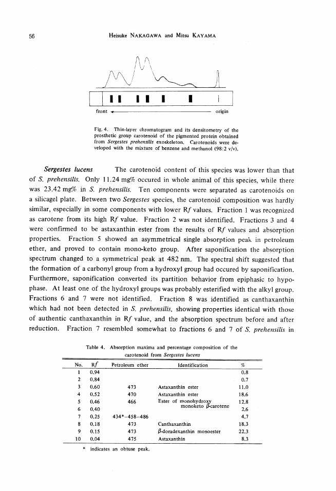
of the prosthetic group extracted from the pigmented protein showed 6 components
on a silicagel plate under the use of benzene and methanol as developing solvent,
although only 2 components showed on a plate developed with a mixture of n-hexane
and ethylacetate. In 6 components, astaxanthin and one unknown pigment were
predominant, while two other components with higher Rf value than astaxanthin were
secondary, and two other components with lower Rf value were subsidiary. The chro
matogram and its densitometry of the carotenoid are illustrated in Fig. 4.
Table 3. Absorption maxima and percentage composition of the
carotenoids from Sergestes prehensilis
No. Rf Pet. ether Chloroform cs2 Identification %
0.93 0.7
2 0.84 0.8
3 0.73 trace
4 0.62 473 486 499 Astaxanthin ester 8.1
5 0.53 465 479 As tax an thin ester 15.5
6 0.45 435*-459-487 457*-480-510* 11.7
7 0.39 433*-457-488 453*-475-502 9.1
8 0.22 472 Astaxanthin 3.6 monoester
9 0.17 460-474 21.8
10 0.14 435*-462-480 16.7
11 0.04 465 483 495 Astaxan thin 11.9
* indicates an obtuse peak.
56 Heisuke NAKAGAWA and Mitsu KAYAMA
I I I I I I I I front +--------------- origin
Fig. 4. Thin-layer chromatogram and its densitometry of the prosthetic group carotenoid of the pigmented protein obtained from Sergestes prehensi/is exoskeleton. Carotenoids were developed with the mixture of benzene and methanol (98:2 v/v).
Sergestes lucens The carotenoid content of this species was lower than that of S. prehensilis. Only 11.24 mg% occured in whole animal of this species, while there was 23.42 mg% in S. prehensilis. Ten components were separated as carotenoids on a silicagel plate. Between two Sergestes species, the carotenoid composition was hardly similar, especially in some components with lower Rf values. Fraction 1 was recognized as carotene from its high Rf value. Fraction 2 was not identified. Fractions 3 and 4 were confirmed to be astaxanthin ester from the results of Rf values and absorption properties. Fraction 5 showed an asymmetrical single absorption peak in petroleum ether, and proved to contain mono-keto group. After saponification the absorption spectrum changed to a symmetrical peak at 482 nm. The spectral shift suggested that the formation of a carbonyl group from a hydroxyl group had occured by saponification. Furthermore, saponification converted its partition behavior from epiphasic to hypophase. At least one of the hydroxyl groups was probably esterified with the alkyl group. Fractions 6 and 7 were not identified. Fraction 8 was identified as canthaxanthin which had not been detected in S. prehensilis, showing properties identical with those of authentic canthaxanthin in Rf value, and the absorption spectrum before and after reduction. Fraction 7 resembled somewhat to fractions 6 and 7 of S. prehensilis in
Table 4. Absorption maxima and percentage composition of the carotenoid from Sergestes /ucens
No. R/ Petroleum ether Identification % 0.94 0.8
2 0.84 0.7 3 0.60 473 Astaxanthin ester 11.0 4 0.52 470 Astaxanthin ester 18.6 5 0.46 466 Ester of monohydrof< 12.8
monoketo carotene 2.6 6 0.40
7 0.25 434*-458-486 4.7 8 0.18 473 Canthaxanthin 18.3
9 0.15 473 13-ctoradexanthin monoester 22.3
10 0.04 475 Astaxanthin 8.3
* indicates an obtuse peak.

Carotenoprotein and carotenoid of micronektonic crustaceans 57
the maximal absorption, but differed in the chromatographic behavior. Dominant com
ponent fraction 9 showed a symmetrical absorption peak at 473 nm in petroleum ether.
The partition behavior and absorption spectrum suggested the presence of di-ke to groups.
Therefore the fraction might be 13-doradexanthin monoester. Fraction I 0 was identified
as astaxanthin.
Euphausia similis The whole extract was applied for the thin-layer chro-
matography. In total 9 components of carotenoid separated on a plate. Fractions I
and 2 were not identified. Fraction 2 of this species was of a similar absorption
maximum with fractions 3 and 6 of A. quadrispinosa. This fraction was likely to be a
derivative from isocarotene. Fraction 3 appeared in large amounts, and did not differ
from authentic echinenone on a thin-layer plate nor in absorption property. Therefore,
it was identified as echinenone. Fractions 4 and 5 were identified as astaxanthin ester
by their Rf values and absorption spectra compared with a well-defined one. Fraction
6 with absorption maximum at 465 nm in petroleum ether was considered to be
astaxanthin monoester from the results of the partition test and chromatography before
and after saponification. Dominant carotenoid in the species, fraction 7, showed the
characteristic absorption maximum and Rf value of canthaxanthin. Fraction 8 revealed
the characteristic of 13-doradexanthin in its absorption spectrum and Rf value. Fraction
9 was confirmed as astaxanthin.
No.
2
3 4
5 6
7 8
9
Table 5. Absorption spectra and percentage composition of the
carotenoids from Euphausia similis
Rf Petroleum ether Identification %
0.85 0.5
0.77 454-473-503 2.3
0.67 473 Echinenone 24.7
0.61 475 Astaxanthin ester 10.5
0.53 Astaxanthin ester 3.4
0.24 465 Astaxanthin monoester 6.1
0.18 472 Canthaxanthin 37.2
0.15 470 /3-doradexanthin monoester 8.1
0.04 472 Astaxanthin 7.4
Table 6. Percentage composition of the carotenoids from
Lucifer sp.
No. Rf Identification %
1 0.87 trace
2 0.57 Astaxanthin ester 10.7
3 0.45 9.5
4 0.18 Canthaxanthin 6.6
5 0.13 41.5
6 0.04 As tax an thin 32,4

58 Heisuke NAKAGAWA and Mitsu KAYAMA
Lucifer sp. The exoskeleton had a pink calor resulting from small red pigmented chromatophores. It should be noted that the concentration of carotenoid in this species was pretty low. The carotenoids were devided into 6 fractions on a silicagel plate. They revealed the Rf values corresponding to the following components; astaxanthin esters, canthaxanthin, astaxanthin, and 3 unidentified components. The polar unidentified xanthophyll differed from phoenicoxanthin or astaxanthin as dominant carotenoid.
DISCUSSION
The recent development of thin-layer chromatographic technique for the examination of carotenoid made possible rapid analysis even for the small amounts of pigment. The carotenoids of 5 species belonging to crustaceans were analyzed on a rather smaller scale. All species examined contained about 10 carotenoid components. Mono- and di-esterified astaxanthin would be distinguished on the thin-layer plate. HERRING 2> found the presence of astaxanthin ester as the major pigments in decapods. A fairly high amount of carotenoids existed in the form of ester. Percentage composition of astaxanthin and its esters ranged from 20-40 per cent in all the species analyzed.
Some unidentified carotenoids found in A. quadrispinosa and E. similis were probably isocarotene derivatives, which had never been reported in crustacea. It is noteworthy that the spectrophotometric determination showed no a-carotene derivatives in these species analyzed, although it is well known that carotenoids such as lutein are distributed widely in crustacean.
The taxonomic proportion of carotenoid distribution have been reported for the phytoplanktonic species19>. The specific pigments are frequently associated with particular phylum or even with a class or order. However, the significance of the specific colors of the exoskeleton and carotenoid composition of various species is not yet clearly accounted for in crustaceans. Between S. prehensilis and S. lucens, the pigment arrays were different. The former showed the carotenoid with a:-doradexanthin esterlike spectrum as major carotenoid, while the latter contained a yet unidentified xanthophyll ester as dominant.
The exoskeleton's carotenoproteins of two deep-sea decapods were analyzed. The brown pigmented protein could be extracted by 0.6 M ammonium sulfate. However, considering that the carotenoid content and color intensity of the pigmented protein were minute, the contribution for calor appearance might be negligible. It is well known that blue or green carotenoproteins are rare in deep-sea decapods2>. A bluish protein did not occur in the exoskeleton of two species analyzed. In S. prehensilis, the oil globule which could be readily separated from the homogenate of exoskeleton contained large amounts of carotenoid, corresponding at least to 25 per cent of the exoskeleton carotenoid. Only one brown protein was detected in the exoskeleton

Carotenoprotein and carotenoid of micronektonic crustaceans 59
of S. prehensilis or A. quadrispinosa. Therefore, the exoskeleton was suggested to be
pigmented by receive its pigmentation from the oil globule. The carapaces of A.
quadrispinosa and S. prehensilis showed an absorption maxima at 465 and 475 nm, which
is somewhat resembling to that of astaxanthin in water in the presence of detergent4 ).
The possibility seemed to exist that a small amount of astaxanthin or other xanthophyl!s
were connected with the large molecular protein in some manners; and almost all
carotenoids existed in free form in the exoskeleton. Therefore, the red exoskeelton
was due to the presence of large amounts of unbound xanthophylls more than to
protein-bound xanthophylls.
The presence of carotenoid-containing protein was confirmed in the exoskeleton
of two species, whereas it was not clear whether the pigmented protein was either
carotenoprotein containing a stoichiometrical amount of carotenoids as prosthetic group,
or carotenoid-containing lipoprotein. It was suggested that the pigmented protein
functioned not for the color appearance, but for the carotenoid-carrier protein. The
presence of carotenoprotein could increase the absorption area of light by hypso- or
bathochromic shift. Thus color of the animal living in feeble light environment was
probable corelated to camouflage or uptake of light.
SUMMARY
Thin-layer chromatographic procedure using silicagel was employed for the exami
nation of the carotenoids of five species of crustacean. The carotenoids were separated
in about 10 components in all species analyzed on a thin-layer plate. Besides astaxanthin
and its esters, several xanthophylls were also detected. The small amounts of carotenoid
containing protein were obtained from the exoskeleton of Acanthephyra quadrispinosa
and Sergestes prehensilis. It seems likely that the pigmented protein has more bio
chemical than pigmentary significance.
ACKNOWLEDGEMENTS
The authors express their gratitude to Associate Prof. K. KusAKA and Prof. G.
Y AMAMOTO of the Ocean Research Institute, Tokyo University, for the joint cruise
of "Tansei Maru", and Drs. M. OHMORI and H. KAWAGUCHI, Ocean Research Institute,
Tokyo University, for the identification of the crustaceans used.
This research was partly supported by the Scientific Research Fund from the Minis
try of Education, Japan.
60 Heisuke NAKAGAWA and Mitsu KAYAMA
REFERENCES
I) MARSHALL, H. B.: in "Aspects of Deep Sea Biology" pp. 162-166, Hutchinson's, London (1954).
2) HERRING, P. J.: l. mar. bioi. Ass. U. K., 53, 539-562 (1973). 3) CHEESMAN, D. F., LEE, W. L., and ZAGALSKY, P. F.: Bioi. Rev., 42, 132-160 (1967). 4) JENCKS, W. P. and BUTEN, B.: Arch.Biochim.Biophys., 107,511-520(1964). 5) LEE, W. L. and ZAGALSKY,P. F.: Biochem. l., 101, 9c-llc (1966). 6) BucHWALD, M. and JENCKS, W. P.: Biochem., 7, 844-859 (1968). 7) ZAGALSKY,P.F.,CECCALDI,H.J.,andDAUMAS, R.: Camp. Biochem. Physiol., 34, 579-607
(1970).
8) ZAGALSKY, P. F. and HERRING, P. J.: Camp. Biochem. Physiol., 41B, 397-415 (1972). 9) NAKAGAWA, H., KAYAMA, M., YAMADA, H., and AsAKAWA, S.: l. Fac. Fish. Anim. Husb.
Hiroshima Univ., 13, 1-13 (1974). 10) KAYAMA, M. and NAKAGAWA, H.: lap. Oil Diem. Soc., (in the press). 11) CECCALDI, H. J. and ALLEMAND, B. H.: Rec. Trav. Stat. Mar. End. Bull., 35, 3-7 (1964). 12) 0RNSTEIN, L.: Ann. N. Y. Acad. Sci., 121, 321-349 {1964). 13) DAVIS, B. J.: Ann. N. Y. Acad. Sci., 121, 402-427 (1964). 14) ASHMAWI, H., HusSEIN, A. A., and SHAKER, M. H.: Bull. Fac. Sci. Cairo Univ., 61, 12
(1955).
15) KARRER, P. andl.EUMAN, E.: Helv. Olim. Acta, 34,445-453 (1951). 16) MATSUNO, T., WATANABE, T., and NAGATA, S.: Bull. lap. Soc. Sci. Fish., 40, 619-624
(1974).
17) KATAYAMA, T., YoKOYAMA, H., and CHICHESTER, C. 0.: Int. J. Biochem., 1, 438-444 (1970).
18) KATAYAMA, T., YoKOYAMA, H., and CHI~HESTER, C. 0.: Bull. lap. Soc. Sci. Fish., 36, 702-708 (1970).
19) RILEY, J. P. and WILSON, T. R. S.: l. mar. bioi. Ass. U. K., 47, 351-362 (1967).
*H.m~. ~iiiJ~£0) 5 ~~(})lfljiUfi!C-:J~'"'Cn o 7-/ 1 l-', &."CH:(})?'!:)(}) 2 ~~IC::-:J~'L'Ef!\'&0)-@.~~ Blf(})7Hfi~rr-:>f.:o ~"'CO) Efl\'&~(})n o 7-/ 1 n.tAJ~:? o < 1- :?. 7 7 -1 -l:~lOfflZ7}1c::7}1ft 1..... -c-0)? '1:) 7 A~ "f--if './"'f- './, 7 A~ "f- -if'./"'!-'./ x. A 7 Jv, &U*F<\l~(})"f--ij- './ 1- 7 -1 JviJi.±.fflZ?}l: if,~ t.:o Acanthephyra quadrispinosa &U Sergestes prehensi/is (}) Ef! \'&il> I? 1J o 7- / 1 r ~-@;- tJ'@.~~ s If ~7} 1ft lA.:tJi, <::. (})~Bifli:l:ft-Jic::~ -c, '@.~ c 1.., -c (J)f'Fjij .t tJ b n o 7- / 1 1-'i!!JIO) .t ? t.r~{t~ft-JtJ:~~~~ :!1;!'9 c~;t l?tL.Oo