
IVIS Lumina XRMS Multi-species Optical and X-ray … · 2 Leading Innovator in Bioluminescence,...
6
The IVIS ® Lumina XRMS Series III from PerkinElmer integrates the best in class in vivo bioluminescence and fluorescence imaging with 2D X-ray capability. The Lumina XRMS offers the flexibility to image small as well as large animals with precise optical and X-Ray overlay, giving anatomical context to the optical signal. The Lumina XRMS includes state of the art spectral unmixing for sensitive multispectral imaging to monitor multiple biological events in the same animal. Multi-species Optical and X-ray Imaging System Pre-clinical in vivo imaging PRODUCT NOTE IVIS Lumina XRMS Series III Key Features • Optical and X-ray imaging • Multi-species imaging including mice and rats • High resolution, low dose digital X-ray • Exquisite sensitivity in bioluminescence • Compute Pure Spectrum (CPS) spectral unmixing for ultimate fluorescence sensitivity • Full fluorescence tunability through the NIR Spectrum
Transcript of IVIS Lumina XRMS Multi-species Optical and X-ray … · 2 Leading Innovator in Bioluminescence,...

The IVIS® Lumina XRMS Series III from PerkinElmer integrates the best in class
in vivo bioluminescence and fluorescence imaging with 2D X-ray capability. The Lumina XRMS offers the flexibility to image small as well as large animals with precise optical and X-Ray overlay, giving anatomical context to the optical signal. The Lumina XRMS includes state of the art spectral unmixing for sensitive multispectral imaging to monitor multiple biological events in the same animal.
Multi-species Optical and X-ray Imaging System
Pre-clinical in vivo imaging
P R O D U C T N O T EIVIS Lumina XRMS Series III
Key Features
• Optical and X-ray imaging
• Multi-species imaging including mice and rats
• High resolution, low dose digital X-ray
• Exquisite sensitivity in bioluminescence
• Compute Pure Spectrum (CPS) spectral unmixing for ultimate fluorescence sensitivity
• Full fluorescence tunability through the NIR Spectrum

2
Leading Innovator in Bioluminescence, Multispectral Fluorescence and Integrated X-Ray Technologies
The IVIS Lumina Series III platform brings together years of leading optical imaging technologies into one easy to use and exquisitely sensitive bench top system. The Lumina XRMS Series III offers the newest technology on the market for industry leading bioluminescence, low dose X-Ray imaging and ultra sensitive two dimensional in vivo fluorescence imaging. With the Lumina XRMS, get an anatomical context to optical signal in mice and rats and other large species.
The system is equipped with up to 26 filters tunable to image fluorescent sources that emit from green to near-infrared. All Lumina Series III systems come with a novel illumination technology that effectively increases fluorescent transmission deep into the near infrared range with full transmission through 900 nm. Moreover, the Lumina III series instruments incorporate PerkinElmer's patented Compute Pure Spectrum (CPS) algorithm
Figure 1. Optical overlay of FolateRSenseTM 680 fluorescence signal on X-ray image at multiple FOV’s in a 560 gram rat. Optional accessory ZFOV lens is needed for zoom image.
Zoom FOV A FOV B FOV C
Large Animal Imaging
Figure 2. Optical overlay of bioluminescent signal with X-ray image at multiple FOV’s in mice.
FOV A FOV B FOV C
Small Animal Imaging
for spectral library generation software tools to ensure accurate autofluorescence removal, unmixing and fluorophore quantitation.
Standard on all IVIS instruments, absolute calibration affords consistent and reproducible results independent of magnification, filter selection from one instrument to any another IVIS instrument within an organization or around the world.
Flexibility-image Small and Large Animals
The Lumina XRMS offers the flexibility to image mice, rats and other animals up to 500-600 g in weight with an accurate optical overlay on X-ray image. The X-ray scintillator can easily be moved to effectively image mice and rats with ease. The Lumina XRMS is the only instrument that can overlay an optical signal to the X-ray image at all Fields of View (FOV’s), as shown in Figures 1 and 2.

3
Figure 4. GI tract infection model was established by feeding contaminated peanut butter, which contained bioluminescence and fluorescence dually labeled Salmonella typhimurium. Bioluminescence and fluorescence (Ex605/Em660 nm) images were taken at 3 hours. A) At 5 hours, tri-modality imaging was performed and the overlaid images were shown B) The GI tract was highlighted due to the presence of barium sulfate (150 mg) in the peanut butter.
Figure 5. Chronic post-arthroplasty infection. Monitoring bacterial burden, inflammation and bone damage longitudinally using optical and X-ray imaging in an orthopedic implant infection model.
Figure 6. Detection of rheumatoid arthritis with fluorescent probe
Applications in Multimodal Imaging
Precise optical and X-ray overlay brings your optical signal into anatomical context. Key applications in oncology, infectious diseases, implant biology or any model that requires anatomical context, the Lumina XRMS Series III will offer complete and rich calibrated datasets for longitudinal studies with supporting analysis software.
C D
Figure 3. A) 5 x 10⁵ 4T1-luc2 cells were intravenously by tail vein injection. Bioluminescence image was taken of cells colonized in the various parts of body. B) Two dimensional overlaid photographic and bioluminescent image. C) Two dimensional overlaid X-ray and bioluminescent image. Red arrows highlight areas osteolysis. D) MicroCT image (Quanutm FX) confirming bone degradation in the right tibia.
A B
Oncology
Infectious Disease
Inflammation

4
Inside the Lumina XRMS Series III
• Back-thinned, back-illuminated grade 1 CCD provides high quantum efficiency over the entire visible to near-infrared spectrum
• Light-tight imaging chamber • Five filter wheel choices for a broad range of fluorescence applications• LED lamps for photographic images• Heated stage to maintain optimum body temperature• Motor controlled stage, filter wheels, lens position, and f-stop
X-Ray Module• Large and Small Animal X-ray• The high sensitivity camera allows fast X-Ray image
acquisition times of 1-10 seconds reducing radiation exposure
• Radiation shielded cabinet• Exceeds standards set by the U.S. FDA Center for Devices
and Radiological Health (21 CFR 1020.40)• Automated image integration to overlay with
Bioluminescence, Fluorescence and Photograph
Optional Accessories• Optical Zoom Lens attachment for close up and high
resolution X-Ray images• Gas anesthesia ports and 3 or 5 position manifold within
imaging chamber allows anesthesia to be maintained during imaging sessions
• Syringe injection system, integrated with Living Image, allows the user to acquire real time functional responses to compounds
IVIS Lumina Series III Software
Living Image® software brings IVIS technology to life by facilitating an intuitive workflow for in vivo optical, X-ray image acquisition, analysis and data organization. The software’s new design creates an intuitive, seamless workflow for researchers of all skill levels. New features include: wizard based guidance for advanced imaging protocols, spectral unmixing tools, expanded fluorescent agent database and a simplified tool palette.
Figure 8. A mouse bearing a subcutaneous 4T1-luc2 tumor in its right flank was injected with 315 μCi of 18F-FDG intravenously. The animal was imaged dynamically starting 55 seconds post-injection to capture the distribution of 18F-FDG in the mouse body via Cerenkov light from positron emission.
Camera
High Collection lens
Filter wheel
Automated scintillator with height adjustments
Heated Stage
Shielded Cabinet
X-ray module
Living Image also supports Dynamic Contrast Enhancement (DyCE™), a new approach to optically based biodistribution analysis and anatomical identification of organs using clearance properties of luminescent, radioisotopic or fluorescent probes. The DyCE technique acquires a series of dynamic images following a bolus injection of an optical agent. The location of major internal organs is derived by proprietary algorithms and displayed in minutes. The DyCE software module includes the Multi-View platform and software that extends the functionality of Living Image and available for all IVIS systems.

54
Features IVIS Lumina IVIS Lumina K IVIS Lumina XRMS IVIS Lumina LT
Bioluminescence ✓ ✓ ✓ ✓
Radioisotopic Cerenkov Imaging ✓ ✓ ✓ ✓
Fluorescence ✓ ✓ ✓ ✓
Compute Pure Spectrum Spectral Unmixing ✓ ✓ ✓
Real-Time Fast Kinetic Imaging (10 ms) ✓
Integrated Small and Large Animal X-Ray ✓
DyCE Imaging (Optional Upgrade) ✓ ✓ ✓ ✓
Extended NIR Range 150W Tungsten EKE ✓ ✓ ✓ ✓
Absolute Calibration to NIST® Standards ✓ ✓ ✓ ✓
The IVIS Lumina Series III platform offers a selection of instruments tailored to your in vivo imaging needs.
Optional IVIS Lumina XRMS Series III Imaging System AccessoriesExpand your Series III Instrument with features when you need them!
XGI-8 Anesthesia System
Cat No. 118918 (120 V) Cat No. 118957 (100 V)
Cat No. 121090 (230 V)
Animal Isolation Chamber Kit XIC-3
Cat No. 123997
ECG Monitoring System
Cat No. 124229ZFOV Zoom Lens
Cat No. 127285
Multi-View Platform
Cat No. CLS134956
IVIS Syringe Injection System
Cat No. 124633
XPM-2 Phantom Mouse for Bioluminescent Imaging
Cat No. 118993XFM-2 Phantom Mouse for
Fluorescent Imaging Cat No. 133803
XRM-5 X-RAY Phantom Mouse Cat No. 133793
XWS-260 WorkbenchCat No. 119207
Camera
High Collection lens
Filter wheel
Automated scintillator with height adjustments
Heated Stage
Shielded Cabinet
X-ray module
For a complete listing of our global offices, visit www.perkinelmer.com/ContactUs
Copyright ©2013-2015, PerkinElmer, Inc. All rights reserved. PerkinElmer® is a registered trademark of PerkinElmer, Inc. All other trademarks are the property of their respective owners. 010794C_01 PKI
PerkinElmer, Inc. 940 Winter Street Waltham, MA 02451 USA P: (800) 762-4000 or (+1) 203-925-4602www.perkinelmer.com
For more information, please visit our website at www.perkinelmer.com/invivo
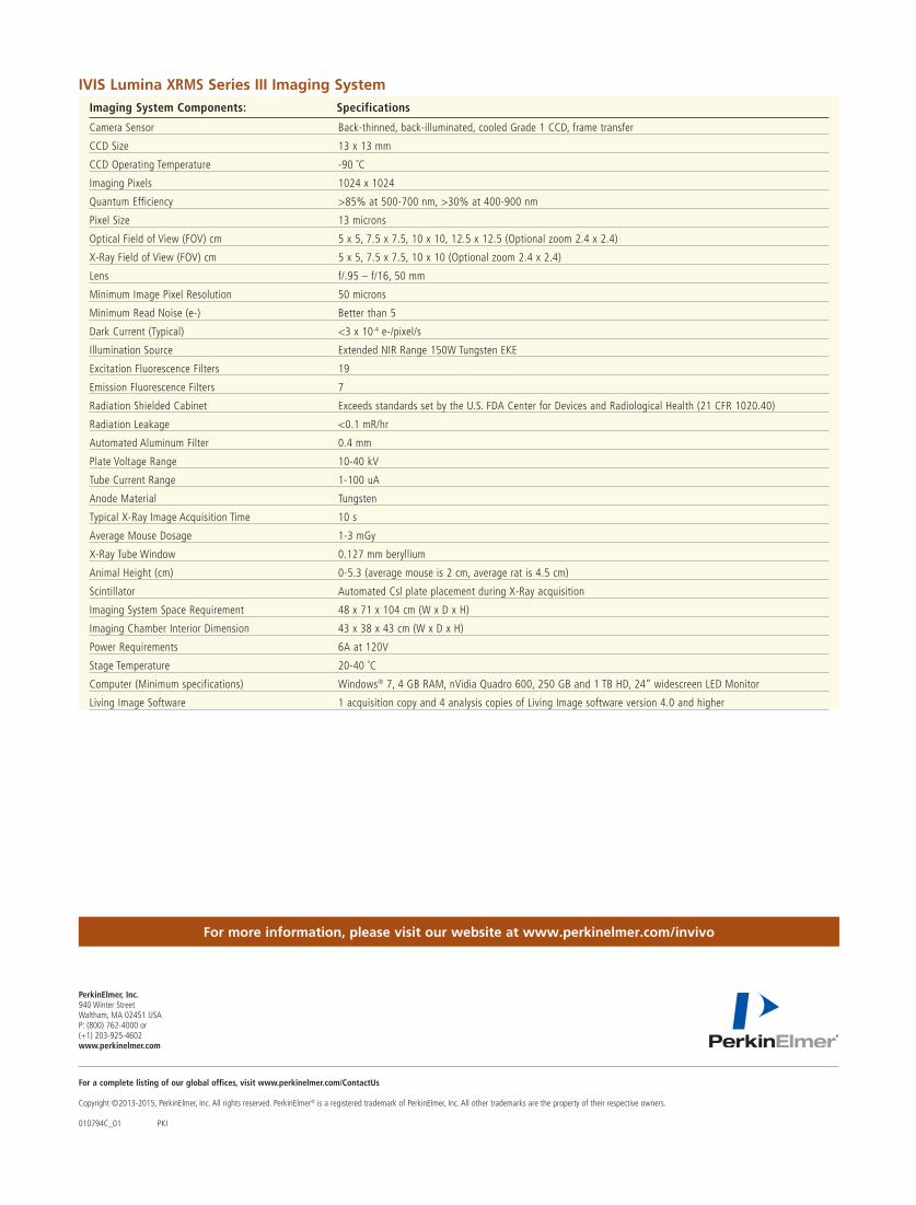
IVIS Lumina XRMS Series III Imaging System Imaging System Components: Specifications
Camera Sensor Back-thinned, back-illuminated, cooled Grade 1 CCD, frame transfer
CCD Size 13 x 13 mm
CCD Operating Temperature -90 ˚C
Imaging Pixels 1024 x 1024
Quantum Efficiency >85% at 500-700 nm, >30% at 400-900 nm
Pixel Size 13 microns
Optical Field of View (FOV) cm 5 x 5, 7.5 x 7.5, 10 x 10, 12.5 x 12.5 (Optional zoom 2.4 x 2.4)
X-Ray Field of View (FOV) cm 5 x 5, 7.5 x 7.5, 10 x 10 (Optional zoom 2.4 x 2.4)
Lens f/.95 – f/16, 50 mm
Minimum Image Pixel Resolution 50 microns
Minimum Read Noise (e-) Better than 5
Dark Current (Typical) <3 x 10⁻⁴ e-/pixel/s
Illumination Source Extended NIR Range 150W Tungsten EKE
Excitation Fluorescence Filters 19
Emission Fluorescence Filters 7
Radiation Shielded Cabinet Exceeds standards set by the U.S. FDA Center for Devices and Radiological Health (21 CFR 1020.40)
Radiation Leakage <0.1 mR/hr
Automated Aluminum Filter 0.4 mm
Plate Voltage Range 10-40 kV
Tube Current Range 1-100 uA
Anode Material Tungsten
Typical X-Ray Image Acquisition Time 10 s
Average Mouse Dosage 1-3 mGy
X-Ray Tube Window 0.127 mm beryllium
Animal Height (cm) 0-5.3 (average mouse is 2 cm, average rat is 4.5 cm)
Scintillator Automated Csl plate placement during X-Ray acquisition
Imaging System Space Requirement 48 x 71 x 104 cm (W x D x H)
Imaging Chamber Interior Dimension 43 x 38 x 43 cm (W x D x H)
Power Requirements 6A at 120V
Stage Temperature 20-40 ˚C
Computer (Minimum specifications) Windows® 7, 4 GB RAM, nVidia Quadro 600, 250 GB and 1 TB HD, 24” widescreen LED Monitor
Living Image Software 1 acquisition copy and 4 analysis copies of Living Image software version 4.0 and higher