ISUOG Basic Training Umbilical and Uterine Artery Doppler ... · ISUOG Basic Training Umbilical and...
45
Editable text here Basic training Basic training ISUOG Basic Training Umbilical and Uterine Artery Doppler Studies Juriy Wladimiroff
Transcript of ISUOG Basic Training Umbilical and Uterine Artery Doppler ... · ISUOG Basic Training Umbilical and...

Editable text here Basic training Basic training
ISUOG Basic Training
Umbilical and Uterine Artery Doppler Studies
Juriy Wladimiroff

Editable text here Basic training Basic training Basic training
Learning objectives At the end of the lecture you will be able to:
• describe how to perform, assess and report an umbilical artery
Doppler examination correctly
• describe how to perform, assess and report a Doppler examination
of the uterine arteries correctly

Editable text here Basic training
Key questions
1. What technique is required to perform a clinically useful Doppler
examination of the umbilical artery ?
2. What are the main pitfalls to be aware of when using Doppler to
sample the umbilical artery?
3. What technique is required to perform a clinically useful Doppler
examination of both uterine arteries?
4. What are the main pitfalls to be aware of when using Doppler to
sample the uterine arteries?

Editable text here Basic training
• umbilical and uterine Doppler

Editable text here Basic training
Bhide A, Acharya G, Bilardo CM, Brezinka C, Cafici D, Hernandez- Andrade E, Kalache K, Kingdom J, Kiserud T,
Lee W, Lees C, Leung KY, Malinger G, Mari G, Prefumo F, Sepulveda W and Trudinger B. on behalf of the ISUOG
Clinical Standards Committee
Editable text here Basic training
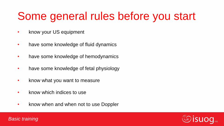
Some general rules before you start
• know your US equipment
• have some knowledge of fluid dynamics
• have some knowledge of hemodynamics
• have some knowledge of fetal physiology
• know what you want to measure
• know which indices to use
• know when and when not to use Doppler

Editable text here Basic training
Fetal circulation
• high heart rate
• low blood pressure
• low peripheral resistance (placenta)
• placental circulation constant
(does not respond to vasoactive substances)
• with advancing gestation fetal BP and
arteriolar
placental bed flow increase, peripheral
resistance decreases

Editable text here Basic training
Fetal and maternal vessels
Fetal side
• umbilical artery
• middle cerebral artery
• ductus venosus
• umbilical vein
Maternal side
• uterine arteries
Editable text here Basic training
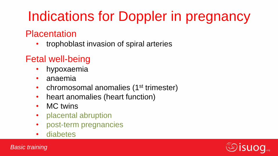
Indications for Doppler in pregnancy Placentation
• trophoblast invasion of spiral arteries
Fetal well-being • hypoxaemia
• anaemia
• chromosomal anomalies (1st trimester)
• heart anomalies (heart function)
• MC twins
• placental abruption
• post-term pregnancies
• diabetes

Editable text here Basic training
Umbilical artery Doppler
Editable text here Basic training
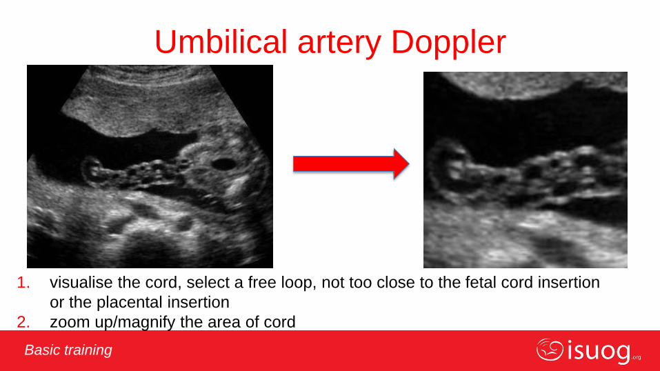
1. visualise the cord, select a free loop, not too close to the fetal cord insertion
or the placental insertion
2. zoom up/magnify the area of cord
Umbilical artery Doppler

Editable text here Basic training
3. switch on the colour Doppler modality (not compulsory)
Umbilical artery Doppler

Editable text here Basic training
3a. optimize the colour flow mapping (CFM) scale
Umbilical artery Doppler

Editable text here Basic training
4. place the sample gate on the umbilical artery
Umbilical artery Doppler
Editable text here Basic training
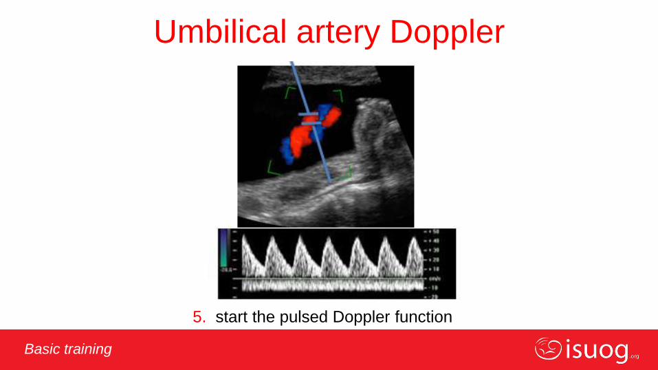
5. start the pulsed Doppler function
Umbilical artery Doppler

Editable text here Basic training
Umbilical artery Doppler
Laurin 1987
Editable text here Basic training
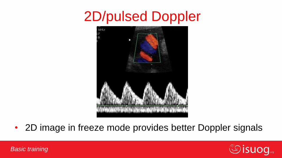
2D/pulsed Doppler
• 2D image in freeze mode provides better Doppler signals

Editable text here Basic training
Irregular umbilical artery flow velocity
pattern due to fetal breathing movements
Editable text here Basic training
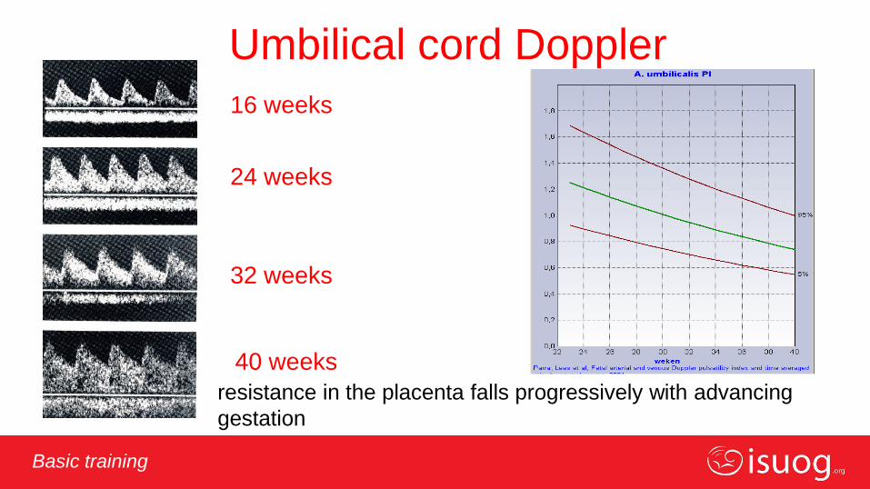
Umbilical cord Doppler
16 weeks
24 weeks
32 weeks
40 weeks
resistance in the placenta falls progressively with advancing
gestation

Editable text here Basic training
Umbilical artery in pathological
pregnancies
high PI
absent end diastolic flow
reversed end diastolic flow

Editable text here Basic training
Abnormal UA findings
Elevated UA index
Baschat AA, Gembruch U,
UOG 2003; 21: 124-7
Trudinger BJ, Giles WB,
Br J Obstet Gynaecol, 1996; 105: 487-9.
REDV 70% of villous vessels are underperfused

Editable text here Basic training Basic training
• there is a significant difference in Doppler indices when measured at the fetal
end, in a free cord loop or at the placental end of the umbilical cord
• for the sake of simplicity and consistency, measurements should be made in a
free cord loop
• in multiple pregnancies, and/or when comparing repeated measurements
longitudinally, recordings from fixed sites (fetal end, placental end or intra-
abdominal portion) may be more reliable
• reference ranges used should be appropriate for the site of interrogation
Variation in umbilical artery waveforms

Editable text here Basic training
When is umbilical artery
assessment indicated?
• reduced fetal growth velocity/fetal growth restriction (FGR)
• MC twins
• fetal hydrops
• EDF (+ve, absent or reversed) more sensitive than PI
Editable text here Basic training
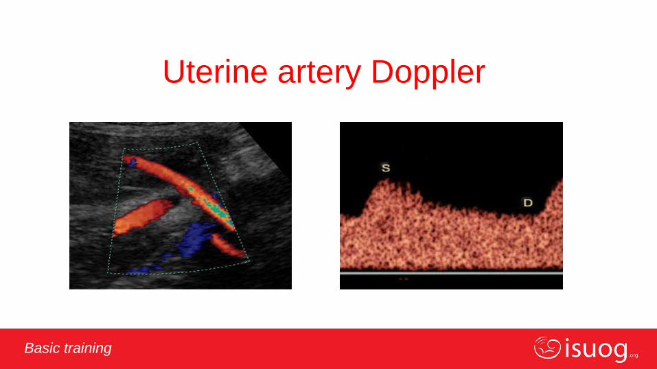
Uterine artery Doppler

Editable text here Basic training

Editable text here Basic training
Trophoblast invasion
uterine artery

Editable text here Basic training
Editable text here Basic training
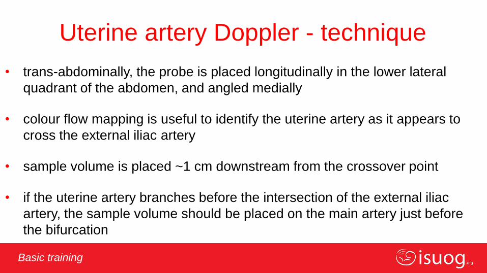
Uterine artery Doppler - technique
• trans-abdominally, the probe is placed longitudinally in the lower lateral
quadrant of the abdomen, and angled medially
• colour flow mapping is useful to identify the uterine artery as it appears to
cross the external iliac artery
• sample volume is placed ~1 cm downstream from the crossover point
• if the uterine artery branches before the intersection of the external iliac
artery, the sample volume should be placed on the main artery just before
the bifurcation

Editable text here Basic training
Uterine artery measurement
Editable text here Basic training
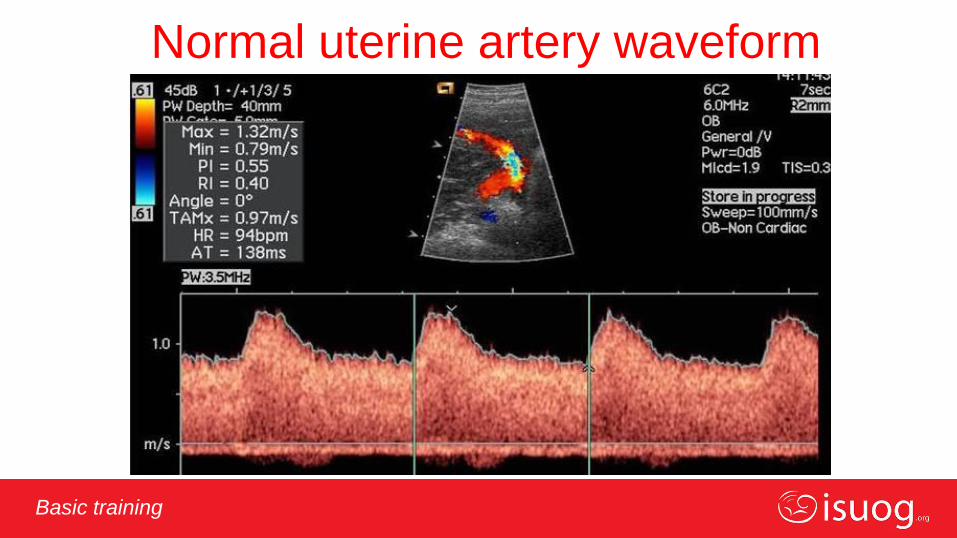
Normal uterine artery waveform
Editable text here Basic training
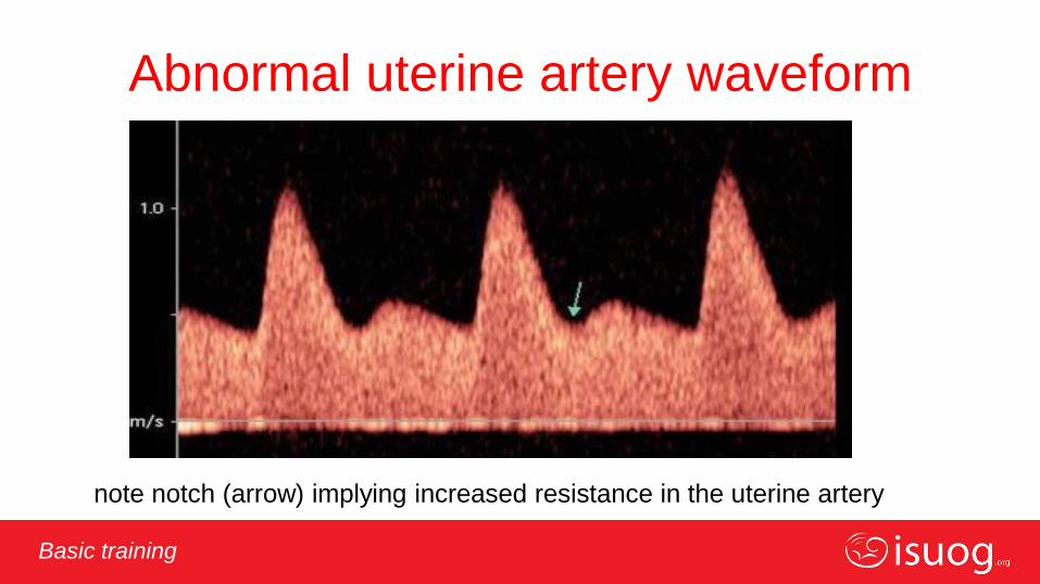
note notch (arrow) implying increased resistance in the uterine artery
Abnormal uterine artery waveform
Editable text here Basic training
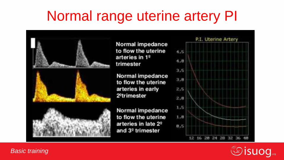
Normal range uterine artery PI

Editable text here Basic training
Uterine artery screening at 22-24 wks
low risk for PE and IUGR high risk for PE and IUGR
Editable text here Basic training
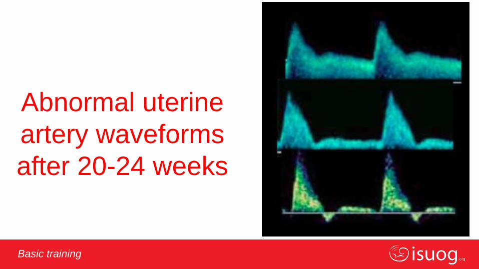
Abnormal uterine
artery waveforms
after 20-24 weeks

Editable text here Basic training
Uterine artery

Editable text here Basic training
Clinical applications

Editable text here Basic training
Editable text here Basic training
Editable text here Basic training
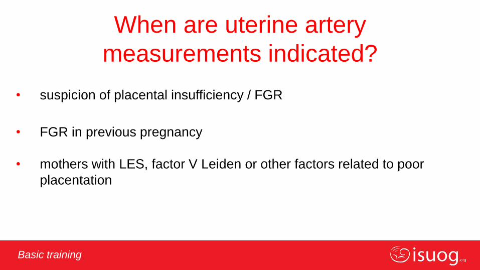
When are uterine artery
measurements indicated?
• suspicion of placental insufficiency / FGR
• FGR in previous pregnancy
• mothers with LES, factor V Leiden or other factors related to poor
placentation

Editable text here Basic training
Papageorghiou et al UOG 2001
Repeatability of transabdominal uterine
artery measurement

Editable text here Basic training
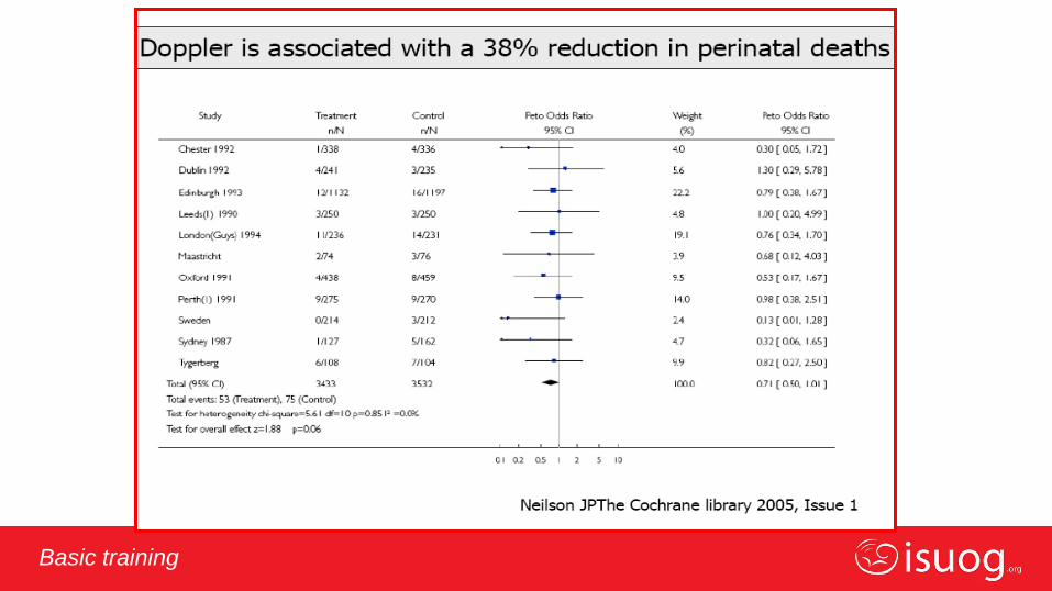
Increased impedance to flow in the uterine arteries in pregnancies attending
for routine antenatal care identifies approximately 40% (L.R. 6) of those who
subsequently develop PE and approximately 20%(L.R. 3,5) of those who
develop fetal growth restriction

Editable text here Basic training
Pre-eclampsia screening
Cnossen JS et al 2008
Editable text here Basic training
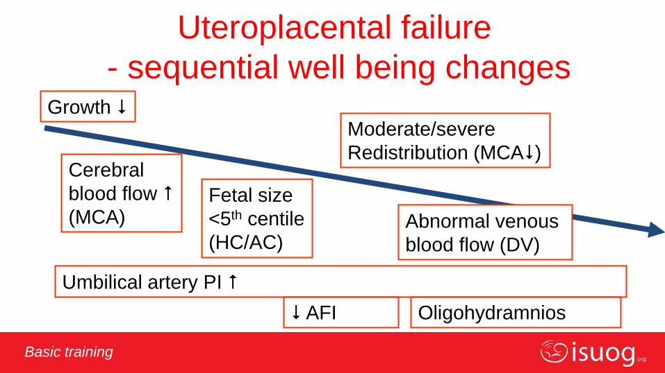
Uteroplacental failure
- sequential well being changes Growth
Moderate/severe
Redistribution (MCA)
Fetal size
<5th centile
(HC/AC)
Umbilical artery PI
Oligohydramnios
Abnormal venous
blood flow (DV)
Cerebral
blood flow
(MCA)
AFI

Editable text here Basic training
Take home messages • Doppler investigations give insight into fetal and pregnancy patho-
physiology
• Doppler is one of the major breakthroughs in Fetal Medicine
• Doppler can be used in all trimesters for different indications
• It can be used as a screening or a diagnostic tool, according to the
circumstances
• In the 2nd and 3rd trimesters it can indicate abnormal placentation, fetal
hypoxemia, fetal anemia and impending heart failure
• Operators should use it skillfully and with knowledge of its potentials,
limitations and dangers

Editable text here Basic training
Thank you for your attention