Evidence-based Prenatal Care: Oxymoron or “Best Practice?” Francesco Leanza, MD FACTS 3/5/04.
Upload
truongxuyenCategory
view
221download
0
Published by Baishideng Publishing Group Inc
World Journal of Stem CellsWorld J Stem Cells 2016 January 26; 8(1): 1-21
ISSN 1948-0210 (online)
EDITORS-IN-CHIEFTong Cao, SingaporeOscar Kuang-Sheng Lee, Taipei
ASSOCIATE EDITORSWei Cui, LondonPaul Lu, La JollaYuko Miyagoe-Suzuki, TokyoSeyed J Mowla, TehranKai C Sonntag, MassachusettsBao-Hong Zhang, Greenville
GUEST EDITORIAL BOARD MEMBERSChia-Ning Chang, TaichungChuck Ching-Kuey Chao, TaoyuanChie-Pein Chen, TaipeiFu-Chou Cheng, TaichungIng-Ming Chiu, JhunanAkon Higuchi, TaoyuanHossein Hosseinkhani, Taipei Yu-Chen Hu, HsinchuYen-Hua Huang, TaipeiJyh-Cherng Ju, TaichungSteven Shoei-Lung Li, KaohsiungFeng-Huei Lin, Zhunan Town Shing-Hwa Liu, TaipeiJui-Sheng Sun, TaipeiTzu-Hao Wang, TaoyuanYau-Huei Wei, New Taipei CityKuo-Liang Yang, HualienChao-Ling Yao, Chung-Li City
MEMBERS OF THE EDITORIAL BOARD
Argentina
Federico J Benetti, Santa Fe
Luis E Politi, Bahia Blanca
Australia
Michael K Atkinson, BrisbanePeter M Bartold, South AustraliaJeremy M Crook, VictoriaSimon A Koblar, South AustraliaKuldip S Sidhu, SydneyPaul J Verma, Clayton VicErnst J Wolvetang, BrisbaneCory J Xian, South AustraliaXin-Fu Zhou, Adelaide
Austria
Ludwig Aigner, SalzburgFerdinand Frauscher, InnsbruckRegina Grillari, ViennaMariann Gyongyosi, ViennaGünter Lepperdinger, InnsbruckPeter Valent, Vienna
Belgium
Yves Beguin, LiegeMieke Geens, BrusselsNajimi Mustapha, Brussels
Brazil
Niels OS Camara, Cidade UniversitáriaArmando DM Carvalho, BotucatuKatherine AT de Carvalho, CuritibaRegina CDS Goldenberg, Rio de Janeiro
Irina Kerkis, Sao PauloAna H da Rosa Paz, Porto AlegreLuís C De Moraes Sobrino Porto, Rio de Janeiro RJRodrigo Resende, Belo HorizonteNaiara Z Saraiva, Jaboticabal
Canada
Borhane Annabi, QuebecLong-Jun Dai, VancouverConnie J Eaves, VancouverSantokh Gill, OttawaJeffrey T Henderson, TorontoRosario Isasi, QuebecXiaoyan Jiang, VancouverSeung U Kim, VancouverWilliam A King, GuelphRen-Ke Li, TorontoZubin Master, EdmontonChristopher Naugler, CalgaryDominique Shum-Tim, QuebecJean-Francois Tanguay, QuebecKursad Turksen, OttawaLisheng Wang, Ontario
China
Xiu-Wu Bian, ChongqingAndrew Burd, Hong KongKai-Yong Cai, ChongqingCHI-KAI Chen, Shantou Ling-Yi Chen, TianjinFu-Zhai Cui, Beijing Yong Dai, Shenzhen Yu-Cheng Dai, NanchangLi Deng, ChengduJian Dong, Shanghai
I
Editorial Board2016-2019
The World Journal of Stem Cells Editorial Board consists of 700 members, representing a team of worldwide experts in infectious diseases. They are from 44 countries, including Argentina (2), Australia (9), Austria (6), Belgium (3), Brazil (9), Canada (16), China (73), Cyprus (2), Czech Republic (5), Denmark (6), Ecuador (1), Egypt (2), Finland (3), France (19), Germany (32), Greece (1), Hungary (3), India (10), Iran (9), Ireland (3), Israel (10), Italy (52), Japan (54), Jordan (1), Malaysia (1), Mexico (1), Morocco (1), Netherlands (6), Norway (2), Portugal (1), Romania (1), Russia (3), Singapore (19), Slovakia (1), South Korea (44), Spain (16), Sweden (3), Switzerland (5), Thailand (1), Tunisia (1), Turkey (5), United Arab Emirates (1), United Kingdom (28), and United States (229).
February 26 2016WJSC|www.wjgnet.com
Jian-Xin Gao, Shanghai Zhi-Liang Gao, GuangzhouZi-Kuan Guo, Beijing Zhong-Chao Han, TianjinLing-Ling Hou, BeijingYi-Ping Hu, ShanghaiJian-Guo Hu, BengbuJian-Hua Huang, YinchuanDong-Sheng Huang, GuangzhouJin-Jun Li, ShanghaiJun Dou, NanjingJiu-Hong Kang, ShanghaiDong-Mei Lai, ShanghaiAnskar Yu-Hung Leung, Hong KongGui-Rong Li, Hong KongXiang-Yun Li, BaodingXiao-Rong Li, TianjinZong-Jin Li, TianjinGang Li, Hong KongQizhou Lian, Hong KongHong Liao, NanjingKai-Yan Liu, Beijing Lei Liu, ChengduPauline Po-Yee Lui, Hong KongCai-Ping Ren, ChangshaRen-Hua Wu, ShantouChun-Meng Shi, ChongqingShu-Ren Zhang, BeijingGuan-Bin Song, ChongqingJing-He Tan, TananJin-Fu Wang, Hangzhou Tie-Qiao Wen, ShanghaiJi Wu, ShanghaiRuian Xu, XiamenXue-Tao Pei, BeijingChuan Ye, GuiyangYi-Jia Lou, HangzhouXi-Yong Yu, GuangzhouHao Zhang, Beijing Yun-Hai Zhang, HefeiLei Zhao, WuhanXiao-Gang Zhong, NanningBo Zhu, ChongqingZhong-Min Zou, Chongqing
Cyprus
Pantelis Georgiades, NicosiaNedime Serakinci, Nicosia
Czech Republic
Eva Bártová, BrnoPetr Dvorak, BrnoJaroslav Mokry, Hradec KraloveJakub Suchánek, Hradec Kralove Holan Vladimir, Videnska
Denmark
Basem M Abdallah, OdenseSoren P Sheikh, OdenseLin Lin, TjelePoul Hyttel, Frederiksberg CMorten Meyer, BlommenslystVladimir Zachar, Aalborg
EcuadorPedro M Aponte, Quito
Egypt
Mohamed A Ghoneim, MansoraAlaa E Ismail, Cairo
Finland
Jukka Partanen, HelsinkiPetri Salven, HelsinkiHeli TK Skottman, Tampere
France
Ez-Zoubir Amri, Nice CedexBernard Binetruy, MarseillePhilippe Bourin, ToulouseAlain Chapel, Fontenay-Aux-RosesYong Chen, ParisDario Coleti, ParisChristelle Coraux, ReimsAnne C Fernandez, Montpellierloic Fouillard, ParisNorbert-Claude Gorin, ParisEnzo Lalli, ValbonneGwendal Lazennec, MontpellierNathalie Lefort, EvryLaurent Lescaudron, NantesDavid Magne, Villeurbanne CedexMuriel Perron, OrsayXavier Thomas, LyonAli G Turhan, VillejuifDidier Wion, Grenoble
Germany
Nasreddin Abolmaali, DresdenJames Adjaye, BerlinHalvard Bonig, FrankfurtSven Brandau, EssenChristian Buske, MunichDenis Corbeil, DresdenHassan Dihazi, GoettingenThomas Dittmar, WittenJuergen Dittmer, HalleFrank Edenhofer, BonnUrsula M Gehling, HamburgAlexander Ghanem, BonnEric Gottwald, KarlsruheGerhard Gross, BraunschweigKaomei Guan, GoettingenChristel Herold-Mende, HeidelbergJorg Kleeff, MunichGesine Kogler, DusseldorfSteffen Koschmieder, MunsterNan Ma, RostockUlrich R Mahlknecht, Homburg/SaarUlrich Martin, HannoverKurt P Pfannkuche, CologneMichael Platten, HeidelbergArjang Ruhparwar, HeidelbergHeinrich Sauer, Giessen
Richard Schafer, TübingenNils O Schmidt, HamburgSonja Schrepfer, HamburgDimitry Spitkovsky, CologneSergey V Tokalov, DresdenWolfgang Wagner, Aachen
Greece
Nicholas P Anagnou, Athens
Hungary
Andras Dinnyes, GodolloBalazs Sarkadi, BudapestFerenc Uher, Budapest
India
Anirban Basu, Haryana Chiranjib Chakraborty, VelloreGurudutta U Gangenahalli, DelhiMinal Garg, LucknowDevendra K Gupta, New Delhi Asok Mukhopadhyay, New Delhi Riaz A Shah, KashmirPrathibha H Shetty, Navi Mumbai Anjali Shiras, MaharashtraMalancha Ta, Bangalore
Iran
Hossein Baharvand, TehranMohamadreza B Eslaminejad, TehranIraj R Kashani, Tehran Mansoureh Movahedin, TehranGhasem Hosseini Salekdeh, TehranMasoud Soleimani, TehranMohammad M Yaghoobi, Ostandari St.KermanArash Zaminy, Rasht
Ireland
Frank P Barry, GalwayLeo Quinlan, GalwayRalf M Zwacka, Galway
Israel
Nadir Askenasy, Petah TiqwaZeev Blumenfeld, HaifaBenayahu Dafna, Tel Aviv Benjamin Dekel, Tel HashomerDan Gazit, JerusalemGal Goldstein, Tel-HashomerEran Meshorer, Jerusalem Rachel Sarig, RehovotAvichai Shimoni, Tel-HashomerShimon Slavin, Tel Aviv
Italy
Andrea Barbuti, Milan
II February 26 2016WJSC|www.wjgnet.com
III February 26 2016WJSC|www.wjgnet.com
Carlo A Beltrami, UdineBruno Bonetti, VeronaPaola Bruni, FlorenceLaura Calzà, BolognaGiovanni Camussi, TurinDomenico Capone, NaplesFrancesco Carinci, FerraraCarmelo Carlo-Stella, MilanClotilde Castaldo, NaplesAngela Chambery, CasertaFrancesco Dieli, Palermo Massimo Dominici, ModenaMassimo De Felici, RomeStefania Filosa, NaplesGuido Frosina, GenovaUmberto Galderisi, NaplesPompilio Giulio, MilanoAntonio Graziano, NapoliBrunella Grigolo, BolognaAnnalisa Grimaldi, VareseAngela Gritti, MilanEnzo Di Iorio, ZelarinoAlessandro Isidori, PesaroGiampiero Leanza, TriesteEnrico Lucarelli, BolognaMargherita Maioli, Sassari Ferdinando Mannello, Urbino Tullia Maraldi, ModenaGianvito Martino, MilanMonica Mattioli-Belmonte, AnconaFabrizio Michetti, RomaGabriella Minchiotti, Naples Roberta Morosetti, RomeGianpaolo Papaccio, NapoliFelicita Pedata, FlorenceMaurizio Pesce, MilanAnna C Piscaglia, RomeVito Pistoia, GenovaFrancesca Pistollato, Ispra Alessandro Poggi, GenoaCaterina AM La Porta, MilanDomenico Ribatti, BariGiampiero La Rocca, PalermoSergio Rutella, RomeSonia Scarfì, GenoaArianna Scuteri, MonzaLuca Steardo, RomeGianluca Vadalà, RomaMaria T Valenti, VeronaCarlo Ventura, RavennaStefania Violini, Lodi
Japan
Manabu Akahane, Nara Yasuto Akiyama, ShizuokaTomoki Aoyama, Kyoto Sachiko Ezoe, OsakaYusuke Furukawa, TochigiMasayuki Hara, OsakaEiso Hiyama, Hiroshima Kanya Honoki, KashiharaYuichi Hori, Kobe Susumu Ikehara, Osaka Masamichi Kamihira, FukuokaYonehiro Kanemura, OsakaHiroshi Kanno, Yokohama Masaru Katoh, Tokyo Eihachiro Kawase, Kyoto Isobe,Ken-ichi, Nagoya
Toru Kondo, Sapporo Toshihiro Kushibiki, Osaka Tao-Sheng Li, NagasakiYasuhisa Matsui, SendaiTaro Matsumoto, Tokyo Hiroyuki Miyoshi, Ibaraki Hiroyuki Mizuguchi, Osaka Hiroshi Mizuno, TokyoTakashi Nagasawa, KyotoKohzo Nakayama, Nagano Tetsuhiro Niidome, Kyoto Toshio Nikaido, Toyama Shoko Nishihara, TokyoHirofumi Noguchi, OkinawaTsukasa Ohmori, Tochigi Katsutoshi Ozaki, Tochigi Kumiko Saeki, Tokyo Kazunobu Sawamoto, Aichi Goshi Shiota, YonagoMikiko C Siomi, Tokyo Yoshiaki Sonoda, Osaka Takashi Tada, Kyoto Miyako Takaki, NaraShihori Tanabe, Tokyo Kenzaburo Tani, Fukuoka Shuji Toda, Saga Atsunori Tsuchiya, NiigataShingo Tsuji, OsakaKohichiro Tsuji, TokyoAkihiro Umezawa, Tokyo Hiroshi Wakao, Sapporo Yoichi Yamada, Aichi Takashi Yokota, Kanazawa Yukio Yoneda, KanazawaKotaro Yoshimura, Tokyo Katsutoshi Yoshizato, HigashihiroshimaLouis Yuge, Hiroshima
Jordan
Khitam SO Alrefu, Karak
Malaysia
Rajesh Ramasamy, Serdang
Mexico
Marco A Velasco-Velazquez, Mexico
Morocco
Radouane Yafia, Ouarzazate
Netherlands
Robert P Coppes, GroningenChristine L Mummery, LeidenVered Raz, LeidenBernard AJ Roelen, UtrechtMarten P Smidt, UtrechtFrank JT Staal, Leiden
Norway
Zhenhe Suo, Oslo
Berit B Tysnes, Bergen
Portugal
Inês M Araújo, Coimbra
Romania
Mihaela C Economescu, Bucharest
Russia
Igor A Grivennikov, Moscow Sergey L Kiselev, MoscowSerov Oleg, Novosibirsk
Singapore
Yu Cai, Singapore Jerry Chan, SingaporeGavin S Dawe, SingaporePeter Droge, Singapore Seet L Fong, Singapore Boon C Heng, SingaporeYunhan Hong, Singapore Chan Woon Khiong, Singapore Chan Kwok-Keung, SingaporeYuin-Han Loh, Singapore Koon G Neoh, Singapore Steve KW Oh, SingaporeKian K Poh, Singapore Seeram Ramakrishna, SingaporeHerbert Schwarz, SingaporeWinston Shim, SingaporeVivek M Tanavde, SingaporeShu Wang, Singapore
Slovakia
Lubos Danisovic, Bratislava
South Korea
Kwang-Hee Bae, Daejeon Hyuk-Jin Cha, Seoul Jong Wook Chang, Seoul Kyu-Tae Chang, Chungcheongbuk-do Chong-Su Cho, SeoulSsang-Goo Cho, Seoul Myung Soo Cho, Seoul Kang-Yell Choi, Seoul HO Jae Han, GwangjuMyung-Kwan Han, JeonjuChanyeong Heo, GyeonggidoKi-Chul Hwang, Seoul Dong-Youn Hwang, SeongnamSin-Soo Jeun, Seoul Youngjoon Jun, Gyeonggi-doJin Sup Jung, Yangsan SiJi-Won Jung, ChungbukKyung-Sun Kang, Seoul Gilson Khang, JeonjuYoon Jun Kim, Seoul
IV February 26 2016WJSC|www.wjgnet.com
Byung Soo Kim, SeoulHyo-Soo Kim, SeoulMoon Suk Kim, Suwon Jong-Hoon Kim, SeoulHaekwon Kim, Seoul Hoeon Kim, Daejeon Sang Gyung Kim, Daegu Song Cheol Kim, SeoulKwang-Bok Lee, Chonbuk Dong Ryul Lee, SeoulSoo-Hong Lee, Gyunggi-do Younghee Lee, ChungbukJong Eun Lee, SeoulDae-Sik Lim, DaejeonKyu Lim, DaejeonDo Sik Min, PusanJong-Beom Park, SeoulByung Soon Park, Seoul Gyu-Jin Rho, Jinju Chun Jeih Ryu, SeoulSun Uk Song, IncheonJong-Hyuk Sung, SeoulJong-Ho Won, Seoul Seung Kwon You, Seoul
Spain
Luis MA Aparicio, A Coruna Angel Ayuso-Sacido, ValenciaFernando Cobo, GranadaJuan AM Corrales, Granada Sabrina C Desbordes, BarcelonaRamon Farre, Barcelona Damian Garcia-Olmo, Madrid Boulaiz Houria, Granada Juan M Hurle, SantanderAntonia A Jiménez, Granada Marta M Llamosas, AsturiasPablo Menendez, GranadaMaria D Minana, Valencia Eduardo Moreno, MadridFelipe Prosper, NavarraManuel Ramírez, Madrid
Sweden
M Quamrul Islam, LinkopingStefan Karlsson, LundRachael V Sugars, Huddinge
Switzerland
Thomas Daikeler, BaselAnis Feki, Geneva Sanga Gehmert, BaselSabrina Mattoli, BaselArnaud Scherberich, Basel
Thailand
Rangsun Parnpai, Nakhon Ratchasima
Tunisia
Faouzi Jenhani, Monastir
TurkeyKamil C Akcali, Ankara Berna Arda, AnkaraAlp Can, Ankara Y Murat Elcin, AnkaraErdal Karaoz, Kocaeli
United Arab Emirates
Sherif M Karam, Al-Ain
United Kingdom
Malcolm R Alison, LondonCharles Archer, CardiffDominique Bonnet, LondonKristin M Braun, London Nicholas R Forsyth, HartshillRasmus Freter, Oxford Hassan T Hassan, Scotland David C Hay, EdinburghWael Kafienah, Bristol Francis L Martin, LancasterStuart McDonald, London Pankaj K Mishra, WolverhamptonAli Mobasheri, Sutton Bonington Michel Modo, LondonDonald Palmer, LondonStefano Pluchino, MilanJulia M Polak, LondonStefan A Przyborski, DurhamJames A Ross, EdinburghAlastair J Sloan, CardiffVirginie Sottile, NottinghamPetros V Vlastarakos, StevenageHong Wan, LondonChristopher M Ward, ManchesterHeping Xu, AberdeenLingfang Zeng, LondonRike Zietlow, Cardiff
United States
Gregor B Adams, Los AngelesArshak R Alexanian, MilwaukeeAli S Arbab, DetroitKinji Asahina, Los AngelesAtsushi Asakura, MinneapolisPrashanth Asuri, Santa ClaraCraig S Atwood, MadisonDebabrata Banerjee, New BrunswickDavid W Barnes, LawrencevilleSurinder K Batra, OmahaAline M Betancourt, New OrleansJohn J Bright, IndianapolisBruce A Bunnell, CovingtonMatthew E Burow, New OrleansRebecca J Chan, IndianapolisAnthony WS Chan, AtlantaJoe Y Chang, HoustonG Rasul Chaudhry, RochesterCaifu Chen, Foster CityKe Cheng, Los Angeles, Herman S Cheung, Coral GablesKent W Christopherson II, Chicago
David W Clapp, IndianapolisClaudius Conrad, BostonCharles S Cox, HoustonRonald G Crystal, New YorkHiranmoy Das, ColumbusDouglas Dean, LouisvilleBridget M Deasy, PittsburghWeiwen Deng, Grand RapidsGoberdhan Dimri, EvanstonDavid Dingli, RochesterJuan Dominguez-Bendala, MiamiSergey V Doronin, Stony BrookFu-Liang Du, VernonGary L Dunbar, PleasantTodd Evans, New YorkToshihiko Ezashi, ColumbiaVincent Falanga, ProvidenceZhongling Feng, CarlsbadMarkus Frank, BostonMohamed A Gaballa, Sun CityG Ian Gallicano, WashingtonYair Gazitt, San AntonioYong-Jian Geng, HoustonJorge A Genovese, Salt Lake CityMehrnaz Gharaee-Kermani, Ann ArborAli Gholamrezanezhad, BaltimoreJoseph C Glorioso, PittsburghW Scott Goebel, IndianapolisBrigitte N Gomperts, Los AngelesKristbjorn O Gudmundsson, FrederickPreethi H Gunaratne, HoustonYan-Lin Guo, HattiesburgRobert G Hawley, WashingtonTong-Chuan He, ChicagoMary JC Hendrix, ChicagoCharles C Hong, Pierce AveYiling Hong, DaytonCourtney W Houchen, Oklahoma CityGeorge TJ Huang, BostonJing Huang, BethesdaJohnny Huard, PittsburghJaclyn Y Hung, San AntonioLorraine Iacovitti, PhiladelphiaTony Ip, WorcesterD Joseph Jerry, AmherstKun-Lin Jin, NovatoLixin Kan, ChicagoWinston W Kao, CincinnatiPartow Kebriaei, HoustonMary J Kelley, PortlandSophia K Khaldoyanidi, San DiegoMahesh Khatri, WoosterJaspal S Khillan, PittsburghKatsuhiro Kita, GalvestonMikhail G Kolonin, HoustonPrasanna Krishnamurthy, ChicagoMarlene A Kristeva, Van Nuys John S Kuo, MadisonMark A LaBarge, BerkeleyUma Lakshmipathy, CarlsbadHillard M Lazarus, Shaker HeightsTechung Lee, BuffaloXudong J Li, CharlottesvilleShaoguang Li, WorcesterJianxue Li, BostonXiao-Nan Li, HoustonShengwen C Li, OrangeMarcos de Lima, HoustonP Charles Lin, NashvilleChing-Shwun Lin, San FranciscoZhenguo Liu, Columbus
V February 26 2016WJSC|www.wjgnet.com
Su-Ling Liu, Ann ArborNing Liu, MadisonAurelio Lorico, Las VegasJean-Pierre Louboutin, PhiladelphiaQing R Lu, DallasBing-Wei Lu, StanfordNadya L Lumelsky, BethesdaHong-Bo R Luo, BostonHinh Ly, AtlantaTeng Ma, TallahasseeKenneth Maiese, NewarkDebra JH Mathews, BaltimoreRobert L Mauck, PhiladelphiaGlenn E Mcgee, New YorkJeffrey A Medin, MilwaukeeLucio Miele, JacksonRobert H Miller, ClevelandDavid K Mills, RustonMurielle Mimeault, OmahaPrasun J Mishra, BethesdaKalpana Mujoo, HoustonMasato Nakafuku, CincinnatiMary B Newman, ChicagoWenze Niu, DallasChristopher Niyibizi, HersheyJon M Oatley, PullmanSeh-Hoon Oh, GainesvilleShu-ichi Okamoto, La JollaNishit Pancholi, ChicagoDeric M Park, CharlottesvilleGregory Pastores, New YorkMing Pei, MorgantownDerek A Persons, MemphisDonald G Phinney, JupiterJohn S Pixley, RenoDimitris G Placantonakis, New YorkGeorge E Plopper, TroyMark EP Prince, Ann ArborApril Pyle, Los AngelesMurugan Ramalingam, GaithersburgGuangwen Ren, New BrunswickBrent A Reynolds, GainesvilleJeremy N Rich, ClevelandShuo L Rios, Los AngelesAngie Rizzino, Omaha,
Fred J Roisen, LouisvilleRouel S Roque, HendersonCarl B Rountree, HersheyClinton T Rubin, MadisonDonald Sakaguchi, AmesPaul R Sanberg, TampaMasanori Sasaki, West HavenStewart Sell, AlbanyIvana de la Serna, ToledoArun K Sharma, ChicagoSusan G Shawcross, ManchesterJinsong Shen, DallasAshok K Shetty, New OrleansYanhong Shi, DuarteSongtao Shi, Los AngelesVassilios I Sikavitsas, NormanIgor I Slukvin, MadisonShay Soker, Winston SalemHong-Jun Song, BaltimoreEdward F Srour, IndianapolisHua Su, San FranciscoJun Sun, RochesterTao Sun, New YorkKenichi Tamama, ColumbusMasaaki Tamura, ManhattanTetsuya S Tanaka, UrbanaDean G Tang, SmithvilleHugh S Taylor, New HavenJonathan L Tilly, BostonJakub Tolar, MinneapolisDeryl Troyer, ManhattanKent KS Tsang, MemphisScheffer C Tseng, MiamiCho-Lea Tso, Los AngelesLyuba Varticovski, BethesdaTandis Vazin, BerkeleyQi Wan, RenoShu-Zhen Wang, BirminghamLianchun Wang, AthensGuo-Shun Wang, New OrleansYigang Wang, CincinnatiZack Z Wang, ScarboroughCharles Wang, Los AngelesLimin Wang, Ann ArborZhiqiang Wang, Duarte
David Warburton, Los AngelesLi-Na Wei, MinneapolisChristof Westenfelder, Salt Lake CityAndre J van Wijnen, WorcesterMarc A Williams, RochesterJ Mario Wolosin, New YorkRaymond C Wong, IrvineJoseph C Wu, StanfordLizi Wu, GainesvilleWen-Shu Wu, ScarboroughSean M Wu, BostonPing Wu, GalvestonXiaowei Xu, PhiladelphiaYan Xu, PittsburghMeifeng Xu, CincinnatiDean T Yamaguchi, Los AngelesJun Yan, LouisvillePhillip C Yang, Stanford Feng-Chun Yang, IndianapolisXiao-Feng Yang, PhiladelphiaXiaoming Yang, SeattleShang-Tian Yang, ColumbusYouxin Yang, BostonJing Yang, OrangeKaiming Ye, FayettevillePampee P Young, NashvilleJohn S Yu, Los AngelesHong Yu, MiamiSeong-Woon Yu, East LansingHui Yu, Pittsburgh Xian-Min Zeng, NovatoMing Zhan, BaltimoreChengcheng Zhang, TexasYing Zhang, BaltimoreQunzhou Zhang, Los AngelesYan Zhang, HoustonX. Long Zheng, PhiladelphiaPan Zheng, Ann ArborXue-Sheng Zheng, CharlestownJohn F Zhong, Los AngelesXianzheng Zhou, MinneapolisBin Zhou, BostonFeng C Zhou, Indianapolis

Contents Monthly Volume 8 Number 1 January 26, 2016
� January 26, 2016|Volume 8|�ssue 1|WJSC|www.wjgnet.com
REVIEW1 Useofbonemorphogeneticproteinsinmesenchymalstemcellstimulationofcartilageandbonerepair
Scarfì S
MINIREVIEWS13 Targetingheadandnecktumoralstemcells:Frombiologicalaspectstotherapeuticperspectives
Méry B, Guy JB, Espenel S, Wozny AS, Simonet S, Vallard A, Alphonse G, Ardail D, Rodriguez-Lafrasse C, Magné N
FLYLEAF
ABOUT COVER
EDITORS FOR THIS ISSUE
Contents
��
AIM AND SCOPE
January 26, 2016|Volume 8|�ssue 1|WJSC|www.wjgnet.com
World Journal of Stem CellsVolume 8 Number 1 January 26, 2016
Room 903, Building D, Ocean International Center, No. 62 Dongsihuan Zhonglu, Chaoyang District, Beijing 100025, ChinaTelephone: +86-10-85381891Fax: +86-10-85381893E-mail: [email protected] Desk: http://www.wjgnet.com/esps/helpdesk.aspxhttp://www.wjgnet.com
PUBLISHERBaishideng Publishing Group Inc8226 Regency Drive, Pleasanton, CA 94588, USATelephone: +1-925-223-8242Fax: +1-925-223-8243E-mail: [email protected] Desk: http://www.wjgnet.com/esps/helpdesk.aspxhttp://www.wjgnet.com
PUBLICATIONDATEJanuary 26, 2016
COPYRIGHT© 2016 Baishideng Publishing Group Inc. Articles published by this Open-Access journal are distributed under the terms of the Creative Commons Attribution Non-commercial License, which permits use, distribution, and reproduction in any medium, provided the original work is properly cited, the use is non-commercial and is otherwise in compliance with the license.
SPECIALSTATEMENTAll articles published in journals owned by the Baishideng Publishing Group (BPG) represent the views and opinions of their authors, and not the views, opinions or policies of the BPG, except where other-wise explicitly indicated.
INSTRUCTIONSTOAUTHORSFull instructions are available online at http://www.wjg-net.com/1948-0210/g_info_20100313165700.htm
ONLINESUBMISSIONhttp://www.wjgnet.com/esps/
NAMEOFJOURNALWorld Journal of Stem Cells
ISSNISSN 1948-0210 (online)
LAUNCHDATEDecember 31, 2009
FREQUENCYMonthly
EDITOR-IN-CHIEFOscar Kuang-Sheng Lee, MD, PhD, Professor, Medical Research and Education of Veterans General Hospital-Taipei, No. 322, Sec. 2, Shih-pai Road, Shih-pai, Taipei 11217, Taiwan
EDITORIALOFFICEJin-Lei Wang, DirectorXiu-Xia Song, Vice DirectorWorld Journal of Stem Cells
EditorialBoardMemberofWorldJournalofStemCells ,ReginaCDSGoldenberg,
PhD,ResearchFellow,DepartmentofCarlosChagasFilhoBiophysicsInstitute,
FederalUniversityofRiodeJaneiro,RiodeJaneiro21941-902,Brazil
World Journal of Stem Cells (World J Stem Cells, WJSC, online ISSN 1948-0210, DOI: 10.4252), is a peer-reviewed open access academic journal that aims to guide clinical practice and improve diagnostic and therapeutic skills of clinicians. WJSC covers topics concerning all aspects of stem cells: embryonic, neural, hematopoietic, mesenchymal, tissue-specific, and cancer stem cells; the stem cell niche, stem cell genomics and proteomics, and stem cell techniques and their application in clinical trials. We encourage authors to submit their manuscripts to WJSC. We will give priority to manuscripts that are supported by major national and international foundations and those that are of great basic and clinical significance.
World Journal of Stem Cells is now indexed in PubMed Central, PubMed, Digital Object Identifier, and Directory of Open Access Journals.
I-V EditorialBoard
Responsible Assistant Editor: Xiang Li Responsible Science Editor: Fang-Fang JiResponsible Electronic Editor: Xiao-Kang Jiao Proofing Editorial Office Director: Xiu-Xia SongProofing Editor-in-Chief: Lian-Sheng Ma
INDEXING/ABSTRACTING
Use of bone morphogenetic proteins in mesenchymal stem cell stimulation of cartilage and bone repair
Sonia Scarfì
Sonia Scarfì, Department of Earth, Environment and Life Sciences, University of Genova, 16132 Genova, Italy
Author contributions: Scarfì S solely contributed to this paper.
Conflict-of-interest statement: The author declares no conflict of interest for this article.
Open-Access: This article is an open-access article which was selected by an in-house editor and fully peer-reviewed by external reviewers. It is distributed in accordance with the Creative Commons Attribution Non Commercial (CC BY-NC 4.0) license, which permits others to distribute, remix, adapt, build upon this work non-commercially, and license their derivative works on different terms, provided the original work is properly cited and the use is non-commercial. See: http://creativecommons.org/licenses/by-nc/4.0/
Correspondence to: Sonia Scarfì, PhD, Associate Professor of Molecular Biology, Department of Earth, Environment and Life Sciences, University of Genova, Via Pastore 3, 16132 Genova, Italy. [email protected]: +39-010-35338227Fax: +39-010-354415
Received: August 21, 2015 Peer-review started: August 24, 2015 First decision: October 13, 2015Revised: December 4, 2015 Accepted: December 18, 2015Article in press: December 21, 2015Published online: January 26, 2016
AbstractThe extracellular matrix-associated bone morphogenetic proteins (BMPs) govern a plethora of biological processes. The BMPs are members of the transforming growth factor-β protein superfamily, and they actively participate to kidney development, digit and limb formation, angiogenesis, tissue fibrosis and tumor development. Since their discovery, they have attracted attention
for their fascinating perspectives in the regenerative medicine and tissue engineering fields. BMPs have been employed in many preclinical and clinical studies exploring their chondrogenic or osteoinductive potential in several animal model defects and in human diseases. During years of research in particular two BMPs, BMP2 and BMP7 have gained the podium for their use in the treatment of various cartilage and bone defects. In particular they have been recently approved for employment in non-union fractures as adjunct therapies. On the other hand, thanks to their potentialities in biomedical applications, there is a growing interest in studying the biology of mesenchymal stem cell (MSC), the rules underneath their differentiation abilities, and to test their true abilities in tissue engineering. In fact, the specific differentiation of MSCs into targeted cell-type lineages for transplantation is a primary goal of the regenerative medicine. This review provides an overview on the current knowledge of BMP roles and signaling in MSC biology and differentiation capacities. In particular the article focuses on the potential clinical use of BMPs and MSCs concomitantly, in cartilage and bone tissue repair.
Key words: Mesenchymal stem cells; Cartilage; Bone repair; Bone morphogenetic protein
© The Author(s) 2016. Published by Baishideng Publishing Group Inc. All rights reserved.
Core tip: Since their first identification, bone morpho-genetic proteins (BMPs) have attracted the attention for their potential therapeutic use in tissue engineering and biomedical regenerative therapies. In particular, BMP2 and BMP7 have been successfully used in the treatment of a number of cartilage and bone defects, although these strategies present a certain number of concerning side effects. Also in the field of mesenchymal stem cell (MSC) biology there is a continually growing interest, especially in the regulation of their differentiation, and in demonstrating their utility in tissue engineering.
REVIEW
� January 26, 20�6|Volume 8|Issue �|WJSC|www.wjgnet.com
Submit a Manuscript: http://www.wjgnet.com/esps/Help Desk: http://www.wjgnet.com/esps/helpdesk.aspxDOI: �0.4252/wjsc.v8.i�.�
World J Stem Cells 20�6 January 26; 8(�): �-�2ISSN �948-02�0 (online)
© 20�6 Baishideng Publishing Group Inc. All rights reserved.

The review focuses on the current knowledge of BMP physiological roles in MSC biology and differentiation capacities. In particular it highlights the potentialities of the concomitant clinical use of BMPs and MSCs in cartilage and bone tissue repair.
Scarfì S. Use of bone morphogenetic proteins in mesenchymal stem cell stimulation of cartilage and bone repair. World J Stem Cells 2016; 8(1): 1-12 Available from: URL: http://www.wjgnet.com/1948-0210/full/v8/i1/1.htm DOI: http://dx.doi.org/10.4252/wjsc.v8.i1.1
INTRODUCTIONThe extracellular matrix (ECM)-associated bone morpho-genetic proteins (BMPs) govern a plethora of biological processes[1]. The BMPs are members of the transforming growth factor-β (TGF-β) protein superfamily[2], and they actively participate to kidney development, digit and limb formation, angiogenesis, tissue fibrosis and tumor development[3]. In particular, these proteins are upregulated in the limb bud epithelium playing a crucial role in the proliferation and differentiation of resident mesodermal progenitors[4]. Thus, the dysregulation of the BMP signaling pathway has dramatic consequences for the development in mammals. As a matter of fact, mutations in BMP receptors impairing the BMP signa-ling are implicated in important vascular conditions and skeletal abnormalities[5]. On the other hand, since BMPs are important morphogens in embryogenesis and development, and also regulate the maintenance of adult tissue homeostasis, their mutations lead to a wide spectrum of both skeletal and extraskeletal abnormalities[3,6]. First of all BMP2 and 4 null mice are embryonic lethal demonstrating the fundamental role of these proteins in the early development. In general, mutations affecting BMPs are associated to various skeletal defects such as the short ear phenotype (BMP5), polydactylity (BMP4 and 7), abnormalities in rib formation (BMP7), smaller long bones (BMP6), chondro-dysplasia (BMP14), bone fusions (BMP13) spontaneous fractures (BMP2 and 5) and osteogenesis imperfecta (BMP1)[3]. For what concerns extraskeletal abnormalities many BMPs are involved in the development of the brain (BMP2, 4, 5 and 11), while BMP4 defects lead to various organ abnormalities. Mutations in BMP7 lead to severe defects in kidney and eye development; BMP6 and BMP8 are associated to decreased fertility and BMP9 to an abnormal lymphatic development[6]. Because of these diverse functions in all organ systems, it has been suggested that BMPs deserve to be called body morphogenetic proteins[7].
BMPs can upregulate growth factors such as platelet-derived growth factor (PDGF), vascular endothelial growth factor and insulin-like growth factor 1 (IGF1)[8]. In particular, the expression of specific BMPs is induced during early recruitment of mesodermal progenitors,
namely mesenchymal stem cells (MSCs) and is sus-tained throughout osteogenic and chondrogenic dif-ferentiation until formation of woven bone[4,9]. MSCs are multipotent cells resident in many tissues such as bone marrow, adipose tissue and periosteum[10]. Thanks to their potential biomedical applications there is a growing interest in studying MSC biology, mainly their differentiation capacities, and in testing their true abilities in tissue engineering[11,12]. In fact, the specific differentiation of MSCs into targeted cell-type lineages for transplantation into sites of injury is a primary goal of the regenerative medicine[12,13].
This review summarizes the current knowledge of BMP roles in MSC biology and lineage differentiation focusing in particular on the potential clinical use of BMPs and MSCs in cartilage and bone tissue repair.
BMPSBMPs were originally shown to induce cartilage form-ation and ectopic bone growth in vivo[14] and are known to set up, foster and support chondrogenesis and osteogenesis[15,16].
Approximately 20 members of the BMP family are known[17,18]. In particular they have been grouped into several subfamilies which members are often redundant: The bona fide BMP subfamily (from BMP1 to BMP15), the osteogenic protein (OP) subfamily (OP1, OP2 and OP3 alias BMP7, BMP8, and BMP8b, respectively), the growth differentiation factor subfamily (GDF1, GDF2/BMP9, GDF3, GDF5/BMP14, GDF6/BMP13, GDF7/BMP12, GDF8, GDF9, GDF10 and GDF11/BMP11) and finally the cartilage-derived morphogenetic proteins (CDMP1 and CDMP2 alias BMP14 and BMP13, respectively)[3,19]. BMPs are synthesized as large inactive precursors containing a N-terminal signal peptide followed by a prodomain controlling appropriate folding and a C-terminal mature polypeptide[20].
Once secreted, BMPs mainly act as homodimers[21] and they can be recognized by homodimeric antagonists like gremlin and noggin, which in turn restrict their biological activity[22].
BMPs bind to two types of serine/threonine kinase receptors, namely type Ⅰ (BMPR-Ⅰ) and type Ⅱ rece-ptors (BMPR-Ⅱ)[23]. BMPs preferentially engage three different type Ⅱ receptors and also three different type Ⅰ receptors[24]. Once bound to a BMPR-Ⅰ, the lig-and/receptor complex recruits BMPR-Ⅱ, which in turn phosphorylates the BMPR-Ⅰ on its cytoplasmic domain containing a glycine/serine rich domain (GS domain)[5]. Upon ligand binding, the BMP signal is transduced to target genes through the Smad-dependent (canonical pathway) or the Smad-independent pathways (Figure 1). The Smad proteins are homologues of D. melanogaster mothers against decapentaplegic and related C. elegans Sma gene[25]. They can be distinguished upon their functions or their activators. In particular, Smad1/5/8 (R-Smads) are so-called receptor Smads
Scarfì S. BMPs and MSCs in cartilage and bone repair
2WJSC|www.wjgnet.com January 26, 20�6|Volume 8|Issue �|
and are triggered by BMPs via BMPR-Ⅰ recruitment and activation. Once the R-Smads have been phosphorylated they form a DNA binding heterodimer with the media-tor Smad4[26,27] (Figure 1A). In the nucleus, the active dimer promotes the transcription of BMP target genes
through recognition of Smad-binding sequences or GC-rich elements present in the promoters of such genes[5]. This specific transduction pathway is finely regulated both by extracellular and intracellular mediators and signals. Intracellular signals encompass proteasome-
�WJSC|www.wjgnet.com January 26, 20�6|Volume 8|Issue �|
BMPs
BMPs
BMPs
R-Smad 4
Nucleus
R-Smad 8
R-Smad 5
Smad target genes
BMP canonlcal pathways
Type Ⅰ Type Ⅰ
Type Ⅰ
Type Ⅱ
Type ⅡType Ⅱ
R-Smad 1
R-Smad 4
R-Smad 4
P
P
P
P
P
P
A
BMPs
BMPs
BMPs
BMPs
Type ⅠType Ⅰ Type Ⅰ
Type Ⅰ
Type Ⅱ
Type Ⅱ
Type Ⅱ
Type Ⅱ
BMP non canonlcal pathways
P
P P
P
PP
P
P
P
P
Reinforce, attenuate and modulate BMP canonical signaling
MAPKp38
AKTJNK
TAK1
TRAF6
Par6
PKC
RhoA
PI3k
Ras
Sos
Grb2
Shc
B
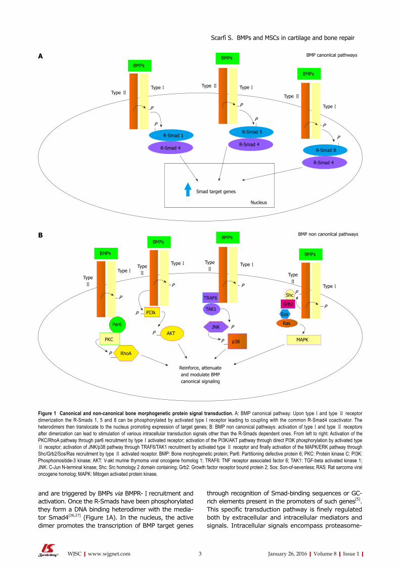
Figure 1 Canonical and non-canonical bone morphogenetic protein signal transduction. A: BMP canonical pathway: Upon type Ⅰ and type Ⅱ receptor dimerization the R-Smads 1, 5 and 8 can be phosphorylated by activated type Ⅰ receptor leading to coupling with the common R-Smad4 coactivator. The heterodimers then translocate to the nucleus promoting expression of target genes; B: BMP non canonical pathways: activation of type Ⅰ and type Ⅱ receptors after dimerization can lead to stimulation of various intracellular transduction signals other than the R-Smads dependent ones. From left to right: Activation of the PKC/RhoA pathway through par6 recruitment by type Ⅰ activated receptor; activation of the PI3K/AKT pathway through direct PI3K phosphorylation by activated type Ⅱ receptor; activation of JNK/p38 pathway through TRAF6/TAK1 recruitment by activated type Ⅱ receptor and finally activation of the MAPK/ERK pathway through Shc/Grb2/Sos/Ras recruitment by type Ⅱ activated receptor. BMP: Bone morphogenetic protein; Par6: Partitioning defective protein 6; PKC: Protein kinase C; PI3K: Phosphoinositide-3 kinase; AKT: V-akt murine thymoma viral oncogene homolog 1; TRAF6: TNF receptor associated factor 6; TAK1: TGF-beta activated kinase 1; JNK: C-Jun N-terminal kinase; Shc: Src homology 2 domain containing; Grb2: Growth factor receptor bound protein 2; Sos: Son-of-sevenless; RAS: Rat sarcoma viral oncogene homolog; MAPK: Mitogen activated protein kinase.
Scarfì S. BMPs and MSCs in cartilage and bone repair
Smad signaling have been demonstrated to alternatively reinforce, attenuate or otherwise modulate downstream BMP cellular responses[32,33].
BMPS IN BONE FORMATIONIn vertebrates, bone formation can be achieved by direct differentiation of osteoblasts in membranous ossifica-tion, or starting from differentiation of chondrocytes in endochondral ossification[34,35]. These two processes are directed by BMPs, with BMP2 and BMP4 acting as the master differentiation triggers of osteoblast and chondrocyte phenotypes leading to bone and cartilage formation[35].
BMP2 and BMP4 drive bone formation through the Smad1/5/8 signaling pathway described earlier. This pathway is common to osteoblasts and chondrocytes and its precursors and is strictly regulated in these cells[1]. BMPs are released in a mature form from osteoblasts and may interact with their cell surface receptors or bind to proteins of the ECM. In the latter case the ECM acts as a “reservoir” of BMPs for future paracrine signaling[5]. In regard to this, a number of transcription factors necessary to cartilage and bone formation have been acknowledged regulating downstream BMP signaling[35].
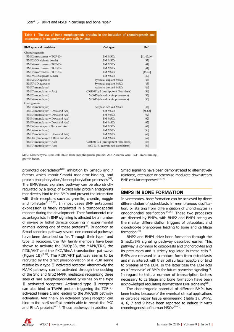
The chondrogenic potential of different BMPs has been tested because of the eventual clinical applications in cartilage repair tissue engineering (Table 1). BMP2, 4, 6, 7 and 9 have been reported to induce in vitro chondrogenesis of human MSCs[36-42].
promoted degradation[28], inhibition by Smad6 and 7 factors which impair Smad4 mediator binding, and protein phosphorylation/dephosphorylation processes[29]. The BMP/Smad signaling pathway can be also strictly regulated by a group of extracellular protein antagonists that directly bind to the BMPs and prevent the interaction with their receptors such as gremlin, chordin, noggin and follistatin[1,17,30]. In most cases BMP antagonist expression is finely regulated in a temporospatial manner during the development. Their fundamental role as antagonists in BMP signaling is attested by a number of severe or lethal defects occurring in experimental animals lacking one of these proteins[1]. In addition to Smad canonical pathway several non canonical pathways have been described so far. Through their type Ⅰ and type Ⅱ receptors, the TGF family members have been shown to activate the JNK/p38, the MAPK/ERK, the PI3K/AKT and the PKC/RhoA transduction pathways (Figure 1B)[6,31]. The PI3K/AKT pathway seems to be recruited by the direct phosphorylation of a PI3K serine residue by a type Ⅱ activated receptor. Alternatively the MAPK pathway can be activated through the docking of the Shc and Grb2 MAPK mediators recognizing three sites of rare autophosphorylated tyrosines on the type Ⅱ activated receptors. Activated type Ⅱ receptor can also bind to TRAF6 protein triggering the TGF-β-activated kinase 1 and leading to the JNK/p38 pathway activation. And finally an activated type Ⅰ receptor can bind to the par6 scaffold protein able to recruit the PKC and RhoA proteins[6,31]. These pathways in addition to
4WJSC|www.wjgnet.com January 26, 20�6|Volume 8|Issue �|
BMP type and conditions Cell type Ref.
Chondrogenesis BMP2 (micromass + TGF-β�) BM MSCs [4�,45,46] BMP2 (�D alginate beads) BM MSCs [�7] BMP4 (micromass + TGF-β�) BM MSCs [4�] BMP6 (micromass + TGF-β�) BM MSCs [4�] BMP7 (micromass + TGF-β�) BM MSCs [45,46] BMP9 (�D alginate beads) BM MSCs [�7] BMP2 (�D agarose) Synovial explant MSCs [4�] BMP7 (�D agarose) Synovial explant MSCs [4�] BMP7 (monolayer) Adipose derived MSCs [44] BMP7 (monolayer + Asc) C3H10T1/2 (multipotent fibroblasts) [54] BMP2 (monolayer) MC6�5 (chondrocyte precursors) [5�] BMP4 (monolayer) MC6�5 (chondrocyte precursors) [5�]Osteogenesis BMP2 (monolayer) Adipose derived MSCs [44] BMP2 (monolayer + Dexa and Asc) BM MSCs [56,62] BMP� (monolayer + Dexa and Asc) BM MSCs [62] BMP4 (monolayer + Dexa and Asc) BM MSCs [62] BMP5 (monolayer + Dexa and Asc) BM MSCs [62] BMP6(monolayer + Dexa and Asc) BM MSCs [62] BMP6 (monolayer) BM MSCs [58] BMP7 (monolayer + Dexa and Asc) BM MSCs [62] BMP8a (monolayer + Dexa and Asc) BM MSCs [62] BMP7 (monolayer + Asc) C3H10T1/2 (multipotent fibroblasts) [55] BMP7 (monolayer + Asc) MC�T�-E� (committed osteoblasts) [54]
Table 1 The use of bone morphogenetic proteins in the induction of chondrogenesis and osteogenesis in mesenchymal stem cells in vitro
MSC: Mesenchymal stem cell; BMP: Bone morphogenetic protein; Asc: Ascorbic acid; TGF: Transforming growth factor.
Scarfì S. BMPs and MSCs in cartilage and bone repair

In human bone marrow-derived (BM) MSCs, BMP2 (in the presence of TGF-β3) was the most efficient inducer of chondrogenesis with the production of a proteoglycan rich cartilage over BMP4 and BMP6[41] while in synovial explants, BMP7 was a more effective trigger of chondrocyte differentiation than BMP2[43]. BMP7 could also stimulate chondrogenic differentiation in adipose tissue-derived stem cells[44] while in other studies Shen et al[45,46] demonstrated that both BMP2 and BMP7 enhance TGF-β3-mediated chondrogenic phenotype of BM MSCs in vitro. In another study, BMP9 and BMP2 used separately and in absence of TGF-β stimulation enhanced the expression of cartilage transcription factor Sox-9 followed by induction of type Ⅱ collagen, aggrecan and cartilage oligomeric matrix protein in BM MSCs[37]. In addition, BMP13 and 14, also called CDMP2 and 1 respectively, demonstrated to be necessary for stimulation of early chondrogenesis and chondrocyte differentiation (BMP14/CDMP1) as well as in the terminal differentiation of chondrocytes in the final stage of hypertrophy and mineralization in vivo[19]. In vivo, BMP7 has also shown a marked anabolic activity in cartilage and bone[47,48] and it has demonstrated to act synergistically with microfractures to boost cartilage repair[49]. Related to this, Mishima and Lotz[50] have more recently demonstrated that BMP4 and 7 elicit a significant chemotactic in vitro response from human MSCs suggesting that the use of these factors in vivo promotes directed cell migration in sites of injury for cartilage repair in transplanted engineered tissues.
For what concerns osteoinduction, studies of pre-n-atal bone development as well as of fracture repair[9,51,52] showed the expression of a plethora of BMP genes with temporospatial variability. In particular, early experiments using human recombinant BMP2, BMP4 BMP6 or BMP7 demonstrated that such proteins are able to individually stimulate osteoblastic (or chondrogenic) phenotypes in a variety of mesenchymal precursor cell lines (Table 1)[53-58]. However, differently from in vitro studies, in vivo investigations indicate that BMPs work in a coordinated fashion[52,59]. In particular, BMP2 can be described as a necessary constituent orchestrating the signaling pathway that regulates fracture repair[60-62]. Differently, BMP7 is undetectable in the MSC differentiating system, but when exogenously added may play the same function of one of the endogenous BMPs physiologically produced by the cells[62].
As a matter of fact, both BMP2 and BMP7 are now approved in clinics for the treatment of non-union fractures as adjunct therapies[63]. In particular human recombinant BMP2 is sold from Medtronic (Minneapolis, MN, United States) with the acronym of In FUSE®, while hrBMP7 is sold from Stryker (Kalamazoo, MI, United States) with the acronym OP-1.
Although the use of these molecules in fracture healing has been welcomed by physicians with great enthusiasm, it must be emphasized that several, clinically relevant, adverse effects have been reported especially at BMP high dosages. The most frequently
described effect is the development of antibodies against BMPs even if this event does not seem to have real adverse consequences[64]. Differently, serious concerns raised from the observation that application of BMPs to a fracture site could result in increased bone resorption as a primary event. As a consequence a higher nonunion rate has been observed in a number of patients leading to termination of BMP use in several clinical settings[65]. Furthermore, local inflammatory responses have also been reported at several anatomical sites, with different degrees of severity[66]. Finally BMP use has also been associated to wound healing complications[66], hematoma formation[67] and several cases of heterotopic bone formation[67]. Thus, we can conclude that the dosage of these powerful molecules needs to be finely calibrated in each clinical setting and in any case reserved to patients in which the risks associated to BMP use are clearly outweighed by the higher risks of fracture healing failure.
MSCSRepair of adult bone involves BM MSCs which serve as a source of osteochondral progenitors able to invade the fracture site, proliferate and differentiate into cartilage and bone. MSCs are multipotent adult cells that have the ability to self-renew and differentiate into multiple lineages[10] that were discovered in 1980[68] but only fully recognized in 1994[69]. MSCs have recently gained increasing attention for their potential in the regenerative medicine. The main reasons for this interest are the relative ease of isolation from several adult tissues and suitable expansion in culture and the high degree of plasticity of these cells. Currently, at least 198 registered MSC clinical trials are ongoing (www.clinicaltrials.gov), as well as autologous and allogeneic MSC products accepted for use in bone repair in a number of inter-national jurisdictions (Mesoblast_Media_Release by Mesoblast Ltd., Melbourne, Australia; Osteocel by Osiris therapeutics Inc., Columbia, MD, United States)[70]. Despite their apparent therapeutic potential, clinical applications of MSCs have been restricted due to the limited understanding of the factors that regulate their fate and activity. Another limiting factor is the lack of knowledge of the complex interplay between these cells and the components of their niche or immediate microenvironment. Due to the disposition of MSC to differentiate into osteoblasts and chondrocytes, and their attested clinical potential in bone tissue engineering, a great amount of research has been centered on the identification of the factors governing osteogenesis in vitro and in vivo (i.e., TGF-β1, 2 and 3, BMPs and PDGF)[71,72].
MSCS IN CHONDROGENIC AND OSTEOGENIC DIFFERENTIATIONThe chondrogenic differentiation occurs when MSCs are
5WJSC|www.wjgnet.com January 26, 20�6|Volume 8|Issue �|
Scarfì S. BMPs and MSCs in cartilage and bone repair
seeded in serum-free, 3D culture format in the presence of one or more TGF-β superfamily members[73]. In this asset, cells abandon the typical fibroblastic morphology and start producing cartilage-specific matrix components. In vitro chondrogenesis is usually obtained by the micromass pellet culture system, allowing the necessary cell-cell interactions which resemble what occurs in pre-chondrogenic condensations in the embryonic develo-pment[74]. In these conditions cells usually differentiate in no more than 2-3 wk into chondrocyte-like cells secreting proteoglycans. Pellets are bordered by a narrow capsule of connective tissue, almost cell-free and rich in type ⅡA collagen. The advancement to terminal differentiation is attested by accumulation of type Ⅹ collagen and matrix mineralization[75]. When BMPs are added in this experimental setting, namely MSCs in micromass culture and in the presence TFG-β, they enhance chondrogenic differentiation and cartilage formation significantly (see Table 1 for the various BMP employed). The 3D culture and the concomitant presence of TGF-β seem to be necessary to attain a real chondrocytic phenotype. Thus, it is possible that in the mesenchymal precursor chondrocyte differentiation occurs only when strict cell-cell interactions are established and when the parallel activation of different R-Smad pathways is achieved by different members of the TGF-β superfamily. In particular, the TGF-β members activating the Smad2/3 and the BMP members activating the Smad1/5/8 (see Figure 1A).
Differently, MSCs undergo an osteogenic differenti-ation when cultured with the opportune osteoinduction factors on two dimensional substrates. In this case, osteogenesis is promoted by a large spread area, while in the same conditions the reduction of the spread area induces adipogenesis[76,77]. In this experimental setting, namely MSCs in 2D wide spread areas, several BMPs used alone or in the presence of ascorbic acid have demonstrated to promote significant osteob-last differentiation (see Table 1 for the various BMP employed). In the presence of BMPs, progenitor cells achieve an osteoblastic phenotype expressing several bone-characterizing ECM proteins. In particular they express type Ⅰ collagen, osteopontin, osteocalcin and bone sialoprotein, and produce high levels of the alkaline phosphatase (ALP) ecto-enzyme. Sustained expression of ALP is required for mineralization of skeletal tissues[78,79], and is induced early during osteoblast differentiation[80,81].
Several studies have explored the use of MSCs encapsulated in osteoinductive scaffolds or morphogenic biomaterials to enhance the natural healing process of bone and cartilage in vivo[82-86]. They overall suggest that these multipotent cells seem both able to differentiate themselves within the scaffolds as well as to secrete factors attracting neighboring autologous progenitors. This behavior can accomplish fracture healing faster and with a superior quality of the resulting new bone respect to the osteoinductive or chondrogenic scaffolds used alone[13]. Thus, these promising results have prompted
the accomplishment of several studies exploring the concomitant use of MSCs and of the most promising members of the BMP family. Namely BMP2 and 7, embedded in suitable scaffolds or carriers, have been used to heal several cartilage defects and bone fractures in experimental animal models hopefully soon to be transferred to human beings.
USE OF BMPS AND MSCS IN CARTILAGE REPAIR Cartilage defects such as degeneration of intervertebral discs and knee joints are ordinary causes of joint disabilities able to affect the quality of life of many people all over the world[87]. It is well known that articular cartilage has a limited capacity of spontaneous repair after damage[88]. Treatments for articular surface lesions usually encompass various clinical approaches like conservation therapies as well as invasive surgery comprising abrasion, debridement and perichondral grafting[87,89]. In recent times also autologous chond-rocyte regeneration has been used. Grafting of autolo-gous chondrocytes to promote cartilage resurfacing has some benefits over allogeneic chondrocyte or solid tissue grafting and other procedures[90]. Unfortunately, its application is hindered by chondrocyte de-differentiation during in vitro expansion and the necessity of large amounts of cartilage samples[91]. Recently, the appea-rance of MSCs in the landscape of the cellular sources for cartilage repair available in quite large quantities raised a great interest and optimism for the treatment of these defects by tissue engineering and cell therapy approaches[92]. As already mentioned, both BMP2 and BMP7 have plenty demonstrated the ability to enhance cartilage repair in vivo as well as the capacity to promote chondrogenic differentiation of MSCs cultured in appropriate inducing media in vitro. Although the outcome of the combined use of precursor cells and BMPs in suitable scaffolds for cartilage repair could be a research field actively persecuted in these years, to date a limited number of studies are present in the literature (Table 2). In particular, one of the major unresolved problems is a durable integration between cartilage and the scaffold[93]. Thus, the presence of chondrogenic precursors releasing chemoattractant factors and of appropriate BMPs stimulating said precursors could ensure the ultimate scaffold remodeling with new cartilaginous tissue formation. Furthermore, both MSCs and BMPs seem able to stimulate endogenous cells to migrate and colonize the artificial graft further promoting the final healing.
In early studies Grande et al[94] transfected MSCs from periosteum with human BMP7 or sonic hedgehog and then seeded them on bioresorbable polymer scaffolds. These implants were used to fill full-thickness osteochondral defects created in the mid-trochlear region of New Zealand white rabbits. The authors observed that, for both genes, their addition significantly
6WJSC|www.wjgnet.com January 26, 20�6|Volume 8|Issue �|
Scarfì S. BMPs and MSCs in cartilage and bone repair
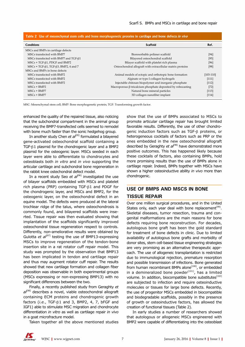
enhanced the quality of the repaired tissue, also noticing that the subchondral compartment in the animal group receiving the BMP7-transfected cells seemed to remodel with bone much faster than the sonic hedgehog group.
In another study Chen et al[95] formulated a bilayered gene-activated osteochondral scaffold containing a TGF-β1 plasmid for the chondrogenic layer and a BMP2 plasmid for the osteogenic layer. MSCs seeded in each layer were able to differentiate to chondrocytes and osteoblasts both in vitro and in vivo supporting the articular cartilage and subchondral bone regeneration in the rabbit knee ostechondral defect model.
In a recent study Seo et al[96] investigated the use of bilayer scaffolds embedded with MSCs and platelet rich plasma (PRP) containing TGF-β1 and PDGF for the chondrogenic layer, and MSCs and BMP2, for the osteogenic layer, on the osteochondral defect in an equine model. The defects were produced at the lateral trochlear ridge of the talus, where osteochondrosis is commonly found, and bilayered scaffolds were inse-rted. Tissue repair was then evaluated showing that implantation of the scaffolds significantly improved osteochondral tissue regeneration respect to controls. Differently, non-ameliorative results were obtained by Gulotta et al[97] testing the use of BMP13-expressing MSCs to improve regeneration of the tendon-bone insertion site in a rat rotator cuff repair model. This study was prompted by the observation that BMP13 has been implicated in tendon and cartilage repair and thus may augment rotator cuff repair. The results showed that new cartilage formation and collagen fiber deposition was observable in both experimental groups (MSCs expressing or non-expressing BMP13) with no significant differences between the two.
Finally, a recently published study from Geraghty et al[98] describes a novel, viable osteochondral allograft containing ECM proteins and chondrogenic growth factors (i.e., TGF-β1 and 3, BMP2, 4, 7, bFGF and IGF1) able to stimulate MSC migration and chondrocyte differentiation in vitro as well as cartilage repair in vivo in a goat microfracture model.
Taken together all the above mentioned studies
show that the use of BMPs associated to MSCs to promote articular cartilage repair has brought limited favorable results. Differently, the use of other chondro-genic induction factors such as TGF-β proteins, or heterogeneous cocktails of factors such as PRP or the ones embedded in the new osteochondral allograft described by Geraghty et al[98] have demonstrated more positive outcomes. This has happened likely because these cocktails of factors, also containing BMPs, hold more promising results than the use of BMPs alone in cartilage repair. Indeed, BMPs together with MSCs have shown a higher osteoinductive ability in vivo more than chondrogenic.
USE OF BMPS AND MSCS IN BONE TISSUE REPAIROver one million surgical procedures, and in the United States only, each year deal with bone replacement[99]. Skeletal diseases, tumor resection, trauma and con-genital malformations are the main reasons for bone defects requiring bone reconstruction. For decades, autologous bone graft has been the gold standard for treatment of bone defects in clinic. Due to limited availability of autologous bone grafts and morbidity of donor sites, stem cell-based tissue engineering strategies are very promising as an alternative therapeutic appr-oach. The use of allogeneic transplantation is restricted due to immunological rejection, premature resorption and possible transmission of infections. Bone generated from human recombinant BMPs alone[100], or embedded in a demineralized bone powder[101], has a limited volume. In addition, biocompatible bone substitutes[102] are subjected to infection and require osteoinductive molecules or tissues for large bone defects. Recently, the use of progenitor MSCs embedded in biocompatible and biodegradable scaffolds, possibly in the presence of growth or osteoinductive factors, has allowed the creation of functional tissues (Table 2).
In early studies a number of researchers showed that autologous or allogeneic MSCs engineered with BMP2 were capable of differentiating into the osteoblast
7WJSC|www.wjgnet.com January 26, 20�6|Volume 8|Issue �|
Conditions Scaffold Ref.
MSCs and BMPs in cartilage defects MSCs transfected with BMP7 Bioresorbable polimer scaffold [94] MSCs transfected with BMP7 and TGF-β� Bilayered osteochondral scaffold [95] MSCs + TGF-β�, PDGF and BMP2 Bilayer scaffold with platelet rich plasma [96] MSCs + TGF-β�, TGF-β�, BMP2, 4 and 7 Osteochondral allograft with extracellular matrix proteins [98]MSCs and BMPs in bone defects MSCs transfected with BMP2 Animal models of ectopic and orthotopic bone formation [�0�-��0] MSCs transfected with BMP2 Alginate or type I collagen hydrogels [���] MSCs transfected with BMP2 Injectable chitosan biopolymer and inorganic phosphate [��2] MSCs + BMP2 Macroporous β-tricalcium phosphate deposited by robocasting [72] MSCs + BMP7 Natural bone mineral particles [���] MSCs + BMP7 3D collagen nanofiber implant [��4]
Table 2 Use of mesenchymal stem cells and bone morphogenetic proteins in cartilage and bone defects in vivo
MSC: Mesenchymal stem cell; BMP: Bone morphogenetic protein; TGF: Transforming growth factor.
Scarfì S. BMPs and MSCs in cartilage and bone repair
lineage and inducing bone formation in several animal models in both ectopic and orthotopic sites in mice, rats, rabbits and pigs[103-107]. In all these systems the authors concluded that combining MSC implantation with BMP2 gene transfer more effectively induced bone formation than MSC implantation alone.
With similar results, but using a different modular expression system approach Moutsatsos et al[108] used a tetracycline-regulated expression vector encoding human BMP2 to transfect a MSC cell line. With such expression system the authors were able to demonstrate that doxycycline controlled BMP2 expression and thus controlled MSC osteogenic differentiation both in vitro and in vivo in a mouse ectopic bone model. Moreover, they showed increased angiogenesis accompanied by bone formation whenever genetically engineered MSCs were induced to express BMP2 in vivo.
In other studies, Chang et al[109] demonstrated the usefulness of BMP2-expressing MSCs in bone repair of large cranial defect in two different animal models: The rabbit model and the swine model[110]. The authors clearly demonstrated near-complete repair of the large cranial defects by the tissue engineered bone containing BMP2-expressing MSCs in the three months of the experiment both in the rabbit[109] and in the swine[110] with respect to the controls.
Thus, the use BMP2 together with MSCs in bone repair, either exogenously added to cells either enabling cells to directly express the protein, has been in the years thoroughly validated by the above mentioned studies. Consequently, the attention has been focused on the use of different scaffolds able to support the MSC colonization and differentiation as well as the temporospatially controlled delivery of the BMPs to quicken bone reconstruction and healing. Thus, in this contest were alternatively tested: (1) alginate or type Ⅰ collagen hydrogels as scaffolds loaded with MSCs expressing BMP2 for bone regeneration in a large cranial defect repair in the swine demonstrating the superiority of BMP2-MSC/collagen type Ⅰ construct over the alginate counterpart[111]; (2) an injectable biopolymer of chitosan and inorganic phosphate seeded with MSCs and BMP2 in a rat calvarial critical size defect demonstrating the superiority of the MSC/BMP2 coupling over the controls[112]; and (3) a macroporous β-tricalcium phosphate (β-TCP) system fabricated by robocasting loaded with MSCs and with BMP2 embedded in microspheres to provide a prolonged BMP release in a critical rat calvarial defect[72]. In the latter case only a minor synergistic effect was demonstrated in the BMP2-MSC group with respect to the BMP2 group alone.
Alternative to these studies only a limited number of works have focused on the concomitant use of BMP7 and MSCs in bone repair (Table 2). In particular Bura-stero et al[113] used the association of human MSCs and BMP7, with natural bone mineral particles as a scaffold to fill the bone loss, to improve bone regeneration in a rat model of critical size segmental bone defect. Indeed a significantly higher score in bone regeneration was
observed in the rats treated with MSCs and BMP7 compared to controls, receiving either MSCs or BMP-7. The data indicated that the association of the two provided a better osteoinductive graft compared to MSCs or BMP7 alone. Finally, Schiavi et al[114] tested a novel 3D collagen nanofiber implant functionalized with BMP7 nanoreservoirs and equipped with human MSC microtissues. The implant was optimized for cell colonization, differentiation and growth. The group clearly demonstrated an acceleration of ectopic bone growth in vivo of the coupled BMP7/MSC microtissues respect to the controls using either BMP7 or MSC microtissues alone.
CONCLUSIONSince their first identification, BMPs have demonstrated great potentialities in the regenerative medicine and tissue engineering fields. They have been tested in numerous preclinical and clinical studies exploring their chondrogenic or osteoinductive potential in several animal model defects and in human diseases. During the years two BMP members in particular, BMP2 and BMP7, have been thoroughly used in the treatment of a number of cartilage and bone defects and have been recently approved for employment in protocols of nonunion fractures as adjunct therapies.
On the other hand, to date the scientific literature provides extensive in vitro evidence of the improvement of the osteoblastic and chondrogenic potential of MSCs, now obtained from many tissues, by treatment with BMPs. Thus, it was just a matter of time for the two, BMPs and MSCs, to be investigated together hopefully to finally achieve the goal of producing the ideal graft for bone replacement. Besides, recently the grafts have evolved including more and more sophisticated scaffolds, appropriate cell precursors and optimal differentiating factors. As outlined in this review, the growing literature in this field and the promising results in recent years suggest that this goal indeed can be achieved and that both BMPs and MSCs in the future will take part to the production of successful avant-garde implants especially designed for bone tissue engineering.
REFERENCES1 Walsh DW, Godson C, Brazil DP, Martin F. Extracellular BMP-
antagonist regulation in development and disease: tied up in knots. Trends Cell Biol 2010; 20: 244-256 [PMID: 20188563 DOI: 10.1016/j.tcb.2010.01.008]
2 Alexander SP, Benson HE, Faccenda E, Pawson AJ, Sharman JL, Spedding M, Peters JA, Harmar AJ. The Concise Guide to PHARMACOLOGY 2013/14: catalytic receptors. Br J Pharmacol 2013; 170: 1676-1705 [PMID: 24528241 DOI: 10.1111/bph.12449]
3 Ducy P, Karsenty G. The family of bone morphogenetic proteins. Kidney Int 2000; 57: 2207-2214 [PMID: 10844590 DOI: 10.1046/j.1523-1755.2000.00081.x]
4 Sakou T. Bone morphogenetic proteins: from basic studies to clinical approaches. Bone 1998; 22: 591-603 [PMID: 9626397 DOI: 10.1016/S8756-3282(98)00053-2]
5 Miyazono K, Kamiya Y, Morikawa M. Bone morphogenetic
8WJSC|www.wjgnet.com January 26, 20�6|Volume 8|Issue �|
Scarfì S. BMPs and MSCs in cartilage and bone repair
protein receptors and signal transduction. J Biochem 2010; 147: 35-51 [PMID: 19762341 DOI: 10.1093/jb/mvp148]
6 Wang RN, Green J, Wang Z, Deng Y, Qiao M, Peabody M, Zhang Q, Ye J, Yan Z, Denduluri S, Idowu O, Li M, Shen C, Hu A, Haydon RC, Kang R, Mok J, Lee MJ, Luu HL, Shi LL. Bone Morphogenetic Protein (BMP) signaling in development and human diseases. Genes Dis 2014; 1: 87-105 [PMID: 25401122 DOI: 10.1016/j.gendis.2014.07.005]
7 Wagner DO, Sieber C, Bhushan R, Börgermann JH, Graf D, Knaus P. BMPs: from bone to body morphogenetic proteins. Sci Signal 2010; 3: mr1 [PMID: 20124549 DOI: 10.1126/scisignal.3107mr1]
8 Nolan K, Thompson TB. The DAN family: modulators of TGF-β signaling and beyond. Protein Sci 2014; 23: 999-1012 [PMID: 24810382 DOI: 10.1002/pro.2485]
9 Cho TJ, Gerstenfeld LC, Einhorn TA. Differential temporal expression of members of the transforming growth factor beta superfamily during murine fracture healing. J Bone Miner Res 2002; 17: 513-520 [PMID: 11874242 DOI: 10.1359/jbmr.2002.17.3.513]
10 Pittenger MF, Mackay AM, Beck SC, Jaiswal RK, Douglas R, Mosca JD, Moorman MA, Simonetti DW, Craig S, Marshak DR. Multilineage potential of adult human mesenchymal stem cells. Science 1999; 284: 143-147 [PMID: 10102814 DOI: 10.1126/science.284.5411.143]
11 Scarfì S. Purinergic receptors and nucleotide processing ectoen-zymes: Their roles in regulating mesenchymal stem cell functions. World J Stem Cells 2014; 6: 153-162 [PMID: 24772242 DOI: 10.4252/wjsc.v6.i2.153]
12 Krampera M, Pizzolo G, Aprili G, Franchini M. Mesenchymal stem cells for bone, cartilage, tendon and skeletal muscle repair. Bone 2006; 39: 678-683 [PMID: 16765663 DOI: 10.1016/j.bone.2006.04.020]
13 Sun H, Yang HL. Calcium phosphate scaffolds combined with bone morphogenetic proteins or mesenchymal stem cells in bone tissue engineering. Chin Med J (Engl) 2015; 128: 1121-1127 [PMID: 25881610 DOI: 10.4103/0366-6999.155121]
14 Urist MR. Bone: formation by autoinduction. Science 1965; 150: 893-899 [PMID: 5319761 DOI: 10.1126/science.150.3698.893]
15 Pizette S, Niswander L. BMPs are required at two steps of limb chondrogenesis: formation of prechondrogenic condensations and their differentiation into chondrocytes. Dev Biol 2000; 219: 237-249 [PMID: 10694419 DOI: 10.1006/dbio.2000.9610]
16 Wozney JM, Rosen V, Celeste AJ, Mitsock LM, Whitters MJ, Kriz RW, Hewick RM, Wang EA. Novel regulators of bone formation: molecular clones and activities. Science 1988; 242: 1528-1534 [PMID: 3201241 DOI: 10.1126/science.3201241]
17 Balemans W, Van Hul W. Extracellular regulation of BMP signaling in vertebrates: a cocktail of modulators. Dev Biol 2002; 250: 231-250 [PMID: 12376100 DOI: 10.1006/dbio.2002.0779]
18 Weiskirchen R, Meurer SK. BMP-7 counteracting TGF-beta1 activities in organ fibrosis. Front Biosci (Landmark Ed) 2013; 18: 1407-1434 [PMID: 23747893 DOI: 10.2741/4189]
19 Chang SC, Hoang B, Thomas JT, Vukicevic S, Luyten FP, Ryba NJ, Kozak CA, Reddi AH, Moos M. Cartilage-derived morphogenetic proteins. New members of the transforming growth factor-beta superfamily predominantly expressed in long bones during human embryonic development. J Biol Chem 1994; 269: 28227-28234 [PMID: 7961761]
20 Xiao YT, Xiang LX, Shao JZ. Bone morphogenetic protein. Biochem Biophys Res Commun 2007; 362: 550-553 [PMID: 17719560 DOI: 10.1016/j.bbrc.2007.08.045]
21 Bragdon B, Moseychuk O, Saldanha S, King D, Julian J, Nohe A. Bone morphogenetic proteins: a critical review. Cell Signal 2011; 23: 609-620 [PMID: 20959140 DOI: 10.1016/j.cellsig.2010.10.003]
22 Guo J, Wu G. The signaling and functions of heterodimeric bone morphogenetic proteins. Cytokine Growth Factor Rev 2012; 23: 61-67 [PMID: 22421241 DOI: 10.1016/j.cytogfr.2012.02.001]
23 Rosenzweig BL, Imamura T, Okadome T, Cox GN, Yamashita H, ten Dijke P, Heldin CH, Miyazono K. Cloning and characterization of a human type II receptor for bone morphogenetic proteins. Proc Natl Acad Sci USA 1995; 92: 7632-7636 [PMID: 7644468 DOI:
10.1073/pnas.92.17.7632]24 Nohe A, Keating E, Knaus P, Petersen NO. Signal transduction
of bone morphogenetic protein receptors. Cell Signal 2004; 16: 291-299 [PMID: 14687659 DOI: 10.1016/j.cellsig.2003.08.011]
25 Riggins GJ, Thiagalingam S, Rozenblum E, Weinstein CL, Kern SE, Hamilton SR, Willson JK, Markowitz SD, Kinzler KW, Vogelstein B. Mad-related genes in the human. Nat Genet 1996; 13: 347-349 [PMID: 8673135 DOI: 10.1038/ng0796-347]
26 Xu L, Chen YG, Massagué J. The nuclear import function of Smad2 is masked by SARA and unmasked by TGFbeta-dependent phosphorylation. Nat Cell Biol 2000; 2: 559-562 [PMID: 10934479 DOI: 10.1038/35019649]
27 Shi W, Chang C, Nie S, Xie S, Wan M, Cao X. Endofin acts as a Smad anchor for receptor activation in BMP signaling. J Cell Sci 2007; 120: 1216-1224 [PMID: 17356069 DOI: 10.1242/jcs.03400]
28 Zhu H, Kavsak P, Abdollah S, Wrana JL, Thomsen GH. A SMAD ubiquitin ligase targets the BMP pathway and affects embryonic pattern formation. Nature 1999; 400: 687-693 [PMID: 10458166]
29 Shi W, Sun C, He B, Xiong W, Shi X, Yao D, Cao X. GADD34-PP1c recruited by Smad7 dephosphorylates TGFbeta type I receptor. J Cell Biol 2004; 164: 291-300 [PMID: 14718519 DOI: 10.1083/jcb.200307151]
30 Nakamura J, Yanagita M. Bmp modulators in kidney disease. Discov Med 2012; 13: 57-63 [PMID: 22284784]
31 Zhang YE. Non-Smad pathways in TGF-beta signaling. Cell Res 2009; 19: 128-139 [PMID: 19114990 DOI: 10.1038/cr.2008.328]
32 Derynck R, Zhang YE. Smad-dependent and Smad-independent pathways in TGF-beta family signalling. Nature 2003; 425: 577-584 [PMID: 14534577 DOI: 10.1038/nature02006]
33 Moustakas A, Heldin CH. Non-Smad TGF-beta signals. J Cell Sci 2005; 118: 3573-3584 [PMID: 16105881 DOI: 10.1242/jcs.02554]
34 Karsenty G. The complexities of skeletal biology. Nature 2003; 423: 316-318 [PMID: 12748648 DOI: 10.1038/nature01654]
35 Nishimura R, Hata K, Matsubara T, Wakabayashi M, Yoneda T. Regulation of bone and cartilage development by network between BMP signalling and transcription factors. J Biochem 2012; 151: 247-254 [PMID: 22253449 DOI: 10.1093/jb/mvs004]
36 Estes BT, Wu AW, Guilak F. Potent induction of chondrocytic differentiation of human adipose-derived adult stem cells by bone morphogenetic protein 6. Arthritis Rheum 2006; 54: 1222-1232 [PMID: 16572454 DOI: 10.1002/art.21779]
37 Majumdar MK, Wang E, Morris EA. BMP-2 and BMP-9 promotes chondrogenic differentiation of human multipotential mesenchymal cells and overcomes the inhibitory effect of IL-1. J Cell Physiol 2001; 189: 275-284 [PMID: 11748585 DOI: 10.1002/jcp.10025]
38 Palmer GD, Steinert A, Pascher A, Gouze E, Gouze JN, Betz O, Johnstone B, Evans CH, Ghivizzani SC. Gene-induced chondro-genesis of primary mesenchymal stem cells in vitro. Mol Ther 2005; 12: 219-228 [PMID: 16043093 DOI: 10.1016/j.ymthe.2005.03.024]
39 Indrawattana N, Chen G, Tadokoro M, Shann LH, Ohgushi H, Tateishi T, Tanaka J, Bunyaratvej A. Growth factor combination for chondrogenic induction from human mesenchymal stem cell. Biochem Biophys Res Commun 2004; 320: 914-919 [PMID: 15240135 DOI: 10.1016/j.bbrc.2004.06.029]
40 Schmitt JM, Hwang K, Winn SR, Hollinger JO. Bone morpho-genetic proteins: an update on basic biology and clinical relevance. J Orthop Res 1999; 17: 269-278 [PMID: 10221845 DOI: 10.1002/jor.1100170217]
41 Sekiya I, Larson BL, Vuoristo JT, Reger RL, Prockop DJ. Comparison of effect of BMP-2, -4, and -6 on in vitro cartilage formation of human adult stem cells from bone marrow stroma. Cell Tissue Res 2005; 320: 269-276 [PMID: 15778851 DOI: 10.1007/s00441-004-1075-3]
42 Xu D, Gechtman Z, Hughes A, Collins A, Dodds R, Cui X, Jolliffe L, Higgins L, Murphy A, Farrell F. Potential involvement of BMP receptor type IB activation in a synergistic effect of chondrogenic promotion between rhTGFbeta3 and rhGDF5 or rhBMP7 in human mesenchymal stem cells. Growth Factors 2006; 24: 268-278 [PMID: 17381068]
43 Shintani N, Hunziker EB. Chondrogenic differentiation of bovine
9WJSC|www.wjgnet.com January 26, 20�6|Volume 8|Issue �|
Scarfì S. BMPs and MSCs in cartilage and bone repair

synovium: bone morphogenetic proteins 2 and 7 and transforming growth factor beta1 induce the formation of different types of cartilaginous tissue. Arthritis Rheum 2007; 56: 1869-1879 [PMID: 17530715 DOI: 10.1002/art.22701]
44 Knippenberg M, Helder MN, Zandieh Doulabi B, Wuisman PI, Klein-Nulend J. Osteogenesis versus chondrogenesis by BMP-2 and BMP-7 in adipose stem cells. Biochem Biophys Res Commun 2006; 342: 902-908 [PMID: 16500625 DOI: 10.1016/j.bbrc.2006.02.052]
45 Shen B, Wei A, Tao H, Diwan AD, Ma DD. BMP-2 enhances TGF-beta3-mediated chondrogenic differentiation of human bone marrow multipotent mesenchymal stromal cells in alginate bead culture. Tissue Eng Part A 2009; 15: 1311-1320 [PMID: 18950289 DOI: 10.1089/ten.tea.2008.0132]
46 Shen B, Wei A, Whittaker S, Williams LA, Tao H, Ma DD, Diwan AD. The role of BMP-7 in chondrogenic and osteogenic differentiation of human bone marrow multipotent mesenchymal stromal cells in vitro. J Cell Biochem 2010; 109: 406-416 [PMID: 19950204 DOI: 10.1002/jcb.22412]
47 Geesink RG, Hoefnagels NH, Bulstra SK. Osteogenic activity of OP-1 bone morphogenetic protein (BMP-7) in a human fibular defect. J Bone Joint Surg Br 1999; 81: 710-718 [PMID: 10463751]
48 Chubinskaya S, Kuettner KE. Regulation of osteogenic proteins by chondrocytes. Int J Biochem Cell Biol 2003; 35: 1323-1340 [PMID: 12798347 DOI: 10.1016/S1357-2725(03)00035-9]
49 Kuo AC, Rodrigo JJ, Reddi AH, Curtiss S, Grotkopp E, Chiu M. Microfracture and bone morphogenetic protein 7 (BMP-7) synergistically stimulate articular cartilage repair. Osteoarthritis Cartilage 2006; 14: 1126-1135 [PMID: 16765606 DOI: 10.1016/j.joca.2006.04.004]
50 Mishima Y, Lotz M. Chemotaxis of human articular chondrocytes and mesenchymal stem cells. J Orthop Res 2008; 26: 1407-1412 [PMID: 18464249 DOI: 10.1002/jor.20668]
51 Lyons KM, Hogan BL, Robertson EJ. Colocalization of BMP 7 and BMP 2 RNAs suggests that these factors cooperatively mediate tissue interactions during murine development. Mech Dev 1995; 50: 71-83 [PMID: 7605753 DOI: 10.1016/0925-4773(94)00326-I]
52 Duprez D, Bell EJ, Richardson MK, Archer CW, Wolpert L, Brickell PM, Francis-West PH. Overexpression of BMP-2 and BMP-4 alters the size and shape of developing skeletal elements in the chick limb. Mech Dev 1996; 57: 145-157 [PMID: 8843392 DOI: 10.1016/0925-4773(96)00540-0]
53 Valcourt U, Ronzière MC, Winkler P, Rosen V, Herbage D, Mallein-Gerin F. Different effects of bone morphogenetic proteins 2, 4, 12, and 13 on the expression of cartilage and bone markers in the MC615 chondrocyte cell line. Exp Cell Res 1999; 251: 264-274 [PMID: 10471312 DOI: 10.1006/excr.1999.4584]
54 Asahina I, Sampath TK, Hauschka PV. Human osteogenic protein-1 induces chondroblastic, osteoblastic, and/or adipocytic differentiation of clonal murine target cells. Exp Cell Res 1996; 222: 38-47 [PMID: 8549671 DOI: 10.1006/excr.1996.0005]
55 Shea CM, Edgar CM, Einhorn TA, Gerstenfeld LC. BMP treatment of C3H10T1/2 mesenchymal stem cells induces both chondrogenesis and osteogenesis. J Cell Biochem 2003; 90: 1112-1127 [PMID: 14635186 DOI: 10.1002/jcb.10734]
56 Rickard DJ, Sullivan TA, Shenker BJ, Leboy PS, Kazhdan I. Induction of rapid osteoblast differentiation in rat bone marrow stromal cell cultures by dexamethasone and BMP-2. Dev Biol 1994; 161: 218-228 [PMID: 8293874 DOI: 10.1006/dbio.1994.1022]
57 Dosch R, Gawantka V, Delius H, Blumenstock C, Niehrs C. Bmp-4 acts as a morphogen in dorsoventral mesoderm patterning in Xenopus. Development 1997; 124: 2325-2334 [PMID: 9199359]
58 Friedman MS , Long MW, Hankenson KD. Osteogenic differentiation of human mesenchymal stem cells is regulated by bone morphogenetic protein-6. J Cell Biochem 2006; 98: 538-554 [PMID: 16317727 DOI: 10.1002/jcb.20719]
59 Macias D, Gañan Y, Sampath TK, Piedra ME, Ros MA, Hurle JM. Role of BMP-2 and OP-1 (BMP-7) in programmed cell death and skeletogenesis during chick limb development. Development 1997; 124: 1109-1117 [PMID: 9102298]
60 Zhang H, Bradley A. Mice deficient for BMP2 are nonviable
and have defects in amnion/chorion and cardiac development. Development 1996; 122: 2977-2986 [PMID: 8898212]
61 Tsuji K, Bandyopadhyay A, Harfe BD, Cox K, Kakar S, Gers-tenfeld L, Einhorn T, Tabin CJ, Rosen V. BMP2 activity, although dispensable for bone formation, is required for the initiation of fracture healing. Nat Genet 2006; 38: 1424-1429 [PMID: 17099713 DOI: 10.1038/ng1916]
62 Edgar CM, Chakravarthy V, Barnes G, Kakar S, Gerstenfeld LC, Einhorn TA. Autogenous regulation of a network of bone morphogenetic proteins (BMPs) mediates the osteogenic differentiation in murine marrow stromal cells. Bone 2007; 40: 1389-1398 [PMID: 17303481 DOI: 10.1016/j.bone.2007.01.001]
63 Gautschi OP, Frey SP, Zellweger R. Bone morphogenetic proteins in clinical applications. ANZ J Surg 2007; 77: 626-631 [PMID: 17635273 DOI: 10.1111/j.1445-2197.2007.04175.x]
64 Lissenberg-Thunnissen SN, de Gorter DJ, Sier CF, Schipper IB. Use and efficacy of bone morphogenetic proteins in fracture healing. Int Orthop 2011; 35: 1271-1280 [PMID: 21698428 DOI: 10.1007/s00264-011-1301-z]
65 Delimar D, Smoljanovic T, Bojanic I. Could the use of bone morphogenetic proteins in fracture healing do more harm than good to our patients? Int Orthop 2012; 36: 683; author reply 685 [PMID: 22052478 DOI: 10.1007/s00264-011-1397-1]
66 Carragee EJ, Hurwitz EL, Weiner BK. A critical review of recom-binant human bone morphogenetic protein-2 trials in spinal surgery: emerging safety concerns and lessons learned. Spine J 2011; 11: 471-491 [PMID: 21729796 DOI: 10.1016/j.spinee.2011.04.023]
67 Boraiah S, Paul O, Hawkes D, Wickham M, Lorich DG. Complications of recombinant human BMP-2 for treating complex tibial plateau fractures: a preliminary report. Clin Orthop Relat Res 2009; 467: 3257-3262 [PMID: 19693635 DOI: 10.1007/s11999-009-1039-8]
68 Friedenstein AJ. Stromal mechanisms of bone marrow: cloning in vitro and retransplantation in vivo. Haematol Blood Transfus 1980; 25: 19-29 [PMID: 7021339]
69 Caplan AI. The mesengenic process. Clin Plast Surg 1994; 21: 429-435 [PMID: 7924141]
70 Parson AB. Stem cell biotech: seeking a piece of the action. Cell 2008; 132: 511-513 [PMID: 18295564 DOI: 10.1016/j.cell.2008.0 2.004]
71 Arthur A, Zannettino A, Gronthos S. The therapeutic applications of multipotential mesenchymal/stromal stem cells in skeletal tissue repair. J Cell Physiol 2009; 218: 237-245 [PMID: 18792913 DOI: 10.1002/jcp.21592]
72 Del Rosario C, Rodríguez-Évora M, Reyes R, Delgado A, Évora C. BMP-2, PDGF-BB, and bone marrow mesenchymal cells in a macroporous β-TCP scaffold for critical-size bone defect repair in rats. Biomed Mater 2015; 10: 045008 [PMID: 26201844 DOI: 10.1088/1748-6041/10/4/045008]
73 Barry F, Boynton RE, Liu B, Murphy JM. Chondrogenic differentiation of mesenchymal stem cells from bone marrow: differentiation-dependent gene expression of matrix components. Exp Cell Res 2001; 268: 189-200 [PMID: 11478845 DOI: 10.1006/excr.2001.5278]
74 Johnstone B, Hering TM, Caplan AI, Goldberg VM, Yoo JU. In vitro chondrogenesis of bone marrow-derived mesenchymal progenitor cells. Exp Cell Res 1998; 238: 265-272 [PMID: 9457080 DOI: 10.1006/excr.1997.3858]
75 Taipaleenmäki H, Suomi S, Hentunen T, Laitala-Leinonen T, Säämänen AM. Impact of stromal cell composition on BMP-induced chondrogenic differentiation of mouse bone marrow derived mesenchymal cells. Exp Cell Res 2008; 314: 2400-2410 [PMID: 18565511 DOI: 10.1016/j.yexcr.2008.04.019]
76 Engler AJ, Sen S, Sweeney HL, Discher DE. Matrix elasticity directs stem cell lineage specification. Cell 2006; 126: 677-689 [PMID: 16923388 DOI: 10.1016/j.cell.2006.06.044]
77 Kabiri M, Kul B, Lott WB, Futrega K, Ghanavi P, Upton Z, Doran MR. 3D mesenchymal stem/stromal cell osteogenesis and autocrine signalling. Biochem Biophys Res Commun 2012; 419: 142-147 [PMID: 22266317 DOI: 10.1016/j.bbrc.2012.01.017]
�0WJSC|www.wjgnet.com January 26, 20�6|Volume 8|Issue �|
Scarfì S. BMPs and MSCs in cartilage and bone repair
78 Fedde KN, Blair L, Silverstein J, Coburn SP, Ryan LM, Weinstein RS, Waymire K, Narisawa S, Millán JL, MacGregor GR, Whyte MP. Alkaline phosphatase knock-out mice recapitulate the metabolic and skeletal defects of infantile hypophosphatasia. J Bone Miner Res 1999; 14: 2015-2026 [PMID: 10620060 DOI: 10.1359/jbmr.1999.14.12.2015]
79 Anderson HC, Sipe JB, Hessle L, Dhanyamraju R, Atti E, Camacho NP, Millán JL. Impaired calcification around matrix vesicles of growth plate and bone in alkaline phosphatase-deficient mice. Am J Pathol 2004; 164: 841-847 [PMID: 14982838 DOI: 10.1016/S0002-9440(10)63172-0]
80 Rickard DJ, Kassem M, Hefferan TE, Sarkar G, Spelsberg TC, Riggs BL. Isolation and characterization of osteoblast precursor cells from human bone marrow. J Bone Miner Res 1996; 11: 312-324 [PMID: 8852942 DOI: 10.1002/jbmr.5650110305]
81 Thies RS, Bauduy M, Ashton BA, Kurtzberg L, Wozney JM, Rosen V. Recombinant human bone morphogenetic protein-2 induces osteoblastic differentiation in W-20-17 stromal cells. Endocrinology 1992; 130: 1318-1324 [PMID: 1311236 DOI: 10.1210/endo.130.3.1311236]
82 Wang T, Dang G, Guo Z, Yang M. Evaluation of autologous bone marrow mesenchymal stem cell-calcium phosphate ceramic composite for lumbar fusion in rhesus monkey interbody fusion model. Tissue Eng 2005; 11: 1159-1167 [PMID: 16144452 DOI: 10.1089/ten.2005.11.1159]
83 Lee LT, Kwan PC, Chen YF, Wong YK. Comparison of the effectiveness of autologous fibrin glue and macroporous biphasic calcium phosphate as carriers in the osteogenesis process with or without mesenchymal stem cells. J Chin Med Assoc 2008; 71: 66-73 [PMID: 18290250 DOI: 10.1016/S1726-4901(08)70077-7]
84 Jafarian M, Eslaminejad MB, Khojasteh A, Mashhadi Abbas F, Dehghan MM, Hassanizadeh R, Houshmand B. Marrow-derived mesenchymal stem cells-directed bone regeneration in the dog mandible: a comparison between biphasic calcium phosphate and natural bone mineral. Oral Surg Oral Med Oral Pathol Oral Radiol Endod 2008; 105: e14-e24 [PMID: 18442730 DOI: 10.1016/j.tripleo.2008.01.010]
85 Chen W, Liu J, Manuchehrabadi N, Weir MD, Zhu Z, Xu HH. Umbilical cord and bone marrow mesenchymal stem cell seeding on macroporous calcium phosphate for bone regeneration in rat cranial defects. Biomaterials 2013; 34: 9917-9925 [PMID: 24054499 DOI: 10.1016/j.biomaterials.2013.09.002]
86 Espitalier F, Vinatier C, Lerouxel E, Guicheux J, Pilet P, Moreau F, Daculsi G, Weiss P, Malard O. A comparison between bone reconstruction following the use of mesenchymal stem cells and total bone marrow in association with calcium phosphate scaffold in irradiated bone. Biomaterials 2009; 30: 763-769 [PMID: 19036434 DOI: 10.1016/j.biomaterials.2008.10.051]
87 Buckwalter JA, Mankin HJ. Articular cartilage: degeneration and osteoarthritis, repair, regeneration, and transplantation. Instr Course Lect 1998; 47: 487-504 [PMID: 9571450]
88 Campbell CJ. The healing of cartilage defects. Clin Orthop Relat Res 1969; 64: 45-63 [PMID: 4894527]
89 Browne JE, Branch TP. Surgical alternatives for treatment of articular cartilage lesions. J Am Acad Orthop Surg 2000; 8: 180-189 [PMID: 10874225]
90 Brittberg M. Autologous chondrocyte transplantation. Clin Orthop Relat Res 1999; 367s: S147-S155 [PMID: 10546643 DOI: 10.1097/00003086-199910001-00016]
91 Hunziker EB. Articular cartilage repair: problems and perspectives. Biorheology 2000; 37: 163-164 [PMID: 10912188]
92 Chen FH, Tuan RS. Mesenchymal stem cells in arthritic diseases. Arthritis Res Ther 2008; 10: 223 [PMID: 18947375 DOI: 10.1186/ar2514]
93 Steinert AF, Ghivizzani SC, Rethwilm A, Tuan RS, Evans CH, Nöth U. Major biological obstacles for persistent cell-based regeneration of articular cartilage. Arthritis Res Ther 2007; 9: 213 [PMID: 17561986 DOI: 10.1186/ar2195]
94 Grande DA, Mason J, Light E, Dines D. Stem cells as platforms for delivery of genes to enhance cartilage repair. J Bone Joint Surg
Am 2003; 85-A Suppl 2: 111-116 [PMID: 12721353]95 Chen J, Chen H, Li P, Diao H, Zhu S, Dong L, Wang R, Guo T,
Zhao J, Zhang J. Simultaneous regeneration of articular cartilage and subchondral bone in vivo using MSCs induced by a spatially controlled gene delivery system in bilayered integrated scaffolds. Biomaterials 2011; 32: 4793-4805 [PMID: 21489619 DOI: 10.1016/j.biomaterials.2011.03.041]
96 Seo JP, Tanabe T, Tsuzuki N, Haneda S, Yamada K, Furuoka H, Tabata Y, Sasaki N. Effects of bilayer gelatin/β-tricalcium phosphate sponges loaded with mesenchymal stem cells, chondrocytes, bone morphogenetic protein-2, and platelet rich plasma on osteochondral defects of the talus in horses. Res Vet Sci 2013; 95: 1210-1216 [PMID: 24054973 DOI: 10.1016/j.rvsc.2013.08.016]
97 Gulotta LV, Kovacevic D, Packer JD, Ehteshami JR, Rodeo SA. Adenoviral-mediated gene transfer of human bone morphogenetic protein-13 does not improve rotator cuff healing in a rat model. Am J Sports Med 2011; 39: 180-187 [PMID: 20956264 DOI: 10.1177/0363546510379339]
98 Geraghty S, Kuang JQ, Yoo D, LeRoux-Williams M, Vangsness CT, Danilkovitch A. A novel, cryopreserved, viable osteochondral allograft designed to augment marrow stimulation for articular cartilage repair. J Orthop Surg Res 2015; 10: 66 [PMID: 25968127 DOI: 10.1186/s13018-015-0209-5]
99 Langer R, Vacanti JP. Tissue engineering. Science 1993; 260: 920-926 [PMID: 8493529 DOI: 10.1126/science.8493529]
100 Zegzula HD, Buck DC, Brekke J, Wozney JM, Hollinger JO. Bone formation with use of rhBMP-2 (recombinant human bone morphogenetic protein-2). J Bone Joint Surg Am 1997; 79: 1778-1790 [PMID: 9409791]
101 Niederwanger M, Urist MR. Demineralized bone matrix supplied by bone banks for a carrier of recombinant human bone morphogenetic protein (rhBMP-2): a substitute for autogeneic bone grafts. J Oral Implantol 1996; 22: 210-215 [PMID: 9524496]
102 Furukawa T, Matsusue Y, Yasunaga T, Nakagawa Y, Okada Y, Shikinami Y, Okuno M, Nakamura T. Histomorphometric study on high-strength hydroxyapatite/poly(L-lactide) composite rods for internal fixation of bone fractures. J Biomed Mater Res 2000; 50: 410-419 [PMID: 10737884 DOI: 10.1002/(SICI)1097-4636(20000605)50:3<410::AID-JBM16>3.0.CO;2-Y]
103 Lieberman JR, Daluiski A, Stevenson S, Wu L, McAllister P, Lee YP, Kabo JM, Finerman GA, Berk AJ, Witte ON. The effect of regional gene therapy with bone morphogenetic protein-2-producing bone-marrow cells on the repair of segmental femoral defects in rats. J Bone Joint Surg Am 1999; 81: 905-917 [PMID: 10428121]
104 Lou J, Xu F, Merkel K, Manske P. Gene therapy: adenovirus-mediated human bone morphogenetic protein-2 gene transfer induces mesenchymal progenitor cell proliferation and differe-ntiation in vitro and bone formation in vivo. J Orthop Res 1999; 17: 43-50 [PMID: 10073646 DOI: 10.1002/jor.1100170108]
105 Riew KD, Wright NM, Cheng S, Avioli LV, Lou J. Induction of bone formation using a recombinant adenoviral vector carrying the human BMP-2 gene in a rabbit spinal fusion model. Calcif Tissue Int 1998; 63: 357-360 [PMID: 9744997 DOI: 10.1007/s002239900540]
106 Cheng SL, Lou J, Wright NM, Lai CF, Avioli LV, Riew KD. In vitro and in vivo induction of bone formation using a recombinant adenoviral vector carrying the human BMP-2 gene. Calcif Tissue Int 2001; 68: 87-94 [PMID: 11310352 DOI: 10.1007/BF02678146]
107 Tsuchida H, Hashimoto J, Crawford E, Manske P, Lou J. Engineered allogeneic mesenchymal stem cells repair femoral segmental defect in rats. J Orthop Res 2003; 21: 44-53 [PMID: 12507579 DOI: 10.1016/S0736-0266(02)00108-0]
108 Moutsatsos IK, Turgeman G, Zhou S, Kurkalli BG, Pelled G, Tzur L, Kelley P, Stumm N, Mi S, Müller R, Zilberman Y, Gazit D. Exogenously regulated stem cell-mediated gene therapy for bone regeneration. Mol Ther 2001; 3: 449-461 [PMID: 11319905 DOI: 10.1006/mthe.2001.0291]
109 Chang SC, Chuang H, Chen YR, Yang LC, Chen JK, Mardini S, Chung HY, Lu YL, Ma WC, Lou J. Cranial repair using BMP-2
��WJSC|www.wjgnet.com January 26, 20�6|Volume 8|Issue �|
Scarfì S. BMPs and MSCs in cartilage and bone repair
gene engineered bone marrow stromal cells. J Surg Res 2004; 119: 85-91 [PMID: 15126087 DOI: 10.1016/j.jss.2003.08.003]
110 Chang SC, Lin TM, Chung HY, Chen PK, Lin FH, Lou J, Jeng LB. Large-scale bicortical skull bone regeneration using ex vivo replication-defective adenoviral-mediated bone morphogenetic protein-2 gene-transferred bone marrow stromal cells and composite biomaterials. Neurosurgery 2009; 65: 75-81; discussion 81-83 [PMID: 19935005 DOI: 10.1227/01.NEU.0000345947.33730.91]
111 Chang SC, Chung HY, Tai CL, Chen PK, Lin TM, Jeng LB. Repair of large cranial defects by hBMP-2 expressing bone marrow stromal cells: comparison between alginate and collagen type I systems. J Biomed Mater Res A 2010; 94: 433-441 [PMID: 20186742 DOI: 10.1002/jbm.a.32685]
112 Stephan SJ, Tholpady SS, Gross B, Petrie-Aronin CE, Botchway
EA, Nair LS, Ogle RC, Park SS. Injectable tissue-engineered bone repair of a rat calvarial defect. Laryngoscope 2010; 120: 895-901 [PMID: 20422682 DOI: 10.1002/lary.20624]
113 Burastero G, Scarfì S, Ferraris C, Fresia C, Sessarego N, Fruscione F, Monetti F, Scarfò F, Schupbach P, Podestà M, Grappiolo G, Zocchi E. The association of human mesenchymal stem cells with BMP-7 improves bone regeneration of critical-size segmental bone defects in athymic rats. Bone 2010; 47: 117-126 [PMID: 20362702 DOI: 10.1016/j.bone.2010.03.023]
114 Schiavi J, Keller L, Morand DN, De Isla N, Huck O, Lutz JC, Mainard D, Schwinté P, Benkirane-Jessel N. Active implant combining human stem cell microtissues and growth factors for bone-regenerative nanomedicine. Nanomedicine (Lond) 2015; 10: 753-763 [PMID: 25816878 DOI: 10.2217/nnm.14.228]
P- Reviewer: Huang W, Jun Y, Liu J, Liu KY S- Editor: Ji FF L- Editor: A E- Editor: Jiao XK
�2WJSC|www.wjgnet.com January 26, 20�6|Volume 8|Issue �|
Scarfì S. BMPs and MSCs in cartilage and bone repair
Targeting head and neck tumoral stem cells: From biological aspects to therapeutic perspectives
Benoîte Méry, Jean-Baptiste Guy, Sophie Espenel, Anne-Sophie Wozny, Stéphanie Simonet, Alexis Vallard, Gersende Alphonse, Dominique Ardail, Claire Rodriguez-Lafrasse, Nicolas Magné
Benoîte Méry, Jean-Baptiste Guy, Sophie Espenel, Alexis Vallard, Nicolas Magné, Department of Radiotherapy, Lucien Neuwirth Cancer Institute, 42271 Saint-Priest en Jarez, France
Benoîte Méry, Jean-Baptiste Guy, Anne-Sophie Wozny, Stéphanie Simonet, Gersende Alphonse, Dominique Ardail, Claire Rodriguez-Lafrasse, Nicolas Magné, Laboratoire de Radiobiologie Cellulaire et Moléculaire de Lyon Sud, Faculté de Médecine Lyon Sud, 69921 Oullins Cedex, France
Author contributions: All authors equally contributed to this paper with conception and design of the study, literature review and analysis, drafting and critical revision and editing, and final approval of the final version.
Conflict-of-interest statement: Authors declare they have no conflict of interest.
Open-Access: This article is an open-access article which was selected by an in-house editor and fully peer-reviewed by external reviewers. It is distributed in accordance with the Creative Commons Attribution Non Commercial (CC BY-NC 4.0) license, which permits others to distribute, remix, adapt, build upon this work non-commercially, and license their derivative works on different terms, provided the original work is properly cited and the use is non-commercial. See: http://creativecommons.org/licenses/by-nc/4.0/
Correspondence to: Nicolas Magné, MD, PhD, Department of Radiotherapy, Lucien Neuwirth Cancer Institute, 108 bis, Avenue Albert Raimond, BP 60008, 42271 Saint-Priest en Jarez, France. [email protected]: +33-4-77917434Fax: +33-4-77917197
Received: August 25, 2015Peer-review started: August 28, 2015First decision: October 27, 2015Revised: November 19, 2015Accepted: December 13, 2015Article in press: December 14, 2015Published online: January 26, 2016
Abstract Head and neck squamous cell cancer (HNSCC) is the sixth most common cancer in the world. Effective therapeutic modalities such as surgery, radiation, chemotherapy and combinations of each are used in the management of the disease. In most cases, treatment fails to obtain total cancer cure. In recent years, it appears that one of the key determinants of treatment failure may be the presence of cancer stem cells (CSCs) that escape currently available therapies. CSCs form a small portion of the total tumor burden but may play a disproporti-onately important role in determining outcomes. CSCs have stem features such as self-renewal, high migration capacity, drug resistance, high proliferation abilities. A large body of evidence points to the fact that CSCs are particularly resistant to radiotherapy and chemotherapy. In HNSCC, CSCs have been increasingly shown to have an integral role in tumor initiation, disease progression, metastasis and treatment resistance. In the light of such observations, the present review summarizes biological characteristics of CSCs in HNSCC, outlines targeted strategies for the successful eradication of CSCs in HNSCC including targeting the self-renewal controlling pathways, blocking epithelial mesenchymal transition, niche targeting, immunotherapy approaches and highli-ghts the need to better understand CSCs biology for new treatments modalities.
Key words: Biology; Head and neck neoplasms; Oral cancer; Neoplastic stem cells; Molecular targeted therapy; Radiation therapy; Chemotherapy
© The Author(s) 2016. Published by Baishideng Publishing Group Inc. All rights reserved.
Core tip: The cancer stem cells (CSCs) theory offers an insight into why currently available therapies for head and neck cancer fail so often. Eradication of cancers may
MINIREVIEWS
13 January 26, 2016|Volume 8|Issue 1|WJSC|www.wjgnet.com
Submit a Manuscript: http://www.wjgnet.com/esps/Help Desk: http://www.wjgnet.com/esps/helpdesk.aspxDOI: 10.4252/wjsc.v8.i1.13
World J Stem Cells 2016 January 26; 8(1): 13-21ISSN 1948-0210 (online)
© 2016 Baishideng Publishing Group Inc. All rights reserved.

require the targeting and elimination of CSCs, especially for head and neck squamous cell cancer (HNSCC). This represents a challenge because many pathways, such as those involved in self-renewal, are shared by CSCs and their normal counterparts and might lead to major toxicities. Developing radio sensitizing strategies is investigated and appears to eliminate CSCs. Overcoming chemo resistance, radio resistance and immune evasion mechanisms of CSCs remains a cornerstone of novel adjuvant therapies specifically targeting CSCs in HNSCC.
Méry B, Guy JB, Espenel S, Wozny AS, Simonet S, Vallard A, Alphonse G, Ardail D, Rodriguez-Lafrasse C, Magné N. Targeting head and neck tumoral stem cells: From biological aspects to therapeutic perspectives. World J Stem Cells 2016; 8(1): 13-21 Available from: URL: http://www.wjgnet.com/1948-0210/full/v8/i1/13.htm DOI: http://dx.doi.org/10.4252/wjsc.v8.i1.13
INTRODUCTIONHead and neck squamous cell carcinoma (HNSCC) remains a major health problem throughout the world, with an estimated 500000 new cases diagnosed yearly[1]. HNSCC refers to a group of cancers that originate in the epithelium of the oral cavity, pharynx and larynx. Currently, therapeutic strategies for HNSCC include surgery, radiotherapy, chemotherapy, concurrent chemoradiation and monoclonal antibodies. Despite progress in the field of oncology, the overall 5year survival rate of HNSCC is below 50%, unchanged in the last 30 years[2]. Local recurrence affects about 60% of patients and metastases develop in 20% of cases. Locoregional failure is linked to unfavorable outcome[3,4]. A new, more strategic approach is needed for the treatment of recurrent head and neck squamous cell carcinoma, as most cases cannot be cured with current therapeutic modalities. The presence of a peculiar subpopulation of cells has been identified in several tumors, including HNSCC: This small population of cancer cells possesses the capability to selfrenewal, is highly tumorigenic, and behaves as tumor progenitor cells. Such characteristics are consistent with the features of cancer stem cells (CSCs)[5,6]. The role of these cells in HNSCC progression and metastasis is a significant point to be further emphasized on for eliminating the disease. Indeed, in addition to their ability for selfrenewal, differentiation, and regeneration, CSCs possess significant resistance to radiochemotherapy[7,8]. Furthermore, by being able to do epithelial mesenchymal transition (EMT), which is a key step in embryogenesis, CSCs might facilitate the metastatic characteristics of tumors[911]. Therefore, targeted elimination of these CSCs could define new therapeutic strategies for head and neck cancer treatment. If the most common method for identifying CSCs relies on the expression of specific cell surface antigens that enrich for cells with CSC properties, their detection within the total tumor
bulk remains a challenge. Indeed, the development of new CSC targeting therapeutic strategies is currently obstructed by the lack of trustworthy markers for the identification of CSCs[1214]. Besides, molecular mechanisms at the basis of CSCs origin are yet not fully understood. Nonetheless, targeting selfrenewal pathways in CSCs, such as the Wnt, Notch, and Hedgehog pathways, or specific CSC markers, such as CD133, CXCR1, and CD44 may offer therapeutic benefits to head and neck cancer therapy[13]. In addition to CSC biomarkers, micro environmental factors, such as nichespecific properties constitute obvious potential targets in order to eradicate highrisk HNSCC cells; to abolish the crosstalk between endothelial cells and CSCs in a targeted manner might be relevant for the treatment of head and neck cancer patients[15]. This review discusses the properties of head and neck tumoral stem cells, outlines initial targeted therapeutic strategies against them, and presents challenges for the future (Figure 1).
CSCS IN HSNCC: IDENTIFICATION, CHARACTERIZATION AND PROPERTIES Role of stem cell molecular markers HNSCC are solid tumors with heterogeneous content. Indeed, into the tumor, not all cells possess the capacity for selfrenewal and unlimited growth. In tumor architecture, it is widely agreed that CSCs are held accountable for tumor growth whereas differentiated cells usually contribute to the tumor bulk[5]. CSC populations are defined by four key features: Only a small portion of intratumoral cancer cells can form a new tumor in an in vivo xenograft assay, particular cell surface markers allow to identify CSC populations from nonCSC populations, the ability to generate endless copies of themselves through selfrenewal, and the potential to give rise to differentiated nonstem cell cancer progeny[16]. As all chemotherapy regimens often damage normal, rapidly dividing cells, CSClike populations, with low turnover and infrequent cell cycling, may escape treatment[17]. Thus, there is an urgent need for early detection of CSCs in the tumor cell population. Identification of CSCs based on increased expression of certain markers in cancerous tissue is the basis of the target therapy which is described later in this review. It is more clear that the development of novel therapeutic strategies will come about through identification of HNSCC CSC populations that regulate tumor growth, metastasis, and treatment resistance. Thanks to the development of immunofluorescence tools, it is possible to more easily isolate CSCs using their surface proteins. The main molecular markers implicated in HNSCC CSC detection are summarized in Table 1.
Role of the CD44 markerOne of the first studies of CSCs in HNSCC using an immunodeficient mouse as model demonstrated that a minor population of CD44+ cancer cells, which account
Méry B et al . Head and neck tumoral stem cells review
14WJSC|www.wjgnet.com January 26, 2016|Volume 8|Issue 1|
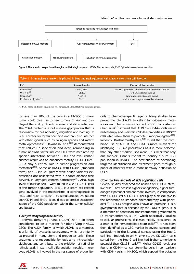
for less than 10% of the cells in a HNSCC primary tumor could give rise to new tumors in vivo and displayed the ability of selfrenewal and differentiation. The CD44 protein is a cell surface glycoprotein that is responsible for cell adhesion, migration and homing. It is a receptor for hyaluronic acid and can also interact with other ligands such as collagen species and matrix metalloproteases[5]. Takahashi et al[18] demonstrated that cellcell dissociation and actin remodeling in tumor necrosis factorinduced EMT were mediated by specific interaction between CD44 and hyaluronan; another result was an enhanced motility. CD44+CD24 CSCs play a critical role in tumor progression and metastasis[19]. Some of HNSCC with CD44s (standard form) and CD44 v6 (alternative splice variant) expressions are associated with a poorer diseasefree survival, in laryngeal cancers particularly[20]. Also, high levels of nuclear BMI1 were found in CD44+CD24 cells of the tumor population. BMI1 is a stem cellrelated gene involved in the mechanisms of carcinogenesis in head and neck cancers[5]. By simultaneous evaluating both CD44 and BMI1, it could lead to precise characterization of the CSC population within the tumor cellular architecture.
Aldehyde dehydrogenase activityAldehyde dehydrogenase (ALDH) has also been considered to be a marker for identifying HNSCC CSCs. The ALDH family, of which ALDH1 is a member, is a family of cytosolic isoenzymes, which are highly ex pressed in many stem and progenitor cells. These enzymes are responsible for oxidizing intracellular aldehydes and contribute to the oxidation of retinol to retinoic acid, in stem cell differentiation notably; moreover, ALDH1 is involved in the resistance of progenitor
cells to chemotherapeutic agents. Many studies have proved the role of ALDH1+ cells in tumorigenesis, metastasis and chemo resistance in HNSCC. For instance, Chen et al[21] showed that ALDH1+ CD44+ cells resist radiotherapy and maintain CSClike properties in HNSCC cells which allow them to promote tumor propagation[22]. Recently, Krishnamurthy et al[15] found that the combined use of ALDH1 and CD44 is more relevant for identifying CSClike populations as it is more selective than any other marker used alone. It is clear that only one marker is not sufficient to identify a pure CSC population in HSNCC. The best chance of developing targeted identification and treatment goes through a panel of markers with a more narrowly definition of CSCs.
Other markers and role of side population cells Several studies evidenced the abilities of CD133+ stemlike cells: They possess higher clonogenicity, higher tumourigenic potential and are more invasive, in comparison with CD133 cells. CD133+ cells play a crucial role in the resistance to standard chemotherapy with paclitaxel[23]. CD133 antigen also known as prominin1 is a glycoprotein that is encoded by the PROM1 gene. It is a member of pentaspan transmembrane glycoproteins (5transmembrane, 5TM), which specifically localize to cellular protrusions. If it was initially considered as a marker for hematopoietic stem cells[24], it has been then identified as a CSC marker in several cancers and particularly in the laryngeal cancer, using the Hep2 cell line. Indeed, in an in vivo study, CD133+ cells sorted from the Hep2 cell line had higher tumorigenic potential than CD133 cells[25]. Higher CD133 levels are found in CD44+ cancer stemlike cells in comparison with CD44 cells in HNSCC, which support the putative
15WJSC|www.wjgnet.com January 26, 2016|Volume 8|Issue 1|
Ref. Stem cell marker Cancer cell lines studied
Prince et al[5] CD44, BMI 1 HNSCC generated in immunodeficient mouse modelWei et al[25] CD133 HNSCC cell lines (hep-2)Chen et al[21] ALDH 1 Immunodeficient mouse modelKrishnamurthy et al[15] ALDH Head and neck squamous cell carcinoma
Table 1 Main molecular markers implicated in head and neck squamous cell cancer cancer stem cell detection
HNSCC: Head and neck squamous cell cancer; ALDH: Aldehyde dehydrogenase.
Targeting head and neck cancer stem cells
Detection of CSCs markers Cell-niche/tumour microenvironment EMT
Vaccination therapy Molecular pathways Induction of immune responses
Figure 1 Therapeutic perspectives through a multistrategic approach. CSCs: Cancer stem cells; EMT: Epithelial mesenchymal transition.
Méry B et al . Head and neck tumoral stem cells review
of Bmi1 increased the effectiveness of radiotherapy and resulted in inhibition of tumor growth in nude mice transplanted with ALDH1+ CSCs[33]. Moreover, Chen et al[32] focused on the Snail superfamily of zincfinger transcription factors, implicated in the regulation of EMT during embryonic development. The importance of SNAI1 in the growth of cancer cells and their metastatic potential has been shown in various malignancies[34]. Chen et al[32] found that the endogenous coexpression of ALDH1+ and Snail resulted in decreased ALDH1 expression, inhibition of CSClike properties, and decreased tumorigenesis in ALDH1+ CD44+ cells. By regulating the EMT, Snail is a key factor in maintaining CSC properties, and could be used as a therapeutic measure for the treatment of HNSCC. Besides, Snail small interfering RNA could reduce resistance to chemo radiotherapy in ALDH1+ cells[32]. Ultimately, the expression of drug efflux pumps by CSCs, another mechanism of chemo resistance remains to be explored in HNSCC. A better understanding of resistance mechanisms in HNSCC CSCs will require future studies and constitutes a prerequisite for improving therapy and possibly preventing tumor spread or recurrence. The main determinants of CSC radioresistance are summarized in Table 2.
Targeting stem cell niches Beyond intrinsic factors, the unique CSC microenvironment could play a crucial role in the radio resistance of CSCSs. Indeed, it has been showed that stromal environment and CSC niche play a vital role in the behavior of cancer cells. As the vast majority of the stem cells are found within a 100 μmradius of a blood vessel in HNSCC, the existence of a perivascular niche was suggested. Using the SCID mouse model of human tumor angiogenesis, it was observed that specific ablation of tumorassociated endothelial cells with an inducible Caspase9 result in the decrease of the fraction of head and neck CSCs[15]. Thus, targeting the stem cell niche directly can weaken the source of nutrition and change the essential signals needed by CSCs to proliferate. Therapeutic strategies as suggested by Tang et al[35] included targeting candidate CSCs and their microenvironment niche, which contributes to selfrenewal of these cells along with the reactive oxygen species status of these cells, and tweaking their intracellular milieu to facilitate apoptotic death signals over proliferative effects may facilitate a new prospective towards target therapy in HNSCC. Similarly,
role of CD133+ as a CSC marker. Using CD133 might serve to identify head and neck cancer patients that are resistant to conventional chemotherapy[26]. Furthermore, side population cells have shown to express stem cell properties when isolated from cancer samples. Their identification does not rely on the relative binding of antibodies but is based on their ability to efflux a fluorescent dye that binds to DNA[27,28]. Side population cells are more tumorigenic, chemoresistant and have displayed selfrenewal in vivo. Besides, side population cells show a more aggressive schema of tumour growth (in vitro)[29]. New strategies to target these cells need to be designed. Above all, further research on the exact role of side population cells and their implication in tumourigenesis is required as the exact mechanisms are not yet fully understood.
MOLECULAR STRATEGIES TO TARGET CSCS IN HSNCC CSCs and therapeutic resistance CSCs have important implications regarding cancer treatment and may lead to new perspectives on therapeutic strategies with a rethink of actual treatment paradigm. Indeed, indiscriminate cytoreduction is the aim of current chemotherapy and radiation treatment for HNSCC whereas the CSC hypothesis suggests that the elimination of CSCs is the only way to treat cancer effectively. Thus, significant reductions in the tumor volume are not enough to prevent tumor recurrence in HNSCC. Moreover, evidence suggests that CSCs have inherent drug and radiation resistance, rendering most conventional therapies ineffective. Radio resistance of CSCs has been attributed to their selfrenewal capacity, DNA repair capacity, freeradical scavenging, upregulation of cell cycle control mechanisms and specific interactions with the stromal microenvironment. Chemotherapy resistance is frequently related to accelerated drug transport and to drug metabolism[30,31]. Bmi1 and CD44 knockdowns have led to an improvement of CSCs chemosensitivity in HNSCC. In particular, knockdown of CD44 increased the sensitivity of HNSCC cells to cisplatin, underlying the crucial of CSCs in the response to chemotherapy[32]. Concerning Bmi1, a stemcellrelated gene, which participates in the selfrenewal of hematopoietic and neuronal stem cells, and has been implicated in the tumorigenesis of various malignancies the experiment showed that that knockdown
16WJSC|www.wjgnet.com January 26, 2016|Volume 8|Issue 1|
Molecular determinants of radioresistance Mechanism
Intrinsic determinants Enhanced DNA repair capability Protection from oxidative DNA damageActivation of the cell survival pathways (PI3K/Akt, WNT/β-catenin, notch)Expression of drug efflux pumps
Extrinsic determinants Hypoxic environment
Table 2 Main determinants of cancer stem cell radioresistance
Méry B et al . Head and neck tumoral stem cells review
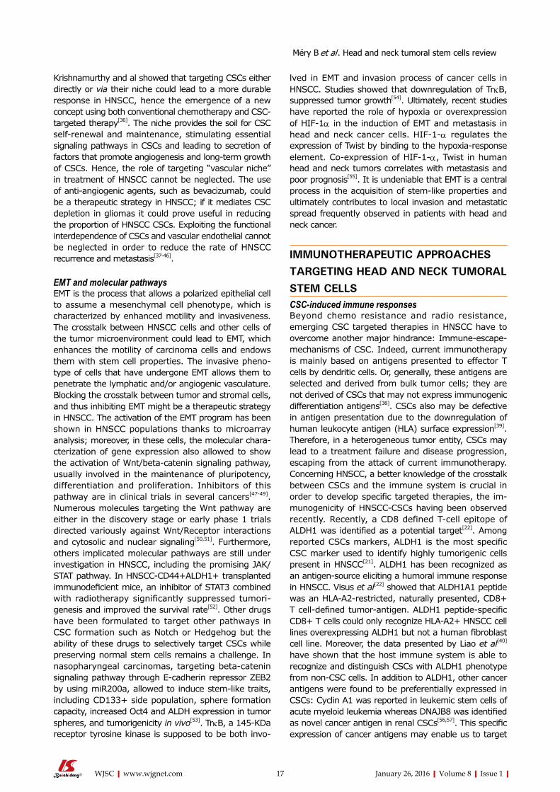
Krishnamurthy and al showed that targeting CSCs either directly or via their niche could lead to a more durable response in HNSCC, hence the emergence of a new concept using both conventional chemotherapy and CSCtargeted therapy[36]. The niche provides the soil for CSC selfrenewal and maintenance, stimulating essential signaling pathways in CSCs and leading to secretion of factors that promote angiogenesis and longterm growth of CSCs. Hence, the role of targeting “vascular niche” in treatment of HNSCC cannot be neglected. The use of antiangiogenic agents, such as bevacizumab, could be a therapeutic strategy in HNSCC; if it mediates CSC depletion in gliomas it could prove useful in reducing the proportion of HNSCC CSCs. Exploiting the functional interdependence of CSCs and vascular endothelial cannot be neglected in order to reduce the rate of HNSCC recurrence and metastasis[3746].
EMT and molecular pathwaysEMT is the process that allows a polarized epithelial cell to assume a mesenchymal cell phenotype, which is characterized by enhanced motility and invasiveness. The crosstalk between HNSCC cells and other cells of the tumor microenvironment could lead to EMT, which enhances the motility of carcinoma cells and endows them with stem cell properties. The invasive phenotype of cells that have undergone EMT allows them to penetrate the lymphatic and/or angiogenic vasculature. Blocking the crosstalk between tumor and stromal cells, and thus inhibiting EMT might be a therapeutic strategy in HNSCC. The activation of the EMT program has been shown in HNSCC populations thanks to microarray analysis; moreover, in these cells, the molecular characterization of gene expression also allowed to show the activation of Wnt/betacatenin signaling pathway, usually involved in the maintenance of pluripotency, differentiation and proliferation. Inhibitors of this pathway are in clinical trials in several cancers[4749]. Numerous molecules targeting the Wnt pathway are either in the discovery stage or early phase 1 trials directed variously against Wnt/Receptor interactions and cytosolic and nuclear signaling[50,51]. Furthermore, others implicated molecular pathways are still under investigation in HNSCC, including the promising JAK/STAT pathway. In HNSCCCD44+ALDH1+ transplanted immunodeficient mice, an inhibitor of STAT3 combined with radiotherapy significantly suppressed tumorigenesis and improved the survival rate[52]. Other drugs have been formulated to target other pathways in CSC formation such as Notch or Hedgehog but the ability of these drugs to selectively target CSCs while preserving normal stem cells remains a challenge. In nasopharyngeal carcinomas, targeting betacatenin signaling pathway through Ecadherin repressor ZEB2 by using miR200a, allowed to induce stemlike traits, including CD133+ side population, sphere formation capacity, increased Oct4 and ALDH expression in tumor spheres, and tumorigenicity in vivo[53]. TrκB, a 145KDa receptor tyrosine kinase is supposed to be both invo
lved in EMT and invasion process of cancer cells in HNSCC. Studies showed that downregulation of TrκB, suppressed tumor growth[54]. Ultimately, recent studies have reported the role of hypoxia or overexpression of HIF1α in the induction of EMT and metastasis in head and neck cancer cells. HIF1α regulates the expression of Twist by binding to the hypoxiaresponse element. Coexpression of HIF1α, Twist in human head and neck tumors correlates with metastasis and poor prognosis[55]. It is undeniable that EMT is a central process in the acquisition of stemlike properties and ultimately contributes to local invasion and metastatic spread frequently observed in patients with head and neck cancer.
IMMUNOTHERAPEUTIC APPROACHES TARGETING HEAD AND NECK TUMORAL STEM CELLS CSC-induced immune responses Beyond chemo resistance and radio resistance, emerging CSC targeted therapies in HNSCC have to overcome another major hindrance: Immuneescapemechanisms of CSC. Indeed, current immunotherapy is mainly based on antigens presented to effector T cells by dendritic cells. Or, generally, these antigens are selected and derived from bulk tumor cells; they are not derived of CSCs that may not express immunogenic differentiation antigens[38]. CSCs also may be defective in antigen presentation due to the downregulation of human leukocyte antigen (HLA) surface expression[39]. Therefore, in a heterogeneous tumor entity, CSCs may lead to a treatment failure and disease progression, escaping from the attack of current immunotherapy. Concerning HNSCC, a better knowledge of the crosstalk between CSCs and the immune system is crucial in order to develop specific targeted therapies, the immunogenicity of HNSCCCSCs having been observed recently. Recently, a CD8 defined Tcell epitope of ALDH1 was identified as a potential target[22]. Among reported CSCs markers, ALDH1 is the most specific CSC marker used to identify highly tumorigenic cells present in HNSCC[21]. ALDH1 has been recognized as an antigensource eliciting a humoral immune response in HNSCC. Visus et al[22] showed that ALDH1A1 peptide was an HLAA2restricted, naturally presented, CD8+ T celldefined tumorantigen. ALDH1 peptidespecific CD8+ T cells could only recognize HLAA2+ HNSCC cell lines overexpressing ALDH1 but not a human fibroblast cell line. Moreover, the data presented by Liao et al[40] have shown that the host immune system is able to recognize and distinguish CSCs with ALDH1 phenotype from nonCSC cells. In addition to ALDH1, other cancer antigens were found to be preferentially expressed in CSCs: Cyclin A1 was reported in leukemic stem cells of acute myeloid leukemia whereas DNAJB8 was identified as novel cancer antigen in renal CSCs[56,57]. This specific expression of cancer antigens may enable us to target
17WJSC|www.wjgnet.com January 26, 2016|Volume 8|Issue 1|
Méry B et al . Head and neck tumoral stem cells review
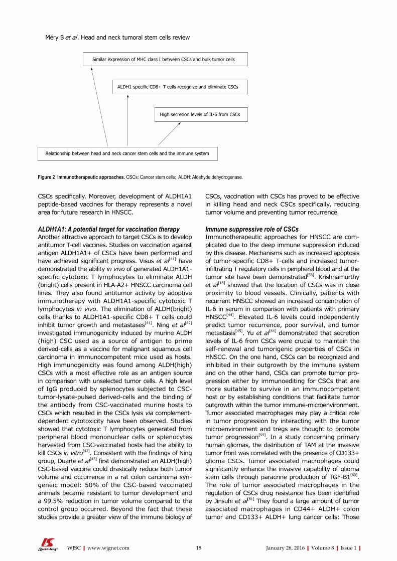
CSCs specifically. Moreover, development of ALDH1A1 peptidebased vaccines for therapy represents a novel area for future research in HNSCC.
ALDH1A1: A potential target for vaccination therapyAnother attractive approach to target CSCs is to develop antitumor Tcell vaccines. Studies on vaccination against antigen ALDH1A1+ of CSCs have been performed and have achieved significant progress. Visus et al[41] have demonstrated the ability in vivo of generated ALDH1A1specific cytotoxic T lymphocytes to eliminate ALDH (bright) cells present in HLAA2+ HNSCC carcinoma cell lines. They also found antitumor activity by adoptive immunotherapy with ALDH1A1specific cytotoxic T lymphocytes in vivo. The elimination of ALDH(bright) cells thanks to ALDH1A1specific CD8+ T cells could inhibit tumor growth and metastases[41]. Ning et al[42]
investigated immunogenicity induced by murine ALDH (high) CSC used as a source of antigen to prime derivedcells as a vaccine for malignant squamous cell carcinoma in immunocompetent mice used as hosts. High immunogenicity was found among ALDH(high) CSCs with a most effective role as an antigen source in comparison with unselected tumor cells. A high level of IgG produced by splenocytes subjected to CSCtumorlysatepulsed derivedcells and the binding of the antibody from CSCvaccinated murine hosts to CSCs which resulted in the CSCs lysis via complementdependent cytotoxicity have been observed. Studies showed that cytotoxic T lymphocytes generated from peripheral blood mononuclear cells or splenocytes harvested from CSCvaccinated hosts had the ability to kill CSCs in vitro[42]. Consistent with the findings of Ning group, Duarte et al[43] first demonstrated an ALDH(high) CSCbased vaccine could drastically reduce both tumor volume and occurrence in a rat colon carcinoma syngeneic model: 50% of the CSCbased vaccinated animals became resistant to tumor development and a 99.5% reduction in tumor volume compared to the control group occurred. Beyond the fact that these studies provide a greater view of the immune biology of
CSCs, vaccination with CSCs has proved to be effective in killing head and neck CSCs specifically, reducing tumor volume and preventing tumor recurrence.
Immune suppressive role of CSCs Immunotherapeutic approaches for HNSCC are complicated due to the deep immune suppression induced by this disease. Mechanisms such as increased apoptosis of tumorspecific CD8+ Tcells and increased tumorinfiltrating T regulatory cells in peripheral blood and at the tumor site have been demonstrated[58]. Krishnamurthy et al[15] showed that the location of CSCs was in close proximity to blood vessels. Clinically, patients with recurrent HNSCC showed an increased concentration of IL6 in serum in comparison with patients with primary HNSCC[44]. Elevated IL6 levels could independently predict tumor recurrence, poor survival, and tumor metastasis[45]. Yu et al[44] demonstrated that secretion levels of IL6 from CSCs were crucial to maintain the selfrenewal and tumorigenic properties of CSCs in HNSCC. On the one hand, CSCs can be recognized and inhibited in their outgrowth by the immune system and on the other hand, CSCs can promote tumor progression either by immunoediting for CSCs that are more suitable to survive in an immunocompetent host or by establishing conditions that facilitate tumor outgrowth within the tumor immunemicroenvironment. Tumor associated macrophages may play a critical role in tumor progression by interacting with the tumor microenvironment and tregs are thought to promote tumor progression[59]. In a study concerning primary human gliomas, the distribution of TAM at the invasive tumor front was correlated with the presence of CD133+ glioma CSCs. Tumor associated macrophages could significantly enhance the invasive capability of glioma stem cells through paracrine production of TGFB1[60]. The role of tumor associated macrophages in the regulation of CSCs drug resistance has been identified by Jinsuhi et al[61] They found a large amount of tumor associated macrophages in CD44+ ALDH+ colon tumor and CD133+ ALDH+ lung cancer cells: Those
18WJSC|www.wjgnet.com January 26, 2016|Volume 8|Issue 1|
Relationship between head and neck cancer stem cells and the immune system
High secretion levels of IL-6 from CSCs
Similar expression of MHC class I between CSCs and bulk tumor cells
ALDH1-specific CD8+ T cells recognize and eliminate CSCs
Figure 2 Immunotherapeutic approaches. CSCs: Cancer stem cells; ALDH: Aldehyde dehydrogenase.
Méry B et al . Head and neck tumoral stem cells review

macrophages allow activating Sonic Hedgehog pathways in CSCs in cooperation with IL6. Targeting tumor associated macrophages by inhibiting either the myeloid cell receptors colonystimulating factor1 receptor or chemokine receptor improves chemotherapeutic efficacy, inhibits metastasis and increases antitumor T cell responses in pancreatic ductal adenocarcinoma[62]. All these findings validate the interplay between CSCs and the tumor immune microenvironment. Therefore, specific targeting of head and neck tumoral stem cells by immunotherapeutic approaches may lead to more efficacious and lasting therapeutic results in the future. Nonetheless, it seems necessary to address several points before immunotherapeutic approaches targeting CSCs can be brought into clinical trials. These include the effective isolation of CSCs from bulk tumor mass to measure potential immunotherapeutic effects on CSC, to determine the antigenprofile presented on CSCs specifically to identify specific CSC targets as well as the induction and enhancement of antigen processing and presentation of CSC epitopes. A lot of work remains to be done to get a better understanding of the immune suppressive role of CSCs in HNSCC. The various immunotherapeutic approaches are displayed in Figure 2.
CONCLUSIONThe CSC theory provides new opening for the treatment of HNSCC. This theory also helps to explain why currently available therapies for head and neck cancer so often fail. Eradication of cancers may require the targeting and elimination of CSCs, especially for HNSCC and thus, there is an urgent need to alter the current paradigm in drug development. Efforts are still advocated to determine specific markers and methods to specifically target these cells, towards a more specific tumor treatment. To date, no antibody selectively targeting CSC has been described in HNSCC yet, but candidates are under investigation. For instance, CD44v6 antibodies either radiolabeled or coupled with a cytotoxic drug entered phase Ⅰ clinical testing in patients with HNSCC. In a phase Ⅰ dose escalation study, the treatment with a radiolabeled antibody showed promising antitumor effects[63]. Clearly, huge variety of approaches to eradicate CSCs is being explored, and particularly in vitro assays; there still remains the issue of how to avoid unwanted toxicity in vivo. Developing radio sensitizing strategies is also being investigated and appears to eliminate CSCs. Overcoming chemo resistance, radio resistance and immune evasion mechanisms of CSCs remains a cornerstone of novel adjuvant therapies specifically targeting CSCs in HNSCC. Bertrand et al[64] demonstrated that the combination of UCN01 (a checkpoint kinase inhibitor) and ATRA (alltrans retinoic acid) with irradiation decreased the survival fraction of CSCs and could be used as a powerful radio sensitizing strategy in HNSCC. Furthermore, advances in nanotechnology could allow a better understanding of the regulatory mechanisms that govern CSC biology in
vivo.
REFERENCES 1 Siegel R, Ward E, Brawley O, Jemal A. Cancer statistics, 2011:
the impact of eliminating socioeconomic and racial disparities on premature cancer deaths. CA Cancer J Clin 2011; 61: 212-236 [PMID: 21685461 DOI: 10.3322/caac.2012]
2 Prince ME, Ailles LE. Cancer stem cells in head and neck squamous cell cancer. J Clin Oncol 2008; 26: 2871-2875 [PMID: 18539966 DOI: 10.1200/JCO.2007.15.1613]
3 Parkin DM, Bray F, Ferlay J, Pisani P. Global cancer statistics, 2002. CA Cancer J Clin 2005; 55: 74-108 [PMID: 15761078 DOI: 10.3322/canjclin.55.2.74]
4 Sjögren EV, Wiggenraad RG, Le Cessie S, Snijder S, Pomp J, Baatenburg de Jong RJ. Outcome of radiotherapy in T1 glottic carcinoma: a population-based study. Eur Arch Otorhinolaryngol 2009; 266: 735-744 [PMID: 18839197 DOI: 10.1007/s00405-008-0803-9]
5 Prince ME, Sivanandan R, Kaczorowski A, Wolf GT, Kaplan MJ, Dalerba P, Weissman IL, Clarke MF, Ailles LE. Identification of a subpopulation of cells with cancer stem cell properties in head and neck squamous cell carcinoma. Proc Natl Acad Sci USA 2007; 104: 973-978 [PMID: 17210912 DOI: 10.1073/pnas.0610117104]
6 Wicha MS, Liu S, Dontu G. Cancer stem cells: an old idea--a paradigm shift. Cancer Res 2006; 66: 1883-1890; discussion 1883-1890 [PMID: 16488983]
7 Diehn M, Clarke MF. Cancer stem cells and radiotherapy: new insights into tumor radioresistance. J Natl Cancer Inst 2006; 98: 1755-1757 [PMID: 17179471 DOI: 10.1093/jnci/djj505]
8 Damek-Poprawa M, Volgina A, Korostoff J, Sollecito TP, Brose MS, O’Malley BW, Akintoye SO, DiRienzo JM. Targeted inhibition of CD133+ cells in oral cancer cell lines. J Dent Res 2011; 90: 638-645 [PMID: 21220361 DOI: 10.1177/0022034510393511]
9 Chen C, Wei Y, Hummel M, Hoffmann TK, Gross M, Kaufmann AM, Albers AE. Evidence for epithelial-mesenchymal transition in cancer stem cells of head and neck squamous cell carcinoma. PLoS One 2011; 6: e16466 [PMID: 21304586 DOI: 10.1371/journal.pone.0016466]
10 Davis SJ, Divi V, Owen JH, Bradford CR, Carey TE, Papagerakis S, Prince ME. Metastatic potential of cancer stem cells in head and neck squamous cell carcinoma. Arch Otolaryngol Head Neck Surg 2010; 136: 1260-1266 [PMID: 21173377 DOI: 10.1001/archoto.2010.219]
11 Sun S, Wang Z. Head neck squamous cell carcinoma c-Met+ cells display cancer stem cell properties and are responsible for cisplatin-resistance and metastasis. Int J Cancer 2011; 129: 2337-2348 [PMID: 21225626 DOI: 10.1002/ijc.25927]
12 Harper LJ, Piper K, Common J, Fortune F, Mackenzie IC. Stem cell patterns in cell lines derived from head and neck squamous cell carcinoma. J Oral Pathol Med 2007; 36: 594-603 [PMID: 17944752 DOI: 10.1111/j.1600-0714.2007.00617.x]
13 Visvader JE, Lindeman GJ. Cancer stem cells in solid tumours: accumulating evidence and unresolved questions. Nat Rev Cancer 2008; 8: 755-768 [PMID: 18784658 DOI: 10.1038/nrc2499]
14 Graziano A, d’Aquino R, Tirino V, Desiderio V, Rossi A, Pirozzi G. The stem cell hypothesis in head and neck cancer. J Cell Biochem 2008; 103: 408-412 [PMID: 17546610 DOI: 10.1002/jcb.21436]
15 Krishnamurthy S, Dong Z, Vodopyanov D, Imai A, Helman JI, Prince ME, Wicha MS, Nör JE. Endothelial cell-initiated signaling promotes the survival and self-renewal of cancer stem cells. Cancer Res 2010; 70: 9969-9978 [PMID: 21098716 DOI: 10.1158/0008-5472.CAN-10-1712]
16 Dalerba P, Cho RW, Clarke MF. Cancer stem cells: models and concepts. Annu Rev Med 2007; 58: 267-284 [PMID: 17002552 DOI: 10.1146/annurev.med.58.062105.204854]
17 Hermann PC, Bhaskar S, Cioffi M, Heeschen C. Cancer stem cells in solid tumors. Semin Cancer Biol 2010; 20: 77-84 [PMID: 20371287 DOI: 10.1016/j.semcancer.2010.03.004]
18 Takahashi E, Nagano O, Ishimoto T, Yae T, Suzuki Y, Shinoda
19WJSC|www.wjgnet.com January 26, 2016|Volume 8|Issue 1|
Méry B et al . Head and neck tumoral stem cells review
T, Nakamura S, Niwa S, Ikeda S, Koga H, Tanihara H, Saya H. Tumor necrosis factor-alpha regulates transforming growth factor-beta-dependent epithelial-mesenchymal transition by promoting hyaluronan-CD44-moesin interaction. J Biol Chem 2010; 285: 4060-4073 [PMID: 19965872 DOI: 10.1074/jbc.M109.056523]
19 Mack B, Gires O. CD44s and CD44v6 expression in head and neck epithelia. PLoS One 2008; 3: e3360 [PMID: 18852874 DOI: 10.1371/journal.pone.0003360]
20 Staibano S, Merolla F, Testa D, Iovine R, Mascolo M, Guarino V, Castellone MD, Di Benedetto M, Galli V, Motta S, Melillo RM, De Rosa G, Santoro M, Celetti A. OPN/CD44v6 overexpression in laryngeal dysplasia and correlation with clinical outcome. Br J Cancer 2007; 97: 1545-1551 [PMID: 17987038 DOI: 10.1038/sj.bjc.6604070]
21 Chen YC, Chen YW, Hsu HS, Tseng LM, Huang PI, Lu KH, Chen DT, Tai LK, Yung MC, Chang SC, Ku HH, Chiou SH, Lo WL. Aldehyde dehydrogenase 1 is a putative marker for cancer stem cells in head and neck squamous cancer. Biochem Biophys Res Commun 2009; 385: 307-313 [PMID: 19450560 DOI: 10.1016/j.bbrc.2009.05.048]
22 Visus C, Ito D, Amoscato A, Maciejewska-Franczak M, Abdels-alem A, Dhir R, Shin DM, Donnenberg VS, Whiteside TL, DeLeo AB. Identification of human aldehyde dehydrogenase 1 family member A1 as a novel CD8+ T-cell-defined tumor antigen in squamous cell carcinoma of the head and neck. Cancer Res 2007; 67: 10538-10545 [PMID: 17974998 DOI: 10.1158/0008-5472.CAN-07-1346]
23 Zhang Q, Shi S, Yen Y, Brown J, Ta JQ, Le AD. A subpopulation of CD133(+) cancer stem-like cells characterized in human oral squamous cell carcinoma confer resistance to chemotherapy. Cancer Lett 2010; 289: 151-160 [PMID: 19748175 DOI: 10.1016/j.canlet.2009.08.010]
24 Yin AH, Miraglia S, Zanjani ED, Almeida-Porada G, Ogawa M, Leary AG, Olweus J, Kearney J, Buck DW. AC133, a novel marker for human hematopoietic stem and progenitor cells. Blood 1997; 90: 5002-5012 [PMID: 9389720]
25 Wei XD, Zhou L, Cheng L, Tian J, Jiang JJ, Maccallum J. In vivo investigation of CD133 as a putative marker of cancer stem cells in Hep-2 cell line. Head Neck 2009; 31: 94-101 [PMID: 18853445 DOI: 10.1002/hed.20935]
26 Okamoto A, Chikamatsu K, Sakakura K, Hatsushika K, Takahashi G, Masuyama K. Expansion and characterization of cancer stem-like cells in squamous cell carcinoma of the head and neck. Oral Oncol 2009; 45: 633-639 [PMID: 19027347 DOI: 10.1016/j.oraloncology.2008.10.003]
27 Wan G, Zhou L, Xie M, Chen H, Tian J. Characterization of side population cells from laryngeal cancer cell lines. Head Neck 2010; 32: 1302-1309 [PMID: 20091690 DOI: 10.1002/hed.21325]
28 Tabor MH, Clay MR, Owen JH, Bradford CR, Carey TE, Wolf GT, Prince ME. Head and neck cancer stem cells: the side population. Laryngoscope 2011; 121: 527-533 [PMID: 21344428 DOI: 10.100 2/lary.21032]
29 Song J, Chang I, Chen Z, Kang M, Wang CY. Characterization of side populations in HNSCC: highly invasive, chemoresistant and abnormal Wnt signaling. PLoS One 2010; 5: e11456 [PMID: 20625515 DOI: 10.1371/journal.pone.0011456]
30 Morrison R, Schleicher SM, Sun Y, Niermann KJ, Kim S, Spratt DE, Chung CH, Lu B. Targeting the mechanisms of resistance to chemotherapy and radiotherapy with the cancer stem cell hypothesis. J Oncol 2011; 2011: 941876 [PMID: 20981352 DOI: 10.1155/2011/941876]
31 Ogawa K, Yoshioka Y, Isohashi F, Seo Y, Yoshida K, Yamazaki H. Radiotherapy targeting cancer stem cells: current views and future perspectives. Anticancer Res 2013; 33: 747-754 [PMID: 23482741]
32 Chen YC, Chang CJ, Hsu HS, Chen YW, Tai LK, Tseng LM, Chiou GY, Chang SC, Kao SY, Chiou SH, Lo WL. Inhibition of tumorigenicity and enhancement of radiochemosensitivity in head and neck squamous cell cancer-derived ALDH1-positive cells by knockdown of Bmi-1. Oral Oncol 2010; 46: 158-165 [PMID: 20036608 DOI: 10.1016/j.oraloncology.2009.11.007]
33 Park IK, Morrison SJ, Clarke MF. Bmi1, stem cells, and senescence regulation. J Clin Invest 2004; 113: 175-179 [PMID: 14722607 DOI: 10.1172/JCI200420800]
34 Zhou BP, Deng J, Xia W, Xu J, Li YM, Gunduz M, Hung MC. Dual regulation of Snail by GSK-3beta-mediated phosphorylation in control of epithelial-mesenchymal transition. Nat Cell Biol 2004; 6: 931-940 [PMID: 15448698 DOI: 10.1038/ncb1173]
35 Tang C, Ang BT, Pervaiz S. Cancer stem cell: target for anti-cancer therapy. FASEB J 2007; 21: 3777-3785 [PMID: 17625071]
36 Krishnamurthy S, Nör JE. Head and neck cancer stem cells. J Dent Res 2012; 91: 334-340 [PMID: 21933937 DOI: 10.1177/0022034511423393]
37 Zhao Y, Bao Q, Renner A, Camaj P, Eichhorn M, Ischenko I, Angele M, Kleespies A, Jauch KW, Bruns C. Cancer stem cells and angiogenesis. Int J Dev Biol 2011; 55: 477-482 [PMID: 21732274 DOI: 10.1387/ijdb.103225yz]
38 Boiko AD, Razorenova OV, van de Rijn M, Swetter SM, Johnson DL, Ly DP, Butler PD, Yang GP, Joshua B, Kaplan MJ, Longaker MT, Weissman IL. Human melanoma-initiating cells express neural crest nerve growth factor receptor CD271. Nature 2010; 466: 133-137 [PMID: 20596026 DOI: 10.1038/nature09161]
39 Busse A, Letsch A, Fusi A, Nonnenmacher A, Stather D, Ochsenreither S, Regenbrecht CR, Keilholz U. Characterization of small spheres derived from various solid tumor cell lines: are they suitable targets for T cells? Clin Exp Metastasis 2013; 30: 781-791 [PMID: 23519726 DOI: 10.1007/s10585-013-9578-5]
40 Liao T, Kaufmann AM, Qian X, Sangvatanakul V, Chen C, Kube T, Zhang G, Albers AE. Susceptibility to cytotoxic T cell lysis of cancer stem cells derived from cervical and head and neck tumor cell lines. J Cancer Res Clin Oncol 2013; 139: 159-170 [PMID: 23001491 DOI: 10.1007/s00432-012-1311-2]
41 Visus C, Wang Y, Lozano-Leon A, Ferris RL, Silver S, Szczepanski MJ, Brand RE, Ferrone CR, Whiteside TL, Ferrone S, DeLeo AB, Wang X. Targeting ALDH(bright) human carcinoma-initiating cells with ALDH1A1-specific CD8+ T cells. Clin Cancer Res 2011; 17: 6174-6184 [PMID: 21856769 DOI: 10.1158/1078-0432.CCR-11-1111]
42 Ning N, Pan Q, Zheng F, Teitz-Tennenbaum S, Egenti M, Yet J, Li M, Ginestier C, Wicha MS, Moyer JS, Prince ME, Xu Y, Zhang XL, Huang S, Chang AE, Li Q. Cancer stem cell vaccination confers significant antitumor immunity. Cancer Res 2012; 72: 1853-1864 [PMID: 22473314 DOI: 10.1158/0008-5472.CAN-11-1400]
43 Duarte S, Momier D, Baqué P, Casanova V, Loubat A, Samson M, Guigonis JM, Staccini P, Saint-Paul MC, De Lima MP, Carle GF, Pierrefite-Carle V. Preventive cancer stem cell-based vaccination reduces liver metastasis development in a rat colon carcinoma syngeneic model. Stem Cells 2013; 31: 423-432 [PMID: 23193035 DOI: 10.1002/stem.1292]
44 Yu CC, Tsai LL, Wang ML, Yu CH, Lo WL, Chang YC, Chiou GY, Chou MY, Chiou SH. miR145 targets the SOX9/ADAM17 axis to inhibit tumor-initiating cells and IL-6-mediated paracrine effects in head and neck cancer. Cancer Res 2013; 73: 3425-3440 [PMID: 23548270 DOI: 10.1158/0008-5472.CAN-12-3840]
45 Duffy SA, Taylor JM, Terrell JE, Islam M, Li Y, Fowler KE, Wolf GT, Teknos TN. Interleukin-6 predicts recurrence and survival among head and neck cancer patients. Cancer 2008; 113: 750-757 [PMID: 18536030 DOI: 10.1002/cncr.23615]
46 Folkins C, Man S, Xu P, Shaked Y, Hicklin DJ, Kerbel RS. Anticancer therapies combining antiangiogenic and tumor cell cytotoxic effects reduce the tumor stem-like cell fraction in glioma xenograft tumors. Cancer Res 2007; 67: 3560-3564 [PMID: 17440065]
47 Sato N, Meijer L, Skaltsounis L, Greengard P, Brivanlou AH. Maintenance of pluripotency in human and mouse embryonic stem cells through activation of Wnt signaling by a pharmacological GSK-3-specific inhibitor. Nat Med 2004; 10: 55-63 [PMID: 14702635]
48 Zechner D, Fujita Y, Hülsken J, Müller T, Walther I, Taketo MM, Crenshaw EB, Birchmeier W, Birchmeier C. beta-Catenin signals regulate cell growth and the balance between progenitor cell
20WJSC|www.wjgnet.com January 26, 2016|Volume 8|Issue 1|
Méry B et al . Head and neck tumoral stem cells review

expansion and differentiation in the nervous system. Dev Biol 2003; 258: 406-418 [PMID: 12798297]
49 Takahashi-Yanaga F, Kahn M. Targeting Wnt signaling: can we safely eradicate cancer stem cells? Clin Cancer Res 2010; 16: 3153-3162 [PMID: 20530697 DOI: 10.1158/1078-0432.CCR-09-2943]
50 Curtin JC, Lorenzi MV. Drug discovery approaches to target Wnt signaling in cancer stem cells. Oncotarget 2010; 1: 552-566 [PMID: 21317452]
51 Takebe N, Ivy SP. Controversies in cancer stem cells: targeting embryonic signaling pathways. Clin Cancer Res 2010; 16: 3106-3112 [PMID: 20530695 DOI: 10.1158/1078-0432.CCR-09-29 34]
52 Chen YW, Chen KH, Huang PI, Chen YC, Chiou GY, Lo WL, Tseng LM, Hsu HS, Chang KW, Chiou SH. Cucurbitacin I suppressed stem-like property and enhanced radiation-induced apoptosis in head and neck squamous carcinoma--derived CD44(+)ALDH1(+) cells. Mol Cancer Ther 2010; 9: 2879-2892 [PMID: 21062915 DOI: 10.1158/1535-7163.MCT-10-0504]
53 Xia H, Cheung WK, Sze J, Lu G, Jiang S, Yao H, Bian XW, Poon WS, Kung HF, Lin MC. miR-200a regulates epithelial-mesenchymal to stem-like transition via ZEB2 and beta-catenin signaling. J Biol Chem 2010; 285: 36995-37004 [PMID: 20826811 DOI: 10.1074/jbc.M110.133744]
54 Kupferman ME, Jiffar T, El-Naggar A, Yilmaz T, Zhou G, Xie T, Feng L, Wang J, Holsinger FC, Yu D, Myers JN. TrkB induces EMT and has a key role in invasion of head and neck squamous cell carcinoma. Oncogene 2010; 29: 2047-2059 [PMID: 20101235 DOI: 10.1038/onc.2009.486]
55 Yang MH, Hsu DS, Wang HW, Wang HJ, Lan HY, Yang WH, Huang CH, Kao SY, Tzeng CH, Tai SK, Chang SY, Lee OK, Wu KJ. Bmi1 is essential in Twist1-induced epithelial-mesenchymal transition. Nat Cell Biol 2010; 12: 982-992 [PMID: 20818389 DOI: 10.1038/ncb2099]
56 Ochsenreither S, Majeti R, Schmitt T, Stirewalt D, Keilholz U, Loeb KR, Wood B, Choi YE, Bleakley M, Warren EH, Hudecek M, Akatsuka Y, Weissman IL, Greenberg PD. Cyclin-A1 represents a new immunogenic targetable antigen expressed in acute myeloid leukemia stem cells with characteristics of a cancer-testis antigen. Blood 2012; 119: 5492-5501 [PMID: 22529286 DOI: 10.1182/blood-2011-07-365890]
57 Nishizawa S, Hirohashi Y, Torigoe T, Takahashi A, Tamura Y, Mori T, Kanaseki T, Kamiguchi K, Asanuma H, Morita R,
Sokolovskaya A, Matsuzaki J, Yamada R, Fujii R, Kampinga HH, Kondo T, Hasegawa T, Hara I, Sato N. HSP DNAJB8 controls tumor-initiating ability in renal cancer stem-like cells. Cancer Res 2012; 72: 2844-2854 [PMID: 22552285 DOI: 10.1158/0008-5472.CAN-11-3062]
58 Hoffmann TK, Dworacki G, Tsukihiro T, Meidenbauer N, Gooding W, Johnson JT, Whiteside TL. Spontaneous apoptosis of circulating T lymphocytes in patients with head and neck cancer and its clinical importance. Clin Cancer Res 2002; 8: 2553-2562 [PMID: 12171883]
59 Duray A, Demoulin S, Hubert P, Delvenne P, Saussez S. Immune suppression in head and neck cancers: a review. Clin Dev Immunol 2010; 2010: 701657 [PMID: 21437225 DOI: 10.1155/2010/701657]
60 Ye XZ, Xu SL, Xin YH, Yu SC, Ping YF, Chen L, Xiao HL, Wang B, Yi L, Wang QL, Jiang XF, Yang L, Zhang P, Qian C, Cui YH, Zhang X, Bian XW. Tumor-associated microglia/macrophages enhance the invasion of glioma stem-like cells via TGF-β1 signaling pathway. J Immunol 2012; 189: 444-453 [PMID: 22664874 DOI: 10.4049/jimmunol.1103248]
61 Jinushi M, Chiba S, Yoshiyama H, Masutomi K, Kinoshita I, Dosaka-Akita H, Yagita H, Takaoka A, Tahara H. Tumor-associated macrophages regulate tumorigenicity and anticancer drug responses of cancer stem/initiating cells. Proc Natl Acad Sci USA 2011; 108: 12425-12430 [PMID: 21746895 DOI: 10.1073/pnas.1106645108]
62 Mitchem JB, Brennan DJ, Knolhoff BL, Belt BA, Zhu Y, Sanford DE, Belaygorod L, Carpenter D, Collins L, Piwnica-Worms D, Hewitt S, Udupi GM, Gallagher WM, Wegner C, West BL, Wang-Gillam A, Goedegebuure P, Linehan DC, DeNardo DG. Targeting tumor-infiltrating macrophages decreases tumor-initiating cells, relieves immunosuppression, and improves chemotherapeutic responses. Cancer Res 2013; 73: 1128-1141 [PMID: 23221383 DOI: 10.1158/0008-5472.CAN-12-2731]
63 Colnot DR, Quak JJ, Roos JC, van Lingen A, Wilhelm AJ, van Kamp GJ, Huijgens PC, Snow GB, van Dongen GA. Phase I therapy study of 186Re-labeled chimeric monoclonal antibody U36 in patients with squamous cell carcinoma of the head and neck. J Nucl Med 2000; 41: 1999-2010 [PMID: 11138685]
64 Bertrand G, Maalouf M, Boivin A, Battiston-Montagne P, Beuve M, Levy A, Jalade P, Fournier C, Ardail D, Magné N, Alphonse G, Rodriguez-Lafrasse C. Targeting head and neck cancer stem cells to overcome resistance to photon and carbon ion radiation. Stem Cell Rev 2014; 10: 114-126 [PMID: 23955575 DOI: 10.1007/s12015-013-9467-y]
P- Reviewer: Economescu MC S- Editor: Qi Y L- Editor: A E- Editor: Jiao XK
21WJSC|www.wjgnet.com January 26, 2016|Volume 8|Issue 1|
Méry B et al . Head and neck tumoral stem cells review

© 2015 Baishideng Publishing Group Inc. All rights reserved.
Published by Baishideng Publishing Group Inc8226 Regency Drive, Pleasanton, CA 94588, USA
Telephone: +1-925-223-8242Fax: +1-925-223-8243
E-mail: [email protected] Desk: http://www.wjgnet.com/esps/helpdesk.aspx
http://www.wjgnet.com