Isolation of Erwinia amylovora Bacteriophage from Aerial ... · Isolation of Erwinia amylovora...
4
Ecology and Epidemiology Isolation of Erwinia amylovora Bacteriophage from Aerial Parts of Apple Trees D. F. Ritchie and E. J. Klos Graduate Assistant and Professor, respectively, Department of Botany and Plant Pathology, Michigan State University, East Lansing, MI 48824. The authors thank D. W. Dye, M. N. Schroth, and T. B. Sutton for kindly providing bacterial cultures. Journal Article No. 7566 of the Michigan State Agricultural Experiment Station. Accepted for publication 16 August 1976. ABSTRACT RITCHIE, D. F., and E. J. KLOS. 1977. Isolation of Erwinia amylovora bacteriophage from aerial parts of apple trees. Phytopathology 67:101-104. Populations of Erwinia amylovora bacteriophage greater a spreading translucent halo and a smaller plaque without a than 106 plaque-forming units (PFU) per gram of tissue were halo. Thirty-five bacterial isolates, consisting of nine genera, isolated without enrichment from diseased aerial parts of 18 species, and 15 strains of E. amylovora were typed; the apple trees during the summer of 1975. Three phage isolates phages lysed only E. amylovora. The burst size of the three were selected from different geographical locations. Two isolates was 40 to 50 PFU per cell. The phages could be stored types of plaques were produced; a clear-centered plaque with at 4 C and -20 C but lost titer when stored at 24 C. Additional key words: fire blight. Studies of bacteriophages of plant pathogenic bacteria procedures, and dilution series were the same as those have dealt primarily with their use as a diagnostic tool (5) described for the isolation of E. amylovora. The plating or in characterization of phage-bacteria interactions (3,6, procedures were as outlined by Adams (1). Plaques, if 11). Phages of phytopathogenic bacteria may be isolated present, were counted after 24 hours incubation at 24 C. from soil in the vicinity of the diseased plant (7) and often Three phage isolates were chosen for further study. Phage from the diseased tissue of the plant (11). PEal was isolated from blighted 'Jonathan' apple Erskine (8) isolated a phage from soil at the base of terminals from southwestern Michigan, PEa2 was Erwinia amylovora-infected trees, but was unable to isolated from soil at MSU, and PEa5 was isolated from a isolate it from infected or healthy aerial tissues. This symptomless 'Jonathan' apple terminal at MSU. The phage (S1) lysed both E. amylovora and a yellow phage isolates were purified by single-plaque isolation saprophytic bacterium which could also be lysogenized. five times using E. amylovora isolate #110 from the MSU This report deals with the isolation and partial orchard as propagating host. characterization of E. amylovora phages from diseased Phage characterization.--Phage lysates were prepared and symptomless plant tissues of aerial parts of apple by scraping the top agar from plates with confluent lysis, trees during the summer of 1975. centrifuged at 12,000 g for 10 minutes, and filtered through Millipore filters (pore size, 0,45 /m). The lysates MATERIALS AND METHODS were stored over a drop of chloroform in 3.54-g (2.0 dram) screw-cap vials covered with aluminum foil. Vials Erwinia amylovora isolation.--Diseased and of each isolate were placed at 24, 4, and -20 C to symptomless aerial tissues of apple trees, Malus sylvestris determine longevity of storage. Mill., at Michigan State University (MSU) and three The double-layer agar technique (1) in standard 100- growers' orchards in southwestern Michigan were mm diameter plastic petri plates was used to examine sampled. No attempt was made to disinfest the tissues. plaque morphology. The bottom layer consisted of 12 to Bacterial populations were quantified on a tissue weight 15 ml of 2.0% nutrient agar supplemented with 0.5% basis unless stated otherwise. Tissues were washed in glucose; the top layer consisted of 2.5 ml of 0.7% nutrient distilled water for 30 to 60 minutes. Serial dilutions agar, 0.5% glucose, and 0.5% yeast extract. The phage ranging from 10- to 10- were made in 0.02 M potassium titers were adjusted to approximately 50 plaque-forming phosphate buffer, pH 6.8, and 0. 1-ml samples were spread units (PFU) per plate. An 18- to 24-hour nutrient broth- over the surface of a differential medium (13). glucose culture of E. amylovora isolate 110 was used as Representative isolates were tested for pathogenicity host. The plates were incubated at 27 C and examined using the seedling technique (12). over a 48-hour period. Bacteriophage isolation.-The tissues, washing The double-layer agar technique was used to type the host-range of the three phage isolates. One-tenth milliliter of a 24-hour nutrient broth-glucose culture of each Copyright © 1977 The American Phytopathological Society, 3340 bacterial isolate was added to the 2.5 ml of warm top agar. Pilot Knob Road, St. Paul, MN 55121. All rights reserved. After the top layer had solidified, one loopful of each 101
Transcript of Isolation of Erwinia amylovora Bacteriophage from Aerial ... · Isolation of Erwinia amylovora...

Ecology and Epidemiology
Isolation of Erwinia amylovora Bacteriophagefrom Aerial Parts of Apple Trees
D. F. Ritchie and E. J. Klos
Graduate Assistant and Professor, respectively, Department of Botany and Plant Pathology, Michigan StateUniversity, East Lansing, MI 48824.
The authors thank D. W. Dye, M. N. Schroth, and T. B. Sutton for kindly providing bacterial cultures.Journal Article No. 7566 of the Michigan State Agricultural Experiment Station.Accepted for publication 16 August 1976.
ABSTRACT
RITCHIE, D. F., and E. J. KLOS. 1977. Isolation of Erwinia amylovora bacteriophage from aerial parts of apple trees.Phytopathology 67:101-104.
Populations of Erwinia amylovora bacteriophage greater a spreading translucent halo and a smaller plaque without athan 106 plaque-forming units (PFU) per gram of tissue were halo. Thirty-five bacterial isolates, consisting of nine genera,isolated without enrichment from diseased aerial parts of 18 species, and 15 strains of E. amylovora were typed; theapple trees during the summer of 1975. Three phage isolates phages lysed only E. amylovora. The burst size of the threewere selected from different geographical locations. Two isolates was 40 to 50 PFU per cell. The phages could be storedtypes of plaques were produced; a clear-centered plaque with at 4 C and -20 C but lost titer when stored at 24 C.
Additional key words: fire blight.
Studies of bacteriophages of plant pathogenic bacteria procedures, and dilution series were the same as thosehave dealt primarily with their use as a diagnostic tool (5) described for the isolation of E. amylovora. The platingor in characterization of phage-bacteria interactions (3,6, procedures were as outlined by Adams (1). Plaques, if11). Phages of phytopathogenic bacteria may be isolated present, were counted after 24 hours incubation at 24 C.from soil in the vicinity of the diseased plant (7) and often Three phage isolates were chosen for further study. Phagefrom the diseased tissue of the plant (11). PEal was isolated from blighted 'Jonathan' apple
Erskine (8) isolated a phage from soil at the base of terminals from southwestern Michigan, PEa2 wasErwinia amylovora-infected trees, but was unable to isolated from soil at MSU, and PEa5 was isolated from aisolate it from infected or healthy aerial tissues. This symptomless 'Jonathan' apple terminal at MSU. Thephage (S1) lysed both E. amylovora and a yellow phage isolates were purified by single-plaque isolationsaprophytic bacterium which could also be lysogenized. five times using E. amylovora isolate #110 from the MSU
This report deals with the isolation and partial orchard as propagating host.characterization of E. amylovora phages from diseased Phage characterization.--Phage lysates were preparedand symptomless plant tissues of aerial parts of apple by scraping the top agar from plates with confluent lysis,trees during the summer of 1975. centrifuged at 12,000 g for 10 minutes, and filtered
through Millipore filters (pore size, 0,45 /m). The lysatesMATERIALS AND METHODS were stored over a drop of chloroform in 3.54-g (2.0
dram) screw-cap vials covered with aluminum foil. VialsErwinia amylovora isolation.--Diseased and of each isolate were placed at 24, 4, and -20 C to
symptomless aerial tissues of apple trees, Malus sylvestris determine longevity of storage.Mill., at Michigan State University (MSU) and three The double-layer agar technique (1) in standard 100-growers' orchards in southwestern Michigan were mm diameter plastic petri plates was used to examinesampled. No attempt was made to disinfest the tissues. plaque morphology. The bottom layer consisted of 12 toBacterial populations were quantified on a tissue weight 15 ml of 2.0% nutrient agar supplemented with 0.5%basis unless stated otherwise. Tissues were washed in glucose; the top layer consisted of 2.5 ml of 0.7% nutrientdistilled water for 30 to 60 minutes. Serial dilutions agar, 0.5% glucose, and 0.5% yeast extract. The phageranging from 10- to 10- were made in 0.02 M potassium titers were adjusted to approximately 50 plaque-formingphosphate buffer, pH 6.8, and 0. 1-ml samples were spread units (PFU) per plate. An 18- to 24-hour nutrient broth-over the surface of a differential medium (13). glucose culture of E. amylovora isolate 110 was used asRepresentative isolates were tested for pathogenicity host. The plates were incubated at 27 C and examinedusing the seedling technique (12). over a 48-hour period.
Bacteriophage isolation.-The tissues, washing The double-layer agar technique was used to type thehost-range of the three phage isolates. One-tenth milliliterof a 24-hour nutrient broth-glucose culture of each
Copyright © 1977 The American Phytopathological Society, 3340 bacterial isolate was added to the 2.5 ml of warm top agar.Pilot Knob Road, St. Paul, MN 55121. All rights reserved. After the top layer had solidified, one loopful of each
101

102 PHYTOPATHOLOGY [Vol. 67
phage isolate (titers ranged from 106 to 107 PFU/ ml) was TABLE 2. Host range of Erwinia amylovora bacteriophagesspotted on sections of the plates. The host-range typing PEal, PEa2, and PEa5was done twice at different times. Thirty-five bacterialisolates representing nine genera, 18 species, and 15 Phage isolate
strains of E. amylovora were typed. Bacterial isolate PEal PEa2 PEa5The one-step growth experiment for each of the three Erwinia amylovora
phage isolates was done in 0.8% nutrient broth, 0.5% Cal Eal hazy hazy hazyglucose, 0.5% yeast extract at 24 C, using E. amylovora Ill 68 +a + +isolate 110 as the host. Phage were added to the bacteria M S U 110 + + +at 1:10 ratio and allowed to adsorb for 10 minutes then M S U 111 + + +diluted to approximately 10' bacteria per milliliter. Traverse City 112 + + +
Beginning at 20 minutes, samples were taken every 10 G. Rapids 113 + + +
minutes through a 90-minute period. The one-step G. Rapids 114 + + +growth experiment was repeated three times for each Paw Paw 115 + + +
growth Spinks Corners 116 + + +phage isolate. Cal Ea5 + + +
Cal Ea38 + + +RESULTS AND DISCUSSION M S U Mac 715 hazy hazy hazy
N.C. EACC512 120 + + +During the monitoring of E. amylovora populations on N.C. EA518 121 + + +
'Jonathan' apple trees at the MSU experimental orchard Paw Paw 122 + + +
during the summer of 1975, E. amylovora phages were Agrobacterium tumefaciens
detected without enrichment. Phages could be isolated UC3416 U - -
easily from three growers' orchards in southwestern A. tumefaciens UC 78 -
Corynebacterium fascians --
Michigan during a fire blight epiphytotic. Phage was C. flaccumfaciens - - -
detected on all tissues sampled where E. amylovora was Enterobacter aerogenes - - -
detected except on symptomless leaves (Table 1). Failure Erwinia atroseptica SR 8 - - -
to detect phage on symptomless leaves may have been due E. carotovora SR 165 - -
to several factors: (i) the phage populations may have E. herbicola ZP-1 - - -
been below the detection levels, (ii) E. amylovora E. herbicola ZP-2 - - -
populations were lower on symptomless leaves than on E. herbicola A-E - - -
other tissues, and (iii) there may be less protection on leaf Escherichia coli - - -
surfaces from factors such as ultraviolet light, effect of Pseudomonas aeruginosa- - -
moisture, etc., than on other tissues. It is not known P. /achrymansP. lachrymans- - -
whether the phages were located internally or externally P. solanacearum - - -
to the plant surface, but they were present wherever E. P. syringae - - -
amylovora was found extensively. Rhizobium sp. - - -
The three phage isolates could be stored for at least six Serratia sp. - - -
months at 4 C and -20 C without significant loss of titer. Xanthomonas juglandis - - -
Titers dropped rapidly when stored at 24 C. X. pruni PF-2 - - -
Two distinct types of plaques were produced by the apIUs (+) indicates lysis.phage isolates (Fig. 1). The first type produced a clear bMinus (-) indicates no lysis.
TABLE 1. The occurrence of Erwinia amylovora and E. amylovora bacteriophages on aerial parts of Jonathan apple trees duringthe summer of 1975 at East Lansing, Michigan
Samples containing E. amylovora Samples containing bacteriophage andTypes and approximate and approximate concentration (CFU)a approximate concentration (PF[J)b of
amounts of tissues sampled of E. amylovora per unit sampled bacteriophage per unit sampled
Symptomless terminals Eight of 25 terminals Four of 25 terminals("-1.0 g/terminal) 10 to 2 X 10 CFU/terminal 102 to 3 X 10⁴ PFU/terminal
3- to 4-week-old infected terminals 25 of 25 terminals 25 of 25 terminalsand leaves ("2 to 4 g/sample) 5 X 10³ to 6 X 10⁶ CFU/terminal 3 X 10' to 7 X 10⁶ PFU/terminal
Newly formed cankers 15 of 15 cankers Nine of 15 cankers('-0.2 to 0.5 g/canker) >106 CFU/g 102 to >106 PFU/g
Symptomless leaves Four of five samples 0 of five samplesFive samples, 10 leaves per sample ""103 CFU/sample None
Three blighted fruits Three of three fruit Three of three fruit("0.5 g/fruit) ,106 CFU/g -1I06 PFU/gaCFU, colony-forming units.'PFU, plaque-forming units.
January 1977] RITCHIE AND KLOS: ERWINIA AMYLOVORA PHAGE 103
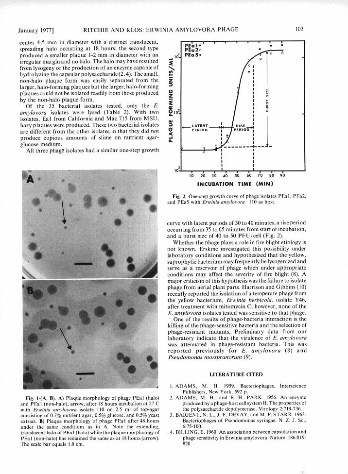
center 4-5 mm in diameter with a distinct translucent, PEl 1spreading halo occurring at 18 hours; the second type PEa2vproduced a smaller plaque 1-2 mm in diameter with an 104 PEa So
irregular margin and no halo. The halo may have resulted
from lysogeny or the production of an enzyme capable ofhydrolyzing the capsular polysaccharide (2, 4). The small, t-non-halo plaque form was easily separated from the Zlarger, halo-forming plaques but the larger, halo-formingplaques could not be isolated readily from those produced 0by the non-halo plaque form. Z
Of the 35 bacterial isolates tested, only the E.amylovora isolates were lysed (Table 2). With two 0101.isolates, Eal from California and Mac 715 from MSU, Ihazy plaques were produced. These two bacterial isolates n LATENT RISE _are different from the other isolates in that they did not PERIOD PERIOD
produce copious amounts of slime on nutrient agar- -vglucose medium. I I
All three phage isolates had a similar one-step growth I Io I II II II I
INCUBATION TIME (MIN)Fig. 2. One-step growth curve of phage isolates PEa I, PEa2,
and PEa5 with Erwinia amylovora 110 as host.
curve with latent periods of 30 to 40 minutes, a rise periodoccurring from 35 to 65 minutes from start of incubation,and a burst size of 40 to 50 PFU/cell (Fig. 2).
Whether the phage plays a role in fire blight etiology is9 ... not known. Erskine investigated this possibility under
laboratory conditions and hypothesized that the yellow,saprophytic bacterium may frequently be lysogenized andserve as a reservoir of phage which under appropriateconditions may affect the severity of fire blight (8). Amajor criticism of this hypothesis was the failure to isolatephage from aerial plant parts. Harrison and Gibbins (10)recently reported the isolation of a temperate phage fromthe yellow bacterium, Erwinia herbicola, isolate Y46,after treatment with mitomycin C; however, none of the
AE. amylovora isolates tested was sensitive to that phage.One of the results of phage-bacteria interaction is the
killing of the phage-sensitive bacteria and the selection ofphage-resistant mutants. Preliminary data from ourlaboratory indicate that the virulence of E. amylovorawas attenuated in phage-resistant bacteria. This wasreported previously for E. amylovora (8) andPseudomonas morsprunorum (9).
LITERATURE CITED
1. ADAMS, M. H. 1959. Bacteriophages. IntersciencePublishers, New York. 592 p.
Fig. 1-(A, B). A) Plaque morphology of phage PEal (halo) 2. ADAMS, M. H., and B. H. PARK. 1956. An enzymeand PEal (non-halo), arrow, after 18 hours incubation at 27 C produced by a phage-host cell system II. The properties ofwith Erwinia amylovora isolate 110 on 2.5 ml of top-agar the polysaccharide depolymerase. Virology 2:719-736.consisting of 0.7% nutrient agar, 0.5% glucose, and 0.5% yeast 3. BAIGENT, N. L., J. E. DEVAY, and M. P. STARR. 1963.extract. B) Plaque morphology of phage PEal after 48 hours Bacteriophages of Pseudomonas syringae. N. Z. J. Sci.under the same conditions as in A. Note the extending, 6:75-100.translucent halo of PEal (halo) while the plaque morphology of 4. BILLING, E. 1960. An association between capsulation andPEal (non-halo) has remained the same as at 18 hours (arrow). phage sensitivity in Erwinia amylovora. Nature 186:819-The scale bar equals 1.0 cm. 820.

104 PHYTOPATHOLOGY [Vol. 67
5. BILLING, E. 1963. The value of phage sensitivity tests for 9. GARRETT, C. M. E., J. E. CROSSE, and A. SLETTEN.the identification of phytopathogenic Pseudomonas spp. 1974. Relations between phage sensitivity and virulenceJ. Appl. Bact. 26:193-210. in Pseudomonas morsprunorum. J. Gen. Microbiol.
6. CIVEROLO, E. L. 1972. Interaction between bacteria and 80:475-483.bacteriophage on plant surfaces and in plant tissues. 10. HARRISON, A. and L. N. GIBBINS. 1975. The isolationPages 25-37 in H. P. Maas Geesteranus, ed. Third Int. and characterization of a temperate phage, Y46/(E2),Conf. Plant Pathgenic Bacteria. Proc., Centre Agric. from Erwinia herbicola Y46. Can. J. Microbiol. 21:937-Publ. Doc. (PUDOC), Wageningen, The Netherlands. 944.365 p. 11. OKABE, N., and M. GOTO. 1963. Bacteriophages of plant
7. CROSSE, J. E., and M. K. HINGORANI. 1958. A method pathogens. Annu. Rev. Phytopathol. 1:397-418.for isolating Pseudomonas mors-prunorum phages from 12. RITCHIE, D. F., and E. J. KLOS. 1974. A laboratorysoil. Nature (Lond.) 181:60-61. method of testing pathogenicity of suspected Erwinia
8. ERSKINE, J. M. 1973. Characteristics of Erwinia amylovora isolates. Plant Dis. Rep. 58:181-183.amylovora bacteriophage and its possible role in the 13. RITCHIE, D. F., and E. J. KLOS. 1976. Selective mediumepidemiology of fire blight. Can. J. Microbiol. 19:837- for isolating Erwinia amylovora from apple and pear845. tissues. Proc. Am. Phytopath. Soc. 3: (Abstr.). (In press).


![The Phytoalexin Resveratrol Regulates the Initiation of Hypersensitive ... · from Erwinia amylovora, the causative agent of fire blight in Rosaceae [19]. In non-host plants, using](https://static.fdocuments.us/doc/165x107/5edb9a3fad6a402d6665e840/the-phytoalexin-resveratrol-regulates-the-initiation-of-hypersensitive-from.jpg)