Isolation and Characterization of the Rat SND p102 Gene Promoter : Putative Role for Nuclear...
14
Isolation and Characterization of the Rat SND p102 Gene Promoter Putative Role for Nuclear Factor-Y in Regulation of Transcription LORENA RODR ´ IGUEZ, NEREA BARTOLOM ´ E, BEGO ˜ NA OCHOA, AND MAR ´ IA J. MART ´ INEZ Department of Physiology, Faculty of Medicine and Dentistry, University of the Basque Country, Sarriena s/n, 48940-Leioa, Bizkaia, Spain ABSTRACT: In this work, we report the isolation and characterization of a 1,688-bp sequence corresponding to the promoter region of the rat endoplasmic reticulum (ER) cholesterol ester hydrolase gene, re- named as staphylococcal nuclease domain–containing protein of 102 kDa (SND p102) in GenBank database according to the structural proper- ties and molecular weight of the protein. The transcription start site was located 216 bases upstream of the ATG start codon by RNA lig- ase mediated-rapid amplification of cDNA ends (RLM-RACE). Bioin- formatic analysis of the isolated sequence revealed a lack of typical promoter TATA box and the presence of GC-rich motifs and CCAAT boxes recognized by Sp 1 and nuclear factor-Y among other puta- tive binding sites for a number of transcription factors implicated in both basal and regulated processes. Electrophoretic mobility shift and supershift assays using nuclear extracts from human (HepG2) and rat (McA-RH7777) hepatoma cells demonstrated that nuclear factor-Y (NF-Y) transcription factor bound to the core sequences at (−257, −253), (−290, −286), and (−370, −366) upstream translation initiation site. The absence of TATA box and the location and reverse orientation of the CCAAT boxes in the promoter region strongly suggest a role for NF-Y in the regulation of transcription of SND p102 gene. KEYWORDS: SND p102; gene promoter; transcription; nuclear factor-Y INTRODUCTION The staphylococcal nuclease domain–containing protein of 102 kDa (SND p102) is a rat protein localized in the liver endoplasmic reticulum (ER) whose Address for correspondence: Mar´ ıa Jos´ e Mart´ ınez, Department of Physiology, Faculty of Medicine and Dentistry, University of the Basque Country, Sarriena s/n, 48940-Leioa, Bizkaia, Spain. Voice: 34-946-012-832; fax: 34-946-015-662. e-mail: [email protected] Ann. N.Y. Acad. Sci. 1091: 282–295 (2006). C 2006 New York Academy of Sciences. doi: 10.1196/annals.1378.074 282
-
Upload
lorena-rodriguez -
Category
Documents
-
view
212 -
download
0
Transcript of Isolation and Characterization of the Rat SND p102 Gene Promoter : Putative Role for Nuclear...

Isolation and Characterizationof the Rat SND p102 Gene Promoter
Putative Role for Nuclear Factor-Yin Regulation of Transcription
LORENA RODRIGUEZ, NEREA BARTOLOME, BEGONA OCHOA,AND MARIA J. MARTINEZ
Department of Physiology, Faculty of Medicine and Dentistry, University of theBasque Country, Sarriena s/n, 48940-Leioa, Bizkaia, Spain
ABSTRACT: In this work, we report the isolation and characterizationof a 1,688-bp sequence corresponding to the promoter region of therat endoplasmic reticulum (ER) cholesterol ester hydrolase gene, re-named as staphylococcal nuclease domain–containing protein of 102 kDa(SND p102) in GenBank database according to the structural proper-ties and molecular weight of the protein. The transcription start sitewas located 216 bases upstream of the ATG start codon by RNA lig-ase mediated-rapid amplification of cDNA ends (RLM-RACE). Bioin-formatic analysis of the isolated sequence revealed a lack of typicalpromoter TATA box and the presence of GC-rich motifs and CCAATboxes recognized by Sp 1 and nuclear factor-Y among other puta-tive binding sites for a number of transcription factors implicated inboth basal and regulated processes. Electrophoretic mobility shift andsupershift assays using nuclear extracts from human (HepG2) andrat (McA-RH7777) hepatoma cells demonstrated that nuclear factor-Y(NF-Y) transcription factor bound to the core sequences at (−257,−253),(−290, −286), and (−370, −366) upstream translation initiation site.The absence of TATA box and the location and reverse orientation of theCCAAT boxes in the promoter region strongly suggest a role for NF-Yin the regulation of transcription of SND p102 gene.
KEYWORDS: SND p102; gene promoter; transcription; nuclear factor-Y
INTRODUCTION
The staphylococcal nuclease domain–containing protein of 102 kDa (SNDp102) is a rat protein localized in the liver endoplasmic reticulum (ER) whose
Address for correspondence: Marıa Jose Martınez, Department of Physiology, Faculty of Medicineand Dentistry, University of the Basque Country, Sarriena s/n, 48940-Leioa, Bizkaia, Spain. Voice:34-946-012-832; fax: 34-946-015-662.
e-mail: [email protected]
Ann. N.Y. Acad. Sci. 1091: 282–295 (2006). C© 2006 New York Academy of Sciences.doi: 10.1196/annals.1378.074
282

RODRIGUEZ et al.: RAT SND p102 GENE PROMOTER 283
function and biological role is still unclear. The purified protein was previ-ously demonstrated to be responsible for more than 90% of the hydrolysisof cholesteryl esters in rat hepatocyte microsomes,1 but very recent evidencepoints to a role for the protein in the secretion of phospholipids into lipoproteinsof very low density and high density.2 Denomination of the protein as SNDp102 (GenBank accession number AAU05374) was based on the presence offour repeated staphylococcal nuclease domains and a single Tudor domain inthe amino acid sequence, and on a polypeptide molecular weight of 102 kDa.The cDNA (GenBank accession number AY697864) and amino acid sequenceof SND p102 showed 98–99% homology with the sequences of rat p105 andhuman p100, two proteins that are members of a highly conserved family ofnuclear transcriptional coactivators. Finding such extensive homology in thesequences of SND p102 and nuclear coactivators was unexpected, since SNDp102 was exclusively immunolocalized in the ER of the liver of rat, mouse,and human as well as in HepG2 cells.3 However, this extensive homology andlocalization is consistent with a number of studies reporting the presence ofhomologues of p100 protein in extranuclear compartments of mammalian cellsand Drosophila,4–6 which indicated additional functions of the protein ratherthan that of a transcriptional coactivator. Thus, a p100-like protein has beenfound in the ER and cytosolic lipid droplets from cow and mouse lactatingmammary gland, in lipid droplets from mouse adipocytes, and in the ER ofmouse liver,4 suggesting a role for the protein in the formation or secretionof milk lipid droplets and/or in the storage of triacylglycerol lipid bodies inadipocytes.4 The presence of p100 and SND p102 in the ER of mouse andrat liver makes likely their participation in the formation of lipoproteins orlipid bodies within hepatocytes. It is well known that the liver is a critical or-gan for the maintenance of whole-body lipid balance and plasma lipoproteinhomeostasis. Hepatocytes produce and secrete very-low-density lipoproteins(VLDL) in a highly complex process. The ER is crucial for the proper re-cruitment of triacylglycerol, cholesteryl ester, phospholipid and cholesterol,and assembly with the apolipoprotein B into the lipoprotein particles to bereleased. In a very recent work using recombinant adenoviruses encoding thecDNA of SND p102 or a specific antisense sequence, we have demonstratedthat the level of expression of SND p102 protein is directly related to the amountof phospholipid released into lipoprotein particles from infected hepatocytes.2
Based on the current knowledge of SND p102 protein, it is relevant tocharacterize the mechanisms of its transcriptional regulation. As an initial steptoward understanding the factors that control SND p102 gene expression, wehave isolated a 1,688-bp region corresponding to the gene promoter (GenBankaccession number AY957585) and studied its characteristics and the putativetranscription factors involved in the expression of the gene. Nuclear factor-Y (NF-Y) is an ubiquitous heterotrimeric transcriptional activator composedof three subunits NF-YA, B, and C, all necessary for DNA binding. NF-Ycomplexes with CCAAT boxes and has a role in the initiation and regulation of

284 ANNALS NEW YORK ACADEMY OF SCIENCES
transcription in TATA-containing and TATA-less promoters.7 We have foundwithin the SND p102 promoter region three binding sites for NF-Y that mightbe involved in the regulation of gene transcription.
MATERIALS AND METHODS
Materials
TaKaRa LA TaqTM polymerase was obtained from TaKaRa Bio, Inc. (Shiga,Japan). Oligonucleotides were synthesized by Isogen Life Science (Ijsselstein,the Netherlands). HepG2 and McA-RH7777 cell lines were purchased fromthe American Type Culture Collection (ATCC, Manassas, VA, USA), as wellas Eagle’s minimum essential medium (EMEM), Dulbecco’s modified Eagle’smedium (DMEM), horse and fetal bovine sera. L-glutamine, penicillin andstreptomycin were obtained from Sigma Aldrich (St. Louis, MO, USA).
Methods
Isolation of the Rat SND p102 Gene Promoter
A double polymerase chain reaction (PCR)-based method using the RatGenome Walker kit (Clontech, Mountain View, CA, USA) was performed.Libraries of rat genomic DNA (Eco RV, Dra I, Pvu II, and Ssp I) were ob-tained by digestion of rat genomic DNA and binding to an adaptor sequence,complementary to sense oligonucleotides (AP). Antisense oligonucleotidesspecific to the cDNA sequence of the rat SND p102 gene (GSP) were de-signed with the Primer 3 program8 (available at http://frodo.wi.mit.edu/cgi-bin/primer3/primer3-www.cgi). First PCR was performed with oligonu-cleotides AP2 (5′ ACTATAGGGCACGCGTGGT 3′) and GSP54-87 (5′CCCAGAGAGGACCATCTTGACGATGCCCCGTTG 3′) and genomic DNAlibraries as template. PCR products were diluted and used as templates fornested PCR with oligonucleotides AP2 and GSP85-56 (5′ GAGGGTGC-CGGTGCTGGTGAGAGGTTGCAG 3′). Nested PCR products were elec-trophoresed, purified from the agarose gel with the High Pure PCR Prod-uct Purification Kit (Roche, Mannheim, Germany) and sequenced with anABI Prism 3100 Genetic Analyzer (Applied Biosystems, Foster City, CA,USA) by the BigDye Terminator method (Faculty of Science and Technol-ogy, University of the Basque Country). New antisense oligonucleotides wereused in order to elongate the isolated promoter sequence. First PCR wasperformed with oligonucleotides AP2 and PRMTER785 (5′ GGAATGTG-GCCGCGAATCTTCCTGCGGTCTGG 3′) and DNA libraries as template.Nested PCR was performed with oligonucleotides AP2 and PRMTER935 (5′CCTGCTGTTCCAGCTGTGGTTAGGAAAAGCCTTGG 3′).

RODRIGUEZ et al.: RAT SND p102 GENE PROMOTER 285
Bioinformatic Analysis of the Sequence
Identification of putative binding sites for transcription factors implicatedin both basal and regulated transcription was done by bioinformatic analysisusing TESS9 (Transcription Element Search System, available at http://www.cbil.upenn.edu/tess), MatInspector10 (Genomatix Software GmbH, Munich,Germany, available at http://www.genomatix.de/cgi-bin/matinspector/matinspector.pl), and Jaspar11,12 (available at http://jaspar.cgb.ki.se/cgi-bin/jaspar-db.pl).
Identification of Transcription Initiation Site by RLM-RACE
Total RNA was extracted from isolated rat hepatocytes13 with Trizol (Invitro-gen Life Technologies, Carlsbad, CA, USA) using 1 mL of reagent per 10 × 106
cells. Ten �g RNA were treated with calf intestine alkaline phosphatase (CIP)to remove 5′-phosphates from fragmented RNA, and with tobacco acid py-rophosphatase (TAP) to remove the cap structure from full-length mRNA. Anadapter oligonucleotide was ligated to RNA by T4 RNA ligase. Both CIP andTAP treatments and adapter ligation were performed using First-Choice RNALigase Mediated-Rapid Amplification of cDNA Ends (RLM-RACE) (Am-bion, TX). RNA was reverse-transcribed using random decamers. cDNA wasused as template for PCR with oligonucleotides 5′ RACE Outer Primer (5′ GCT-GATGGCGATGAATGAACACTG 3′), which binds to the adaptor sequencepreviously ligated to RNA, and GSP55-77 (5′ ACCATCTTGACGATGCCC-CGTTG 3′) complementary to region (+55, +77) of the rat SND p102 cDNA(A of ATG start codon is considered as +1). A nested PCR was performedwith oligonucleotides 5′ RACE Inner Primer (5′ CGCGGATCCGAACACT-GCGTTTGCTGGCTTTGATG 3′) and GSP5-17 (5′ CTTTGCGCGAATTC-CATGTGTG 3′), complementary to the region (−5, +17) of the cDNA. ThePCR product was digested with Eco RI and Bam HI (Roche, Germany) andcloned into pUC19 for sequencing as mentioned above.
Preparation of Nuclear Extracts
Extracts were prepared from HepG2 or McA-RH7777 cell cultures usingthe Nuclear Extraction kit (Panomics, Fremont, CA, USA). Human hepato-blastoma HepG2 cells were cultured in EMEM containing 2 mM L-glutamine,100 U/mL penicillin, 100 mg/mL streptomycin and 10% fetal bovine serumfor 96 h before nuclear extracts were obtained. Rat hepatoma McA-RH777cells were grown in DMEM supplemented with 20% horse serum and 5% fetalbovine serum for 96 h prior to obtaining nuclear extracts. Protein concentra-tion was determined by the Bradford method,14 using bovine seroalbumin asstandard.

286 ANNALS NEW YORK ACADEMY OF SCIENCES
Electrophoretic Mobility-Shift Assay (EMSA)
Probes were obtained by annealing complementary single-stranded oligonu-cleotides corresponding to three different putative NF-Y binding sites presentin the SND p102 gene promoter. Bicatenary probes were labeled at the 3′end with digoxigenin using the DIG Gel shift kit second generation (Roche,Germany). For mobility shift assays, 3 �g of HepG2 nuclear extracts or 6 �g ofMcA-RH7777 nuclear extracts were incubated at room temperature for 15 minwith 160 fmol of the 3′-DIG-labeled dimerized probe, 1 �g poly[d(I-C)], 100ng poly-L-lysine in 20 mM HEPES (pH 7,6), 1 mM EDTA, 10 mM (NH4)2SO4,1 mM DTT, 0,2% (w/v) Tween 20, 30 mM KCl. For specific or nonspecificcompetition assays (sequences of the probes are shown below), 25- to 125-foldmolar excess of unlabeled probe was used.
NFY (−271, −239).wt 5′ GGTTCTGGACGCTAATTGGCCAGAGGGAGGTGG 3′
NFY (−303, −271).wt 5′ AAACAGAGTGCTATTGGCTTGAGTTCTAGCAG 3′
NFY (−384, −352).wt 5′ TTTGCCCGTCACCGCAAATCAGCTCTTTCAGCG 3′
NFY (−270, −241).mut 5′ GTTCTGGACGCTACCTGTCCAGAGGG-AGG 3′
NFY (−303, −275).mut 5′ AAACAGAGTGCCTCCTGTCTTGAGTTCTA 3′
NFY (−381, −353).mut 5′ GCCCGTCACCGAAAGGCAGCTCTTTCAGC 3′
For supershift experiments, 1.5 �g of anti-NF-Y antibody (BD BiosciencesPharmingen; Franklin Lakes, NJ, USA) was added, and incubated for 15 addi-tional minutes at room temperature. Electrophoresis was performed at 4◦C in6% nondenaturing polyacrylamide gels in 45 mM Tris-borate, 1 mM EDTA(pH 8) at 110V for 70 min. Complexes were transferred to a HyBond N+ ny-lon membrane (Amersham Biosciences, Buckinghamshire, UK) at 300 mA for30 min. Oligonucleotides were cross-linked at 120 mJ in a Stratalinker 1800(Stratagene, LaJolla, CA, USA) and visualized by an enzyme immunoassayusing anti-digoxigenin-AP. Chemilumiscence signal detection was performedin HyperfilmTM ECL (Amersham Biosciences).
RESULTS
Isolation of the Rat SND p102 Gene Promoter Region
A genome walking method based on two consecutive PCRs was used to iso-late the promoter region of the rat SND p102 gene. Rat genomic DNA libraries,obtained by digestion of genomic DNA with different endonucleases (Eco RV,Dra I, Pvu II, and Ssp I) and ligation to an adaptor sequence, were used astemplate for the first PCR. Both DNA libraries and sense oligonucleotideswere part of the kit used. Antisense oligonucleotides complementary to the 5′end of the rat SND p102 cDNA were designed. For the first PCR, oligonu-cleotides AP2, complementary to the adaptor sequence of genomic DNA, andGSP54-87, complementary to region (+54, +87) of the SND p102 cDNA

RODRIGUEZ et al.: RAT SND p102 GENE PROMOTER 287
(A from ATG translation start codon is considered as +1) were used. PCRproducts of different sizes were amplified from each library, and they wereused as templates for nested PCR with oligonucleotides AP2 and GSP85-56,complementary to region (−85, −56) of the SND p102 cDNA. Fragments of1.27 kb (Eco RV), 1.29 kb (Dra I), 0.89 kb (Pvu II), and 1.20 kb (Ssp I) wereamplified and purified. Sequence analysis showed that shorter fragments wereincluded in the 1.29-kb amplicon. A region of 1,256 pairs of bases upstreamof the ATG start codon was obtained, whose last 155 bases corresponded tothe 5′ region of the cDNA sequence reported for the rat SND p102 gene and1,101 bases corresponded to a new sequence upstream of the cDNA sequence.
A second double-PCR run was performed in order to elongate the isolatedsequence. Oligonucleotides AP2 and PRMTER785, complementary to theregion (−785, −754) of the new isolated sequence, were used for the firstPCR, and AP2 and PRMTER935, complementary to the region (−935, −901)for the second PCR. Fragments of 0.58 kb (Eco RV), 1.07 kb (Dra I), and0.50 kb (Pvu II) were amplified. Sequence analysis showed again that theshorter fragments were included in the 1.07-kb amplicon. A new fragmentof 989 bases was obtained whose last 411 bases corresponded to the 5′ endof the previously isolated promoter sequence.
In this way, a region of 1,899 bases was isolated (FIG. 1) whose last 211bases corresponded to the cDNA sequence for SND p102 gene (GenBankaccession number AY697864) and 1,688 bases corresponded to a new sequenceupstream the cDNA. Sequence analysis using the Blast Rat Genome tool fromNCBI established the chromosomal location of the new isolated sequenceon chromosome 4, coinciding with the previously reported location for theSND p102 gene. Considering all these data, the new isolated sequence wasconsidered as part of the promoter region for the SND p102 gene.
Determination of Transcription Initiation Siteof Rat SND p102 Gene in Hepatocytes
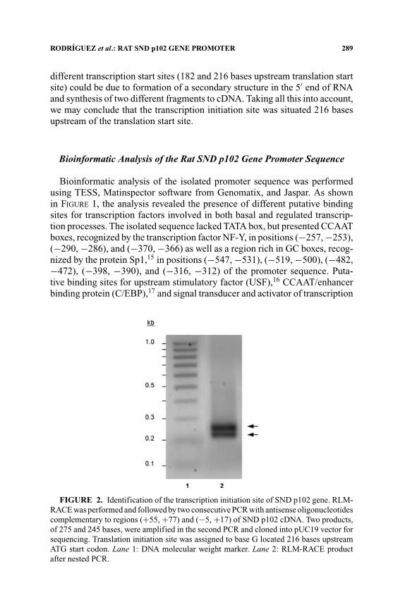
RLM-RACE using RNA isolated from rat hepatocytes was performed. Bythis method, retrotranscription to cDNA was selectively achieved from full-length capped mRNA, as explained in the MATERIALS AND METHODS section,and was used to determine the transcription initiation site of the rat SND p102gene. Two specific antisense oligonucleotides complementary to regions (+55,+77) and (−5, +17) of the 5′ end of the SND p102 cDNA were used for ampli-fication from cDNA. Two products, differing only in 30 bases, were amplifiedin the nested PCR (FIG. 2) and cloned into pUC19 vector for sequencing. Se-quences from seven clones of RLM-RACE products were analyzed. The mostupstream 5′ ends for these fragments were −177, −179, −182 for the shorterfragment, and −208, −212 (2), and −216 for the longer fragment. These mul-tiple 5′ ends may reflect incomplete reverse transcription. The existence of two

288 ANNALS NEW YORK ACADEMY OF SCIENCES
FIGURE 1. Isolated promoter region of the rat SND p102 gene (GenBank AY957585).Translation start site (A) is considered as position +1 of the sequence. Initiation of tran-scription was localized at position −216 by RLM-RACE and is marked in the sequencewith an arrow. Binding sites for some of the transcription factors predicted by bioinformaticanalysis are underlined. Binding sequences for NF-Y confirmed by EMSA are shown in abox. HNF-4 hepatocyte nuclear factor 4; C/EBP CCAAT/enhancer binding protein; STATsignal transducer and activator of transcription; USF upstream stimulatory factor.
RODRIGUEZ et al.: RAT SND p102 GENE PROMOTER 289
different transcription start sites (182 and 216 bases upstream translation startsite) could be due to formation of a secondary structure in the 5′ end of RNAand synthesis of two different fragments to cDNA. Taking all this into account,we may conclude that the transcription initiation site was situated 216 basesupstream of the translation start site.
Bioinformatic Analysis of the Rat SND p102 Gene Promoter Sequence
Bioinformatic analysis of the isolated promoter sequence was performedusing TESS, Matinspector software from Genomatix, and Jaspar. As shownin FIGURE 1, the analysis revealed the presence of different putative bindingsites for transcription factors involved in both basal and regulated transcrip-tion processes. The isolated sequence lacked TATA box, but presented CCAATboxes, recognized by the transcription factor NF-Y, in positions (−257, −253),(−290, −286), and (−370, −366) as well as a region rich in GC boxes, recog-nized by the protein Sp1,15 in positions (−547, −531), (−519, −500), (−482,−472), (−398, −390), and (−316, −312) of the promoter sequence. Puta-tive binding sites for upstream stimulatory factor (USF),16 CCAAT/enhancerbinding protein (C/EBP),17 and signal transducer and activator of transcription
FIGURE 2. Identification of the transcription initiation site of SND p102 gene. RLM-RACE was performed and followed by two consecutive PCR with antisense oligonucleotidescomplementary to regions (+55, +77) and (−5, +17) of SND p102 cDNA. Two products,of 275 and 245 bases, were amplified in the second PCR and cloned into pUC19 vector forsequencing. Translation initiation site was assigned to base G located 216 bases upstreamATG start codon. Lane 1: DNA molecular weight marker. Lane 2: RLM-RACE productafter nested PCR.

290 ANNALS NEW YORK ACADEMY OF SCIENCES
(STATs)18 were also found. The analysis also revealed several DNA elementsthat matched the consensus binding sequences for liver-enriched transcriptionfactors, such as hepatocyte nuclear factor 4 (HNF-4).19
Binding of NF-Y to the SND p102 Gene Promoter Region
Binding of transcription factor NF-Y to the isolated promoter sequencewas analyzed by electrophoretic mobility shift and supershift assays. Oligonu-cleotides complementary to regions (−271, −239), (−303, −271), and (−384,−352), which contain CCAAT boxes recognized by NF-Y, were designed andlabeled with digoxigenin. Incubation of the probe for region (−384, −352) withnuclear extracts of HepG2 (FIG. 3) or McA-RH7777 (FIG. 4) cells resulted ina shifted band. Competitive assays with increasing amounts of specific, non-specific and mutated unlabeled probes were performed, confirming the shift(FIG. 3A, lanes 2–5; FIG. 4, lanes 4–9). The same binding pattern of NF-Y toregion (−384, −352) of the promoter was observed for regions (−271, −239)and (−303, −271) (data not shown). To confirm the specificity of NF-Y bind-ing to the three analyzed CCAAT boxes, supershift analysis was performed.
FIGURE 3. Electrophoretic mobility shift (A) and supershift (B) assays to analyzeNF-Y binding to the CCAAT box located at (−370, −366) of the rat SND p102 genepromoter using nuclear extracts from HepG2 cells. Incubation of the double-strandedoligonucleotide labeled with digoxigenin, corresponding to the region (−384, −352) of thepromoter, with nuclear extracts resulted in a shift (lanes 1 and 6). Incubation with a spe-cific antibody for NF-Y resulted in a supershift (lane 7). Competition assays using 30- and120-fold molar excess of specific (wild-type) and nonspecific (Oct2A) competitors wereperformed. Incubation with excess of specific unlabeled probe resulted in the absence of theDNA–protein complex formation (lanes 2 and 3), whereas incubation with excess of non-specific probe for transcription factor Oct2A (lanes 4 and 5) did not inhibit the formationof the DNA–protein complex.

RODRIGUEZ et al.: RAT SND p102 GENE PROMOTER 291
FIGURE 4. Electrophoretic mobility shift and supershift assays for NF-Y binding tothe CCAAT box located at (−370, −366) of the rat SND p102 gene promoter. Nuclearextracts from HepG2 (lane 1) and McA-RH7777 (lanes 2–9) were incubated with double-stranded oligonucleotide corresponding to the region (−384, −352) of the promoter andlabeled with digoxigenin. A DNA–protein complex resulting in a shifted band was observedin the presence of HepG2 (lane 1) or McA-RH7777 (lane 2) nuclear extracts. Incubationwith a specific antibody for NF-Y resulted in a supershift (lane 3). Competition assaysusing 30- and 125-fold molar excess of different competitors were performed. Incubationwith excess of specific unlabeled probe NF-Y (−384, −352).wt resulted in the absenceof the complex formation (lanes 4 and 5), whereas incubation with excess of nonspecificprobe for transcription factor Oct2A (lanes 6 and 7) or mutated specific probe NF-Y (−381,−353).mut (lanes 8 and 9) did not inhibit the formation of the complex.
The addition of a monoclonal antibody anti-NF-YA produced a single super-shifted complex in HepG2 (FIG. 3B, lane 7) and McA-RH7777 (FIG. 4, lane 3)nuclear extracts.
DISCUSSION
In this report, we isolated and characterized a region of 1,688 bases corre-sponding to the promoter region of the rat SND p102 gene which was registeredin GenBank with the accession number AY957585. The isolated promoter re-gion was extensively analyzed with bioinformatic tools and some elementswere identified as putative binding sites for transcription factors implicatedin basal or regulated transcription. Homology studies showed the chromo-somal location of the promoter region at the rat chromosome four genomiccontig NW-047689.4. It matched 98% with the 5´ flanking region of the geneidentified as Snd1. This is the name given by NCBI to the Staphylococcalnuclease domain–containing 1 gene, including SND p102 and p105 coactiva-tor genes (GenBank accession numbers AY697864 and U83883). The proteins

292 ANNALS NEW YORK ACADEMY OF SCIENCES
encoded by these two genes displayed similar molecular weights and structuraldomains (four staphylococcal nuclease domains and one Tudor domain).
The biological function of SND p102 is yet poorly understood. As far aswe know, homologues of SND p102 can act as transcriptional coactivators inthe nuclei of cow, mouse and rat cells,4,6 but additional functions have to beconsidered since several homologue proteins have been identified in subcellu-lar fractions other than nuclei. In the cytosol of adipocytes and mammary glandand in the ER of mammary gland and liver, the p100 protein has been describedas a participant in the formation, growth or secretion of lipid bodies.4 This isconsistent with recent evidence that pointed to a role for the ER SND p102 inVLDL generation and secretion.2 It was found that adenovirus-mediated dif-ferential expression of SND p102 in rat hepatocytes modulated the amount ofphospholipid secreted into lipoprotein particles of very low and high density.Assuming a potential involvement of this protein in lipid homeostasis in theliver, it is of great importance to characterize the requirements and mechanismfor SND p102 gene transcription. Thus, bioinformatic analysis of the isolatedsequence allowed us to identify several putative elements involved in both basaland regulated transcription. The promoter has no consensus TATA box or ini-tiator element, but in contrast, there are GC-rich regions with putative bindingsites for Sp1 transcription factor and several CCAAT boxes recognized by NF-Y. Both Sp1 and NF-Y are ubiquitous nuclear factors which usually functionsynergistically and are effective for basal transcription of several genes.20–23
TATA-less promoters are typical of housekeeping genes, together with thepresence of GC-boxes and multiple transcriptional start sites. The SND p102promoter matches some of the characteristics of these housekeeping gene,such as the lack of TATA box and the presence of GC-boxes. However, themultiplicity of transcriptional start sites is a point of discussion because wefound a main initiation site at −216 bp and a secondary start point 30 bpdownstream. RLM-RACE was used to localize the transcription initiation siteof SND p102 gene. The amplification of two products in nested PCR could beprobably due to the formation of an internal structure in the 5′ end of RNA,which presents a high percentage of GC and autocomplementarity, more thanthe presence of two transcription initiation sites. For this reason, we haveassigned the origin of transcription to base G located 216 bases upstreamtranslation initiation site.
In addition to GC-boxes recognized by Sp1, the promoter also containsseveral potential binding sites for NF-Y, which recognizes CCAAT boxes.7
These are common elements in eukaryotic promoters that can act differentlyin promoters with or without TATA box. In the absence of TATA box, NF-Y canfunction as a recruiting factor for the general transcriptional machinery that al-lows polymerase II to find the start site.24 In most of the TATA-less promoters,CCAAT boxes appear in reverse orientation, ATTGG, and are generally dis-tributed in (−40, −80) region. This is consistent with the location of CCAAT

RODRIGUEZ et al.: RAT SND p102 GENE PROMOTER 293
boxes in the SND p102 promoter at positions −42 and −75. These two ATTGGelements appear in close proximity and have been demonstrated to be specificbinding sites for NF-Y by EMSA experiments. Another CCAAT box is foundin forward direction in a more distant region −165 which is also bound specif-ically by NF-Y. Because there are a wide range of proteins that recognize andbind CCAAT boxes, it is recommended that NF-Y be identified as the proteininvolved in the DNA–protein complex generated in the promoter sequence.When we tested nuclear extracts from different species, human HepG2 andrat McA-RH7777 hepatoma cells, different DNA–protein complexes resultedin EMSA competition experiments, depending on the source of nuclear pro-teins. However, in both cases a common band is directly supershifted by theanti-NF-Y antibody, demonstrating that NF-Y is really the protein bound tothe CCAAT elements included in the probes. These results reinforce the ideathat CCAAT/ATTGG boxes may represent functional promoter elements andthat the binding of NF-Y could be essential for transcription of SND p102gene. Nevertheless, further studies have to be performed to confirm the roleof NF-Y and to clarify which other transcription factors might be crucial forthe basal and regulated transcriptional activity of this promoter.
ACKNOWLEDGMENTS
The authors thank Montse Busto for her expert technical help with cellcultures. This work was supported by DGICYT (BMC2001-0067), FIS(Red G03-015) and the Basque Government (IE05-147). Lorena Rodrıguez,and Nerea Bartolome, are recipients of predoctoral fellowships from theSpanish Ministry of Education and Science and the Basque Government,respectively.
REFERENCES
1. CRISTOBAL, S., B. OCHOA & O. FRESNEDO. 1999. Purification and characterizationof a cholesteryl ester hydrolase from rat liver microsomes. J. Lipid Res. 40:715–725.
2. PALACIOS, L., B. OCHOA, M.J. GOMEZ-LECHON, et al. 2006. Overexpression of SNDp102, a rat homologue of p100 coactivator, promotes the secretion of lipoproteinphospholipids in primary hepatocytes. Biochim. Biophys. Acta 1761: 698–708.
3. FRESNEDO, O., M. LOPEZ DE HEREDIA, M.J. MARTıNEZ, et al. 2001. Inmunolocal-ization of a novel cholesteryl ester hydrolase in the endoplasmic reticulum ofmurine and human hepatocyte. Hepatology 3: 662–667.
4. KEENAN, T.W., S. WINTER, H.R. RACKWITZ & H.W. HEID. 2000. Nuclear coactivatorprotein p100 is present in endoplasmic reticulum and lipid droplets of milksecreting cells. Biochim. Biophys. Acta 1523: 84–90.

294 ANNALS NEW YORK ACADEMY OF SCIENCES
5. CAUDY, A.A., R.F. KETTING, S.M. HAMMOND, et al. 2003. A micrococcal nucleasehomologue in RNAi effector complexes. Nature 425: 411–414.
6. BROADHURST, M.K., R.S.F. LEE, S. HAWKINS & T.T. WHEELER. 2005. The p100EBNA-2 coactivator: a highly conserved protein found in a range of exocrineand endocrine cells and tissues in cattle. Biochim. Biophys. Acta 1681: 126–133.
7. MANTOVANI, R. 1998. A survey of 178 NF-Y binding CCAAT boxes. NucleicAcids Res. 26: 1135–1143.
8. ROZEN, S. & H.J. SKALETSKY. 2000. Primer3 on the WWW for general users andfor biologist programmers. In Bioinformatics Methods and Protocols: Methodsin Molecular Biology. S. Krawetz & S. Misener, Eds.: 365–386. Humana Press,Totowa, NJ.
9. SCHUG, J. & G.C. OVERTON. 1997. TESS: Transcription Element Search Softwareon the WWW. Technical Report CBIL-TR-1997-1001-v0.0. School of Medicine.University of Pennsylvania.
10. CARTHARIUS, K., K. FRECH, K. GROTE, et al. 2005. MatInspector and beyond:promoter analysis based on transcription factor binding sites. Bioinformatics21: 2933–2942.
11. SANDELIN, A., W. ALKEMA, P. ENGSTROM, et al. 2004. JASPAR: an open accessdatabase for eukaryotic transcription factor binding profiles. Nucleic Acids Res.32: D91–D94 [Database issue].
12. LENHARD, B. & W. WASSERMAN. 2002. TFBS: computational framework for tran-scription factor binding site analysis. Bioinformatics 18: 1135–1136.
13. ISUSI, E., P. ASPICHUETA, M. LIZA, et al. 2000. Short- and long-term effects ofatorvastatin, lovastatin and simvastatin on the cellular metabolism of cholesterylesters and VLDL secretion in hepatocytes. Atherosclerosis 153: 283–294.
14. BRADFORD, M.M. 1976. A rapid and sensitive method for the quantitation of mi-crogram quantities of protein utilizing the principle of protein-dye binding. Anal.Biochem. 72: 248–254.
15. BOUWMAN, P. & S. PHILIPSEN. 2002. Regulation of the activity of Sp1-relatedtranscription factors. Mol. Cell. Endocrinol. 195: 27–38.
16. CORRE, S. & M.D. GALIBERT. 2005. Upstream stimulating factors: highly versatilestress-responsive transcription factors. Pigment Cell Res. 18: 337–348.
17. ROESLER, W.J. 2001. The role of C/EBP in nutrient and hormonal regulation ofgene expression. Annu. Rev. Nutr. 21: 141–165.
18. BROMBERG, J. & J.E. DARNELL JR. 2000. The role of STATs in transcriptionalcontrol and their impact on cellular function. Oncogene 19: 2468–2473.
19. SCHREM, H., J. KLEMPNAUER & J. BORLAK. 2002. Liver-enriched transcription fac-tors in liver function and development. Part I: the hepatocyte nuclear factornetwork and liver-specific gene expression. Pharmacol. Rev. 54: 129–158.
20. RODER, K., S.S. WOLF, K.J. LARKIN & M. SCHWEIZER.1999. Interaction betweenthe two ubiquitously expressed transcription factors NF-Y and Sp1. Genes 234:61–69.
21. SUNYAKUMTHORN, P., T. BOONSAEN, V. BOONSAENG, et al. 2005. Involvement ofspecific proteins (Sp1/Sp3) and nuclear factor Y in the basal transcription ofthe distal promoter of the rat pyruvate carboxylase gene in beta-cells. Biochem.Biophys. Res. Commun. 329: 188–196.
22. TAPIAS, A., P. MONASTERIO, C.J. CIUDAD & V. NOE. 2005. Characterization of the5′-flanking region of the human transcription factor Sp3 gene. Biochim. Biophys.Acta 1730: 126–136.

RODRIGUEZ et al.: RAT SND p102 GENE PROMOTER 295
23. XIONG, S., S.S. CHIRALA & S.J. WAKIL. 2000. Sterol regulation of human fatty acidsynthase promoter I requires nuclear factor-Y and Sp1 binding sites. Proc. Natl.Acad. Sci. USA 97: 3948–3953.
24. MANTOVANI, R. 1999. The molecular biology of the CCAAT-binding factor NF-Y.Gene 239: 15–27.