
Isolation and characterization of channel-forming … and characterization of channel-forming...
108
Isolation and characterization of channel-forming proteins in the outer membrane of E. coli and Borrelia species Dissertation zur Erlangung des naturwissenschaftlichen Doktorgrades der Fakultät für Biologie an der Bayerischen Julius-Maximilians-Universität Würzburg vorgelegt von Katrin Denker Hansestadt Lübeck Würzburg, 2005
Transcript of Isolation and characterization of channel-forming … and characterization of channel-forming...

1
Isolation and characterization of channel-forming proteins
in the outer membrane of E. coli and Borrelia species
Dissertation
zur Erlangung des naturwissenschaftlichen Doktorgrades
der Fakultät für Biologie
an der Bayerischen Julius-Maximilians-Universität Würzburg
vorgelegt von
Katrin Denker
Hansestadt Lübeck
Würzburg, 2005

2

3
Eingereicht am: Mitglieder der Promotionskommission: Vorsitzender: Prof. Dr. ____________ 1. Gutachter: Prof. Dr. R. Benz 2. Gutachter: Prof. Dr. S. Bergström Tag des Promotionskolloquiums: Doktorurkunde ausgehändigt am:

4

5
Diese Dissertation wurde von mir selbständig und
nur mit den angegebenen Quellen und Hilfsmitteln angefertigt.
..........................................

6

7
TO MY GRANNY (in loving memory)

8

9
PUBLICATIONS
REGULAR PAPERS
• Denker K., Orlik F., Schiffler B., and Benz R. Site-directed mutagenesis of the greasy
slide aromatic residues within the LamB (maltoporin) channel of Escherichia coli: effect
on ion and maltopentaose transport. J Mol Biol. 2005 Sep 23;352(3):534-50.
Reprinted with permission from Elsevier.
• Denker K., Thein M., Larsson C., Mentele R., Lottspeich F., Bergström S., and Benz
R. Discovery of a channel-forming protein in the cell wall of the relapsing fever pathogen
Borrelia duttonii. (submitted).
• Denker K., Andersen C., Pinne M., Bunikis I., Bergström S., Sickmann A., and Benz
R. Identification of a 300 pS channel-forming protein in the p66 knock out mutant of
Borrelia burgdorferi senso stricto strain HB19. (submitted).

10

11
CONTENTS
Publications ........................................................................................................................................................ 9 Contents............................................................................................................................................................ 11 CHAPTER 1.................................................................................................................................................... 14
1.1 INTRODUCTION ............................................................................................................................. 14 CHAPTER 2.................................................................................................................................................... 23 Discovery of a channel-forming protein in the cell wall of the relapsing fever pathogen Borrelia duttonii ................................................................................................................................................................ 23
2.1 SUMMARY........................................................................................................................................... 23 2.2 INTRODUCTION ............................................................................................................................. 23 2.2 MATERIAL AND METHODS ....................................................................................................... 26
2.2.1 Bacterial strain and growth conditions ...................................................................................... 26 2.2.2 Separation of outer membrane proteins and purification of the 80 pS channel ................. 26 2.2.3 Protein electrophoresis ................................................................................................................ 26 2.2.4 Sequencing of the 27 kDa protein peptides and 27 kDa antibodies ..................................... 27 2.2.5 Lipid bilayer experiments ............................................................................................................ 27
2.3 RESULTS.............................................................................................................................................. 29 2.3.1 Channel-forming activities in the outer membrane fractions of different Borrelia strains . 29 2.3.2 Single channel analysis of the 80 pS channel ............................................................................ 30 2.3.3 The 80 pS channel is anion selective.......................................................................................... 32 2.3.4 The 80 pS channel does not contain a binding site for substrates ........................................ 33 2.3.5 Voltage-dependence of the 80 pS channel................................................................................ 34 2.3.6 Attempts to identify of the protein responsible for the 80 pS channel ................................ 35
2.4 DISCUSSION ...................................................................................................................................... 37 CHAPTER 3.................................................................................................................................................... 41 Identification of a 300 pS pore forming protein in the p66 knock out mutant of Borrelia burgdorferi sensu stricto strain HB19 ............................................................................................................................... 41
3.1 SUMMARY........................................................................................................................................... 41 3.2 INTRODUCTION ............................................................................................................................. 41

Contents
12
3.3 MATERIAL AND METHODS........................................................................................................43 3.3.1 Bacterial strain and culture conditions .......................................................................................43 3.3.2 Preparation of outer membrane fraction and purification of BB0142..................................43 3.3.3 Protein precipitation, electrophoresis and immunoblotting ...................................................44 3.3.4 Mass spectrometry (MS) of BB0142 ..........................................................................................44
3.4 RESULTS ..............................................................................................................................................45 3.4.1 Porin activity in the outer membrane fraction of p66 knock-out mutant.............................45 3.4.2 Identification of proteins involved in channel forming activity.............................................47 3.4.3 Analysis of the channels formed by BB0142 ............................................................................49 3.4.4 The channels formed by BB0142 are not selective..................................................................50 3.4.5 Alignment of BB0142 and E. coli TolC......................................................................................51
3.5 DISCUSSION.......................................................................................................................................54 CHAPTER 4 ....................................................................................................................................................57 Site-directed mutagenesis of the greasy slide aromatic residues within the LamB (maltoporin) channel of Escherichia coli: Effect on ion and maltopentaose transport....................................................57
4.1 SUMMARY ...........................................................................................................................................57 4.2 INTRODUCTION..............................................................................................................................58 4.3 MATERIALS AND METHODS......................................................................................................60
4.3.1 Materials .........................................................................................................................................60 4.3.2 Plasmids and DNA manipulations .............................................................................................61 4.3.3 Growth of bacteria and purification of LamB-mutants ..........................................................62 4.3.4 Growth experiments with the strains containing LamB or the LamB-mutants ..................63 4.3.5 Lipid bilayer experiments.............................................................................................................63
4.4. RESULTS .............................................................................................................................................66 4.4.1 Growth experiments with KS26 strains containing wildtype LamB and different greasy slide mutants............................................................................................................................................66 4.4.2. Effect of the aromatic residues of the greasy slide on single-channel conductance ..........66 4.4.3 Evaluation of the stability constants for maltopentaose binding to the different greasy slide mutants............................................................................................................................................71 4.4.3. Measurement of current noise with the greasy slide mutants................................................76
4.5. DISCUSSION......................................................................................................................................81 4.5.1 The replacement of the aromatic residues of the greasy slide by alanine increases ion flux through LamB.........................................................................................................................................81 4.5.2 The mutation of greasy slide residues has a major impact on maltopentaose binding affinity.......................................................................................................................................................82 4.5.3 The additional mutation Y118W results in an increase of the binding affinity of the greasy slide mutants............................................................................................................................................83 4.5.4. Effect of greasy slide mutation on maltopentaose binding kinetics as derived from the analysis of carbohydrate-induced current noise. ................................................................................84

Contents
13
4.5.5 The additional mutation Y118W allows the derivation of the maltopentaose binding kinetics for the greasy slide mutants.................................................................................................... 85 4.5.6 Implication of greasy slide mutations for the carbohydrate transport in vivo....................... 86
CHAPTER 5.................................................................................................................................................... 91 CONCLUSION .............................................................................................................................................. 91
5.1 Conclusion and Outlook ..................................................................................................................... 91 CHAPTER 6.................................................................................................................................................... 94 SUMMARY...................................................................................................................................................... 94
6.1 Summary ................................................................................................................................................ 94 6.2 Zusammenfassung................................................................................................................................ 96
CHAPTER 7.................................................................................................................................................... 99 APPENTIX ..................................................................................................................................................... 99
7.1 REFERENCES.................................................................................................................................... 99 7.2 Curriculum vitae .................................................................................................................................107
ACKNOWLEDMENTS.............................................................................................................................108
14
CHAPTER 1
1.1 INTRODUCTION
Traditionally, the eubacteria are divided in the three subgroups (Mycoplasma, gram-negative and
gram-positive bacteria) based on differences of their cell envelope. All three have the
cytoplasmic membrane in common. The cytoplasmic membrane contains many transporter
systems and determines what goes in and out of the organism (Madigan et al., 2003). All cells
must take in and retain all the various substances needed for their metabolism.
One subgroup of the eubacteria is the cell wall free Mycoplasma and has just a cytoplasmic
membrane. The major difference regarding the cell wall between the other two subgroups, gram-
negative and gram-positive bacteria, is the restricted diffusion of hydrophilic solutes across the
cell envelope. This is the result of a second permeability barrier besides the cytoplasmic
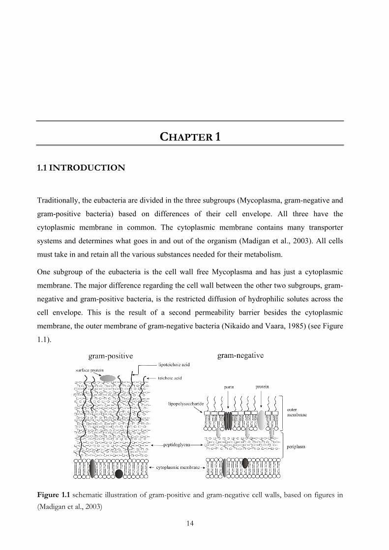
membrane, the outer membrane of gram-negative bacteria (Nikaido and Vaara, 1985) (see Figure
1.1).
Figure 1.1 schematic illustration of gram-positive and gram-negative cell walls, based on figures in (Madigan et al., 2003)
Chapter 1
15
A main component of the cell wall is the peptidogylcan. However, it occurs in different amounts
in these two subgroups and indicates a second major structural difference between the two
subgroups. The gram staining procedure makes the different properties of gram-negative and
gram-positive cell walls visible.
Gram-positive bacteria have a thick mesh-like cell wall made of up to 40 layers of
peptidoglycan, which is capable of retaining the crystal violet–iodine complex (violet dye) when
it is dehydrated in the gram staining procedure. Gram-negative bacteria have a thinner cell wall
than gram-positive bacteria. In addition to the cytoplasmic (inner) membrane, they have the outer
membrane which contains lipids, lipopolysaccharides (LPS), and proteins and is separated from
the cell wall by the periplasmic space, where just several layer of peptidoglygan are located. The
thin layer of peptidoglycan is unable to retain the crystal violet–iodine complex and therefore
gram-negative bacteria are not stained. Usually a counter stain with fuchsine dye is utilized to
make gram-negative bacteria visible.
Two Gram-negative bacteria of different appearance: E. coli versus Borrelia Within the group of gram-negative bacteria structural differences in the outer membrane and
differences in other morphologic details have developed through evolution.
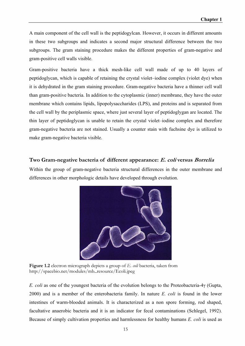
Figure 1.2 electron micrograph depicts a group of E. coli bacteria, taken from http://spacebio.net/modules/mb_resource/Ecoli.jpeg
E. coli as one of the youngest bacteria of the evolution belongs to the Proteobacteria-4γ (Gupta,
2000) and is a member of the enterobacteria family. In nature E. coli is found in the lower
intestines of warm-blooded animals. It is characterized as a non spore forming, rod shaped,
facultative anaerobic bacteria and it is an indicator for fecal contaminations (Schlegel, 1992).
Because of simply cultivation properties and harmlessness for healthy humans E. coli is used as

Chapter 1
16
a model organism for bacteria in general. The genomes of several E. coli strains are sequenced.
The E. coli genome on a circular chromosome has a size of app. 4.6 Mbp (basepairs) and has a
G-C content of around 50% (Blattner et al., 1997).
In some cases the generally harmless bacteria E. coli can become infectious, when they gain
additional genetic information encoding a variety of virulence factors such as adhesins, capsular
genes, siderophores or toxins. Uropathogenic E. coli strains (UPEC) can cause “honeymoon
cystitis” when the bacteria enter the urinary tract. Furthermore some E. coli strains can produce
very harmful toxins. In summary, it can be said that E. coli is a very important bacteria for the
genetic and medical research.
Figure 1.3 micrographs of Borrelia burgdorferi sento lato, taken from http://www.microbelibrary.org/images/jnelson/Images/borrelia.jpg)
The genus Borrelia belongs to the eubacterial phyla of the spirochetes, which is genetically very
far related to the Proteobacteria (Gupta, 2000; Woese, 2000). Spirochetes have a characteristic
shap (see Figure 1.3). They are helically shaped and very flexible. All species of the genus
Borrelia are microaerophilic and transmitted to vertebrates by hematophagous arthropods
(Barbour and Hayes, 1986). In the case of B. burgdorferi Ixodidae ticks are used as vectors for
transmission of Borrelia to their reservoir, which are vertebrates (mostly small rodents and deer).
The infection of humans happens accidentally and for B. burgdorferi it means a dead end.
Infection of humans is called Lyme borreliosis or Lyme disease and can be treated with
antibiotics (Nadelman et al., 2001; Steere et al., 2004). Diagnosis is not easy, if the typical
symopton of early Lyme disease, an erythema migrans (red circular skin lesion) is absent,
because other symptoms can be unspecific fever or flu like symptoms. In late states of infection
the treatment with antibiotics can be problematic, because Borrelia disseminate to other organs,
for example joints, which are not well supplied with blood (Steere et al., 2004). For the genus
Borrelia B. burgdorferi is a model organism, because the genome of B. burgdorferi B31 is

Chapter 1
17
sequenced and genetic manipulations have been established in it. The B. burgdorferi genome
differs completely from any known bacterial genomes. It consists of a linear chromosome of 0.9
Mbp and at least 17 linear and circular plasmids, which have together an additional size of 0.5
Mbp (Casjens et al., 2000; Fraser et al., 1997). In contrast to E. coli, the G-C content in B.
burgdorferi lies between 27-32%, which is low for a Gram-negative organism. One special
feature of B. burgdorferi is the loss of some of its plasmids during cultivation concomitant with
loss of virulence (Purser and Norris, 2000).
Another infectious Borrelia family causes relapsing fever. In contrast to B. burgdorferi,
relapsing fever B. recurrentis is transmitted by lice and is spread over the whole world (Dodge,
1973; Dodge, 1973). B. duttonii, the relapsing fever agent in Africa, is transmitted by soft body
ticks (Ornithodorus moubata) and it has been reported that humans are their only reservoir
(Barclay and Coulter, 1990; Dutton JE, 1905). B. hermsii is the best studied relapsing fever agent
causing relapsing fever in the US and it is transmitted by the soft body ticks Ornithodorus
hermsii (Porcella et al., 2005).
Gram-negative cell wall (E. coli versus Borrelia)
The outer cell envelope of E. coli and B. burgdorferi are quite similar, however there are also
characteristic differences (see Figure 1.4).
Figure 1.4 schematic illustration of the cell walls of E. coli and B. burgdorferi sento lato, based on (Tokuda and Matsuyama, 2004) and (Bergström et al., 2002)
Chapter 1
18
In E. coli the outer leaflet of the outer membrane, which is the contact area to the environment is
composed of lipopolysaccharides (LPS). LPS are completely missing in Borrelia (Takayama et
al., 1987). B. burgdorferi in contrast has a huge amount of lipoproteins anchored in the outer
leaflet.
Another tpyical feature of spirochaetes are endoflagella. The characteristic helical shape as well
motility of Borrelia is established by flagella, which is located in the periplasmic space. The
numbers of flagella in one cell varies in the genus Borrelia. For B. burgdorferi 7-11 flagella can
be present (Barbour and Hayes, 1986).
Integral outer membrane proteins with the function as porins have also been reported for
Borrelia (Ostberg et al., 2002; Shang et al., 1998; Skare et al., 1996; Skare et al., 1997).
However, a structure of any channel-forming protein (porin) has so far not been determined.
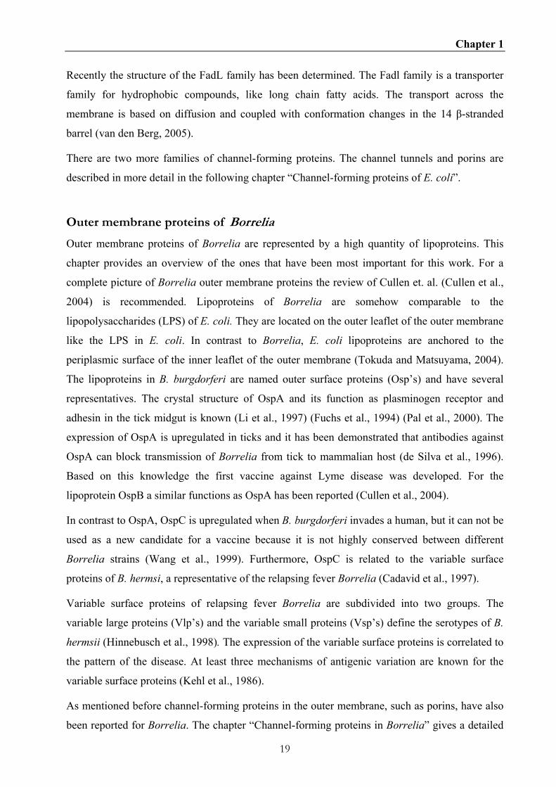
Outer membrane proteins of E. coli
Outer membrane proteins of E. coli are well studied and the model organism E. coli can provide
one prototype for each of the six functionally and structurally different families. While α-helical
bundles have only been found in the cytoplasmic membranes, for E. coli it has been shown that
β-barrels are restricted to the outer membrane and all six groups are structurally formed as β-
barrels (Koebnik et al., 2000).
The first group of the outer membrane proteins are the small β-barrel membrane anchors
composed of eight β-strands. A classical representative is OmpA, which serves as an anchor
between outer membrane and peptidoglycan (Koebnik, 1999; Ried et al., 1994). Whereas OmpX
belongs because of its structure to this group, however its function is the neutralization of the
host defense mechanisms (Heffernan et al., 1994; Vogt and Schulz, 1999).
The second group are the membrane integral enzymes built by 12 transmembrane β-strands, like
the outer membrane phospholipase A (OMPLA) (Snijder et al., 1999).
The barrel interior of OmpA, OmpX, and OMPLA is too small to allow the passage of ions or
substrates. Thus, they do not act as pore-forming proteins.
Thirdly, the group of TonB-dependent receptors has the biggest β-barrel known so far. A
representative of this group is FhuA, which is built out of 22 β-strands. These huge barrels of this
group are necessary for the uptake of iron-siderophore complexes (FhuA) and large substrates
like vitamin B12 (BtuB) (Chimento et al., 2003; Ferguson et al., 1998; Killmann et al., 2002;
Killmann et al., 1995).
Chapter 1
19
Recently the structure of the FadL family has been determined. The Fadl family is a transporter
family for hydrophobic compounds, like long chain fatty acids. The transport across the
membrane is based on diffusion and coupled with conformation changes in the 14 β-stranded
barrel (van den Berg, 2005).
There are two more families of channel-forming proteins. The channel tunnels and porins are
described in more detail in the following chapter “Channel-forming proteins of E. coli”.
Outer membrane proteins of Borrelia
Outer membrane proteins of Borrelia are represented by a high quantity of lipoproteins. This
chapter provides an overview of the ones that have been most important for this work. For a
complete picture of Borrelia outer membrane proteins the review of Cullen et. al. (Cullen et al.,
2004) is recommended. Lipoproteins of Borrelia are somehow comparable to the
lipopolysaccharides (LPS) of E. coli. They are located on the outer leaflet of the outer membrane
like the LPS in E. coli. In contrast to Borrelia, E. coli lipoproteins are anchored to the
periplasmic surface of the inner leaflet of the outer membrane (Tokuda and Matsuyama, 2004).
The lipoproteins in B. burgdorferi are named outer surface proteins (Osp’s) and have several
representatives. The crystal structure of OspA and its function as plasminogen receptor and
adhesin in the tick midgut is known (Li et al., 1997) (Fuchs et al., 1994) (Pal et al., 2000). The
expression of OspA is upregulated in ticks and it has been demonstrated that antibodies against
OspA can block transmission of Borrelia from tick to mammalian host (de Silva et al., 1996).
Based on this knowledge the first vaccine against Lyme disease was developed. For the
lipoprotein OspB a similar functions as OspA has been reported (Cullen et al., 2004).
In contrast to OspA, OspC is upregulated when B. burgdorferi invades a human, but it can not be
used as a new candidate for a vaccine because it is not highly conserved between different
Borrelia strains (Wang et al., 1999). Furthermore, OspC is related to the variable surface
proteins of B. hermsi, a representative of the relapsing fever Borrelia (Cadavid et al., 1997).
Variable surface proteins of relapsing fever Borrelia are subdivided into two groups. The
variable large proteins (Vlp’s) and the variable small proteins (Vsp’s) define the serotypes of B.
hermsii (Hinnebusch et al., 1998). The expression of the variable surface proteins is correlated to
the pattern of the disease. At least three mechanisms of antigenic variation are known for the
variable surface proteins (Kehl et al., 1986).
As mentioned before channel-forming proteins in the outer membrane, such as porins, have also
been reported for Borrelia. The chapter “Channel-forming proteins in Borrelia” gives a detailed

Chapter 1
20
description.
Channel-forming proteins of E. coli
Channel-forming proteins are necessary for the uptake of several substrates, like phosphate,
sugars and nucleotides, but also for the efflux of antibiotics, dyes, or heavy metals and secretions
of proteins like toxins or proteases.
For efflux processes channel tunnels are important as the outer membrane component of an
export machinery spanning the whole cell envelope. They interact with inner membrane
complexes comprising ABC transporter for secretion or proton antiporter for efflux (Andersen,
2003). The channel tunnel of E. coli TolC is a homotrimer with a 140 Ǻ long cannon-shape
structure. Each of the three monomers contributes four β-strands to form a single 40 Ǻ long β-
barrel, which is anchored in the outer membrane and is a single-channel forming domain
(Koronakis et al., 2000). The β-barrel is prolonged by a 100 Ǻ long tunnel domain formed
exclusively by α-helices (see Figure 5). TolC homologues are ubiquitous among gram-negative
bacteria, and already nearly a hundred have been identified (Koronakis, 2003).
The outer membrane protein family of the porins is necessary for the uptake of substrates and
they are also usually trimers, but of totally different architecture as TolC. Porins form one barrel
of 16 or 18 β-strands per monomer and therefore the trimeric assembly contains three separate
channels (Koebnik et al., 2000). They can be subdivided into general (nonspecific) porins
(OmpF) and substrate specific porins (LamB). OmpF is a classical diffusion pore and allows the
diffusion of molecules smaller than 600 Da through a β-barrel build by 16 β-strands spanning the
outer membrane (Garavito, 1994). An example for a specific pore is the well studied LamB,
which is the outer membrane component of the mal regulon (Benz et al., 1986; Schirmer et al.,
1995). The channel forms like OmpF a homotrimer and it is specific for maltose and
maltodextrins. The specificity is caused by a special arrangement of amino acids within the
channel. The loop 3 with the residue Y118 is inwardly folded and constricts the diameter of the
channel. Furthermore inside the channel is a belt of six aromatic amino acids (“greasy slide”)
lined up with polar amino acids (“polar tracks”). These aromatic residues form a pathway from
the vestibule to the periplasmic outlet of the porin (see Figure 4.1B). Sugar binding obstructs the
channel constriction leading to a blockage of the ion flow in black lipid bilayer experiments. By
moving through the channel the sugar “glides” along the aromatic residues and hydrogen bonds
between the sugar and the “polar tracks” are formatted and disrupted (Meyer and Schulz, 1997).
Recently the crystal structure of the nucleoside specific porin of E. coli Tsx was published (Ye

Chapter 1
21
and van der Berg, 2004). The structure of Tsx is an exception within the channel forming protein
family, because it forms monomers with 14 β-strands instead of trimers.
Figure 1.5 Porins and TolC: (a) view down the outer membrane channels, (b) side view, at right angles to the plane of the outer membrane, and (c) cross-section of TolC near the tunnel entrance; modified figure of (Andersen et al., 2001); with permission from Elsevier
Channel-forming proteins of Borrelia
By now, three pore-forming proteins of B. burgdorferi are described in literature. P66 is a 66
kDa protein, which has an integrin αvβ3 binding property and a channel forming ability with a
huge single-channel conductance of 11.5 nS in 1 M KCl (Coburn et al., 1999; Skare et al., 1997).
Oms28 is a pore forming protein with a small single-channel conductance of 0.6 nS in 1 M KCl.
Details about the function of this protein are not known so far (Skare et al., 1996).
P13, a 13 kDa integral outer membrane protein of B. burgdorferi, was identified as a porin using
the black lipid bilayer assay (Ostberg et al., 2002). P13 has an unusually small molecular mass
compared with other known porins having usually molecular masses of more than 30 kDa. The
P13 protein is processed at the amino (N)-terminus and carboxyl (C)-terminus (Noppa et al.,
2001). As for the other pore-forming proteins of Borrelia, the structure of P13 is not determined,
but it would be of great interest to investigate how such a small protein arranges to build a
transmembrane spanning barrel with a conductance of 3.5 nS.
Single-channel conductance measurements of the outer membrane fraction shows that B.
Chapter 1
22
burgdorferi contains many other pore-forming proteins besides P66, Oms28, and P13. Various
sizes of single channel conductance could be detected. Some were of very small size which leads
to the conclusion that B. burgdorferi has like E. coli general diffusion porins and specific porins.
A channel tunnel such as TolC most likely is present in B. burgdorferi. A BLAST search
revealed that a homologue (BB0142) is present in B. burgdorferi genome.
The relapsing fever agent B. hermsii was also studied for channel forming proteins. A P66
homologue was found and single-channel conductance fluctuations of various magnitudes were
observed in the outer membrane preparation (Shang et al., 1998).
Aims of the work
The aim of the work was to functionally characterize outer membrane channels of the relapsing
fever pathogen B. duttonii and the Lyme disease agent B. burgdorferi.
I have characterized an 80 pS channel of B. duttonii.
Furthermore I have identified the B. burgdorferi TolC homologue (BB0142) from a p66 knock-
out strain HB19/K02. The channel has single-channel conductance of 300 pS and shows
different properties to E. coli TolC.
In addition, point mutations in the maltose binding protein LamB of E. coli could demonstrate
the importance of the “greasy slide” for the sugar specificity of this channel.

23
CHAPTER 2
Discovery of a channel-forming protein in the cell wall of the relapsing fever
pathogen Borrelia duttonii
2.1 SUMMARY
Outer membrane preparations of three Borrelia relapsing fever species have been studied for
pore forming activity in the black lipid bilayer assay. Histograms of single-channel conductions
fluctuations were obtained from single-channel experiments with outer membrane preparations
of B. hermsii, B. recurrentis and B. duttonii. All strains had a different conductance fluctuation
pattern with a broad range of single-channel conductance values varying from 0.5 ns – 11 nS. All
three strains have a high pore forming activity at around 0.5 nS in common. Furthermore for one
of the species, B. duttonii, a channel-forming protein with a single-channel conductance of 80 pS
in 1M KCl could be isolated from the outer membrane preparation. Characterization of the 80 pS
channel showed that it is slightly anionic selective and voltage independent.
2.2 INTRODUCTION
Old world tick-borne relapsing fever (RF), caused by the spirochetal pathogen Borrelia duttonii,
is a common endemic disease in Central, East and South Africa. B. duttonii is transmitted to
humans by the bite of an infected soft body tick Ornithodorus moubata (Barclay and Coulter,
1990). This was first time described in literature 1904 by Dutton (Dutton JE, 1905). Most
infected people are farmers who live in traditional huts constructed of mud with flat, earth
covered roofs. They sleep on the floor on animal skin and frequently share their accommodation
with small livestock (Cutler et al., 1999). The bite by a tick occurs normally unrecognized,
because they feed quickly within 30 minutes during night (Schwan et al., 2003); (Walton, 1962).
Chapter 2
24
The pathogen is transmitted when infectious salivary and coxal secretions contaminate the
feeding site (Barclay and Coulter, 1990). Man is the only reservoir for B. duttonii and the disease
is characterized by high fever with chills, headache, myalgia and coughing. Each episode of high
fever is followed by a period of well-being but several other episodes of high fever and other
effects like abdominal pain, vomiting, diarrhea and photophobia may follow. The symptoms and
recurrent fever of the disease have been linked to antigenic variation of and antibody production
against different surface antigens presented by B. duttonii in the blood of the patient. The
mechanism of antigenic variation has been thoroughly investigated in the closely related RF
species B. hermsii and the recurring pattern of fever coincides with massive spirochetemia in the
blood and is a consequence of the antigenic variation of the most abundant surface protein,
variable major protein (Vmp), used by the bacterium to evade host antibody-mediated immune
response (Barbour, 1990; Burman et al., 1990; Meier et al., 1985). The host develops immune
response against these lipoproteins recently divided into two different families, Vsp (variable
small proteins) and Vlp (variable large proteins) (Hinnebusch et al., 1998; Parola and Raoult,
2001). Also for the louse borne species B. recurrentis, which is very close related to B. duttonii,
the Vmp is an important factor for infection. The Vmp is identified as the TNF-inducing factor
(Scragg et al., 2000). A unique property of B. duttonii, is the ability to form erythrocyte
aggregates which block precapillary blood vessels causing haemorrhage and inducing cell death
and hypoxia (Burman et al., 1998; Shamaei-Tousi et al., 1999).
The extracellular pathogen B. duttonii is classified as a gram-negative microaerophilic mobile
spirochete. The cell envelope consists of an outer membrane, periplasmic space and cytoplasmic
membrane, similar to the cell envelope architecture of other gram-negative bacteria. However,
the class Spirochaetes differs from other gram-negative bacteria because of the localization of
the flagella in the periplasmic space (endo flagella). Furthermore, the outer membrane contains
lipoproteins instead of lipopolysaccharides (LPS) (Takayama et al., 1987).
The cell wall of gram-negative bacteria acts as a molecular filter for hydrophilic solutes. These
molecular sieving properties are the result of channel forming proteins in their outer membranes,
called porins. Porins are integral proteins which form large water filled pores through the outer
membrane (Benz et al., 1978). They are involved in the uptake of substances from their
environment and can be subdivided into two classes. The porins of the first class can be
described as general diffusion pores, such as OmpF of E.coli K12 (Benz et al., 1985). These
porins sort mainly according the molecular mass of the solutes. The second class contains pores
with a binding site inside the channel. These specific porins are responsible for the rapid uptake
of classes of solutes such as carbohydrates (Benz et al., 1986; Ferenci et al., 1980), nucleosides
Chapter 2
25
(Maier et al., 1988) or phosphate (Benz and Hancock, 1987).
Within the class Spirochaetes the species B. burgdorferi lacks genes involved in the synthesis of
certain amino acids, fatty acids, co factors and nucleotides (Fraser et al., 1997). Consequently, B.
burgdorferi needs complex media containing all these nutrients for effective growth. It has been
demonstrated that these bacteria grow best in Barbour-Stoenner-Kelly media (BSK)
supplemented with rabbit serum (Barbour et al., 1984). This suggests that B. burgdorferi and
other Borrelia species may need special or specific porins (transport proteins) for uptake of these
nutrients.
Only a few integral membrane proteins have previously been identified and characterized for
Borrelia spp. Three porins of B. burgdorferi, the Lyme disease agent, are identified. These are
P66 (Skare et al., 1997), P13 (Ostberg et al., 2002) and Oms28 (Skare et al., 1996). A homolog
of P66 was identified in the outer membrane of the causative agent of new world relapsing fever
B. hermsii (Shang et al., 1998).
Pore forming outer membrane proteins of gram-negative bacteria form ß-barrel cylinders. A
prominent example is maltoporin of E. coli that is formed by a trimer of three identical
polypeptide subunits (Schirmer et al., 1995). Porins of gram-negative bacteria are normally
processed at the N-terminal end. P13 of B. burgdorferi represents in this respect an exception
because it is processed at the N-terminal and the C-terminal end. Secondary structure prediction
showed possible α-helices as transmembrane spanning domain of P13 (Noppa et al., 2001; Pinne
et al., 2004).
To date, little is known about outer membrane proteins of B. duttonii and their involvement in
pathogenesis of relapsing fever and life cycle besides the Vsp’s and Vlp’s. In this study we
compared the channel forming activity of the outer membrane protein fraction of B. duttonii, B.
hermsii (tick borne) and B. recurrentis (louse borne). Clear differences were detectable in the
appearance of single channel conductance. Here we also describe the purification and
characterization of a channel forming protein of B. duttonii. The biophysical characterization
was performed using the black lipid bilayer assay. The channel forming protein has a single-
channel conductance of 80 pS and is slightly anion selective. Parts of the amino acid sequence of
a putative channel forming candidate were identified by mass spectrometric analysis. Search
within the known protein sequences of other relapsing fever agents such as B. hermsii and B.
turicatae demonstrated that this protein is a possible homologue of the lipoprotein of the Vsp
family but it is presumably not a channel former.

Chapter 2
26
2.2 MATERIAL AND METHODS
2.2.1 Bacterial strain and growth conditions
The relapsing fever Borrelia strains used in this study were Borrelia duttonii 1120 and B.
recurrentis A1, a kind gift of Guy Baranton, Institut Pasteur, Paris, France and Borrelia hermsii
ATCC 35209, a kind gift of Alan Barbour, University of California Irvine, USA. Bacteria were
grown at 37°C in Barbour-Stoenner-Kelly-II (BSKII) medium (Barbour, 1984) or BSK-H
(Sigma, St. Louis, Mo.) supplemented with 10% rabbit serum and 1.4% gelatine.
2.2.2 Separation of outer membrane proteins and purification of the 80 pS channel
Outer membrane fractions (so called B-fraction) of B. duttonii, B. recurrentis and B. hermsii
were prepared as described elsewhere (Magnarelli et al., 1989). Purification of the native 80 pS
channel was performed with a dry hydroxyapatite (HTP) Bio-gel (Biorad) column. 100 µl of B-
fraction of B. duttonii (app. 100 ng proteins) was diluted with 400 µl 2% Genapol X-080 (Fluka)
and preheated for 10 min at 50 °C. Subsequently the mixture was applied to a dry HTP column
made from 0.3 g hydroxyapatite in an Econa-Column (Biorad) with the dimensions 0.5 cm
diameter and 5 cm length. The column was washed with six column volumes of a buffer
containing 2% Genapol (Roth), 10 mM Tris-HCl (pH 8.0). The bound 80 pS channel protein was
released from the column by increasing the ionic strength of the elution buffer. Three column
volumes of a buffer containing 2% Genapol (Roth), 1 M KCl and 10 mM Tris-HCl (pH 8.0)
(Merck) were passed through the column. Fractions of 1.0 ml were collected. Similarly, the 80
pS channel could also be obtained by using immobilised metal ion affinity chromatography
(IMAC). About 100 ng of B. duttonii outer membrane B-fraction was dissolved in 400 µl 1%
Genapol, 0.02 M Na2HPO4, 0.75 M NaCl (pH 7) (Merck) and subsequently applied to an Econa-
Column (Biorad) with the dimensions 0.5 cm diameter and 5 cm length filled with 5 ml Cu2+
loaded Chelating Sepharose Fast Flow (Amersham Bioscience). The column was washed with
1% Genapol, 0.02 M Na2HPO4, 0.75 M NaCl (pH 7) until the zero line was reached again. The
bound 80 pS channel protein was eluted with a solution containing 10 mM imidazol (Roth), 1%
Genapol, 0.02 M Na2HPO4, 0.75 M NaCl (pH 7). Fractions of 1.0 ml were collected.
2.2.3 Protein electrophoresis
Proteins samples were precipitated by the protocol of Wessel and Flügge (Wessel and Flugge,
1984) and loaded with 4x sample buffer on SDS polyacrylamide gels (SDS-PAGE). Proteins
were separated on 12 % SDS-PAGE or 4-15 % gradient SDS acryl gels (Biorad) under native or
Chapter 2
27
denatured conditions (boiled for 5 min in 4x SDS sample buffer before loading the gel) by using
a Biorad electrophoresis system according to the Laemmli method (Laemmli, 1970). The gels
were stained either with Coomassie R250 (Sigma) or silver stain (Blum et al., 1987).
2.2.4 Sequencing of the 27 kDa protein peptides and 27 kDa antibodies
For peptide sequencing SDS-PAGE was performed as described above using 12% gels. Protein
bands were cut out of the gel and digested with trypsin, endoproteinnases Lys-G and Asp-N
(Roche Diagnostics) according to Eckerskorn and Lottspeich (Eckerskorn and Lottspeich, 1993).
The resulting peptides were separated by reversed-phase HPLC (Sykam) with Luna C-18
column, 150 x 1 mm and subjected to N-terminal sequence and MALDI-TOF mass
spectrometry. Microsequencing was performed using a gas-phase sequencer Procise 492 cLC
(Applied Biosystems GmbH) according to the instructions of the manufacturer. Mass
spectrometry was performed using a Bruker reflex III MALDI-TOF mass spectrometer equipped
with a 337 nm nitrogen laser (Bruker-Franzen).
The 27 kDa protein was purified by preparative SDS-PAGE (Jordy et al., 1996). Polyclonal
antibodies against the purified 27 kDa protein were raised in rabbits and obtained by Agrisera
(Sweden).
2.2.5 Lipid bilayer experiments
The methodology for the black lipid bilayer experiments was described earlier (Benz et al.,
1978). The membrane cell consisted of a Teflon chamber with two aqueous compartments
connected by small circular holes (surface area 0.5 mm²). Membranes were formed by painting
onto the hole a 1% solution of diphytanoyl phosphatidylcholine (Avanti Polar Lipids) in n-
decane. The aqueous salt solutions (Merck) were used unbuffered and had a pH around 6. The
temperature was kept at 20°C throughout. Small amounts of the different protein fractions were
added to the aqueous phase on one or both sides of black lipid bilayer membranes.
The membrane current was measured with a pair of Ag/AgCl electrodes switched in series with a
voltage source and an electrometer (Keithley 617). For single-channel recordings the
electrometer was replaced by a highly sensitive current amplifier (Keithley 427). The amplified
signal was recorded with a strip chart recorder.
The zero-current membrane potentials were measured as described previously (Benz et al.,
1979). The membranes were formed in a 100 mM salt solution containing a predetermined
Chapter 2
28
protein concentration so that the membrane conductance increased about 100-1000 fold within
10-20 min after membrane formation. At this time the instrumentation was switched to the
measurements of the zero-current potential and the salt concentration on one side of the
membrane was raised by adding small amounts of concentrated salt solutions. The zero-current
membrane potential reached its final value 2-5min after addition of the concentrated salt
solution.
Titration experiments were performed with multi-channel experiments. The protein sample
containing the 80 pS channel was added to black diphytanoyl phosphatidylcholine/n-decane
membranes at concentration of about approximate 50 ng/ml. 30 min after the addition of the
proteins, the rate increase in conductance had slowed down considerably. At this time small
amounts of concentrated solutions of different substrates (see Table 3) were added to the
aqueous phase to both sides of the membrane, with constant stirring to allow equilibration. The
conductance data of the titration experiments were analyzed using the following equations used
earlier for the carbohydrate-induced block of LamB from Escherichia coli (Benz et al., 1986;
Benz et al., 1987). The conductance, G(c), of a membrane containing many channels in the
presence of a substrate with the stability constant, K (half saturation constant KS) and the
carbohydrate concentration, c, is given by the maximum conductance (without substrate), Gmax
times the probability that the binding site is free:
( ) =
( + ) G c G
K cmax
1 ⋅ (1)
Equation 1 may also be written as:
( - ( ) ) =( + )
G G cG
K cK c
max
max
⋅⋅ 1 (2)
which means that the conductance is a function of the substrate concentration and can be
analyzed using Lineweaver-Burke plots.
The voltage dependence of the porin channel was checked as described elsewhere (Riess and
Benz, 2000) by using membrane potentials as high as –150 to + 150 mV.

Chapter 2
29
Single-channel measurements of incubated protein sample with 27 kDa protein antibodies (in a
ratio 2:1 for 1h at 20 °C) were performed to check is the 27 kDa protein is the channel former in
the black lipid bilayer.
2.3 RESULTS
2.3.1 Channel-forming activities in the outer membrane fractions of different Borrelia strains
The outer membrane fractions obtained from the three RF Borrelia species were dissolved in 1%
Genapol and investigated for channel-forming activity in the black lipid bilayer assay. All outer
membrane preparations contained porins. Figure 2.1 shows histograms of conductance
fluctuations obtained from single-channel experiments with outer membrane preparations of the
three Borrelia strains. The histograms show a broad range of single-channel conductance values
caused presumably by different channel-forming proteins reconstituted into the lipid bilayer
membranes. The three Borrelia strains showed a different conductance fluctuation patterns. The
B-fraction of B. duttonii (A) formed channels with single-channel conductance centered on 0.25
nS, 2.5 nS, 5.5 nS and 11 nS. For B. hermsii (B) highest single-channel conductance was found
to be around 4 nS, which is different to that reported previously by Shang et al. (Shang et al.,
1998). The histogram obtained for total outer membrane of B. duttonii and B. recurrentis (panel
C) showed also high single-channel conductance above 4 nS. Both contained large channels with
conductance around 9.5 nS and 11 nS. Smaller channels could not be observed for outer
membrane preparations of the three strains because channels in the range below 200 pS are
masked by the appearance of the high conductance channels.

Chapter 2
30
Figure 2.1 Histogram of the probability P(G) for the occurrence of given conductivity steps observed with membranes formed of 1% diphytanoyl phosphatidylcholine/n-decane in the presence of outer membrane fractions from B. duttonii (A), B. hermsii (B) and B. recurrentis (C). P(G) is the probability that a given conductance step G is observed in the single-channel experiments. It was calculated by dividing the number of fluctuations with a given conductance step by the total number of conductance fluctuations. The aqueous phase contained 1 M KCl. The applied membrane potential was 20 mV; T = 20°C. The total numbers of conductance steps were 195 for B. duttonii (A), 116 for B. hermsii (B) and 135 for B. recurrentis (C).
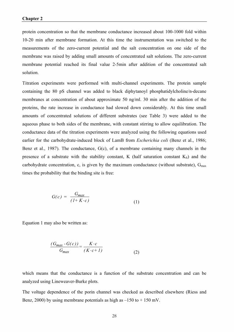
2.3.2 Single channel analysis of the 80 pS channel
When the proteins of the B-fraction of B. duttonii was separated using a dry HTP column or
immobilized metal ion affinity chromatography (IMAC) the high ionic strength or the
competitive ligand eluates contained a channel-forming protein that formed stable channels with
a single-channel conductance of 80 pS in 1 M KCl (see Figure 2.2A). Control experiments with
Genapol alone showed that the addition of the detergent did not result in channel formation. It is
noteworthy that the 80 pS channels could not be observed in the lipid bilayer assay when the B-
fractions of B. hermsii and B. recurrentis were separated in the same way (data not shown).
Figure 2.2B shows a histogram of 121 conductance steps of the 80 pS channel of B. duttonii in 1
Chapter 2
31
M KCl at a membrane potential of 20 mV. The average single-channel conductance was about
80 pS, therefore the channel forming protein was named 80 pS channel for further reference.
Figure 2.2 (A) Single channel recording of a 1% diphytanoyl phosphatidylcholine/n-decane membrane in the presence the HTP protein fraction of B. duttonii, which forms 80 pS channels. 50 ng/ml protein was added to the aqueous phase bathing a black lipid bilayer membrane. (B) Histogram of the conductance steps observed in the presence of the protein sample of B. duttonii forming the 80 pS channel. The aqueous phase contained 1 M KCl. The applied voltage was 20 mV; T = 20°C. The average single-channel conductance was about 80 pS for 121 steps.
The conductance of the 80 pS channel was studied as a function of different electrolytes and
concentrations. The results are summarized in Table 2.1. The single-channel conductance was a
linear function of the KCl-concentration between 0.1 and 3 M demonstrating that the channel did
not contain a binding site for ions inside the channel. The channel conductance did not vary
much when the salts contained different cations. The replacement of K+ by Li+ resulted in a
decrease from 80 pS to 50 pS (1.6-fold). However, replacement of chloride by the less mobile
Chapter 2
32
acetate anion had a more substantial effect on the single channel conductance. It decreased from
80 pS to 35 pS by a factor of 2.7. This result suggested that the 80 pS channel is more specific
for anions than for cations.
Table 2.1 Average single channel conductance, G, of the 80 pS channel as a function of concentration c of different salt solutions
Salt Concentration Average single channel
conductance
M pS LiCl 1 50 KCl 0.1 15 0.3 30 1 80 3 250 CH3COOK, pH 7 1 35
The membranes were formed from 1% diphytanoyl phoshatidylcholine dissolved in n-decane. The pH of the aqueous salt solutions was approximatively 6. The single-channel conductance is given as the mean of at least 100 single steps. The applied voltage was 20 mV and the temperature was 20°C.
2.3.3 The 80 pS channel is anion selective
Zero-current membrane potential experiments were carried out in the presence of salt gradients
to study the selectivity of the 80 pS channel. After the insertion of 100 - 1000 channels into the
PC membranes, the KCl concentration was raised on one side from 100 to 500 mM by addition
of 3 M KCl, while stirring to allow equilibration. Table 2.2 shows the results of these
measurements using KCl, LiCl and potassium acetate. In all three cases we observed negative
potentials at the more diluted side of the membrane (100 mM) indicating preferential movements
of anions through the channel. The zero-current membrane potentials for the three salts had
values between –23 mV (LiCl), –14.4 mV (KCl) and –8.4 mV (potassium acetate). Analysis of
these potentials using the Goldman-Hodgkin-Katz equation (Benz et al., 1979) suggested that
cations also have a certain permeability through the channel because of the radios of
permeability Pcation to Panion were 0.24 (LiCl), 0.41 (KCl) and 0.58 (potassium acetate). These
results demonstrate that the 80 pS channel is weakly anion selective, which agrees with the
Chapter 2
33
single-channel experiments.
Table 2.2 Zero-current membrane potential, Vm, of diphytanoylphoshatidylcholine/n-decane membranes in the present of the HTP protein fraction measured for a tenfold gradient of different salts.
Salt Vm Permeability ratio Pcation/Panion
mV KCl - 14.4 0.41 LiCl - 23 0.24 KCH3COO, pH 7 - 8.4 0.58
Vm is defined as the difference between the potential at the diluted side (100 mM) and the potential at the concentrated side (500 mM). The pH of the aqueous salt solution was 6 unless otherwise indicated. T = 20°C. Pcation/Panion was calculated with the Goldman-Hodgkin-Katz equation (Benz et al., 1978; Benz et al., 1985) from at least three experiments.
2.3.4 The 80 pS channel does not contain a binding site for substrates
Because of the small conductance and anion selectivity of the 80 pS channel it seems possible
that the channel is specific for anionic and other substrates. To check such a possibility titration
experiments were performed to determine specific binding of anionc and other substrates to the
channel. In particular, we tested substrates that are reported to bind to small specific porins such
as LamB (Benz et al., 1987) and Tsx (Maier et al., 1988). Table 2.3 contains a list of substrates
that are available in the host or vector of B. duttonii and may be transported across its outer
membrane. So far we did not find a pronounced conductance decrease for these compounds
within the concentration range, which could be used here. Assuming that we would have noticed
10% decrease of conductance Gmax following addition of the compounds we calculated the lower
limit of the half saturation constant; KS, from eqn. (2) (see Table 2.3). This means that the half
saturation constant for binding of these molecules, if it existed, is in most cases above 100 mM,
which is outside the physiologically relevant concentration range. Taken together it is clear that
the 80 pS channel does not show a significant specificity for the tested substrates.

Chapter 2
34
Table 2.3 Lower limit of the half saturation constant KS for the binding of different substrates to the B. duttonii 80 pS channel as derived from titration experiments.
Substrate KS [M]
Maltopentaose >0.1 Glutamate, pH7 >0.4 KH2PO4 >0.8 Adenosintriphosphate >0.04 Citrate, pH6 >0.8
The membranes were formed from 1% diphytanoyl phosphatidylcholine/n-decane. The aqueous phase contained 1 M KCl and small amounts of the HTP protein fraction containing the 80 pS channel. The lower limit of the half saturation constants, KS, was calculated using equation (2) and assuming that a 10% decrease of conductance Gmax followed the addition of the highest concentration of the compounds.
2.3.5 Voltage-dependence of the 80 pS channel
Certain outer membrane porins show voltage-dependent closure despite the fact that no voltage-
dependent closure was observed so far in in vivo experiments, which make it likely that the
voltage dependence is a reconstitution artifact (Lakey and Pattus, 1989; Sen et al., 1988, Benz,
1994). To check if also the 80 pS outer membrane channel of B. duttonii is voltage-dependent we
performed experiments with many 80 pS channels. The results demonstrate that this channel did
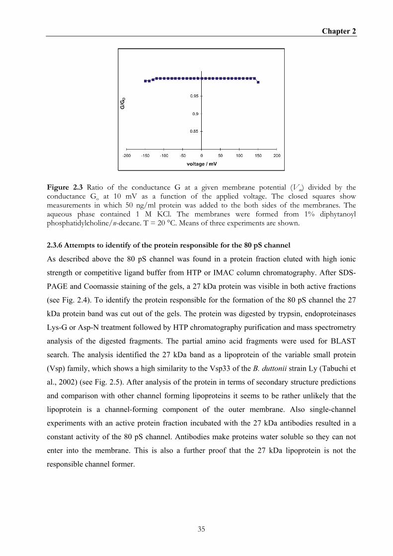
not show any voltage-induced closure even at voltages as high as ± 150 mV (see Fig. 2.3).
Chapter 2
35
Figure 2.3 Ratio of the conductance G at a given membrane potential (Vm) divided by the conductance Go at 10 mV as a function of the applied voltage. The closed squares show measurements in which 50 ng/ml protein was added to the both sides of the membranes. The aqueous phase contained 1 M KCl. The membranes were formed from 1% diphytanoyl phosphatidylcholine/n-decane. T = 20 °C. Means of three experiments are shown.
2.3.6 Attempts to identify of the protein responsible for the 80 pS channel
As described above the 80 pS channel was found in a protein fraction eluted with high ionic
strength or competitive ligand buffer from HTP or IMAC column chromatography. After SDS-
PAGE and Coomassie staining of the gels, a 27 kDa protein was visible in both active fractions
(see Fig. 2.4). To identify the protein responsible for the formation of the 80 pS channel the 27
kDa protein band was cut out of the gels. The protein was digested by trypsin, endoproteinases
Lys-G or Asp-N treatment followed by HTP chromatography purification and mass spectrometry
analysis of the digested fragments. The partial amino acid fragments were used for BLAST
search. The analysis identified the 27 kDa band as a lipoprotein of the variable small protein
(Vsp) family, which shows a high similarity to the Vsp33 of the B. duttonii strain Ly (Tabuchi et
al., 2002) (see Fig. 2.5). After analysis of the protein in terms of secondary structure predictions
and comparison with other channel forming lipoproteins it seems to be rather unlikely that the
lipoprotein is a channel-forming component of the outer membrane. Also single-channel
experiments with an active protein fraction incubated with the 27 kDa antibodies resulted in a
constant activity of the 80 pS channel. Antibodies make proteins water soluble so they can not
enter into the membrane. This is also a further proof that the 27 kDa lipoprotein is not the
responsible channel former.

Chapter 2
36
Figure 2.6 Protein separation by 12% SDS-PAGE of different protein preparations of B. duttonii. Lane OM shows total outer membrane proteins. Lane HTP shows the high ionic strength eluate of the HTP column and lane IMAC shows the competitive ligand eluate of the IMAC column. In each lane, 5 µg proteins were separated and visualized by Coomassie staining. Molecular mass standard in kilodaltons is indicated at the left of the gel. The HTP and the IMAC proteins resulted in formation of the 80 pS channel in lipid bilayer experiments.
10 20 30 40 50 60 | | | | | |
peptide KGDGSVIDLGV-SKKIEDAVAFAES Vsp33Bd -------MVLGILMVVMGCCNSGGGIKEGDEGKAKKGDGSVIDLKVIGEKIKSAVEFAGS Vsp2Bhe MKKNTLSAILMTLFLFISCNNGGPELKKGE---VTKSDGTVLDLSKISANIKNAVTFAAS VspABtu MKRITLSALLMTLFLLMSCNNSGTSPKDGQ---AAKSDGTVIDLATISKNIKDTVAFAKS OspCBbu MKKNTLSAILMTLFLFISCNNSGGDSASTN---PDES-AKGPNLTVISKKITDSNAFLLA 70 80 90 100 110 120 | | | | | | peptide VKEVKTLVKSVD DNGSLLAGVHSVISAVNTKL Vsp33Bd VKEVHALVRSVGEFAKAIGKKVTQNTGVIAADAGGNNNGGLIAGAYSLISELNTKVEVLG Vsp2Bhe VQEVETLVKSIDELAKAIGQKV--NADGLTAEA--NKNDSLVAGVYQLISDVQGKLTKLE VspABtu VKDVHTLVKSIDELAKAIGQKIQQNSDQFANDG--AHNGSLISGAFQVILTVETKLKSLE OspCBbu VKEVEALLSSIDELSKAIGKKIK-NDGTLDNEA--NRNESLIAGAYEISKLITQKLSVLN 130 140 150 160 170 180 | | | | | | peptide DELKVKIVAVAKEGKAFLDK TKDKGAT Vsp33Bd KKDGNSSELKSKFDDLNKKCKAFLDKVKGD-AELCKKDVTDENAQKALDVNNATKDKGAS Vsp2Bhe IGASKFAGLKEKVVAAKKGSDDFLTKVKAQHNNLGQS----AEAPKAIKKGNADSTKGAE VspABtu DTVGLSDTLKTKVTSSKIASRAFLDKVKSKHTELGKEGASDADAKAAILVSNGTKDKGVD OspCBbu -----SEELKEKIKEAKDCSQKFTTKLKDSHAELGIQSVQDDNAKKAILKTHGTKDKGAK 190 200 210 220 | | | | peptide ELAKLNGTIDELLKASNKILSDVLAELVVKPTT Vsp33Bd ELDALNTSIDGLLIVIKKLVEDAVNELTTSS---------- Vsp2Bhe ELGKLNTAIDELLTAAKDAVEAAIADVTAAPAKPATPVKP- VspABtu ELVKLNTEIDALLTAAEAAVTAAI-NALSTPAKSDAPAQSN OspCBbu ELEELFKSLESLSKAAQAALTNSVKELTN-PVVAESPKKP-
Figure 2.5 Amino acid comparison of the full-length Vsp33 protein of B. duttonii Ly, Vsp protein of B. hermsii, VspA protein of B. turicatae and OspC protein of B. burgdorferi using Pole Bioinformatique Lyonnaise Network Protein Sequence Analysis (http://npsa-pbil.ibcp.fr). Identical amino acids are given in red. Partial peptide sequences of the 27 kDa protein (see Figure 4) were obtained by mass spectrometry.
Chapter 2
37
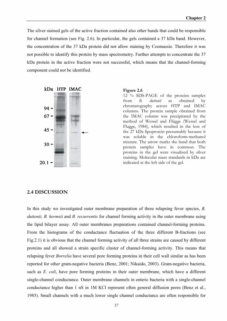
The silver stained gels of the active fraction contained also other bands that could be responsible
for channel formation (see Fig. 2.6). In particular, the gels contained a 37 kDa band. However,
the concentration of the 37 kDa protein did not allow staining by Coomassie. Therefore it was
not possible to identify this protein by mass spectrometry. Further attempts to concentrate the 37
kDa protein in the active fraction were not successful, which means that the channel-forming
component could not be identified.
Figure 2.6 12 % SDS-PAGE of the proteins samples from B. duttonii as obtained by chromatography across HTP and IMAC columns. The protein sample obtained from the IMAC column was precipitated by the method of Wessel and Flügge (Wessel and Flugge, 1984), which resulted in the loss of the 27 kDa lipoprotein presumably because it was soluble in the chloroform-methanol mixture. The arrow marks the band that both protein samples have in common. The proteins in the gel were visualized by silver staining. Molecular mass standards in kDa are indicated at the left side of the gel.
2.4 DISCUSSION
In this study we investigated outer membrane preparation of three relapsing fever species, B.
duttonii, B. hermsii and B. recurrentis for channel forming activity in the outer membrane using
the lipid bilayer assay. All outer membranes preparations contained channel-forming proteins.
From the histograms of the conductance fluctuation of the three different B-fractions (see
Fig.2.1) it is obvious that the channel forming activity of all three strains are caused by different
proteins and all showed a strain specific cluster of channel-forming activity. This means that
relapsing fever Borrelia have several pore forming proteins in their cell wall similar as has been
reported for other gram-negative bacteria (Benz, 2001; Nikaido, 2003). Gram-negative bacteria,
such as E. coli, have pore forming proteins in their outer membrane, which have a different
single-channel conductance. Outer membrane channels in enteric bacteria with a single-channel
conductance higher than 1 nS in 1M KCl represent often general diffusion pores (Benz et al.,
1985). Small channels with a much lower single channel conductance are often responsible for

Chapter 2
38
uptake of specific substrates such as carbohydrates or nucleosides (Benz et al., 1986; Maier et
al., 1988). This should also be possible for Borrelia when the different habitat and the metabolic
needs of these bacteria are considered. Thus it seems conceivable that they also developed
individual uptake mechanisms for specific substrates. The histogram of channel-forming proteins
from the outer membranes of B. hermsii differs from earlier published data. This may be
explained either by genetic differences in the investigated strains or by differences between the
methods used to prepare outer membrane proteins (Shang et al., 1998).
In the second part of the study we investigated the biophysical properties of an 80 pS channel of
B. duttonii in the lipid bilayer assay. The 80 pS channel of B. duttonii was discovered in a
fraction obtained by high ionic strength elution from the HTP column and by competitive ligand
elution from an IMAC column of the outer membrane preparation. We could demonstrate that
the 80 pS channel forms a water filled channel with a conductance in the range of the specific
outer membrane porins such as LamB (150 pS in 1 M KCl) (Benz et al., 1993) and Tsx (10 pS in
1 M KCl) (Maier et al., 1988). Its single-channel conductance was a linear function of the
aqueous salt concentration. Interestingly, the 80 pS channel of B. duttonii showed no voltage
dependence and was anion selective with a cation/anion permeability ratio of 0.41 in KCl
solution. This indicates a specificity of the channel for anionic substrates. Voltage independence
is frequently found for specific outer membrane channels, because oppositely charged residues
are distributed in a more balanced way within the channel in contrast to general diffusion pores
(Koebnik et al., 2000). However, our titration experiments demonstrated that the 80 pS channel
does not contain a binding site for a variety of substrates such as maltopentaose, glutamate,
KH2PO4, adenosintriphosphate or citrate (Table 3). This was concluded from the experimental
observation that the membrane conductance was not influenced by the addition of these
substrates. When we take a previously proposed formalism for the binding of these substrates to
a specific channel into consideration it is possible to calculate an upper limit for the stability
constant of substrate binding to the 80 pS channel. For this we assume that the 80 pS channel is a
single-file channel (Benz et al., 1986). This means that the 80 pS channel is open when no
substrate is bound, and closed when it is occupied. The conductance G(c) of a 80 pS channel
containing membrane in the presence of a substrate (concentration c), which binds with the
stability constant, K, is given in equation (2) in the Material and Method part, where Gmax is the
membrane conductance before the start of the substrate addition to the aqueous phase. When we
assume that we could observe at least a 10 % decrease of the conductance during the addition of
the substrate we can calculate an upper limit for the stability constant for substrate binding by
using Eqn. (2) and the maximum substrate concentration given above. For maltopentaose,

Chapter 2
39
glutamate, KH2PO4, adenosintriphosphate and citrate we calculated stability constants that
should be below 10, 2.5, 1.25, 25 and 1.25 1/M, respectively. The corresponding half saturation
constants (lower limits) should be higher than 0.1, 0.4, 0.8, 0.04 and 0.8 M, respectively.
These possible half saturation constants are in a range that is not of physiological relevance for
effective substrate scavenging because the concentration of these substrates is in the low
millimolar range in the hosts of Borrelia. This means that the substrate concentration probably
never reaches those of the half-saturation constants if these are about 0.04 M and higher. This
result suggests that the 80 pS channel does not contain a binding site for these substrates inside
the channel. Taken together it is clear that the 80 pS channel is unspecific with respect to the
tested substrates. Nevertheless, there exist indications that the channel could be specific for a not
identified substrate.
We tried to identify the protein responsible for channel forming activity of the 80 pS channel.
During our search we identified a 27 kDa protein of B. duttonii as a possible candidate (see
figures 4 and 5). Secondary structure analysis of this protein led to the conclusion that it is
presumably not a channel former because its secondary structure lacks the typical amphipatic
transmembrane β-strands of gram-negative bacterial porins (data not shown). By BLAST search
and amino acid sequence alignment it was shown that the 27 kDa protein belongs presumably to
the Vsp family. It has a high amino acid identity to Vsp33 of B. duttonii Ly strain (Tabuchi et al.,
2002). In addition, Vsp-proteins share a high degree of amino acid and DNA sequence identity
with OspC of B. burgdorferi (Carter et al., 1994). The structure of OspC of B. burgdorferi is
known and shows no transmembrane domain that could form a channel. Furthermore,
inactivation of the channel forming activity by antibodies against the 27 kDa lipoprotein was not
possible. Our attempts to relate the channel-forming activity porin activity of the 80 pS channel
to bands excised and eluted from preparative SDS-PAGE failed also because of the general
susceptibility of Borrelia porins towards SDS-treatment (data not shown). Comparison of silver
stained SDS-PAGE of the active fractions obtained from the two purification procedures
(hydroxyapatite and IMAC columns) finally led to the conclusion that a 37 kDa protein could be
the channel former, responsible for the 80 pS activity (Fig. 6). This seems to be possible because
outer membrane porins of gram-negative bacteria have molecular masses between 30 and 50
kDa, which is sufficient to form β-barrel structures of either 16 or 18 β-strands (Koebnik et al.,
2000).

Chapter 2
40
The identification of this candidate as a channel forming protein was difficult because the
genome of B. duttonii is not known. The analysis of the primary structure of the 37 kDa protein
could have solved this problem. However, it was not possible to obtain this protein in sufficient
quantity (at least a visible protein band on a gel stained by Coomassie) to perform N-terminal
sequencing (Eckerskorn and Lottspeich, 1993). Small protein amounts which are visible by
silver staining can be used for identification of proteins using peptide mass fingerprints
(Sickmann et al., 2002). However, this procedure needs the knowledge of the entire chromosome
of B. duttonii. This means that further attempts are necessary to identify the protein responsible
for the formation of the 80 pS channel.
This study demonstrates that the relapsing fever strains, B. duttonii, B. hermsii and B. recurrentis
contain channel forming proteins in their outer membrane. Further investigations are needed to
understand the role of porins such as the 80 pS channel in the physiology and virulence of these
relapsing fever Borrelia.

41
CHAPTER 3
Identification of a 300 pS pore forming protein in the p66 knock out mutant of
Borrelia burgdorferi sensu stricto strain HB19
3.1 SUMMARY
Outer membrane preparations of B. burgdorferi HB19 and p66 knock-out strain HB19/K02 were
characterized in the black lipid bilayer assay. Comparing the histograms of single-channel
conductions fluctuations of both strains showed no single-channel activity at 11.5 nS for the p66
knock-out strain. This verifies earlier studies that P66 is a pore-forming protein in B.
burgdorferi. Furthermore, one fraction obtained by anion exchange chromatography of the p66
knock out outer membrane preparation showed a uniform channel forming activity with a single
channel conductance of 300 pS. The biophysically characterization showed that the 300 pS
channel is ionselective and not voltage dependent. By mass spectrometry using peptide mass
finger prints, BB0142 could be identified as the sole channel forming candidate in the active
fraction. A BLAST search and a conserved domain search showed that BB0142 is a putative
TolC homologue in B. burgdorferi. For the first time a putative drug efflux system of B.
burgdorferi could be identified and the outer membrane component BB0142 biophysically
characterized.
3.2 INTRODUCTION
Lyme borreliosis is a medical term of an infection caused by the pathogen Borrelia burgdorferi
sensu lato transmitted by the bite of an infected hard body tick of the genus Ixodes. B.

Chapter 3
42
burgdorferi, was primarily described as a causative agent for Lyme borreliosis in 1975 by
Burgdorfer in Lyme, Connecticut USA (Burgdorfer et al., 1982). Beside this, two other species,
B. afzelii (Canica et al., 1993) and B. garinii (Baranton et al., 1992), are also known to cause a
similar disease in Eurasia (Hubalek and Halouzka, 1997); (Baranton et al., 1998; Li et al., 1998).
Today it is clear that Lyme borreliosis is a bacterial infection limited to the northern hemisphere.
The disease is closely correlated to the distribution of the Ixodes hard bodied ticks (Eisen et al.,
2002; Hudson et al., 1998).
Infection of humans by B. burgdorferi results in a wide variety of clinical manifestations (Steere
et al., 2004). The genus Borrelia represents highly mobile spirochetes. Following an initial local
infection of the skin (erythema migrans) the bacteria spread into other tissues, where they trigger
inflammation in distant dermis, nervous tissue, joints, and the heart (Steere et al., 2004). The
bacteria can survive for a long time in humans by evading the immune system. Chronic
infections result in some cases in arthritis, dermatitis or neuroborreliosis. Diagnosis of Borrelia
infections is sometimes difficult and problematic, in particular because of ambiguous symptoms
in late infection state. To date antibiotic treatment with tetracycline or β-lactam is successful in
most cases (Steere et al., 2004), especially when diagnosed at the early stage. However, it is also
known that B. burgdorferi has a natural resistance towards several antibiotics. This resistance
may rely on different mechanism known from other bacteria, such as enzymes, differences of the
antibiotic target, and efflux pumps. On the other hand, the cause of antibiotic resistance of
Borrelia is still an open question.
Until now only little is known about the physiology and metabolic of B. burgdorferi in different
hosts. However, the recently published genome of B. burgdorferi sensu stricto provides an
interesting insight into its metabolism (Fraser et al., 1997). Gene products necessary for the
synthesis of several essential metabolic substrates are lacking. Nevertheless, B. burgdorferi can
adapt in its tick vector (Ixodes) and escape from the immune system of its mammalian host, such
as humans, small rodents, and deer.
In comparison to enteric gram-negative bacteria, the density of transmembrane proteins is low.
Nevertheless, B. burgdoferi has within the group of spirochetes the highest concentration of
transmembrane proteins (Radolf et al., 1994; Walker et al., 1991). This means presumably that
Borrelia has an effective uptake mechanism for several amino acids, fatty acids, co-factors and
nucleotides, which it cannot synthesize (Fraser et al., 1997). The proteins responsible for solute
transport across the outer membrane of gram-negative bacteria are porins. Further knowledge on
the structure and function of these porins would be useful for understanding of the pathogenesis

Chapter 3
43
during Lyme borreliosis.
Porins can be subdivided into two classes. General diffusion pores such as OmpF of E. coli are
needed for the uptake of smaller solutes because these porins sort according to the molecular
mass (Benz et al., 1985). Specific porins like the maltoporin of E. coli, containing binding sites
(Benz et al., 1986), are necessary parts of uptake systems for classes of solutes. Furthermore,
porins often serve as receptors for bacteriophages (Wang et al., 2000).
Three putative porins are known for B. burgdorferi, Oms28 (Skare et al., 1996), P13 (Noppa et
al., 2001; Ostberg et al., 2002) and P66 (Skare et al., 1997).
In this study we investigated the outer membrane fraction of a p66 knock out mutant strain of B.
burgdorferi HB19/K02 (Coburn and Cugini, 2003) for its channel forming properties. Channel
activity with 11.5 nS single channel conductance as reported for P66 could not be detected in
outer membrane fractions of the mutant. However, it contained a channel with a single-channel
conductance of 300 pS in 1 M KCl, which could only be detected because the giant P66 pore was
absent in the lipid bilayer assay. We determined the biophysical properties of the 300 pS channel
and identified the protein by mass spectrometry. It is very likely that the 300 pS channel is
caused by a TolC homolog, which could mean that B. burgdorferi possesses also one or several
export systems for harmful drugs, which could be part of its antibiotic resistance (Andersen,
2004)
3.3 MATERIAL AND METHODS
3.3.1 Bacterial strain and culture conditions
The Borrelia burgdorferi strains used in this study were Borrelia burgdorferi HB19 and the P66
knock out strain B. burgdorferi HB19/K02, a kind gift of Jennifer Coburn, Tufts- New England
Medical Center, Boston, USA. The bacteria were cultured in Barbour-Stoenner-Kelly (BSK-II)
(Barbour and Hayes, 1986) or BSK-H (Sigma, St. Louis, Mo.) supplemented with 6 % rabbit
serum at 32°C. In the case of B. burgdorferi HB19/K02, the growth media contained also 200
µg/ml kanamycin.
3.3.2 Preparation of outer membrane fraction and purification of BB0142
Outer membrane fractions (fraction B) of B. burgdorferi HB19 and HB19/K02 were prepared as
described previously (Magnarelli et al., 1989). Purification of native BB0142 was performed

Chapter 3
44
with an anion exchange MonoQ column in combination with fast protein liquid chromatography
(FPLC) (Amersham Biosciences). About 200 µg of B. burgdorferi HB19/K02 fraction B were
dissolved in 800 µl 2% lauryl-dimethyl-amine-oxide (LDAO, Sigma) and were applied to the
column. The column was first washed with 7.5 ml 0.4% (LDAO) buffered with 10 mM Tris-HCl
(pH 8.0). Bound proteins were eluted with a linear NaCl gradient (0 to 1 M) containing 0.4%
LDAO buffered with 10 mM Tris-HCl (pH 8.0). Fractions showing a peak in the FPLC protocol
(see Fig. 2.3) were further analyzed by SDS-PAGE and the black lipid bilayer assay.
3.3.3 Protein precipitation, electrophoresis and immunoblotting
Two hundred µl of each FPLC fraction 33, 35, and 36 was added to 500 µl ice cold ethanol
(analytical grade, Merck) and stored for 18 h at –20 °C followed by centrifugation for 45 minutes
at 20,000 g and 4°C. The supernatant was discarded and the pellet was dried under vacuum
centrifugation (Speedvac, Eppendorf). Protein samples dissolved at 30°C and 100°C (5 min) in
sample buffer were separated by SDS polyarcrylamide gel electrophoresis (SDS-PAGE) using
the method of Lämmli (Laemmli, 1970). SDS-polyacrylamid gel were stained either with
Coomassie blue R-250 (Sigma) or silver stain (Blum et al., 1987). The monomeric band of
BB0142 (51 kDa) was purified by preparative SDS-PAGE (Jordy et al., 1996). Polyclonal
antibodies against BB0142 were raised in rabbits and obtained by Agrisera (Sweden). The
BB0142 antiserum was used for Western blots of the protein bands transferred on a
polyvinylidene difluoride membrane (PDVF, Roth) using standard procedures (Towbin et al.,
1979). For detection of bind BB0142 antibodies secondary antibodies raised against rabbit
antibodies conjugated with horseradish peroxidase (Amersham Pharmacia Biotech) were used.
The blot was developed with the ECL Western Blotting Detection kit (Amersham Pharmacia
Biotech) according to the manufacturer’s instructions.
3.3.4 Mass spectrometry (MS) of BB0142
The FPLC fractions showing channel-forming activity were subjected to SDS-PAGE followed
by silver staining (see Fig. 3.3). The different bands were analyzed by nano LC-mass
spectrometry as described elsewhere (Sickmann et al., 2003). Data interpretation of the obtained
MS/MS datasets was performed by the mascot algorithm (Perkins et al., 1999).
3.3.5 Planar lipid bilayer assay experiments
The methods used for the black lipid bilayer experiments have been described previously (Benz
Chapter 3
45
et al., 1978). The instrumentation consisted of a Teflon chamber with two aqueous compartments
connected by small circular holes (surface area about 0.5 mm²). Membranes were formed by
painting onto the hole a 1% solution of diphytanoyl phosphatidylcholine (Avanti Polar Lipids) in
n-decane. All salts (analytical grade) were obtained from Merck, Germany). The aqueous salt
solutions were used without buffering and had a pH around 6. The temperature was kept at 20°C
throughout. The channel forming protein solutions were highly diluted in 1% Genapol X-080
(Fluka) and added to the aqueous phase after the membrane turned black.
The membrane current was measured with a pair of calomel electrodes switched in series with a
voltage source and an electrometer (Keithley 617). For single-channel recordings the
electrometer was replaced by a highly sensitive current amplifier (Keithley 427). The amplified
signal was recorded with a strip chart recorder. The zero-current membrane potentials were
measured as described previously (Benz et al., 1979). The membranes were formed in a 100 mM
salt solution containing a predetermined protein concentration so that the membrane conductance
increased about 100-1000 fold within 10-20 min after membrane formation. At this time the
instrumentation was switched to the measurements of the zero-current potential and the salt
concentration on one side of the membrane was raised by adding small amounts of concentrated
salt solutions. The zero-current membrane potential reached its final value after 2-5min.
The voltage dependence of the porin channel was checked as described elsewhere (Riess and
Benz, 2000) by using membrane potentials as high as –150 to + 150 mV.
3.4 RESULTS
3.4.1 Porin activity in the outer membrane fraction of p66 knock-out mutant
In a first approach we checked the outer membrane preparation (fraction B) of B. burgdorferi
strain HB19 (wildtype) and p66 knock-out strain HB19/K02 for channel forming activity in the
lipid bilayer assay. For these outer membrane fractions of B. burgdorferi strains HB19
(wildtype) and HB19/K02 were diluted 1:1000 in 1% Genapol and added to the 1 M KCl
solution bathing a lipid bilayer membrane. Figure 3.1A and B show the histograms of
conductance fluctuations obtained in single-channel experiments with both B-fractions. The
histograms illustrate a broad range of single-channel events caused by different channel-forming
proteins reconstituted into the membranes. For the wildtype B-fraction a high channel-forming
activity with a conductance around 11-12 nS was detected, whereas in the B-fraction of the p66
knock out mutant strain HB19/K02 only a small channel-forming activity was observed with a
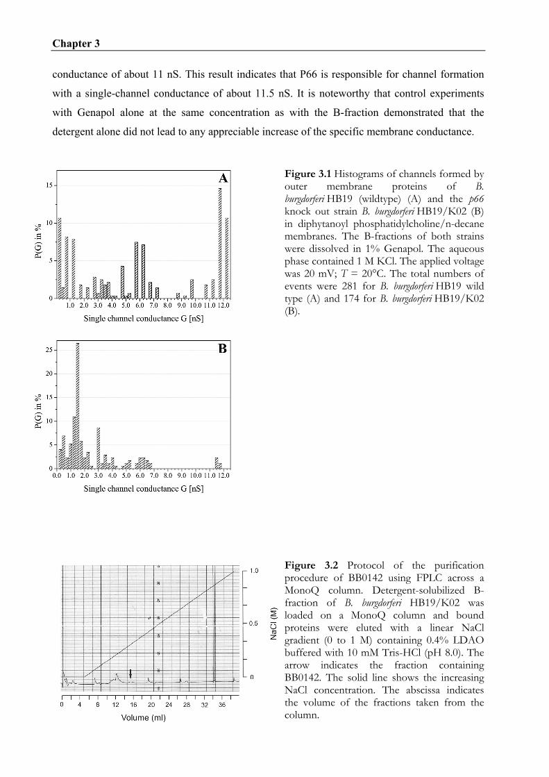
Chapter 3
46
conductance of about 11 nS. This result indicates that P66 is responsible for channel formation
with a single-channel conductance of about 11.5 nS. It is noteworthy that control experiments
with Genapol alone at the same concentration as with the B-fraction demonstrated that the
detergent alone did not lead to any appreciable increase of the specific membrane conductance.
Figure 3.1 Histograms of channels formed by outer membrane proteins of B. burgdorferi HB19 (wildtype) (A) and the p66 knock out strain B. burgdorferi HB19/K02 (B) in diphytanoyl phosphatidylcholine/n-decane membranes. The B-fractions of both strains were dissolved in 1% Genapol. The aqueous phase contained 1 M KCl. The applied voltage was 20 mV; T = 20°C. The total numbers of events were 281 for B. burgdorferi HB19 wild type (A) and 174 for B. burgdorferi HB19/K02 (B).
Figure 3.2 Protocol of the purification procedure of BB0142 using FPLC across a MonoQ column. Detergent-solubilized B-fraction of B. burgdorferi HB19/K02 was loaded on a MonoQ column and bound proteins were eluted with a linear NaCl gradient (0 to 1 M) containing 0.4% LDAO buffered with 10 mM Tris-HCl (pH 8.0). The arrow indicates the fraction containing BB0142. The solid line shows the increasing NaCl concentration. The abscissa indicates the volume of the fractions taken from the column.

Chapter 3
47
3.4.2 Identification of proteins involved in channel forming activity
Outer membrane proteins of the p66 knock out mutant B. burgdorferi HB19/K02 were separated
by chromatography across a MonoQ anion exchange column. Proteins were eluted with elution
buffer supplemented with increasing concentration of NaCl and checked in the lipid bilayer
assay for channel forming activity. The fractions eluted at 250 mM NaCl showed a uniform
channel-forming activity with a single-channel conductance of about 300 pS (see below). The
protocol of the elution taken at 280 nm showed a small protein peak when the elution buffer
contained 250 mM NaCl (see Fig. 3.2). To check the protein content of the corresponding
fractions 200 µl of the active fraction were precipitated and separated by SDS-PAGE. The
proteins in the silver stained bands were analyzed by mass spectrometry and identified using
peptide mass fingerprints (see Fig. 3.3). Several proteins of known and unknown function were
identified (data not shown). The protein of great interest was BB0142, which is a potential
homologue of the outer membrane protein TolC of E. coli. To verify that the corresponding
fractions contained BB0142 we performed a Western blot with polyclonal antibodies against
BB0142 (see Fig. 3.4). The BB0142 monomer was detected in the membrane-active FPLC
fraction and in the B-fraction of B. burgdorferi strain HB19/K02. When the membrane-active
protein sample was solubilized at 30°C a high molecular mass band was observed with an
apparent molecular mass of approx. 115 kDa on 12% SDS-PAGE that was not visible when the
samples were boiled (see Fig. 3.5). This band reflects presumably the trimer of BB0142 similar
to the formation of trimers by TolC on SDS-PAGE (Benz et al., 1993; Andersen et al., 2000).
However, the protein concentration of the oligomeric band was not high enough to be identified
by mass spectrometry or Western blot (data not shown).

Chapter 3
48
Figure 3.3 Fraction 35 of the MonoQ FPLC that was active in the lipid bilayer assay was precipitated and separated by 10% SDS-PAGE followed by silver staining. The arrow indicates the band where BB0142 was found by mass spectrometry. Molecular mass standards in kilodaltons (kDa) are indicated to the left.
Figure 3.4 Western blot of the B-fraction of B. burgdorferi HB19/K02 (first two lines from left), of the fractions 35, 36 and 33 of the MonoQ FPLC (middle three lines) and of B. burgdorferi B31 lp (right two lanes). The proteins were dissolved in sample buffer at room temperature (RT) or 100°C (100), separated by 12 % SDS-PAGE and transferred to a polyvinylidene difluoride membrane. The proteins were probed with polyclonal rabbit serum (RS) against BB0142. Molecular mass standards in kilodaltons (kDa) are indicated to the left. Figure 3.5 12% SDS-PAGE of fraction 35 of the separation of the B-fraction of B. burgdorferi HB19/K02 using MonoQ FPLC. The protein sample was loaded on the SDS polyarcylamid gel after dilution with sample buffer at room temperature (RT) and 100°C. The gel was stained with silver. The arrow marks the possible trimer of BB0142. Molecular mass standards in kilodaltons (kDa) are indicated to the left.

Chapter 3
49
3.4.3 Analysis of the channels formed by BB0142
Single-channel experiments with FPLC fractions containing BB0142 resulted in a stepwise
increase of the membrane conductance. Figure 3.6A shows a single-channel recording of a lipid
bilayer membrane in the presence of a very low concentration of BB0142 at a membrane voltage
of 20 mV. Panel B shows a histogram of 122 conductance steps observed in single-channel
experiments with BB0142 containing FPLC fractions. This protein forms obviously channels
with an average single-channel conductance of 300 pS in 1 M KCl. Channel-formation by the
300 pS channel could be completely block by incubation with polyclonal antibodies against
BB0142. After addition of the polyclonal antibodies to the protein fraction in a ratio 1:2 and
incubation for 1h at room temperature, the mixture showed no channel-forming activity in the
lipid bilayer assay. Interestingly, the 300 pS pore did not show any voltage dependence even at
voltages as high as ± 150 mV (data not shown). This demonstrates that the 300 pS channel did
not exhibit voltage dependent closure.
Figure 3.6 (A) Single-channel recording of a diphytanoyl phosphatidylcholine/n-decane membrane observed in the presence of 100 ng/ml of fraction 35 of the MonoQ-FPLC of the B-fraction of B. burgdorferi HB19 K02. The aqueous phase contained 1 M KCl; Vm = 20 mV; T = 20°C. (B) Histogram of the probability P(G) for the occurrence of a given conductivity unit observed with membranes formed of 1% diphytanoyl phosphatidylcholine/n-decane in the presence of fraction 35 of the MonoQ-FPLC of the B-fraction of B. burgdorferi HB19 K02. P(G) is the probability that a given conductance increment G is observed in the single-channel experiments. It was calculated by dividing the number of fluctuations with a given conductance increment by the total number of conductance fluctuations. The average single-channel conductance for 122 single-channel events was 300 pS

Chapter 3
50
The channels formed by BB0142 were permeable for a variety of different ions. The
conductance of the 300 pS channel was found to be dependent on the type of electrolyte and its
concentration (see Table 3.1). The KCl concentration varied from 0.1 to 3 M and the
conductance behaved as a linear function of the electrolyte concentration. This means that the
channel does not contain a binding site for potassium ions or chloride. Replacement of chloride
against the less mobile acetate had a strong effect on the single-channel conductance, which
decreased by 50% from 300 pS for 1 M KCl to 150 pS for 1 M potassium acetate. The exchange
of the cation from K+ to Li+ had in contrast to this a relatively small effect: the single-channel
conductance decreased only from 300 pS (1 M KCl) to 250 pS (1 M LiCl). This result
demonstrates that the 300 pS channel shows some preference for anions over cations.
Table 3.1 Average single channel conductance, G, of the channels formed by BB0142 as a function of different salt solutions of concentration c
Salt Concentration Average single channel
conductance
M pS LiCl 1 250 KCl 0.1 30 0.3 90 1 300 3 900 CH3COOK, pH 7 1 150
The membranes were formed from 1% diphytanoylphoshatidylcholine dissolved in n-decane. The pH of the aqueous salt solutions was 6 if not indicated otherwise. The single-channel conductance is given as the mean of at least 100 single events. The applied voltage was 20 mV and the temperature was 20°C.
3.4.4 The channels formed by BB0142 are not selective
Zero-current membrane potential experiments were performed for the analysis of the ion
selectivity. Table 3.2 shows the results of the measurements in the presence of 5-fold salt
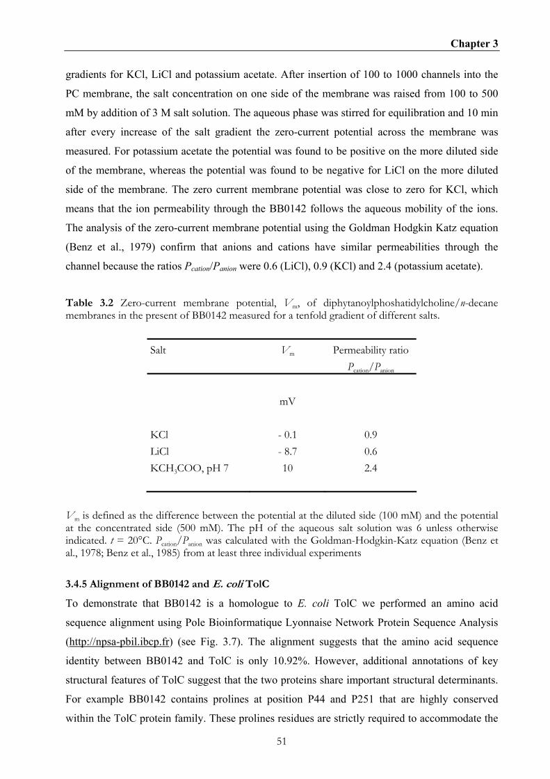
Chapter 3
51
gradients for KCl, LiCl and potassium acetate. After insertion of 100 to 1000 channels into the
PC membrane, the salt concentration on one side of the membrane was raised from 100 to 500
mM by addition of 3 M salt solution. The aqueous phase was stirred for equilibration and 10 min
after every increase of the salt gradient the zero-current potential across the membrane was
measured. For potassium acetate the potential was found to be positive on the more diluted side
of the membrane, whereas the potential was found to be negative for LiCl on the more diluted
side of the membrane. The zero current membrane potential was close to zero for KCl, which
means that the ion permeability through the BB0142 follows the aqueous mobility of the ions.
The analysis of the zero-current membrane potential using the Goldman Hodgkin Katz equation
(Benz et al., 1979) confirm that anions and cations have similar permeabilities through the
channel because the ratios Pcation/Panion were 0.6 (LiCl), 0.9 (KCl) and 2.4 (potassium acetate).
Table 3.2 Zero-current membrane potential, Vm, of diphytanoylphoshatidylcholine/n-decane membranes in the present of BB0142 measured for a tenfold gradient of different salts.
Salt Vm Permeability ratio Pcation/Panion
mV KCl - 0.1 0.9 LiCl - 8.7 0.6 KCH3COO, pH 7 10 2.4
Vm is defined as the difference between the potential at the diluted side (100 mM) and the potential at the concentrated side (500 mM). The pH of the aqueous salt solution was 6 unless otherwise indicated. t = 20°C. Pcation/Panion was calculated with the Goldman-Hodgkin-Katz equation (Benz et al., 1978; Benz et al., 1985) from at least three individual experiments
3.4.5 Alignment of BB0142 and E. coli TolC
To demonstrate that BB0142 is a homologue to E. coli TolC we performed an amino acid
sequence alignment using Pole Bioinformatique Lyonnaise Network Protein Sequence Analysis
(http://npsa-pbil.ibcp.fr) (see Fig. 3.7). The alignment suggests that the amino acid sequence
identity between BB0142 and TolC is only 10.92%. However, additional annotations of key
structural features of TolC suggest that the two proteins share important structural determinants.
For example BB0142 contains prolines at position P44 and P251 that are highly conserved
within the TolC protein family. These prolines residues are strictly required to accommodate the

Chapter 3
52
abrupt turn that links the β-barrel to the α-helical motifs. Similarly, the glycines residues at
positions 151 and 370 are also conserved and are situated in the turns near the closed end of the
TolC channel. However there exist also differences in the amino acid sequence between both
proteins. The BB0142 protein, which has 405 residues, is 71 amino acids shorter than the E. coli
TolC. This depends on a gap of eight residues in the loop 2 in BB0142. In addition, the
equatorial domain is missing because BB0142 ends directly after helix 8. It is noteworthy that
variations in the loop regions and the deletion of the equatorial domain have been reported also
for other TolC homologues (Andersen et al., 2000). Moreover differences in the amino acid
sequence are that none of the reported links for intra and intermolecular connections between the
monomers are conserved. Also the aspartate ring at the TolC tunnel entrance from the periplasm
is not conserved (Andersen et al., 2002; Koronakis et al., 2000).
Furthermore an NCBI conserved domain search has been performed and produced a significant
alignment (E value = 10-21) of BB0142 with the outer membrane efflux protein (OEP) family,
including the E. coli outer membrane protein TolC (Marchler-Bauer and Bryant, 2004). This is
an important hint for BB0412 being a TolC homologue.
One further observation on the genome level is that the gene bb0142 is located in an operon
encoding an efflux system. The gene bb0142 encodes the outer membrane compartment of the
efflux system which interacts with the acriflavine resistance protein (acrB) BB0140 and a
membrane fusion protein (mtrC) BB0141 (Fraser et al., 1997).
Figure 3.7 10 20 30 40 50 60 | | | | | | H1 H2 S1 L1 BB0142 EIIQISPKQAVNMALENSLDSENALYKENIKKLYKNNAWNVFVPNVNLSSTLSRNPSALS TolC ----ENLMQVYQQARLSNPELRKSAADRDAAFEKINEARSPLLPQLGLGADYTYSNGYRD Consensus enlkQay#qArennl#lrnaaadr#aakekiN#ArnpllP#lnLgadlsrnngard Prim.cons. --------Q----A---------------------N-A-----P---L------------ 70 80 90 100 110 120 | | | | | | S2 H3 BB0142 ELERDYWGFGFGVGINLSL-SPSVLKRMQLVMLEYESAKIERESAVRNIKLNVLKSYNQL TolC ANGINSNATSASLQLTQSIFDMSKWRALTLQEKAAGIQDVTYQTDQQTLILNTATAYFNV Consensus aner#snafgaglqinqSi dmSklra$qLqelaaeiad!er#saqrniiLNtakaYn#l Prim.cons. -----------------S-----------L--------------------LN----Y--- 130 140 150 160 170 180 | | | | | | T1 H4 BB0142 IALKSTLKVFESQIQNSKLKFEQARIAYNNGLISEIDFLDAQLKYKKSQPDLDGHIINFE TolC LNAIDVLSYTQAQKEAIYRQLDQTTQRFNVGLVAITDVQNARAQYDTVLANEVTARNNLD Consensus iaaidtLkyf#aQi#aikrql#Qarqa%NnGL!aeiDfq#AraqYdksqa#edgarnNl# Prim.cons. ------L-----Q---------Q-----N-GL----D---A---Y------------N--
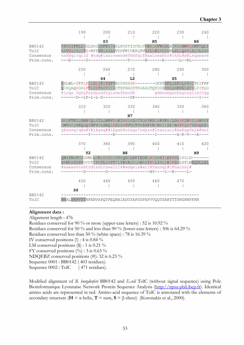
Chapter 3
53
190 200 210 220 230 240 | | | | | | S3 H5 H6 BB0142 KSKEIFKLLIGLDHDQDFEIIGELPDETIDFSLFNEALNFNESLEIKDLNMRLKMTEQLI TolC NAVEQLRQITG-NYYPELAALNVENFKTDKPQPVNALLKEAEKRNLSLLQARLSQDLARE Consensus nakEqlrqiiG #hdq#laaineendeTddfqlfNaaLneaEkr#ikdL#aRLkqdeare Prim.cons. ---E------G----------------T------N------E------L--RL------- 250 260 270 280 290 300 | | | | | | S4 L2 S5 BB0142 DSLWL-DTYLPSLSLSFSYSPYKSFHENSK--------GFSTGFLASFSLNYGLTEIFPF TolC QIRQAQDGHLPTLDLTASTGISDTSYSGSKTRGAAGTQYDDSNMGQNKVGLSFSLPIYQG Consensus #irqa DghLPsLdLsaStgisdsfhenSK gddsnmganksgnsglleI%qg Prim.cons. ------D--LP-L-L—S-----------SK--------------------------I--- 310 320 330 340 350 360 | | | | | | H7 BB0142 SKSFTKIQDNNYQLKILQNNVEGKIRNLKSSIVQKRKDIRRYKAILDASKINVELANKNY TolC GMVNSQVKQAQYNFVGASEQLESAHRSVVQTVRSSFNNINASISSINAYKQAVVSAQSSL Consensus gksnsq!q#a#Y#lkgaq##lEgahRnlkqs!rqkrn#Irasiaii#AsKqaVelA#knl Prim.cons. -----------Y---------E----------------I--------A-K—V---A---- 370 380 390 400 410 420 | | | | | | T2 H8 H9 BB0142 QMAFNAFNSGVMDLSKLNDIELVYKQSDLKFIEDKLNYANSILEYKNLINSLD------- TolC DAMEAGYSVGTRTIVDVLDATTTLYNAKQELANARYNYLINQLNIKSALGTLNEQDLLAL Consensus #aaeaa%nsGtrdisdlnDaeltlk#adqela#arlNYannqL#iKnainsL# Prim.cons. ---------G--------D-----------------NY----L--K-----L- 430 440 450 460 470 | | | | | S6 BB0142 -------------------------------------------------------- TolC NNALSKPVSTNPENVAPQTPEQNAIADGYAPDSPAPVVQQTSARTTTSNGHNPFRN
Alignment data : Alignment length : 476 Residues conserved for 90 % or more (upper-case letters) : 52 is 10.92 % Residues conserved for 50 % and less than 90 % (lower-case letters) : 306 is 64.29 % Residues conserved less than 50 % (white space) : 78 is 16.39 % IV conserved positions (!) : 4 is 0.84 % LM conserved positions ($) : 1 is 0.21 % FY conserved positions (%) : 3 is 0.63 % NDQEBZ conserved positions (#): 32 is 6.23 % Sequence 0001 : BB0142 ( 403 residues). Sequence 0002 : TolC ( 471 residues).
Modified alignment of B. burgdorferi BB0142 and E.coli TolC (without signal sequence) using Pole Bioinformatique Lyonnaise Network Protein Sequence Analysis (http://npsa-pbil.ibcp.fr). Identical amino acids are represented in red. Amino-acid sequence of TolC is annotated with the elements of secondary structure (H = α-helix, T = turn, S = β-sheet) (Koronakis et al., 2000).
Chapter 3
54
3.5 DISCUSSION
Pore forming activity of Borrelia outer membrane proteins has been described in previous
publications (Ostberg et al., 2002; Shang et al., 1998; Skare et al., 1996; Skare et al., 1997). The
previously indentified channel-forming proteins are P66, Oms28 and P13. The channel-forming
activity of the latter one has been demonstrated by comparison of the current fluctuations of
outer membrane protein preparations of B. burgdorferi wildtype and a p13 knock out strain,
respectively (Ostberg et al., 2002). In the first part of our study we performed similar
experiments with a p66 knock out B. burgdorferi strain. This strain was used by Coburn and
Cugini (2003) to demonstrate the function of P66 as integrin αvβ3 adhesion protein (Coburn and
Cugini, 2003). We observed for outer membrane protein preparations of the knock out strain a
clear reduction of the 11.5 nS activity that is typical for reconstitution experiments with P66
(Skare et al., 1997). This suggests that the outer membrane protein P66 is a bifunctional protein.
It is an integrin αvβ3 adhesion protein but it acts also as a channel forming protein with a single
channel conductance of 11.5 nS in 1 M KCl. P66 is the largest outer membrane channel present
in B. burgdorferi. Electrophysiological analysis of its properties suggest that it is a general
diffusion pore such as the 1.7 nS OmpF of E. coli and in contrast to the smaller outer membrane
channels such as LamB and Tsx, which are solute-specific (Benz et al., 1985; Benz et al., 1986;
Maier et al., 1988; Maier et al., 2001). P66 has a very high single-channel conductance for a
general diffusion pore as compared to the channels of the outer membrane of enteric bacteria.
However it is not the largest outer membrane pore reported in literature. Thermus thermophilus
outer membrane contains a giant pore with a single-channel conductance of 20 nS in 1 M KCl
(Maier et al., 2001).
Absence of the giant P66 outer membrane channel represents a considerable advantage for
purification of other B. burgdorferi channel forming proteins. Separation of outer membrane
proteins of the p66 knock out strain by anionic exchange chromatography resulted in a fraction,
which showed a uniform channel forming activity in the black lipid bilayer assay with a single-
channel conductance of 300 pS in 1 M KCl. By mass spectrometry using peptide mass
fingerprints (Sickmann et al., 2002) we identified the protein BB0142 as the sole protein with a
putative pore forming function. A BLAST search and a NCBI conserved domain search
identified BB0142 as a homologue of E. coli TolC. TolC is the outer membrane component of
type I secretion systems and multidrug efflux pumps (Koronakis, 2003). By itself, TolC forms
channels with a single-channel conductance of 80 pS in 1 M KCl, which is in the similar range as
the observed channel in the isolated FPLC fraction (Andersen, 2004; Benz et al., 1993;

Chapter 3
55
Koronakis, 2003; Koronakis et al., 2000). TolC is a homotrimer with a 140 Ǻ long cannon-shape
structure. Each of the three monomers contributes four β-strands to form a single 40 Ǻ long β-
barrel, the so-called channel domain, which is anchored in the outer membrane (Koronakis et al.,
2000). The β-barrel is prolonged by a 100 Ǻ long tunnel domain formed exclusively by α-
helices. The alignment of BB0142 with TolC (see Fig. 7) depicts conserved residues, which are
important for the correct folding of the channel-tunnel family. Among these are prolines and
glycines, which are important for the transitions between the β-sheets of the channel and the α-
helices of the tunnel (Andersen et al., 2001). An immunoblot analysis with BB0142 antibodies
showed that BB0142 is present in the FPLC fraction, which is active in the black lipid bilayer.
When the FPLC fraction was incubated with BB0142 antibodies and added to the black lipid
bilayer assay the channel forming activity was completely abolished, confirming that BB0142 is
responsible for the channel forming activity. A further purification of the putative oligomer
protein band of BB0142 by preparative SDS-PAGE failed (data not shown). We assume that this
occurs because of the sensitivity of BB0142 to SDS, as it is reported for other channel forming
protein of B. burgdorferi (Ostberg et al., 2002).
For TolC of E. coli it is known that residues lining the periplasmic entrance are critical for the
electrophysiological behavior. The aspartate ring consisting of six aspartates residues at the TolC
tunnel entrance from the periplasm is responsible for its high cation selectivity (Andersen et al.,
2002). In BB0142 this aspartate ring is replaced by positive lysine residues (K366) and negative
aspartate residues (D369) resulting in an unselective BB0142 channel, which is in contrast to the
selective TolC channel. The lysine residue (K366) and the aspartate residue (D369) could
compensate each other and might form a salt bridge. This means that no net charge exists in the
tunnel entrance. This consideration is supported by titration experiments with multivalent
cations, which do not block the BB0142 channel in contrast to the E. coli TolC (data not shown)
(Andersen et al., 2002). Another difference between BB0142 and TolC is that the BB0142
channel has a nearly four times higher conductance in 1M KCl than TolC (80 pS in 1M KCl)
(Benz et al., 1993). TolC of E. coli contains a circular network of inter- and intramolecular
connections between the three monomers that is presumably responsible for the stability of the
closed state of the channel-tunnel (Andersen et al., 2002). None of the residues forming these
inter- and intramolecular connections are conserved in BB0142. Thus it is possible that other
residues build a circular network of inter and intramolecular connections, keeping the tunnel
entrance closed otherwise the single-channel conductance should be much higher. Structural
modeling or crystal structure determinations of BB0142 could answer the questions what kind of
network is responsible for the closed state of the channel.

Chapter 3
56
Looking at the location of the bb0142 gene within the chromosome of B. burgdorferi it is evident
that it is a part of an operon coding for a multidrug efflux pump. This means that BB0142 is
interacting with the putative inner membrane complex formed by the AcrB homologue BB0140,
which serves as a inner membrane transporter, and the MtrC homologue BB0141, a periplasmic
accessory protein (Pos et al., 2004; Veal et al., 1998). These three components are necessary to
form a transport system spanning the two membranes for pumping out noxious compounds from
the cytoplasm (Andersen, 2003; Sulavik et al., 2001). So far, the finding of a multidrug efflux
system in B. burgdorferi makes sense, because B. burgdorferi is carrying a natural resistance for
several antibiotics (Hunfeld et al., 2001). Resistance can originate from multidrug efflux pumps,
as it is demonstrated for several other gram-negative bacteria (Eswaran et al., 2004; Grkovic et
al., 2002; Mata et al., 2000; Sulavik et al., 2001). Expression of BB0142 occurs also under
cultivation conditions and this shows that the only drug efflux pump found in the B. burgdorferi
B31 genome is active. For further studies it would be interesting to investigate, if the protein is
expressed in all phases of the Borrelia life cycle or if expression is special for certain conditions.
Another future interest would be to identify the substrates of the multidrug efflux pump. This
could be done by investigation of a bb0142 knock out mutant.
Taken together we have here in demonstrated that P66 is a channel forming protein besides its
function as integrin αvβ3 adhesion protein because the typical 11.5 nS channels were absent in
the B-fraction of the B. burgdorferi knock out strain. In addition we identified and characterized
BB0142 as a TolC homologue of B. burgdorferi in the black lipid bilayer assay. In future studies
it would be of interest to investigate the role of the TolC homologue BB0142 in virulent and in
drug resistant Borrelia strains as well as performing complementation studies in other bacterial
species.

57
CHAPTER 4
Site-directed mutagenesis of the greasy slide aromatic residues within the
LamB (maltoporin) channel of Escherichia coli: Effect on ion and
maltopentaose transport.
4.1 SUMMARY
The 3D-structure of the maltooligosaccharide-specific LamB-channel of Escherichia coli
(also called maltoporin) is known from X-ray crystallography. The 3D-structure suggest that a
number of aromatic residues (Y6, Y41, W74, F229, W358 and W420) within the channel
lumen is involved in carbohydrate and ion transport. All aromatic residues were replaced by
alanine (A) scanning mutagenesis. Furthermore, LamB mutants were created in which one,
two, three, four, and five aromatic residues were replaced to study their effects on ion and
maltopentaose transport through LamB. The purified mutant proteins were reconstituted into
lipid bilayer membranes and the single-channel conductance of the mutants was studied in
conductance experiments. The results suggest that all aromatic residues provide some steric
hindrance for ion transport through LamB. Highest impact is provided by Y6 and Y41 that are
localized opposite to Y118, which form the central constriction of the LamB channel.
Stability constants for binding of maltopentaose to the mutant channels were measured using
titration experiments with the carbohydrate. The mutation of one or several aromatic amino
acid led to a substantial decrease of the stability constant of binding. The highest effect was
observed when all aromatic amino acids were replaced by alanine because no binding of
maltopentaose could be detected in such a case. However, binding was again possible when
Y118 was replaced by tryptophane (W). The carbohydrate-induced block of the channel
function could also be used for the study of current noise through the different mutant LamB-
Chapter 4
58
channels. The analysis of the power density spectra of some of the mutants allowed the
evaluation of the on- and off-rate constants (k1 and k-1) of carbohydrate binding to the
binding-site inside the channels. The results suggest that both on- and off-rate constants were
affected by the mutations. For most mutants k1 decreased and k-1 increased. The possible
influence of the aromatic residues of the greasy slide on carbohydrate and ion transport
through LamB is discussed.
4.2 INTRODUCTION
The cell envelope of gram-negative bacteria consists of different layers. The inner or
cytoplasmic membrane contains the respiration chain, proteins for the transport of nutrients
and proteins involved in the synthesis of phospholipids, peptidoglycan and
lipopolysaccharides (Beveridge, 1981; Nikaido and Vaara, 1985). The periplasmic space
between the membranes is an aqueous compartment isoosmolar to the cytoplasm(Benz, 1994).
It contains the peptidoglycan and a large number of different proteins. The outer membrane is
composed of protein, lipid and lipopolysaccharide (Nikaido and Vaara, 1985). It contains only
a few major proteins. At least one of constitutive outer membrane proteins called porin forms
a general diffusion pore with a defined exclusion limit for hydrophilic solutes within the outer
membrane (Benz and Bauer, 1988; Danelon and Winterhalter, 2004; Hancock et al., 1986;
Nikaido and Vaara, 1985). In addition to the constitutive porins the outer membrane may
contain porins, which are induced under special growth conditions (Brass et al., 1985;
Hancock, 1981; Szmelcman et al., 1976; Tommassen and Lugtenberg, 1980). They often form
solute-specific channels and contain binding sites for neutral substrates such as carbohydrates
(Benz et al., 1986; Ferenci et al., 1980), nucleosides(Maier et al., 1988) and charged solutes
such as phosphate (Benz and Hancock, 1987; Hancock, 1981). Many of these specific porins
are part of uptake and degradation systems, such as the mal-system of Escherichia coli
(Schwartz, 1987) or the single copy plasmid pUR400 of enteric bacteria (Schmid et al., 1988).
These systems confer to the bacteria the possibility of growth on maltose and
maltooligosaccharides and on sucrose as carbon source (Schmid et al., 1982; Szmelcman et
al., 1976).
The proteins coded on the maltose operon contain also a specific outer membrane porin that is

Chapter 4
59
specific for maltose and maltooligosaccharides. LamB of Escherichia coli and other enteric
bacteria is essential for the uptake of maltooligosaccharides across the outer membrane
(Szmelcman et al., 1976), but it has a much lower efficiency for the uptake of
sucrose(Andersen et al., 1998; Schmid et al., 1982). LamB has been crystallized and its 3D-
structure is known from X-ray analysis (Schirmer et al., 1995). The individual channel within
a LamB-trimer is formed by 18 antiparallel β-strands, which form a cylinder with a diameter
of about 2.5 nm. The diameter of the channel is reduced by the external loop 3 folding into the
channel lumen to form a central constriction with a size of about 0.5 x 0.3 nm (see Figure
4.1). Carbohydrate transport through the channel is mediated partially by van der Waals
interaction of the maltooligosaccharides with six aromatic residues lining up the channel
interior (the greasy slide, Y6, Y41, W74, W358, W420 and F227) and by hydrogen bonds
between the hydroxy groups of the carbohydrates and amino acid residues within the
constriction zone such as R8 (Dutzler et al., 1996; Jordy et al., 1996; Schirmer et al., 1995).
The aromatic amino acids are lined up from the external surface in the series W74 (provided
by a neighboring monomer), Y41, Y6, W420, W358 and F227 close to the periplasmic space
(see Figure 1). Localized opposite to these amino acids of the greasy slide is tyrosine 118
(Y118) (see Figure 1). This amino acid has a major impact of ion and carbohydrate transport
through LamB (Orlik et al., 2002; Orlik et al., 2002). Furthermore, alanine-scanning
mutagenesis of single aromatic residues of the greasy slide has shown that they influence
carbohydrate binding in vitro and maltose uptake in vivo (Van Gelder et al., 2002).
Figure 4.1 Cross-section of the E. coli LamB monomer (maltoporin) (A, left side panel) and arrangement of the greasy slide together with a bound maltopentaose (B, right side panel). The left side panel shows loop 3 (dark gray) and tyrosine 118 (denoted with its number from the mature N-terminal end) that are relevant for passage of carbohydrates and ions through the
Chapter 4
60
central constriction. The amino acids of the greasy slide are localized opposite to Y118 and are given in dark gray. The strands of the β-barrel cylinders of the porins are given in light gray. The coordinates of maltoporin were taken from the crystallographic data of Schirmer et al. (1995) (Schirmer et al., 1995). The right side panel shows the arrangement of the aromatic residues of the greasy slide (denoted with their numbers from the N-terminal end and of the deletions) with respect to a bound maltopentaose molecule. Note that the amino acids W74 (D1; denoted with an asterix) is provided from a neighboring LamB monomer. The coordinates of bound maltopentaose were taken from the crystallographic data of Dutzler et al. (1995)(Dutzler et al., 1996).
Carbohydrate binding to the LamB channel has been studied in detail by assuming a
symmetrical one-site two-barrier model for sugar transport and titrating the ion current
through LamB with increasing concentrations of different carbohydrates (Benz et al., 1987).
Carbohydrate binding affinity increases with the chain length of the maltooligosaccharides.
The kinetics of carbohydrate transport can be derived from carbohydrate-induced current
noise of the LamB-channel (Andersen et al., 1995; Jordy et al., 1996; Nekolla et al., 1994). In
this study we give a quantitative description of the effect of alanine scanning mutagenesis of
all aromatic residues of the greasy slide on ion transport and carbohydrate binding through the
LamB-channel. Besides single mutations, two, three, four, five and all six amino acids of the
greasy slide were replaced by alanine. Some of the mutants included also mutagenesis of
Y118W together with mutations of the greasy slide residues. All mutants were purified to
homogeneity and ion transport as well as maltopentaose binding was studied in detail using
the lipid bilayer assay. In addition, for some of the mutants it was possible to evaluate
maltopentaose-binding kinetics by the measurement of the current noise. The results suggest
that all amino acids of the greasy slide contribute to certain extent to ion and carbohydrate
transport properties of LamB. Highest impact on channel function have the aromatic amino
acids Y41, Y6 and W420 localized opposite Y118 and Y118 itself. When all six residues of
the greasy slide are mutated to alanine, the mutation Y118W is sufficient to confer to LamB
maltopentaose transport in vivo and maltopentaose binding in vitro.
4.3 MATERIALS AND METHODS
4.3.1 Materials
Diphytanoyl phosphatidylcholine (DiphPC) was obtained from Avanti Polar Lipids
(Alabaster, AL). All salts were analytical grade (Merck, Darmstadt, Germany). Ultrapure
water was obtained by passing deionized water through a Milli-Q equipment (Millipore,
Bedford, MA).

Chapter 4
61
The QuikChange™ site-directed mutagenesis kit and the One Shot® Top10F’ competent cells
were purchased from Stratagene (Amsterdam, The Netherlands). Maltopentaose was obtained
from Seikagaku (Japan) distributed by Medac (Germany).
4.3.2 Plasmids and DNA manipulations
The generation of LamB mutant Y118W was described previously (Orlik et al., 2002). All
other LamB mutants were constructed according to standard genetic manipulations using the
QuikChange™ site directed mutagenesis kit. Table 1 contains a list of the mutants and the
primers used for the mutation. Plasmid pAM117 encoding wild-type LamB (Heine et al.,
1988) was used as a template for in vitro site directed mutagenesis. For each mutant two
synthetic oligonucleotide primers were designed (purchased from Carl Roth, Karlsruhe,
Germany) each complementary to opposite strands of the plasmid and containing the desired
mutation (see Table 1). The oligonucleotide primers were extended during temperature
cycling by using Pfu-turbo DNA polymerase. Incorporation of the primers generates a
mutated plasmid containing staggered nicks. After temperature cycling the product was
treated with DpnI. The DpnI endonuclease (target sequence: 5’-Gm6ATC-3’) is specific for
methylated and hemimethylated DNA and is used to digest the parental DNA template. This
led to the selection of the mutation-containing synthesized DNA. The nicked vector DNA was
then transformed into One Shot® Top10F’ competent cells, where the nicks in the mutated
plasmid were repaired. For the generation of double mutations the plasmid coding for a single
mutation was used as a template for further in vitro site directed mutagenesis. Similarly, using
the already existing plasmids coding for mutant porins as templates triple and higher
mutations were created. The mutants containing the mutation Y118W were likewise created.
All mutated plasmids were controlled by DNA sequencing.

Chapter 4
62
Table 4.1 Primers for the greasy slide mutants of LamB
4.3.3 Growth of bacteria and purification of LamB-mutants
The plasmids that contained the genes of the LamB-mutants were transformed via
electroporation into competent cells of the strain E. coli KS26 or Omp8 (which both lack most
of the outer membrane porins (LamB-, OmpF-, OmpC- and TolC-) (Schulein et al., 1995).
The strains harboring the plasmids were grown in DYT-Medium at 37 °C. Although the
LamB gene in this plasmid has no controllable promoter, the expression of the LamB gene is
sufficient and is not lethal for the cells (Heine et al., 1988). The cells containing the LamB
mutants were harvested at an OD of 1.0 and passed three times through a French pressure cell
at 900 psi. Unbroken cells were removed by centrifugation. The cell envelopes were pelleted
in an ultracentrifuge (Beckman Omega XL90) at 48.000 rpm for 60 min. Further isolation
procedure has been described in detail elsewhere (Jordy et al., 1996). Briefly, the LamB
mutants were isolated by (i) extraction of cell envelopes with sodium dodecyl sulfate (SDS) at
30 °C and (ii) release of LamB mutants from the protein-peptidoglycan complex by treatment
with 0.4 M NaCl-solution. The supernatant of a subsequent centrifugation step was applied to
a starch column (starch coupled to Sepharose 6B as described by Ferenci et al.(1980)11). The
column was washed first with a buffer containing 0.1 M NaHCO3, 1 % Triton X-100, pH 8.6
(buffer 1) and then with the same buffer supplemented with 1 % SDS (buffer 2) to remove
unspecifically bound proteins. The column was eluted with buffer 2 containing in addition
Mu-
tant
Muta-
tion
Primer
D1 W74A 5´-GCA CAA CAG AAT GAC GCG GAA GCT ACC GAT CCG-3
5´-CGG ATC GGT AGC TTC CGC GTC ATT CTG TTG TGC-3´
D2 Y41A 5´-GT CTT GGC AAC GAA TGT GAA ACT GCT GCT GAA TTA AAA TTG GG-3´
5´-CC CAA TTT TAA TTC AGC AGC AGT TTC ACA TTC GTT GCC AAG AC-3´
D3 Y6A 5´-GTT GAT TTC CAC GGC GCT GCA CGT TCC GGT ATT G-3´
5´-C AAT ACC GGA ACG TGC AGC GCC GTG GAA ATC AAC-3´
D4 W420A 5´-GGT GCC CAG ATG GAA ATC GCG TGG TAA TAG CAA AAC-3´
5´-GTT TTG CTA TTA CCA CGC GAT TTC CAT CTG GGC ACC-3´
D5 W358A 5´-GC GAC AGC ATC GCG TCA CGC CCG GCT ATT C-3´ 5´-G AAT AGC CGG GCG TGA CGC GAT GCT GTC GC-3´
D6 F227A 5´-CAG AGT GTC CTG AAG GGC GCT AAC AAG TTT GTT GTT C-3´
5´-G AAC AAC AAA CTT GTT AGC GCC CTT CAG GAC ACT CTG-3´
Chapter 4
63
20% maltose to remove the bound mutant protein from the column. Some of the mutants did
not bind sufficiently tight to the starch column to allow the use of this protocol for
purification. In these cases purification of the LamB mutants was performed by preparative
SDS-PAGE as has been described previously (Jordy et al., 1996). All mutant LamB proteins
were pure and in their trimeric form as judged by SDS-PAGE using a solubilization
temperature of 30 °C.
4.3.4 Growth experiments with the strains containing LamB or the LamB-mutants
The growth experiments with the LamB mutant strains were performed in M9 minimal
medium containing 6.4 % Na2HPO4, 1.5 % KH2PO4, 0.25 % NaCl, 0.5 % NH4Cl, (all
weight/vol), 2 mM MgSO4 and 0.1 mM CaCl2. The salt solution was autoclaved and then
supplemented with sterile filtered 0.4% (4.8 mM) maltopentaose, 5mg/l thiamin and 50 mg/
ampicillin. The wild-type lamB gene and all the mutated alleles were expressed from
pAM117 derived plasmids in KS26, a strain, which lacks most of the outer membrane porins
(Orlik et al., 2002; Schulein et al., 1995). It is noteworthy that the LamB and the LamB
mutant genes are not under the control of an inducible promoter and have an expression rate
of about 40 % of the level of fully induced, wildtype, chromosomally encoded protein (Heine
et al., 1988).
4.3.5 Lipid bilayer experiments
Single channel conductance measurements Black lipid bilayer membranes were formed as described previously (Benz et al., 1978). The
instrumentation consisted of a Teflon chamber with two aqueous compartments connected by
a small circular hole with a surface area of about 0.4 mm2. Membranes were formed by
painting onto the hole a 1 % solution of diphytanoyl phosphatidylcholine in n-decane across
the holes. The aqueous KCl solutions were used unbuffered and had a pH of 6. The LamB
mutants were added from concentrated stock solution to the aqueous phase bathing a
membrane in the black state. The temperature was kept at 20°C throughout. The membrane
current was measured with a pair of Ag/AgCl electrodes switched in series with a voltage
source and a current amplifier (a current to voltage converter home made using a Burr Brown
operational amplifier). The amplified signal was monitored with an oscilloscope and recorded
with a strip chart recorder or a PC. For all conditions used here at least 100 conductance steps
were recorded and averaged to give the single-channel conductance value. Its standard

Chapter 4
64
deviation was generally below 10 % of its mean value.
Titration experiments Wildtype LamB can be blocked for ion transport when a carbohydrate is bound to the binding
site inside the channel(Benz et al., 1986). We studied whether the mutant channels also bind
carbohydrates. These measurements were performed with multi-channel experiments under
absolutely stationary conditions (Andersen et al., 1995; Nekolla et al., 1994). The LamB
mutants were added to black diphytanoyl phosphatidylcholine/n-decane membranes at
concentration of about 500 ng/ml. After about 30 minutes the current was stationary. At that
time small amounts of concentrated solutions of maltopentaose were added to the aqueous
phase to both sides of the membrane, with stirring to allow equilibration. In these experiments
we observed a strong dose-dependent decrease of the membrane conductance. The
conductance data of the titration experiments were analyzed using the following equations
used earlier for the carbohydrate-induced block of wildtype LamB (Benz et al., 1986; Benz et
al., 1987). The conductance, G(c), of a LamB mutant channel in the presence of
maltopentaose with the stability constant, K (half saturation constant KS) and the carbohydrate
concentration, c, is given by the maximum conductance (without carbohydrate), Gmax times
the probability that the binding site is free:
( ) = ( + )
G c GK c
max1 ⋅
(1)
Equation 1 may also be written as:
( - ( ) ) =( + )
G G cG
K cK c
max
max
⋅⋅ 1
(2)
which means that the conductance as a function of the carbohydrate concentration can be
analyzed using Lineweaver-Burke plots.
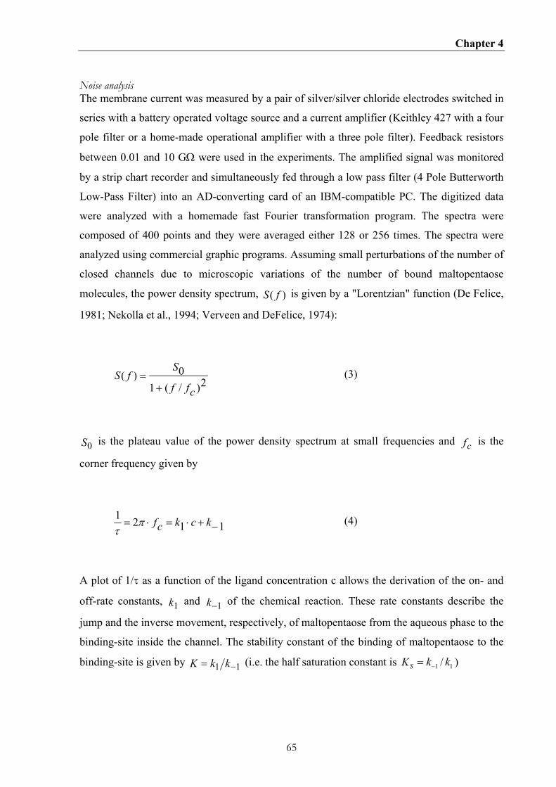
Chapter 4
65
Noise analysis The membrane current was measured by a pair of silver/silver chloride electrodes switched in
series with a battery operated voltage source and a current amplifier (Keithley 427 with a four
pole filter or a home-made operational amplifier with a three pole filter). Feedback resistors
between 0.01 and 10 GΩ were used in the experiments. The amplified signal was monitored
by a strip chart recorder and simultaneously fed through a low pass filter (4 Pole Butterworth
Low-Pass Filter) into an AD-converting card of an IBM-compatible PC. The digitized data
were analyzed with a homemade fast Fourier transformation program. The spectra were
composed of 400 points and they were averaged either 128 or 256 times. The spectra were
analyzed using commercial graphic programs. Assuming small perturbations of the number of
closed channels due to microscopic variations of the number of bound maltopentaose
molecules, the power density spectrum, )( fS is given by a "Lorentzian" function (De Felice,
1981; Nekolla et al., 1994; Verveen and DeFelice, 1974):
2)/(1
0)(cff
SfS
+= (3)
0S is the plateau value of the power density spectrum at small frequencies and cf is the
corner frequency given by
1121−+⋅=⋅= kckcfπ
τ (4)
A plot of 1/τ as a function of the ligand concentration c allows the derivation of the on- and
off-rate constants, 1k and 1−k of the chemical reaction. These rate constants describe the
jump and the inverse movement, respectively, of maltopentaose from the aqueous phase to the
binding-site inside the channel. The stability constant of the binding of maltopentaose to the
binding-site is given by 11 −= kkK (i.e. the half saturation constant is 11 / kkKS −= )

Chapter 4
66
4.4. RESULTS
4.4.1 Growth experiments with KS26 strains containing wildtype LamB and different
greasy slide mutants
To study the functional integrity of the greasy slide mutants of LamB in vivo we performed
growth experiments of some of the mutant strains in an synthetic M9 medium supplemented
with 0.4% maltopentaose, which means that it contained maltopentaose as sole carbon source.
For these investigations the plasmid pAM117 carrying the genes for wildtype LamB and the
different LamB greasy slide mutants (Heine et al., 1988) were transferred into the KS26 strain
that lacks most outer membrane porins (Schulein et al., 1995). An aliquot corresponding to
0.02 OD of an overnight culture, grown in LB media was added to 10 ml M9 minimal
medium and the growth was followed over at least one day. The growth curves showed a
substantial difference between the growth of the porin deficient strain KS26 and KS26
transfected with the plasmids encoding for wildtype LamB and the LamB mutants. KS26
showed absolutely no growth on maltopentaose, but the strains that expressed wildtype LamB
and most of the LamB greasy slide mutants showed approximately the same growth rate. This
result indicated that most of the LamB mutants were able to transport maltopentaose, i.e. the
mutants were functional. However, we observed one exception. The six-fold greasy slide
mutant (D1/2/3/4/5/6), which lacks all aromatic residues of the greasy slide showed no growth
similar to KS26. Interestingly, the six-fold greasy slide mutant that contains the additional
mutation Y118W shows again growth. This means presumably that maltopentaose transport
sufficient for growth is restored when tyrosine 118 is mutated to tryptophane (see also
Discussion). For most of the mutants the outer membrane was not rate limiting under the
conditions employed here because 0.4% maltopentaose used in the growth experiments
corresponds to a concentration of 4.8 mM maltopentaose, which is above the half saturation
of most of the LamB mutants used in this study (see below).
4.4.2. Effect of the aromatic residues of the greasy slide on single-channel conductance
In a first set of experiments we investigated the effect of the replacement of single greasy
slide amino acids by alanine. The data were compared to wildtype LamB (Benz et al., 1986)
Y118W and Y118A, which had been studied previously (Orlik et al., 2002). All mutants
formed well-defined channels in lipid bilayer membranes indicating no gross perturbation of
the channel structure induced by the mutations. Examples for single channel recordings are

Chapter 4
67
shown in Figure 4.2 for the mutants D1 (A), D1/2 (B), D1/2/3/6+Y118W (C) and
D1/2/3/4/5/6 (D). The single-channel conductance of the mutants was analyzed in histograms
(see Figure 3) showing a homogeneous distribution for all LamB mutants. Table 2 shows a
summary of the single-channel conductance of the six single mutant channels in 1 M KCl
solution. The data indicate that the single-channel conductance was in many cases influenced
by the mutation of the aromatic residues.
Figure 4.2 Single-channel recordings of diphytanoyl phosphatidylcholine/n-decane membranes in the presence of 4 different maltoporin greasy slide mutants D6 (A), D1/2 (B), D1/2/3/4+Y118W (C) and D1/2/3/4/5/6 (D). The aqueous phase contained 1 M KCl (pH 6) and 10 ng/ml LamB mutants. The applied membrane potential was 20 mV; T = 20°C. Note that current noise of the single-channel recording of wildtype LamB is similar to that of the mutants indicating that the mutation did not induce a major change of the channel structure.
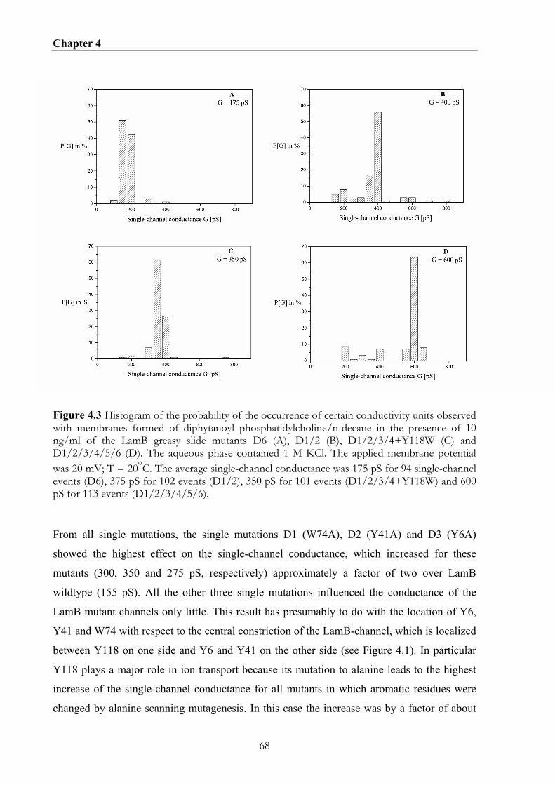
Chapter 4
68
Figure 4.3 Histogram of the probability of the occurrence of certain conductivity units observed with membranes formed of diphytanoyl phosphatidylcholine/n-decane in the presence of 10 ng/ml of the LamB greasy slide mutants D6 (A), D1/2 (B), D1/2/3/4+Y118W (C) and D1/2/3/4/5/6 (D). The aqueous phase contained 1 M KCl. The applied membrane potential was 20 mV; T = 20°C. The average single-channel conductance was 175 pS for 94 single-channel events (D6), 375 pS for 102 events (D1/2), 350 pS for 101 events (D1/2/3/4+Y118W) and 600 pS for 113 events (D1/2/3/4/5/6).
From all single mutations, the single mutations D1 (W74A), D2 (Y41A) and D3 (Y6A)
showed the highest effect on the single-channel conductance, which increased for these
mutants (300, 350 and 275 pS, respectively) approximately a factor of two over LamB
wildtype (155 pS). All the other three single mutations influenced the conductance of the
LamB mutant channels only little. This result has presumably to do with the location of Y6,
Y41 and W74 with respect to the central constriction of the LamB-channel, which is localized
between Y118 on one side and Y6 and Y41 on the other side (see Figure 4.1). In particular
Y118 plays a major role in ion transport because its mutation to alanine leads to the highest
increase of the single-channel conductance for all mutants in which aromatic residues were
changed by alanine scanning mutagenesis. In this case the increase was by a factor of about

Chapter 4
69
six as compared to wildtype LamB (Orlik et al., 2002).
The effect of double mutations on single-channel conductance was in all three cases not much
different compared to the single mutations (see Table 4.2). The combination D1/2 resulted in
the highest conductance, which is well understood because the single mutations D1 and D2
showed already a high conductance. The two triple mutations (D1/2/3 and D4/5/6), in which
the outer and the inner aromatic residues were deleted, respectively, follow this line. The
effect of the deletion of the outer residues on ion permeability is higher than that of the inner
ones. Similar results were obtained for the mutants where more than three aromatic residues
were deleted (see Table 4.2). Ion permeability through the mutant LamB-channels was high
when all three of the outer residues were mutated together with one or two of the inner
aromatic amino acids (W420, W359 and F227; see Table 4.2). Highest single-channel
conductance was observed, when all six aromatic residues were removed. This result indicates
that all these aromatic amino acids represent some steric hindrance for ion transport though
LamB.
In previous papers, it has been demonstrated that the replacement of Y118 by phenylalanine
and tryptophane had the smallest influence on ion transport through LamB probably because
the bulky tyrosine side chain was replaced by other bulky groups (Jordy et al., 1996; Orlik et
al., 2002). The mutation Y118W had a substantial effect on carbohydrate binding (Orlik et al.,
2002). Therefore some mutants were investigated where besides greasy slide mutations Y118
was replaced by tryptophane. Single-channel experiments with these mutants suggested that
the mutation Y118W had a substantial influence on the conductance of the mutants. In all
cases the single-channel conductance decreased when Y118W was introduced in addition to
greasy slide mutations (see Table 4.2). This means that the bulky side chain of tryptophane
controls ion flux through the central constriction in a similar way as phenylalanine or tyrosine
(Orlik et al., 2002).
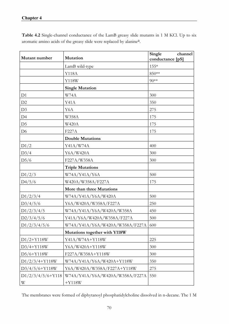
Chapter 4
70
Table 4.2 Single-channel conductance of the LamB greasy slide mutants in 1 M KCl. Up to six aromatic amino acids of the greasy slide were replaced by alaninea.
Mutant number Mutation Single channel conductance [pS]
LamB wild-type 155* Y118A 850** Y118W 90** Single Mutation D1 W74A 300 D2 Y41A 350 D3 Y6A 275 D4 W358A 175 D5 W420A 175 D6 F227A 175 Double Mutations D1/2 Y41A/W74A 400 D3/4 Y6A/W420A 300 D5/6 F227A/W358A 300 Triple Mutations D1/2/3 W74A/Y41A/Y6A 500 D4/5/6 W420A/W358A/F227A 175 More than three Mutations D1/2/3/4 W74A/Y41A/Y6A/W420A 500 D3/4/5/6 Y6A/W420A/W358A/F227A 250 D1/2/3/4/5 W74A/Y41A/Y6A/W420A/W358A 450 D2/3/4/5/6 Y41A/Y6A/W420A/W358A/F227A 500 D1/2/3/4/5/6 W74A/Y41A/Y6A/W420A/W358A/F227A 600 Mutations together with Y118W D1/2+Y118W Y41A/W74A+Y118W 225 D3/4+Y118W Y6A/W420A+Y118W 300 D5/6+Y118W F227A/W358A+Y118W 300 D1/2/3/4+Y118W W74A/Y41A/Y6A/W420A+Y118W 350 D3/4/5/6+Y118W Y6A/W420A/W358A/F227A+Y118W 275 D1/2/3/4/5/6+Y118W
W74A/Y41A/Y6A/W420A/W358A/F227A +Y118W
550
The membranes were formed of diphytanoyl phosphatidylcholine dissolved in n-decane. The 1 M
Chapter 4
71
KCl solutions were unbuffered and had a pH of 6. The applied voltage was 20 mV, and the temperature was 20°C. The average single-channel conductance was calculated from at least 100 single events: The single-channel conductance of wildtype LamB* (Benz et al., 1986) and the Y118A** mutant (Orlik et al., 2002) are given for comparison.
4.4.3 Evaluation of the stability constants for maltopentaose binding to the different
greasy slide mutants
The stability constants for maltopentaose binding to LamB-mutants were evaluated from
titration experiments similar to those performed previously to study carbohydrate binding to
LamB wildtype and ScrY (Benz et al., 1987; Schulein et al., 1995). Figure 4.4 shows an
example for such experiments. LamB mutant Y6A (D3) was added while stirring from a
concentrated stock solution to the aqueous phase (concentration about 500 ng/ml) bathing a
black lipid bilayer membrane. The corresponding current increase was monitored on a strip
chart recorder. After about 30 min the conductance was almost stationary. The titration
experiment was started by the addition of concentrated solution of maltopentaose to both
sides of the membrane. This led to a decrease of membrane conductance in a dose dependent
way as is shown in Figure 4.4. At a maltopentaose concentration of 5.4 mM the current
through the membrane almost decreased to zero, which means that the LamB mutant Y6A
(D3) channels were almost totally blocked for the flux of ions caused by the binding of
maltopentaose to the binding site. The stability constant K for the binding of maltotriose to
the binding site inside the Y6A (D3) channel was evaluated using Lineweaver-Burke plots
according to eqn. (2). An example for the fit of the data of Figure 4.3 is given in Figure 4.5.
The stability constant for maltopentaose binding to the Y6A (D3) mutant was 973±16 1/M
(half saturation constant 1.03 mM). This has to be compared with a stability constant of
14,000 1/M (half saturation constant about 70 µM) for maltopentaose binding to wildtype
LamB (Benz et al., 1987).

Chapter 4
72
Figure 4.4 Titration of LamB-mutant Y6A (D3) induced membrane conductance with maltopentaose. The membrane was formed from diphytanoyl phosphatidylcholine/n-decane. The aqueous phase contained 500 ng/ml protein, 1 M KCl, and maltopentaose at the concentrations shown at the top of the figure. The temperature was 20°C and the applied voltage was 20 mV.
Figure 4.5 Lineweaver-Burke plot of the inhibition of the LamB Y6A (D3) mutant-induced membrane conductance by maltopentaose. The data taken from Figure 4.4 and of two similar experiments were analyzed using eqn. (2). The data points reflect the mean of the measurement ± SD. Note that the standard deviation was for most of the measurements so small that it lies within the data points. The straight line corresponds to a least squares fit of the data points. The fit corresponds to a stability constant K, for maltopentaose binding to LamB mutant Y6A (D3) of 973±16 l/mol (KS = 1.03 mM).
It is noteworthy that replacement of the individual aromatic amino acids of the greasy slide by
alanine led to different effects on the stability constants for maltopentaose binding. The
mutation of W74A (D1), Y41A (D2) and F227A (D6) had a small influence on the stability
constant. The effect of D3 (Y6A), D4 (W358A) and D5 (W420A) on maltopentaose binding

Chapter 4
73
was more substantial (see Table 4.3) and K decreased by factors between about 7 and 30 as
compared to wildtype LamB (Benz et al., 1987). This result indicated that the influence of the
inner amino acids of the greasy slide (which are localized opposite to the Y118 amino acids in
the central constriction of the LamB channel) on carbohydrate binding is higher than that of
the aromatic amino acids localized at the periphery of the channel. This result agrees nicely
with a previous study where Y118 was exchanged by a variety of other amino acids (Orlik et
al., 2002). The effect of the mutation of Y118 on carbohydrate binding was much higher as
measured here for the single greasy slide mutation. Y118A led to a dramatic decrease of
maltopentaose binding by a factor of about 20, whereas the mutation Y118W resulted in very
strong increase of the stability constant by a factor of more than 200 (see Table 4.3) (Orlik et
al., 2002).
Table 4.3 Parameters of maltopentaose induced transport noise and stability constants of maltopentaose binding to different LamB greasy slide mutant channels.
Mutant k1/106 M-1s-1 k-1/s-1 K/103M-1 K*/103M-1 KS*/mM
LamB wild type* 5.3 420 13 14 0.07 Y118W** 16.5 9 1,800 3,100 0.0003 Y118A** 1.5 970 1.8 0.7 1.4 D1 0.08 53 1.5 2.1 0.48 D2 3.8 2,880 1.3 2 0.5 D3 0.8 1,890 0.4 1.1 0.9 D4 5.2 490 10.6 14 0.07 D5 6.5 670 9.9 8.9 0.1 D6 10.4 830 12.5 20.8 0.05 D1/2 0.05 275 0.4 0.8 1.25 D3/4 n.d. 0.16 6.25 D5/6 n.d. 0.15 6.7 D1/2/3 n.d. 0.05 20 D4/5/6 n.d. 0.38 2.6 D1/2/3/4 n.d. 0.02 50 D3/4/5/6 n.d. <0.014 >70 D1/2/3/4/5 n.d. <0.03 >33 D2/3/4/5/6 n.d. <0.03 >33 D1/2/3/4/5/6 n.d. <0.015 >67

Chapter 4
74
D1/2+Y118W 10.8 120 92 91 0.01 D3/4+Y118W 1.8 365 5.0 6.9 0.15 D5/6+Y118W 2.0 445 4.6 7.1 0.14 D1/2/3/4+Y118W 3.7 1,200 3.1 3.9 0.26 D3/4/5/6+Y118W n.d. 0.11 9.1 D1/2/3/4/5/6+Y118W 0.09 1,030 0.09 0.05 20 The membranes were formed from diphytanoyl phosphatidylcholine/n-decane. The aqueous phase contained 1 M KCl and about 500 ng/ml LamB mutants. K* was derived from titration experiments similar to that shown in Figure 4.4 by using Lineweaver-Burke-plots (Figure 4.5). The data represent means of at least three individual titration experiments. The standard deviation was typically less than 10% of the mean value. The results of previous titration experiments with wild type LamB*(Benz et al., 1986) and the Y118W mutant**(Orlik et al., 2002) are given for comparison. The rate constants k1 and k−1 were derived from a fit of the corner frequencies as a function of the maltopentaose concentration (compare eqn. (4)). K is the stability constant for maltose binding derived from the ratio k k1 1− . The data represent the mean of at least three individual noise experiments. The standard deviation of the mean value was always smaller than 20% of the mean. The parameters for *wildtype LamB (Andersen et al., 1995) and the **Y118W mutant (Orlik et al., 2002) are given for comparison. n.d. means not detectable
Titration experiments were performed with all greasy slide mutants used in this study. The
results for K and the half saturation constant, KS, ( = 1/K) are listed in Table 4.3. The results
suggest that the affinity of maltopentaose binding decreased for all double mutations within
the greasy slide. The stability constants derived for the mutants D3/4 and D5/6 were about
100 times smaller than LamB wildtype. Only the D1/2 mutant showed a somewhat higher
binding constant for maltopentaose that was only a factor of about 20 smaller than that of
LamB wildtype. From the mutants with four mutations only binding could be measured for
the D1/2/3/4 mutant. The D3/4/5/6 did not show any maltopentaose-mediated current
decrease (data not shown). This was also the case for all other greasy slide mutants with more
than four mutations (see Table 4.3). This means that the half saturation constant for binding of
maltopentaose, if it existed, is at least above 100 mM, which is outside the physiologically
relevant concentration range. It is noteworthy that no decrease of membrane conductance was
observed when maltopentaose was added to the six-fold mutant D1/2/3/4/5/6 (data not
shown). Even the addition of about 14 mM of maltoheptaose (which has a much higher
affinity to LamB (Andersen et al., 1995; Benz et al., 1986)) to the mutant channels of this
type was not able to block the current through these mutant channels to any appreciable extent
(see Figure 4.6).

Chapter 4
75
Figure 4. 6 Titration of LamB-mutant induced membrane conductance with carbohydrates. The membranes were formed from diphytanoyl phosphatidylcholine/n-decane. The aqueous phase contained 500 ng/ml protein, 1 M KCl, and maltoheptaose or maltopentaose at the concentrations shown at the top of the figure. The temperature was 20°C and the applied voltage was 20 mV. A: The experiment was performed using the D1/2/3/4/5/6 mutant and maltoheptaose. B: The experiment was performed using the D1/2/3/4/5/6+Y118W mutant and maltopentaose.
The LamB mutants Y118F and Y118W show increased affinity for carbohydrate binding as
compared to wildtype LamB (Jordy et al., 1996; Orlik et al., 2002). To study the effect of the
mutation Y118W in some more detail, greasy slide mutants were studied that contained in
addition the replacement of Y118 by tryptophane (see Table 4.3). The results suggest that this
mutation led to a strong increase of the affinity of maltopentaose binding to the LamB
mutants (see Table 4.3). In particular, for those mutants, which did not bind maltopentaose to
any appreciable extent (D1/2/3/4 and D3/4/5/6) the additional mutation Y118W resulted in

Chapter 4
76
restoration of binding. The same was observed for the six-fold mutant (see Figure 4.6). The
additional mutation allowed the evaluation of the binding of maltopentaose (K = 50 1/M, KS =
20 mM). This result agrees nicely with the growth experiments. Whereas no growth could be
detected for KS26 expressing the mutant D1/2/3/4/5/6, growth was reestablished when the
additional mutation Y118W was introduced.
4.4.3. Measurement of current noise with the greasy slide mutants.
The equilibrium experiments do not allow the evaluation of the on- and off-rate constants for
maltopentaose binding to LamB or the LamB mutants (Andersen et al., 1995; Nekolla et al.,
1994). To receive some information on the binding kinetics we used the measurement of the
current noise, which is based on the principle of the microscopic reversibility. Parallel to the
titration measurements the frequency-dependence of the spectral density of the current noise
was measured using fast Fourier transformation. The reference spectrum was taken before
addition of carbohydrates to obtain the current noise of the open LamB-mutant channels,
which exhibit 1 f noise in the frequency range between 1 and 50 Hz (Andersen et al., 1995;
Bezrukov and Winterhalter, 2000; Nekolla et al., 1994; Wohnsland and Benz, 1997). An
example is given in Figure 7 for the measurement of current noise of 1329 D6 mutant
channels without maltopentaose (trace 1, 0 mM). At small frequencies up to about 100 Hz the
spectral density was dependent on 1 f , which is typical for open bacterial porin
channels(Bezrukov and Winterhalter, 2000; Wohnsland and Benz, 1997). The increase of the
spectral density at frequencies above about 200 Hz was caused by the intrinsic noise of the
preamplifier that produces frequency-dependent current noise through the membrane capacity
Cm . It is observed also with membranes without reconstituted LamB-channels. The time
resolution of the instrumentation was approximately 10 kHz, which was limited in the
experiments of Figure 4.7 and similar experiments by the bandwidth of the current amplifier
and a low pass filter (0.3 ms). The reference spectrum was subtracted from each spectrum
taken after the successive addition of maltopentaose in increasing concentration, which led to
a considerable increase of the spectral density of the current noise as Figure 4.7 clearly
indicates.

Chapter 4
77
Figure 4.7 A: Power density spectra of maltopentaose-induced current noise of 1329 LamB D6 mutant channels. Trace 1 shows the control (1 M KCl). Trace 2: the aqueous phase contained 0.02 mM maltopentaose and the power density spectrum of Trace 1 was subtracted (τ = 2.0 ms;
S0 = 0.26·10-24 A2s). Trace 3: the aqueous phase contained 0.3 mM maltopentaose and the
power density spectrum of Trace 1 was subtracted (τ = 0.55 ms; S0 = 0.05·10-24 A2s); T = 20°C; Vm= 20 mV. B: Power density spectra of maltopentaose-induced current noise of 215 LamB D1/2/3/4/5/6+Y118W mutant channels. Trace 1 shows the control (1 M KCl). Trace 2: the aqueous phase contained 0.5 mM maltopentaose and the power density spectrum of Trace 1 was
subtracted (τ = 1.8 ms; S0 = 0.03·10-24 A2s). Trace 3: the aqueous phase contained 3.7 mM maltopentaose and the power density spectrum of Trace 1 was subtracted (τ = 1.4 ms; S0 =
0.11·10-24 A2s); T = 20°C; Vm= 20 mV.

Chapter 4
78
Figure 4.7A trace 2 shows a spectrum taken after addition of maltopentaose ( c = 0.02 mM;
the reference spectrum of curve 1 was subtracted) to the membrane containing 1329 LamB
D6 mutant channels. The current noise spectrum of the LamB-mutant channel D6 after the
addition of carbohydrates could be fitted to a single Lorentzian function (see Figure 4.7A,
trace 2). This result agrees well with earlier investigations with LamB (maltoporin) wildtype
of E. coli (Andersen et al., 1995; Nekolla et al., 1994) and Salmonella typhimurium (Jordy et
al., 1996) and also Y118 LamB-mutants (Orlik et al., 2002), where also one single Lorentzian
has been observed without any exception. A spectrum taken few minutes later did not show
any systematic variation compared to spectrum taken before. In further experiments the
concentration of maltopentaose was increased in defined steps. At another concentration of
maltopentaose (c = 0.3 mM) the power density spectrum corresponded to that of trace 3 in
Figure 4.7A, which could also be fitted to a single Lorentzian. Such a type of noise is
expected for a random switch with different on- and off-probabilities and can be fitted to eqn.
(3) with sufficient accuracy (Conti and Wanke, 1975; De Felice, 1981; Verveen and DeFelice,
1974).
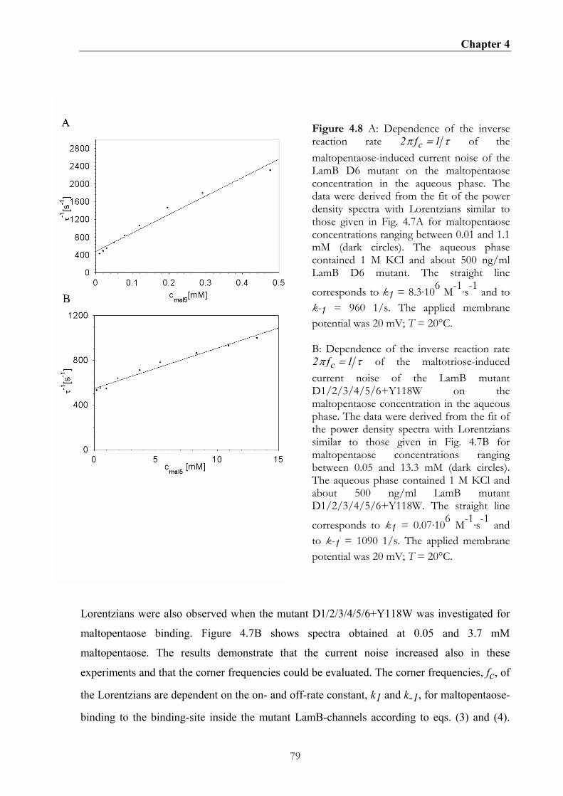
Chapter 4
79
Figure 4.8 A: Dependence of the inverse reaction rate 2 1π τfc = of the maltopentaose-induced current noise of the LamB D6 mutant on the maltopentaose concentration in the aqueous phase. The data were derived from the fit of the power density spectra with Lorentzians similar to those given in Fig. 4.7A for maltopentaose concentrations ranging between 0.01 and 1.1 mM (dark circles). The aqueous phase contained 1 M KCl and about 500 ng/ml LamB D6 mutant. The straight line
corresponds to k1 = 8.3·106 M-1·s-1 and to k-1 = 960 1/s. The applied membrane potential was 20 mV; T = 20°C. B: Dependence of the inverse reaction rate 2 1π τfc = of the maltotriose-induced current noise of the LamB mutant D1/2/3/4/5/6+Y118W on the maltopentaose concentration in the aqueous phase. The data were derived from the fit of the power density spectra with Lorentzians similar to those given in Fig. 4.7B for maltopentaose concentrations ranging between 0.05 and 13.3 mM (dark circles). The aqueous phase contained 1 M KCl and about 500 ng/ml LamB mutant D1/2/3/4/5/6+Y118W. The straight line
corresponds to k1 = 0.07·106 M-1·s-1 and to k-1 = 1090 1/s. The applied membrane potential was 20 mV; T = 20°C.
Lorentzians were also observed when the mutant D1/2/3/4/5/6+Y118W was investigated for
maltopentaose binding. Figure 4.7B shows spectra obtained at 0.05 and 3.7 mM
maltopentaose. The results demonstrate that the current noise increased also in these
experiments and that the corner frequencies could be evaluated. The corner frequencies, fc, of
the Lorentzians are dependent on the on- and off-rate constant, k1 and k-1, for maltopentaose-
binding to the binding-site inside the mutant LamB-channels according to eqs. (3) and (4).
Chapter 4
80
This means that the corner frequencies, fc, should increase with increasing maltopentaose
concentration as they did in the experiments shown in Figure 4.7A and 4.7B. The reaction rate
1/ι was plotted as a function of the carbohydrate concentration in the aqueous phase to
evaluate the off- and the off-rate constants for maltopentaose binding. Figures 4.8A and 4.8B
show the fit of the corner frequencies, fc , of the experiments shown in Figure 4.7A and 4.7B
and of other maltopentaose concentrations (data not shown) to eqn. (4). The rate constants for
the binding of maltopentaose to the LamB D6 mutant channel were k1 = 8.3·106 1/(M⋅s) and
k−1 = 960 1/s. This corresponds to stability constant, K , for the binding of maltopentaose to
the binding-site inside the LamB D6 mutant-channel of 1329 1/M, which agrees well with the
stability constant for the same system derived from the titration experiments (see Table 4.3).
Similarly the corner frequencies of the experiments shown in Figure 4.7B were plotted as a
function of the maltopentaose concentration and yielded the on- and off-rate constants for
maltopentaose binding to the D1/2/3/4/5/6+Y118W mutant (k1 = 0.07·106 1/(M⋅s) and k−1 =
1090 1/s). We repeated these experiments several times and yielded similar results for both
the rate constants of maltopentaose binding and for the stability constants. It is noteworthy
that the on- and off-rate constants of maltopentaose-binding were fairly independent from the
experimental conditions including the number of reconstituted channels. The rate constants
describing the movement of maltopentaose to and from the binding-site inside the mutant
channels are not identical to the unidirectional flux through the channel. It is possible,
however, to calculate this flux through the channels from the rate constants (see Discussion).
With some of the greasy slide mutants it was not possible to evaluate the corner frequency of
the spectral density. In particular, when more than 4 residues of the greasy slide were replaced
by alanine-scanning mutagenesis it was not possible to fit the spectral density of the current
noise to a Lorentzian function. One reason for this could be that the kinetics of maltopentaose
binding was too fast. Another possibility is that the amplitude of the spectral density of the
maltopentaose-mediated current noise was too small. The evaluation of the binding kinetics
was possible, however, when besides the greasy slide mutations Y118 was also mutated to
tryptophane. This was possible for all double mutants (D1/2, D3/4 and D5/6), for one of the
mutant with four mutations (D1/2/3/4) and when all aromatic residues of the greasy slide were
removed (D1/2/3/4/5/6) (see above and Table 4.3). The results of all noise measurements
demonstrate that the mutations of the greasy slide preferentially affect the on-rate of

Chapter 4
81
maltopentaose binding. The additional mutation Y118W led to an increase of the on-rate,
which means that the kinetics could again be evaluated from the experiments (see Table 4.3).
This result demonstrates again the central role of Y118 in carbohydrate transport through
LamB.
4.5. DISCUSSION
4.5.1 The replacement of the aromatic residues of the greasy slide by alanine increases
ion flux through LamB
Alanine scanning mutagenesis of the amino acids of the greasy slide resulted in increased ion
flux through LamB. Highest effect for the single mutation was observed for the mutants
where the aromatic residues opposite Y118 were removed. This result appears to be
reasonable because tyrosine 118 is localized in the central constriction at the external loop 3
and controls there the flux of ions as previous experiments with the mutant Y118A have
demonstrated (Orlik et al., 2002). Experiments with multiple greasy slide mutants tend to
support this view, because for additional mutation the single-channel conductance increases.
Highest conductance was observed for the mutant where all aromatic residues were removed
(D1/2/3/4/5/6; see Table 4.2). Nevertheless its conductance was with 600 pS in 1 M KCl still
lower than that of the mutant Y118A (850 pS). Introduction of the mutation Y118W in
addition to the alanine scanning mutagenesis of greasy slide amino acids always decreased ion
flux through LamB, which agrees well with the situation within the cross section of the
monomers (see Figure 4.1). This means that aromatic residues within the channel lumen
control ion transport through LamB and probably also permeability for neutral solutes. It is
noteworthy that the sucrose-specific ScrY-channel of enteric bacteria (sucroseporin)
representing a natural mutant of LamB (Schmid et al., 1991), has besides a carbohydrate
binding site also general diffusion properties (Schulein et al., 1991). It has a high single-
channel conductance of about 700 pS in 1 M KCl similar to those of some of the LamB
mutants described here. In position 201 of ScrY (corresponding to Y118 in LamB) is an
aspartate. This is the reason for the increased size of ScrY wildtype as compared to LamB
wildtype (see Figure 4.1).

Chapter 4
82
4.5.2 The mutation of greasy slide residues has a major impact on maltopentaose binding
affinity
In the preceding study by Orlik et al. (2002)(Orlik et al., 2002) and above we have
demonstrated that tyrosine 118 and the aromatic residues of the greasy slide have a major
effect on ion permeation through LamB of E. coli. The effect on ion conductance is definitely
caused by the size of the bulky side chain of tyrosine, phenylalanine or tryptophane, which
protrudes in the channel lumen and limits its size. LamB functions in the outer membrane of
enteric bacteria as a channel for carbohydrates in particular for the uptake of
maltooligosaccharides although it is induced by maltose (Schwartz, 1987). In the growth
experiments we could demonstrate that this is also the case for most of the mutants (D1/2,
D1/2/3/4/5/6, D1/2/3/4/5/6+Y118W) and LamB wildtype used in this study. All with the
exception of D1/2/3/4/5/6 were able to confer to the porin-deficient KS26 strain the growth on
maltopentaose and the growth rate was very similar to that when the strain KS26 contained a
plasmid for the expression of wildtype LamB. This result means that some of the mutant
channels have the same function as wildtype LamB with the exception of D1/2/3/4/5/6. The
latter mutant is not able to bind maltopentaose or maltoheptaose to any appreciable extent,
which suggests that these carbohydrates are also not transported (see below).
Alanine scanning mutagenesis of the single amino acids of the greasy slide suggests that they
all contribute to maltopentaose binding. Highest impact on binding was observed, however,
when the three aromatic residues (D3, D4 and D5) localized opposite Y118 were mutated.
The stability constant for binding dropped in these cases by at least a factor of 10 (D3 and D5)
and at maximum a factor of 30 (D4). These results are in qualitative agreement with another
investigation of greasy slide mutants (Van Gelder et al., 2002). In this study maltose and
maltohexaose binding to the same mutants was also drastically decreased as compared to
wildtype LamB. These results suggest that the central part of the LamB channel has an
important function in carbohydrate binding. The aromatic residues in the central part form a
binding pocket about three glucose residues long that allows together with hydrogen bonds to
other residues within the channel lumen (D116, E43, R8, R109, R82 and R33) optimum
interaction between carbohydrates and the channel interior (Dutzler et al., 1996; Jordy et al.,
1996; Meyer et al., 1997; Orlik et al., 2002; Orlik et al., 2002). The other three amino acids of
the greasy slide (D1, D2 and D6) have only a relatively small effect on maltopentaose
binding.

Chapter 4
83
The affinity for maltopentaose binding decreased further when greasy slide mutants with two
mutations were studied. Highest affinity was observed for the D1/2 mutant (K = 790 1/M).
The stability constants for maltopentaose binding were for the D3/4 and the D5/6 even
smaller and did not exceed about 160 1/M. From all other greasy slide mutants including
three, four, five and all six residues only maltopentaose binding could be detected for the
D1/2/3/4 mutant (K = 20 1/M). For the other mutants only a lower limit for maltopentaose
binding could be evaluated from the titration experiments (K<10 1/M). This means that in all
these cases the half saturation constant was much higher than 100 mM, if there was any
binding at all.
4.5.3 The additional mutation Y118W results in an increase of the binding affinity of the
greasy slide mutants
In a previous study we could demonstrate that the mutation Y118W resulted in a substantial
increase of maltopentaose binding by more than a factor of 200 (see also Table 4.3).
Similarly, this mutation was also able to increase the affinity of maltopentaose binding to the
greasy slide mutants. Table 4.3 demonstrates that the stability constant of maltopentaose
binding increased in most cases by more than a factor of 100 when Y118W was introduced in
addition to the greasy slide mutations. In particular high effects were observed when Y118W
was introduced in the double mutants D1/2, D3/4 and D5/6. In these cases maltopentaose
binding affinity was almost the same as for LamB wildtype or considerably higher (D1/2
+Y118W). For other mutants with three, four and six greasy slide mutations the stability
constant for maltopentaose binding decreased but was still much higher than without the
Y118W exchange, which means that maltopentaose binding could be detected for these
mutants. This result again demonstrates the importance of the central part of the LamB-
channel for carbohydrate binding. The estimation of the stability constant for maltopentaose
binding to the LamB greasy slide mutants allows also the calculation of the free energy for
maltopentaose binding, calculated according to ΔG R T K0 = ⋅ ln . The free energy, ΔG0, of
binding for maltopentaose binding decreased considerably for all greasy slide mutants studied
here from 23.2 kJ/mol for LamB wildtype to less than 5.5 kJ/mol for many of the greasy slide
mutants.
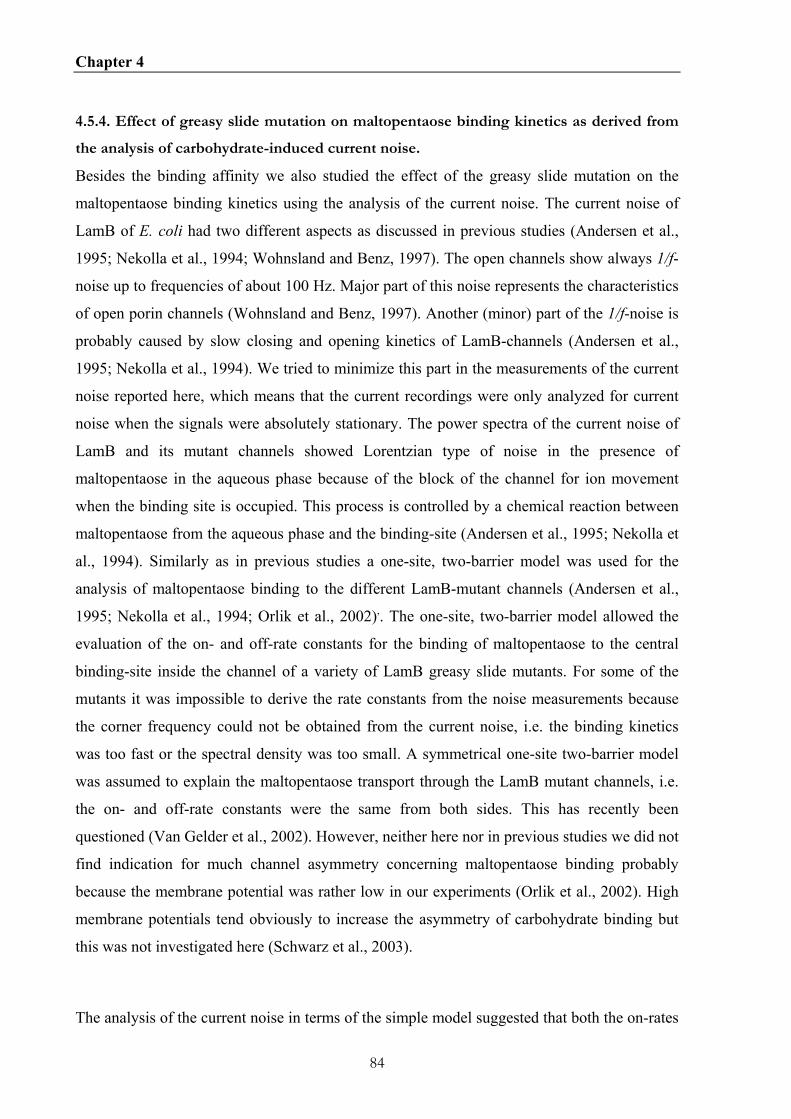
Chapter 4
84
4.5.4. Effect of greasy slide mutation on maltopentaose binding kinetics as derived from
the analysis of carbohydrate-induced current noise.
Besides the binding affinity we also studied the effect of the greasy slide mutation on the
maltopentaose binding kinetics using the analysis of the current noise. The current noise of
LamB of E. coli had two different aspects as discussed in previous studies (Andersen et al.,
1995; Nekolla et al., 1994; Wohnsland and Benz, 1997). The open channels show always 1/f-
noise up to frequencies of about 100 Hz. Major part of this noise represents the characteristics
of open porin channels (Wohnsland and Benz, 1997). Another (minor) part of the 1/f-noise is
probably caused by slow closing and opening kinetics of LamB-channels (Andersen et al.,
1995; Nekolla et al., 1994). We tried to minimize this part in the measurements of the current
noise reported here, which means that the current recordings were only analyzed for current
noise when the signals were absolutely stationary. The power spectra of the current noise of
LamB and its mutant channels showed Lorentzian type of noise in the presence of
maltopentaose in the aqueous phase because of the block of the channel for ion movement
when the binding site is occupied. This process is controlled by a chemical reaction between
maltopentaose from the aqueous phase and the binding-site (Andersen et al., 1995; Nekolla et
al., 1994). Similarly as in previous studies a one-site, two-barrier model was used for the
analysis of maltopentaose binding to the different LamB-mutant channels (Andersen et al.,
1995; Nekolla et al., 1994; Orlik et al., 2002),. The one-site, two-barrier model allowed the
evaluation of the on- and off-rate constants for the binding of maltopentaose to the central
binding-site inside the channel of a variety of LamB greasy slide mutants. For some of the
mutants it was impossible to derive the rate constants from the noise measurements because
the corner frequency could not be obtained from the current noise, i.e. the binding kinetics
was too fast or the spectral density was too small. A symmetrical one-site two-barrier model
was assumed to explain the maltopentaose transport through the LamB mutant channels, i.e.
the on- and off-rate constants were the same from both sides. This has recently been
questioned (Van Gelder et al., 2002). However, neither here nor in previous studies we did not
find indication for much channel asymmetry concerning maltopentaose binding probably
because the membrane potential was rather low in our experiments (Orlik et al., 2002). High
membrane potentials tend obviously to increase the asymmetry of carbohydrate binding but
this was not investigated here (Schwarz et al., 2003).
The analysis of the current noise in terms of the simple model suggested that both the on-rates

Chapter 4
85
and the off-rates are influenced by the greasy slide mutations. The on-rate generally
decreased, whereas the off-rate tended to increase by at maximum a factor of about 2 with one
exception (D3, (Y6A)). When the single greasy slide mutations are considered k1 decreased
about 70-fold from wildtype (k1 = 5.3·106 1/(Ms)) to the smallest on-rate (D3 (Y6A), k1 =
8·104 1/(Ms)) for maltopentaose binding. This means that the on-rates became considerably
smaller than those of diffusion-controlled reaction processes (Eigen et al., 1964). To explain
these smaller on-rates it has to be assumed that the carbohydrates hit many times the channel
before they are bound to the binding site. The carbohydrates can only be bound to LamB and
its mutants when they are in a special order with respect to the binding-site. In a previous
study we could demonstrate that the carbohydrates bind to LamB with the non-reducing end
in advance from the surface exposed side and vice versa for the periplasmic side (Andersen et
al., 1999), which agrees well with crystallographic studies (Dutzler et al., 1996).
From all the other greasy slide mutations only the kinetics of maltopentaose binding to the
D1/2 mutant could be measured with sufficient accuracy. The on-rate showed in this case an
even stronger decrease as compared to the single mutations D1 and D2. The off-rate was on
the other hand comparable to those of the single mutations. It is possible that for all the other
greasy slide mutants with more than one mutation the on-rate was so small combined with an
increased off-rate that the current noise was only little increased with respect to the open
channel noise. This means that it was impossible to separate the Lorentzian from the 1/f-noise
of the open channels with sufficient accuracy.
4.5.5 The additional mutation Y118W allows the derivation of the maltopentaose binding
kinetics for the greasy slide mutants
Previous studies demonstrated that the mutations Y118F and Y118W had a substantial
influence on the kinetics of carbohydrate binding (Jordy et al., 1996; Orlik et al., 2002). In
particular, the on-rate increased slightly whereas the off-rate showed a dramatic decrease. The
introduction of the additional mutation Y118W in some of the greasy slide mutants changed
the binding kinetics in such a way that it was again possible to measure the current noise. This
allowed for a full set of mutations to measure the influence of the greasy slide mutations on
the on- and off-rate constants of maltopentaose binding, which was not possible for LamB
Chapter 4
86
wildtype (see above). Even for the six-fold greasy slide mutation D1/2/3/4/5/6 it was possible
to measure the binding kinetics when Y118W was additionally introduced. This allowed for
many of the mutants to judge what happens to the rate constants of binding, k1 and k-1, when
the aromatic residues of the greasy slide were changed to alanine. The data of Table 2 clearly
demonstrate that the on-rate constants dramatically decreased while the off-rate constants
increased. The data with the mutant D1/2/3/4/5/6+Y118W clearly indicate that the central
tryptophane in position 118 is already sufficient for low affinity carbohydrate binding and
also transport as the growth experiments with this mutant LamB channel clearly demonstrate.
4.5.6 Implication of greasy slide mutations for the carbohydrate transport in vivo
The porin-deficient KS26 strain has a small growth rate on maltose as sole carbon source and
did not grow on the medium containing maltopentaose. The expression plasmids containing
the genes for wildtype LamB and some of the mutants confer to the strain KS26 the
possibility to grow on maltopentaose as sole carbon sources. These experiments suggest that
maltopentaose can pass through the mutant channels, which means that they are functional.
This allows the calculation of maltopentaose flux through the channel using the one-site two-
barrier model. The LamB channel shows only a small asymmetry if any at small voltages
(Andersen et al., 1995; Benz et al., 1986; Benz et al., 1987; Orlik et al., 2002; Van Gelder et
al., 2000). This means that a symmetrical channel can be assumed. The net flux of sugar
molecules, Φ, through the channel under stationary conditions as the result of a concentration
gradient c" - c' across the membrane is given by the net movement of sugar across one barrier
of the two identical potential energy barriers (Benz et al., 1987):
Φ= k c" (1 + K' ) - k K' (1 + K' )1 -1⋅ ⋅ (5)
K' is under symmetrical conditions given by:
K' = K ( c' +c" ) / 2 ⋅ (6)
In equation (5) the rate constants k1 and k-1 are multiplied by the probabilities that the
Chapter 4
87
binding site is free or occupied, respectively. The flux has in the case c" = c, c' = 0, when
carbohydrates are only present on one side of the channel the form:
Φ= k c / ( 2 + K c )1 ⋅ ⋅ (7)
Eqn. (7) suggest that the maximum permeability of the channel for a carbohydrate with an on-
rate k1 for its binding to the binding site is k1 2, which is obtained for wildtype LamB at very
small carbohydrate concentration (c ≤ 10 µM). The flux strongly saturates at high
carbohydrate concentration since the half saturation constant for the sugar flux is KS = 1/K.
The maximum turnover number of the channel (similar to the maximum turnover number of
an enzyme that is substrate saturated) is reached at high carbohydrate concentration on one
side of the membrane and is given by k−1. This means that the flux through LamB is limited
at high maltose and maltooligosaccharide concentration. It is noteworthy; however, that this is
not a serious restriction since the substrate concentration is normally low under physiological
conditions. For the effective scavenging of nutrients at very small concentrations it seems to
be more important to have a high permeability (i.e. a high k1), which is indeed given for the
transport of maltooligosaccharides through the wildtype LamB channel (Andersen et al.,
1995).
Our data allow a comparison of the flux of maltopentaose through wildtype and some of the
LamB-mutant channels. Figure 4.9A shows the maximum flux of maltopentaose through a
single LamB wildtype, D3 (Y6A) and D4 (W420A) mutant channels calculated on the basis
of eqn. (7). The curves were calculated using the rate constants given in Table 4.3. Fig. 4.9A
shows the substantial effect of a single amino acid mutation within the primary sequence of
LamB on the transport of maltopentaose. The carbohydrate-specific porins have their
maximum permeability in the linear range of the Figure. This means LamB wildtype has the
highest permeability, followed by D4 and D3. The D4 mutant had the highest turnover
number of 1,890 1/s as compared to wildtype (420 1/s) and it was lowest for the D3 mutant
(53 1/s).
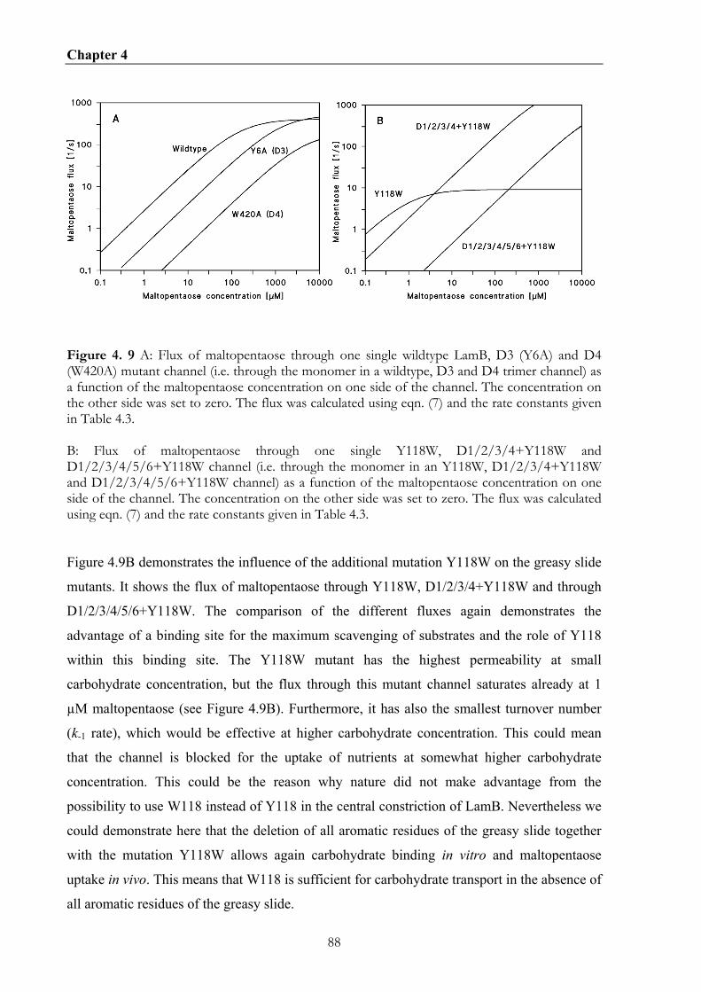
Chapter 4
88
Figure 4. 9 A: Flux of maltopentaose through one single wildtype LamB, D3 (Y6A) and D4 (W420A) mutant channel (i.e. through the monomer in a wildtype, D3 and D4 trimer channel) as a function of the maltopentaose concentration on one side of the channel. The concentration on the other side was set to zero. The flux was calculated using eqn. (7) and the rate constants given in Table 4.3. B: Flux of maltopentaose through one single Y118W, D1/2/3/4+Y118W and D1/2/3/4/5/6+Y118W channel (i.e. through the monomer in an Y118W, D1/2/3/4+Y118W and D1/2/3/4/5/6+Y118W channel) as a function of the maltopentaose concentration on one side of the channel. The concentration on the other side was set to zero. The flux was calculated using eqn. (7) and the rate constants given in Table 4.3.
Figure 4.9B demonstrates the influence of the additional mutation Y118W on the greasy slide
mutants. It shows the flux of maltopentaose through Y118W, D1/2/3/4+Y118W and through
D1/2/3/4/5/6+Y118W. The comparison of the different fluxes again demonstrates the
advantage of a binding site for the maximum scavenging of substrates and the role of Y118
within this binding site. The Y118W mutant has the highest permeability at small
carbohydrate concentration, but the flux through this mutant channel saturates already at 1
µM maltopentaose (see Figure 4.9B). Furthermore, it has also the smallest turnover number
(k-1 rate), which would be effective at higher carbohydrate concentration. This could mean
that the channel is blocked for the uptake of nutrients at somewhat higher carbohydrate
concentration. This could be the reason why nature did not make advantage from the
possibility to use W118 instead of Y118 in the central constriction of LamB. Nevertheless we
could demonstrate here that the deletion of all aromatic residues of the greasy slide together
with the mutation Y118W allows again carbohydrate binding in vitro and maltopentaose
uptake in vivo. This means that W118 is sufficient for carbohydrate transport in the absence of
all aromatic residues of the greasy slide.
Chapter 4
89
The expression of LamB in enteric bacteria is always induced with the expression of the
periplasmic maltose binding protein MBP or MalE. MalE is present in the periplasmic space
in a concentration in the range of millimolar and confers this space into a sink for
carbohydrates although it does not modulate LamB channel function (Benz et al., 1986; Brass
et al., 1985; Schwartz, 1987). This property is an essential part of carbohydrate uptake across
the outer membrane because a carbohydrate bound from the cell surface to the binding site
inside the LamB channel has still two possibilities with equal probabilities for further
movement because the channel is symmetric with respect to its transport properties. The
maltooligosaccharides bound to LamB can move back to the cell surface or further on to the
periplasmic space. In the latter case they are bound to MBP with a half saturation constant of
considerably less than 10 µM, which means that they are trapped within the periplasmic space
and as long as inner membrane transport functions it is unlikely that the
maltooligosaccharides can be lost through the outer membrane.
The calculation of the maximum flux of maltopentaose molecules through LamB and its
mutants allows also a meaningful comparison with the in vivo requirements of maximum
growth of E. coli cells. The minimum carbon source supply for maximum growth rate is 20
nmol glucose/(min x 109 cells) (Freundlieb et al., 1988). The expression of the LamB mutants
studied here is about 40% of full expression (Heine et al., 1988). This means that a single cell
has about 104 channels (3.3⋅103 trimers) in the outer membrane. The flux of maltopentaose
through one single D1/2/3/4/5/6+Y118W monomer at a concentration of 4.8 mM (the
conditions of the growth experiments) is 179 s-1, which means that the maximum flux of
maltopentaose in 109 cells is about 1.79⋅1015 s-1 or 1.07⋅1017 min-1. This number
corresponds to a maximum flux of about 172 nmol maltopentaose/(min x 109 cells). The
carbon source supply under these conditions in the cells expressing the D1/2/3/4/5/6+Y118W
mutant is 863 nmol glucose/(min x 109 cells). This result suggests indeed that the KS26 cells
expressing the LamB mutant D1/2/3/4/5/6+Y118W can show maximum growth under the
conditions of our growth experiments. On the other hand it is clear that the flux of
maltopentaose through a single channel under the conditions of the D1/2/3/4/5/6 mutant
should be much below 4 s-1, otherwise some growth would be observed. This result clearly
indicates that binding is in the case of LamB mutant channels a prerequisite for transport. The
aromatic residues of the greasy slide and tyrosine 118 are both important for carbohydrate

Chapter 4
90
transport through LamB.
The separation of the hydrate water and the sugar can be done only effective with the
interaction of the greasy slide and the residue Y118.

91
CHAPTER 5
CONCLUSION
5.1 Conclusion and Outlook
In our research group the focus lies on the biophysical characterization of pore-forming
proteins by the use of the black lipid bilayer assay. In this study, pore-forming proteins of
gram-negative bacteria, such as E.coli and Borrelia, were studied. The strains used in this
study have been studied in science with different effort and thus different amounts of
background information of these strains were available. For example, E. coli as a model
organism for genetic engineering of bacteria is completely sequenced whereas the genome of
B. duttonii is not. Out of this, future studies of pore-forming proteins of these strains will
differ from each other.
Especially in the field of pore-forming proteins of Borrelia, there are many studies still to be
done to gain more knowledge of Borrelia. Neither for Lyme borreliosis nor for relapsing fever
vaccines are available on the market and it is of great interest to find targets for vaccine
development against these pathogens.
Although outer membrane proteins like porins are potential targets for vaccine development
the aim of this thesis was not directly to identifiy a new target for a vaccine, but it could be a
first step in this direction. Furthermore the characterization of porins would results in a better
understanding of the uptake and efflux mechanism of gram-negative Borrelia across their
permeability barrier, the outer membrane.

Chapter 5
92
In the case of the 80 pS channel-forming protein of B. duttonii, which could be characterized
electrophysically, the next step will be to determine parts of the amino acid sequence of the
putative 80 pS channel forming protein by N-terminal sequencing (Edman degradation). By a
following BLAST search homologues in other bacteria strains can be found. Further
experiments make sense when more background knowledge is available. Exceedingly relevant
would be the sequencing of the genome, which would bring much needed information. When
the sequenced genome is available, then knock-out mutants can be generated and be
investigated for their channel forming activity. And also identification of B. duttonii proteins
by mass spectrometry would be easier.
However, the situation for the 300 pS channel of B. burgdorferi is different. The genome of B.
burgdorferi B31 is fully sequenced and methods to generate knock-out mutants are
established. Hence, BB0142 could be identified as a TolC homologue in B. burgdorferi. The
next step should be the biophysical characterization of the outer membrane preparation of a
bb0142 knock-out mutant using the black lipid bilayer, to show that the 300 pS activity is
gone. Furthermore the substrate spectrum of the drug efflux pump would be interesting to
know for a more effective treatment of Lyme borreliosis. Also it would be very interesting to
investigate the expression levels of BB0142 in the different hosts of B. burgdorferi. Because
if BB0142 is expressed in all stages, it can be used as a putative target for vaccine
development. Moreover, the expression of recombinant BB0142 in E. coli would help in
obtaining enough protein material for crystallization of BB0142 for 3D structure
determination by X-ray. With the knowledge of the BB0142 3D structure, it would be
possible to generate point mutations within the BB0142 channel and perform functional
studies in vitro to verify the presented theory about electrophysical properties of BB0142 in
this study.
However, first studies with outer membrane protein preparations from different Borrelia
species were done in parallel to this study and they showed a wide spectrum of pore-forming
proteins. It would be interesting to get a complete profile of pore-forming proteins appearing
under different growth conditions in the Lyme borreliosis and relapsing fever Borrelia. So we
could see which porins Borrelia has in common and which porins are expressed under
different conditions.
Chapter 5
93
For LamB, the best studied specific pore in E. coli, it is hard to think of any further black lipid
bilayer experiments to gain more information about the channel and its properties. In this
thesis it was shown in a very detailed study of point mutants of the “greasy slide”, how
important the aromatic residues are for the sugar binding. All three factors (central
constriction by residue Y118, “polar tracks” and the “greasy slide”) together build a finely
tuned system for the sugar binding, which was determined by point mutations within the
channel already in several studies. For the future it would be very interesting to verify the
obtained black lipid bilayer kinetic data by other approaches. One technique of choice could
be isothermal titration calorimetry (ITC), which is a direct experimental access to the enthalpy
of the binding process, to gain data of the affinity constant, stoichiometry of the complex and
the entropy of the binding process. Another interesting technique is the surface plasmon
resonance biomolecular interaction analysis (SPR-BIA). Sensorgram data are a function of
analyte concentration and can be used to determine kinetic parameters and dissociation
constants of the interaction. Taken together kinetic data are important for a better
understanding of the specific binding and they can be used as basis background knowledge to
interpret other specific bindings between molecules.

94
CHAPTER 6
SUMMARY
6.1 Summary
Structural differences in the cell envelope of gram-negative bacteria are a consequence of
evolutionary development. One conserved feature of all gram-negative bacteria is the
incidence of pore forming channels in the outer membrane for the uptake and efflux of
substrates through their cell wall.
In this study pore forming proteins of the gram-negative bacteria B. burgdorferi, B. duttonii
and E.coli were investigated. Therefore the study is subdivided into three parts. In the first
part outer membrane preparation of three relapsing fever Borrelia were investigated. In the
second part the putative TolC homologue BB0124 of B. burgdorferi, the Lyme borreliosis
agent, was studied. In the last part the influence of point mutants within the greasy slide of the
maltose specific porin (LamB) of E. coli were shown.
In the first part of this study outer membrane preparations of three Borrelia relapsing fever
strains have been studied for pore-forming activity in the black lipid bilayer assay.
Histograms of conductance fluctuations were obtained from single-channel experiments with
outer membrane preparations of B. hermsii, B. recurentis and B. duttonii. All strains had a
different conductance fluctuation pattern with a broad range of single-channel conductance
values varying from 0.5 nS – 11 nS. Common for all three strains was a high pore-forming
activity at around 0.5 nS. Furthermore the proteins of the outer membrane of B. duttonii were
separated by chromatographic methods. Some eluate fractions contained a channel-forming
Chapter 6
95
protein, which was forming stable channels with a single-channel conductance of 80 pS in 1
M KCl. Characterization of this channel showed that it is slightly anionic selective and
voltage independent. The small single-channel conductance suggests that it is a specific pore.
However, a substrate specificity could not be determined.
In the second part, for the B. burgdorferi HB19 and p66 knock out strain HB19/K02, their
outer membrane preparations were characterized in the black lipid bilayer assay. Comparing
the histograms of single-channel conductions fluctuations of both strains showed no single-
channel activity at 11.5 nS for the p66 knock out strain. This verifies earlier studies that P66
is a pore-forming protein in B. burgdorferi. Furthermore, one fraction obtained by anion
exchange chromatography of the p66 knock out outer membrane protein preparation showed a
uniform channel-forming activity with a single channel conductance of 300 pS. The
electrophysically characterization of the 300 pS channel showed that it is not ionselective or
voltage dependent. By mass spectrometry using peptide mass finger prints, BB0142 could be
identified as the sole channel forming candidate in the active fraction. A BLAST search and a
conserved domain search showed that BB0142 is a putative TolC homologue in B.
burgdorferi. Furthermore the location of the bb0142 gene within the chromosome is in an
operon encoding a multidrug efflux pump. In this study the expression of an outer membrane
component of a putative drug efflux system of B. burgdorferi was shown for the first time.
In the third part functional studies of the maltooligosaccharide-specific LamB channel were
performed. The 3D-structure of LamB suggests that a number of aromatic residues (Y6, Y41,
W74, F229, W358 and W420) within the channel lumen is involved in carbohydrate and ion
transport. All aromatic residues were replaced by alanine (A) scanning mutagenesis.
Furthermore, LamB mutants were created in which one, two, three, four and five aromatic
residues were replaced to study their effects on ion and maltopentaose transport through
LamB. The purified mutant proteins were reconstituted into lipid bilayer membranes and the
single-channel conductance was studied. The results suggest that all aromatic residues provide
some steric hindrance for ion transport through LamB. Highest impact is provided by Y6 and
Y41, which are localized opposite to Y118, which forms the central constriction of the LamB
channel. Stability constants for binding of maltopentaose to the mutant channels were
measured using titration experiments with the carbohydrate. The mutation of one or several
aromatic amino acids led to a substantial decrease of the stability constant of binding. The

Chapter 6
96
highest effect was observed when all aromatic amino acids were replaced by alanine because
no binding of maltopentaose could be detected in this case. However, binding was again
possible when Y118 was replaced by tryptophane (W). The carbohydrate-induced block of the
channel function could also be used for the study of current noise through the different mutant
LamB-channels. The analysis of the power density spectra of some of the mutants allowed the
evaluation of the on- and off-rate constants (k1 and k-1) of carbohydrate binding to the
binding-site inside the channels. The results suggest that both on- and off-rate constants were
affected by the mutations. For most mutants k1 decreased and k-1 increased.
6.2 Zusammenfassung
Strukturelle Unterschiede in dem Zellwandaufbau von gram-negativen Bakterien sind eine
evolutionäre Entwicklung. Eine konservierte Eigenschaft von gram-negativen Bakterien ist
das Vorkommen von porenformenden Kanälen in der Außenmembrane für die Aufnahme und
den Export von Substraten durch die Zellwand.
In dieser Studie wurden porenformende Proteine der gram-negativen Bakterien B.
burgdorferi, B. duttonii und E. coli untersucht. Daher wurde die Arbeit in drei Teile
untergliedert. Im ersten Teil wurden zuerst die Außenmembranpräparationen von drei
Rückfallfieber auslösenden Borrelien Stämmen untersucht. Der zweite Teil der Arbeit
behandelt das TolC homologe Protein BB0142 des Erreger der Lyme Borreliose, B.
burgdorferi. Im letzten Teil wurde der Einfluss von Punktmutationen innerhalb der „greasy
slide“ auf die zuckerspezifische Pore (LamB) von E. coli untersucht.
Die Außenmembranpräparationen der drei Rückfallfieber Borrelien wurden auf
porenformende Aktivität im Black Lipid Bilayer untersucht. Leitfähigkeitshistograme wurden
nach Einzelkanalmessungen der Außenmembranepräparation von B. hermsii, B. recurrentis
und B. duttonii erstellt. Alle Stämme zeigten ein anderes Leitfähigkeitsfluktuationsmuster,
wobei die Werte der Einzelleitfähigkeit von 0,5 nS bis 11 nS variierten. Auffällig war das alle
drei Stämme eine hohe Aktivität um 0,5 nS gemeinsan hatten. Darauffolgend wurden die
Proteine der Außenmembran von B. duttonii durch verschiedene chromatographische
Methoden aufgetrennt. Einige Eluatfraktionen enthielten ein kanalformendes Protein, das
Chapter 6
97
stabile Kanäle mit einer Einzelleitfähigkeit von 80 pS in 1 M KCl formt. Die Bestimmung der
Kanaleigenschaften zeigte, dass er leicht anionenselektive und spannungsunabhängig ist. Die
geringe Einzelleitfähigkeit suggeriert, dass der Kanal zudem eine spezifische Pore sein
könnte. Bisher konnte aber keine Substratespezifität festgestellt werden.
Im zweiten Abschnitt erfolgte die Untersuchung von Außenmembranpräparationen von B.
burgdorferi HB19 und dem p66 knock-out Stamm HB19/K02 mit Hilfe des Black Lipid
Bilayers. Im Vergleich der beiden erhalten Einzelleitfähigkeitshistogramme konnte gezeigt
werden, dass die Einzelleitfähigkeit von 11,5 nS nicht mehr im p66 knock out Stamm
vorkam. Dies belegt frühere Studien, dass P66 ein poreformendes Protein von B. burgdorferi
ist. Durch Anionenaustauscher-Chromatographie der Außenmembranpräparation des p66
knock out Stammes wurde zudem eine Proteinfraktion mit einer einheitlichen
porenformenden Aktivität von 300 pS in 1 M KCl erhalten. Die elektrophysikalische
Charakterisierung der 300 pS Pore zeigte, dass der Kanal nicht ionenselektiv oder
spannungsabhängig ist. Mit Hilfe von Massenspektrometrie und Peptidemassenbestimmung,
konnte BB0142 als einziger möglicher kanalformender Kandidat identifiziert werde. Eine
BLAST Suche sowie eine „conserved domain“ Suche zeigten, dass BB0142 ein putatives
TolC homologes Protein von B. burgdorferi ist. Darüber hinaus ist das bb0142 Gen in einem
Operon lokalisiert, das eine putative Efflux Pumpe codiert. In der vorliegenden Arbeit konnte
das erste Mal gezeigt weden, dass in B. burgdorferi die Außenmembrankomponente einer
Efflux Pumpe expremiert wird.
Im dritten Teil wurden Funktionsstudien an dem zuckerspezifischen Kanal LamB
durchgeführt. Die 3D Struktur zeigte, dass einige aromatische Aminosäuren (Y6, Y41, W74,
F229, W358 und W420) innerhalb des Kanallumen in den Kohlenhydrat- und Ionentransport
involviert sind. Alle aromatischen Reste wurden bei Punktmutation durch Alanin ersetzt.
Zudem wurden LamB Mutanten erzeugt in denen ein, zwei, drei, vier oder fünf aromatische
Reste durch Alanin ersetzt. Die aufgereinigten LamB Mutanten wurden im Black Lipid
Bilayer untersucht und ihre Einzelleitfähigkeit bestimmt. Die Ergebnisse zeigen, dass alle
aromatischen Reste eine gewisse sterische Hinderung beim Ionentransport durch LamB
bewirken. Den größten Einfluss haben die Reste Y6 und Y41, die gegenüber dem Rest Y188
liegen, der die zentrale Verengung des LamB Kanal darstellt. Stabilitätskonstanten der
Zuckerbindung in den Mutanten wurde durch Titrationsexperimente mit Maltopentaose und -

Chapter 6
98
heptaose bestimmt. Mutation an einem oder mehreren aromatischen Resten führt zu einer
deutlichen Abnahme der Bindungsstabilitätskonstanten. Den stärksten Effekt zeigte die
Mutante, in der alle aromatischen Reste durch Alanin ersetzt wurden. Es konnte dort keine
Zuckerbindung mehr festgestellt werden. Die Bindung wurde wieder hergestellt nachdem
Y118 durch ein Tryptophan ersetzt wurde. Durch das zuckerinduzierte Öffnen und Schließen
des Kanals ergiebt sich ein Stromrauschen. Die Analyse des Rausch Spektrum von einigen
Mutanten erlaubte die Bestimmung der Dissoziations- (k-1) und der Assoziationskonstaten (k1)
der Zuckerbindung an der Bindestelle innerhalb des Kanals. Die Ergebnisse zeigten, dass die
Geschwindigkeitskonstanten durch die Mutationen beeinflusst wurden. Für die meisten
Mutanten sank der k1 Wert während der k-1 Wert anstieg.

99
CHAPTER 7
APPENTIX
7.1 REFERENCES
Andersen, C. (2003) Channel-tunnels: outer membrane components of type I secretion systems and multidrug efflux pumps of Gram-negative bacteria. Rev Physiol Biochem Pharmacol, 147, 122-165.
Andersen, C. (2004) Drug efflux and protein export through channel-tunnels. WILEY-VCH Verlag GmbH, Weinheim/Germany.
Andersen, C., Bachmeyer, C., Tauber, H., Benz, R., Wang, J., Michel, V., Newton, S.M., Hofnung, M. and Charbit, A. (1999) In vivo and in vitro studies of major surface loop deletion mutants of the Escherichia coli K-12 maltoporin: contribution to maltose and maltooligosaccharide transport and binding. Mol Microbiol, 32, 851-867.
Andersen, C., Cseh, R., Schulein, K. and Benz, R. (1998) Study of sugar binding to the sucrose-specific ScrY channel of enteric bacteria using current noise analysis. J Membr Biol, 164, 263-274.
Andersen, C., Hughes, C. and Koronakis, V. (2000) Chunnel vision. Export and efflux through bacterial channel-tunnels. EMBO Rep, 1, 313-318.
Andersen, C., Hughes, C. and Koronakis, V. (2001) Protein export and drug efflux through bacterial channel-tunnels. Curr Opin Cell Biol, 13, 412-416.
Andersen, C., Jordy, M. and Benz, R. (1995) Evaluation of the rate constants of sugar transport through maltoporin (LamB) of Escherichia coli from the sugar-induced current noise. J Gen Physiol, 105, 385-401.
Andersen, C., Koronakis, E., Bokma, E., Eswaran, J., Humphreys, D., Hughes, C. and Koronakis, V. (2002) Transition to the open state of the TolC periplasmic tunnel entrance. Proc Natl Acad Sci U S A, 99, 11103-11108.
Andersen, C., Koronakis, E., Hughes, C. and Koronakis, V. (2002) An aspartate ring at the TolC tunnel entrance determines ion selectivity and presents a target for blocking by large cations. Mol Microbiol, 44, 1131-1139.
Baranton, G., Marti Ras, N. and Postic, D. (1998) Molecular epidemiology of the aetiological agents of Lyme borreliosis. Wien Klin Wochenschr, 110, 850-855.
Baranton, G., Postic, D., Saint Girons, I., Boerlin, P., Piffaretti, J.C., Assous, M. and Grimont, P.A. (1992) Delineation of Borrelia burgdorferi sensu stricto, Borrelia garinii sp. nov., and group VS461 associated with Lyme borreliosis. Int J Syst Bacteriol, 42, 378-383.

Chapter 7
100
Barbour, A.G. (1984) Isolation and cultivation of Lyme disease spirochetes. Yale J Biol Med, 57, 521-525.
Barbour, A.G. (1990) Antigenic variation of a relapsing fever Borrelia species. Annu Rev Microbiol, 44, 155-171.
Barbour, A.G. and Hayes, S.F. (1986) Biology of Borrelia species. Microbiol Rev, 50, 381-400. Barbour, A.G., Tessier, S.L. and Hayes, S.F. (1984) Variation in a major surface protein of Lyme
disease spirochetes. Infect Immun, 45, 94-100. Barclay, A.J. and Coulter, J.B. (1990) Tick-borne relapsing fever in central Tanzania. Trans R Soc
Trop Med Hyg, 84, 852-856. Benz, R. (1994) Uptake of solutes through bacterial outer membranes. Elsevier, Amsterdam. Benz, R. (2001) Porins - structure and function. In Winkelmann, G. (ed.), Microbial Transport
Systems. Wiley-VCH, Weinheim, pp. 227-246. Benz, R. and Bauer, K. (1988) Permeation of hydrophilic molecules through the outer membrane
of gram-negative bacteria. Review on bacterial porins. Eur J Biochem, 176, 1-19. Benz, R. and Hancock, R.E. (1987) Mechanism of ion transport through the anion-selective
channel of the Pseudomonas aeruginosa outer membrane. J Gen Physiol, 89, 275-295. Benz, R., Janko, K., Boos, W. and Lauger, P. (1978) Formation of large, ion-permeable
membrane channels by the matrix protein (porin) of Escherichia coli. Biochim Biophys Acta, 511, 305-319.
Benz, R., Janko, K. and Lauger, P. (1979) Ionic selectivity of pores formed by the matrix protein (porin) of Escherichia coli. Biochim Biophys Acta, 551, 238-247.
Benz, R., Maier, E. and Gentschev, I. (1993) TolC of Escherichia coli functions as an outer membrane channel. Zentralbl Bakteriol, 278, 187-196.
Benz, R., Schmid, A. and Hancock, R.E. (1985) Ion selectivity of gram-negative bacterial porins. J Bacteriol, 162, 722-727.
Benz, R., Schmid, A., Nakae, T. and Vos-Scheperkeuter, G.H. (1986) Pore formation by LamB of Escherichia coli in lipid bilayer membranes. J Bacteriol, 165, 978-986.
Benz, R., Schmid, A. and Vos-Scheperkeuter, G.H. (1987) Mechanism of sugar transport through the sugar-specific LamB channel of Escherichia coli outer membrane. J Membr Biol, 100, 21-29.
Bergström, S., Noppa, L., Gylfe, A. and Östberg, Y. (2002) Molecular and Cellular Biology of Borrelia burgdorferi sensu lato. CAB, International.
Beveridge, T.J. (1981) Ultrastructure, chemistry, and function of the bacterial wall. Int Rev Cytol, 72, 229-317.
Bezrukov, S.M. and Winterhalter, M. (2000) Examining noise sources at the single-molecule level: 1/f noise of an open maltoporin channel. Phys Rev Lett, 85, 202-205.
Blattner, F.R., Plunkett, G., 3rd, Bloch, C.A., Perna, N.T., Burland, V., Riley, M., Collado-Vides, J., Glasner, J.D., Rode, C.K., Mayhew, G.F., Gregor, J., Davis, N.W., Kirkpatrick, H.A., Goeden, M.A., Rose, D.J., Mau, B. and Shao, Y. (1997) The complete genome sequence of Escherichia coli K-12. Science, 277, 1453-1474.
Blum, H., Beier, H. and Gross, H.J. (1987) Improved silver staining of plant proteins, RNA and DNA in polyacrylamide gels. Electrophoresis, 8, 93-99.
Brass, J.M., Bauer, K., Ehmann, U. and Boos, W. (1985) Maltose-binding protein does not modulate the activity of maltoporin as a general porin in Escherichia coli. J Bacteriol, 161, 720-726.
Burgdorfer, W., Barbour, A.G., Hayes, S.F., Benach, J.L., Grunwaldt, E. and Davis, J.P. (1982) Lyme disease-a tick-borne spirochetosis? Science, 216, 1317-1319.
Burman, N., Bergstrom, S., Restrepo, B.I. and Barbour, A.G. (1990) The variable antigens Vmp7 and Vmp21 of the relapsing fever bacterium Borrelia hermsii are structurally analogous to the VSG proteins of the African trypanosome. Mol Microbiol, 4, 1715-1726.
Burman, N., Shamaei-Tousi, A. and Bergstrom, S. (1998) The spirochete Borrelia crocidurae causes

Chapter 7
101
erythrocyte rosetting during relapsing fever. Infect Immun, 66, 815-819. Cadavid, D., Pennington, P.M., Kerentseva, T.A., Bergstrom, S. and Barbour, A.G. (1997)
Immunologic and genetic analyses of VmpA of a neurotropic strain of Borrelia turicatae. Infect Immun, 65, 3352-3360.
Canica, M.M., Nato, F., du Merle, L., Mazie, J.C., Baranton, G. and Postic, D. (1993) Monoclonal antibodies for identification of Borrelia afzelii sp. nov. associated with late cutaneous manifestations of Lyme borreliosis. Scand J Infect Dis, 25, 441-448.
Carter, C.J., Bergstrom, S., Norris, S.J. and Barbour, A.G. (1994) A family of surface-exposed proteins of 20 kilodaltons in the genus Borrelia. Infect Immun, 62, 2792-2799.
Casjens, S., Palmer, N., van Vugt, R., Huang, W.M., Stevenson, B., Rosa, P., Lathigra, R., Sutton, G., Peterson, J., Dodson, R.J., Haft, D., Hickey, E., Gwinn, M., White, O. and Fraser, C.M. (2000) A bacterial genome in flux: the twelve linear and nine circular extrachromosomal DNAs in an infectious isolate of the Lyme disease spirochete Borrelia burgdorferi. Mol Microbiol, 35, 490-516.
Chimento, D.P., Kadner, R.J. and Wiener, M.C. (2003) The Escherichia coli outer membrane cobalamin transporter BtuB: structural analysis of calcium and substrate binding, and identification of orthologous transporters by sequence/structure conservation. J Mol Biol, 332, 999-1014.
Coburn, J., Chege, W., Magoun, L., Bodary, S.C. and Leong, J.M. (1999) Characterization of a candidate Borrelia burgdorferi beta3-chain integrin ligand identified using a phage display library. Mol Microbiol, 34, 926-940.
Coburn, J. and Cugini, C. (2003) Targeted mutation of the outer membrane protein P66 disrupts attachment of the Lyme disease agent, Borrelia burgdorferi, to integrin alphavbeta3. Proc Natl Acad Sci U S A, 100, 7301-7306.
Conti, F. and Wanke, E. (1975) Channel noise in nerve membranes and lipid bilayers. Q Rev Biophys, 8, 451-506.
Cullen, P.A., Haake, D.A. and Adler, B. (2004) Outer membrane proteins of pathogenic spirochetes. FEMS Microbiol Rev, 28, 291-318.
Cutler, S.J., Akintunde, C.O., Moss, J., Fukunaga, M., Kurtenbach, K., Talbert, A., Zhang, H., Wright, D.J. and Warrell, D.A. (1999) Successful in vitro cultivation of Borrelia duttonii and its comparison with Borrelia recurrentis. Int J Syst Bacteriol, 49 Pt 4, 1793-1799.
Danelon, C. and Winterhalter, M. (2004) Reconstitution of general diffusion pores from bacterial outer membranes. Wiley-VCH, Weinheim.
De Felice, L.J. (1981) Introduction to Membrane Noise. Plenum Press, New York. de Silva, A.M., Telford, S.R., 3rd, Brunet, L.R., Barthold, S.W. and Fikrig, E. (1996) Borrelia
burgdorferi OspA is an arthropod-specific transmission-blocking Lyme disease vaccine. J Exp Med, 183, 271-275.
Dodge, R.W. (1973) Culture of Ethiopian strains of Borrelia recurrentis. Appl Microbiol, 25, 935-939. Dodge, R.W. (1973) Human serological response to louse-borne relapsing fever. Infect Immun, 8,
891-895. Dutton JE, T.J. (1905) The nature of tick fever in the eastern part of the Conga Free State, with
notes on the distribution and bionomics of the tick. Br Med J, 2, 1259-1260. Dutzler, R., Wang, Y.F., Rizkallah, P., Rosenbusch, J.P. and Schirmer, T. (1996) Crystal structures
of various maltooligosaccharides bound to maltoporin reveal a specific sugar translocation pathway. Structure, 4, 127-134.
Eckerskorn, C. and Lottspeich, F. (1993) Structural characterization of blotting membranes and the influence of membrane parameters for electroblotting and subsequent amino acid sequence analysis of proteins. Electrophoresis, 14, 831-838.
Eigen, M., Kruse, W., Maass, G. and De Maeyer, L. (1964) Rate constants of protolytic reactions in aqueous solutions. Prog. React. Kinet., 2, 287-318.
Eisen, L., Eisen, R.J. and Lane, R.S. (2002) Seasonal activity patterns of Ixodes pacificus nymphs in

Chapter 7
102
relation to climatic conditions. Med Vet Entomol, 16, 235-244. Eswaran, J., Koronakis, E., Higgins, M.K., Hughes, C. and Koronakis, V. (2004) Three's
company: component structures bring a closer view of tripartite drug efflux pumps. Curr Opin Struct Biol, 14, 741-747.
Ferenci, T., Schwentorat, M., Ullrich, S. and Vilmart, J. (1980) Lambda receptor in the outer membrane of Escherichia coli as a binding protein for maltodextrins and starch polysaccharides. J Bacteriol, 142, 521-526.
Ferguson, A.D., Hofmann, E., Coulton, J.W., Diederichs, K. and Welte, W. (1998) Siderophore-mediated iron transport: crystal structure of FhuA with bound lipopolysaccharide. Science, 282, 2215-2220.
Fraser, C.M., Casjens, S., Huang, W.M., Sutton, G.G., Clayton, R., Lathigra, R., White, O., Ketchum, K.A., Dodson, R., Hickey, E.K., Gwinn, M., Dougherty, B., Tomb, J.F., Fleischmann, R.D., Richardson, D., Peterson, J., Kerlavage, A.R., Quackenbush, J., Salzberg, S., Hanson, M., van Vugt, R., Palmer, N., Adams, M.D., Gocayne, J., Weidman, J., Utterback, T., Watthey, L., McDonald, L., Artiach, P., Bowman, C., Garland, S., Fuji, C., Cotton, M.D., Horst, K., Roberts, K., Hatch, B., Smith, H.O. and Venter, J.C. (1997) Genomic sequence of a Lyme disease spirochaete, Borrelia burgdorferi. Nature, 390, 580-586.
Freundlieb, S., Ehmann, U. and Boos, W. (1988) Facilitated diffusion of p-nitrophenyl-alpha-D-maltohexaoside through the outer membrane of Escherichia coli. Characterization of LamB as a specific and saturable channel for maltooligosaccharides. J Biol Chem, 263, 314-320.
Fuchs, H., Wallich, R., Simon, M.M. and Kramer, M.D. (1994) The outer surface protein A of the spirochete Borrelia burgdorferi is a plasmin(ogen) receptor. Proc Natl Acad Sci U S A, 91, 12594-12598.
Garavito, R.M. (1994) The X-ray structures of Escherichia coli porin OmpF and ovine prostaglandin synthase: implications for membrane structure. Ren Physiol Biochem, 17, 219-222.
Grkovic, S., Brown, M.H. and Skurray, R.A. (2002) Regulation of bacterial drug export systems. Microbiol Mol Biol Rev, 66, 671-701, table of contents.
Gupta, R.S. (2000) The phylogeny of proteobacteria: relationships to other eubacterial phyla and eukaryotes. FEMS Microbiol Rev, 24, 367-402.
Hancock, R.E. (1981) Aminoglycoside uptake and mode of action--with special reference to streptomycin and gentamicin. I. Antagonists and mutants. J Antimicrob Chemother, 8, 249-276.
Hancock, R.E., Schmidt, A., Bauer, K. and Benz, R. (1986) Role of lysines in ion selectivity of bacterial outer membrane porins. Biochim Biophys Acta, 860, 263-267.
Heffernan, E.J., Wu, L., Louie, J., Okamoto, S., Fierer, J. and Guiney, D.G. (1994) Specificity of the complement resistance and cell association phenotypes encoded by the outer membrane protein genes rck from Salmonella typhimurium and ail from Yersinia enterocolitica. Infect Immun, 62, 5183-5186.
Heine, H.G., Francis, G., Lee, K.S. and Ferenci, T. (1988) Genetic analysis of sequences in maltoporin that contribute to binding domains and pore structure. J Bacteriol, 170, 1730-1738.
Hinnebusch, B.J., Barbour, A.G., Restrepo, B.I. and Schwan, T.G. (1998) Population structure of the relapsing fever spirochete Borrelia hermsii as indicated by polymorphism of two multigene families that encode immunogenic outer surface lipoproteins. Infect Immun, 66, 432-440.
Hubalek, Z. and Halouzka, J. (1997) Distribution of Borrelia burgdorferi sensu lato genomic groups in Europe, a review. Eur J Epidemiol, 13, 951-957.
Hudson, B.J., Stewart, M., Lennox, V.A., Fukunaga, M., Yabuki, M., Macorison, H. and Kitchener-Smith, J. (1998) Culture-positive Lyme borreliosis. Med J Aust, 168, 500-502.
Hunfeld, K.P., Weigand, J., Wichelhaus, T.A., Kekoukh, E., Kraiczy, P. and Brade, V. (2001) In
Chapter 7
103
vitro activity of mezlocillin, meropenem, aztreonam, vancomycin, teicoplanin, ribostamycin and fusidic acid against Borrelia burgdorferi. Int J Antimicrob Agents, 17, 203-208.
Jordy, M., Andersen, C., Schulein, K., Ferenci, T. and Benz, R. (1996) Rate constants of sugar transport through two LamB mutants of Escherichia coli: comparison with wild-type maltoporin and LamB of Salmonella typhimurium. J Mol Biol, 259, 666-678.
Kehl, K.S., Farmer, S.G., Komorowski, R.A. and Knox, K.K. (1986) Antigenic variation among Borrelia spp. in relapsing fever. Infect Immun, 54, 899-902.
Killmann, H., Herrmann, C., Torun, A., Jung, G. and Braun, V. (2002) TonB of Escherichia coli activates FhuA through interaction with the beta-barrel. Microbiology, 148, 3497-3509.
Killmann, H., Videnov, G., Jung, G., Schwarz, H. and Braun, V. (1995) Identification of receptor binding sites by competitive peptide mapping: phages T1, T5, and phi 80 and colicin M bind to the gating loop of FhuA. J Bacteriol, 177, 694-698.
Koebnik, R. (1999) Structural and functional roles of the surface-exposed loops of the beta-barrel membrane protein OmpA from Escherichia coli. J Bacteriol, 181, 3688-3694.
Koebnik, R., Locher, K.P. and Van Gelder, P. (2000) Structure and function of bacterial outer membrane proteins: barrels in a nutshell. Mol Microbiol, 37, 239-253.
Koronakis, V. (2003) TolC--the bacterial exit duct for proteins and drugs. FEBS Lett, 555, 66-71. Koronakis, V., Sharff, A., Koronakis, E., Luisi, B. and Hughes, C. (2000) Crystal structure of the
bacterial membrane protein TolC central to multidrug efflux and protein export. Nature, 405, 914-919.
Laemmli, U.K. (1970) Cleavage of structural proteins during the assembly of the head of bacteriophage T4. Nature, 227, 680-685.
Li, H., Dunn, J.J., Luft, B.J. and Lawson, C.L. (1997) Crystal structure of Lyme disease antigen outer surface protein A complexed with an Fab. Proc Natl Acad Sci U S A, 94, 3584-3589.
Li, M., Masuzawa, T., Takada, N., Ishiguro, F., Fujita, H., Iwaki, A., Wang, H., Wang, J., Kawabata, M. and Yanagihara, Y. (1998) Lyme disease Borrelia species in northeastern China resemble those isolated from far eastern Russia and Japan. Appl Environ Microbiol, 64, 2705-2709.
Madigan, M.T., J.M., M. and Parker, J. (2003) Biology of Microorganisms. Pearson Education LTD, London.
Magnarelli, L.A., Anderson, J.F. and Barbour, A.G. (1989) Enzyme-linked immunosorbent assays for Lyme disease: reactivity of subunits of Borrelia burgdorferi. J Infect Dis, 159, 43-49.
Maier, C., Bremer, E., Schmid, A. and Benz, R. (1988) Pore-forming activity of the Tsx protein from the outer membrane of Escherichia coli. Demonstration of a nucleoside-specific binding site. J Biol Chem, 263, 2493-2499.
Maier, E., Polleichtner, G., Boeck, B., Schinzel, R. and Benz, R. (2001) Identification of the outer membrane porin of Thermus thermophilus HB8: the channel-forming complex has an unusually high molecular mass and an extremely large single-channel conductance. J Bacteriol, 183, 800-803.
Marchler-Bauer, A. and Bryant, S.H. (2004) CD-Search: protein domain annotations on the fly. Nucleic Acids Res, 32, W327-331.
Mata, M.T., Baquero, F. and Perez-Diaz, J.C. (2000) A multidrug efflux transporter in Listeria monocytogenes. FEMS Microbiol Lett, 187, 185-188.
Meier, J.T., Simon, M.I. and Barbour, A.G. (1985) Antigenic variation is associated with DNA rearrangements in a relapsing fever Borrelia. Cell, 41, 403-409.
Meyer, J.E., Hofnung, M. and Schulz, G.E. (1997) Structure of maltoporin from Salmonella typhimurium ligated with a nitrophenyl-maltotrioside. J Mol Biol, 266, 761-775.
Meyer, J.E. and Schulz, G.E. (1997) Energy profile of maltooligosaccharide permeation through maltoporin as derived from the structure and from a statistical analysis of saccharide-protein interactions. Protein Sci, 6, 1084-1091.
Nadelman, R.B., Nowakowski, J., Fish, D., Falco, R.C., Freeman, K., McKenna, D., Welch, P.,
Chapter 7
104
Marcus, R., Aguero-Rosenfeld, M.E., Dennis, D.T. and Wormser, G.P. (2001) Prophylaxis with single-dose doxycycline for the prevention of Lyme disease after an Ixodes scapularis tick bite. N Engl J Med, 345, 79-84.
Nekolla, S., Andersen, C. and Benz, R. (1994) Noise analysis of ion current through the open and the sugar-induced closed state of the LamB channel of Escherichia coli outer membrane: evaluation of the sugar binding kinetics to the channel interior. Biophys J, 66, 1388-1397.
Nikaido, H. (2003) Molecular basis of bacterial outer membrane permeability revisited. Microbiol Mol Biol Rev, 67, 593-656.
Nikaido, H. and Vaara, M. (1985) Molecular basis of bacterial outer membrane permeability. Microbiol Rev, 49, 1-32.
Noppa, L., Ostberg, Y., Lavrinovicha, M. and Bergstrom, S. (2001) P13, an integral membrane protein of Borrelia burgdorferi, is C-terminally processed and contains surface-exposed domains. Infect Immun, 69, 3323-3334.
Orlik, F., Andersen, C. and Benz, R. (2002) Site-directed mutagenesis of tyrosine 118 within the central constriction site of the LamB (Maltoporin) channel of Escherichia coli. I. Effect on ion transport. Biophys J, 82, 2466-2475.
Orlik, F., Andersen, C. and Benz, R. (2002) Site-directed mutagenesis of tyrosine 118 within the central constriction site of the LamB (maltoporin) channel of Escherichia coli. II. Effect on maltose and maltooligosaccharide binding kinetics. Biophys J, 83, 309-321.
Ostberg, Y., Pinne, M., Benz, R., Rosa, P. and Bergstrom, S. (2002) Elimination of channel-forming activity by insertional inactivation of the p13 gene in Borrelia burgdorferi. J Bacteriol, 184, 6811-6819.
Pal, U., de Silva, A.M., Montgomery, R.R., Fish, D., Anguita, J., Anderson, J.F., Lobet, Y. and Fikrig, E. (2000) Attachment of Borrelia burgdorferi within Ixodes scapularis mediated by outer surface protein A. J Clin Invest, 106, 561-569.
Parola, P. and Raoult, D. (2001) Ticks and tickborne bacterial diseases in humans: an emerging infectious threat. Clin Infect Dis, 32, 897-928.
Perkins, D.N., Pappin, D.J., Creasy, D.M. and Cottrell, J.S. (1999) Probability-based protein identification by searching sequence databases using mass spectrometry data. Electrophoresis, 20, 3551-3567.
Pinne, M., Ostberg, Y., Comstedt, P. and Bergstrom, S. (2004) Molecular analysis of the channel-forming protein P13 and its paralogue family 48 from different Lyme disease Borrelia species. Microbiology, 150, 549-559.
Porcella, S.F., Raffel, S.J., Anderson, D.E., Jr., Gilk, S.D., Bono, J.L., Schrumpf, M.E. and Schwan, T.G. (2005) Variable tick protein in two genomic groups of the relapsing fever spirochete Borrelia hermsii in western North America. Infect Immun, 73, 6647-6658.
Pos, K.M., Schiefner, A., Seeger, M.A. and Diederichs, K. (2004) Crystallographic analysis of AcrB. FEBS Lett, 564, 333-339.
Purser, J.E. and Norris, S.J. (2000) Correlation between plasmid content and infectivity in Borrelia burgdorferi. Proc Natl Acad Sci U S A, 97, 13865-13870.
Radolf, J.D., Bourell, K.W., Akins, D.R., Brusca, J.S. and Norgard, M.V. (1994) Analysis of Borrelia burgdorferi membrane architecture by freeze-fracture electron microscopy. J Bacteriol, 176, 21-31.
Ried, G., Koebnik, R., Hindennach, I., Mutschler, B. and Henning, U. (1994) Membrane topology and assembly of the outer membrane protein OmpA of Escherichia coli K12. Mol Gen Genet, 243, 127-135.
Riess, F.G. and Benz, R. (2000) Discovery of a novel channel-forming protein in the cell wall of the non-pathogenic Nocardia corynebacteroides. Biochim Biophys Acta, 1509, 485-495.
Schirmer, T., Keller, T.A., Wang, Y.F. and Rosenbusch, J.P. (1995) Structural basis for sugar translocation through maltoporin channels at 3.1 A resolution. Science, 267, 512-514.
Schlegel, H.G. (1992) Allgemeine Mikrobiologie. Georg Thieme Verlag, Stuttgart, New York.

Chapter 7
105
Schmid, K., Ebner, R., Altenbuchner, J., Schmitt, R. and Lengeler, J.W. (1988) Plasmid-mediated sucrose metabolism in Escherichia coli K12: mapping of the scr genes of pUR400. Mol Microbiol, 2, 1-8.
Schmid, K., Ebner, R., Jahreis, K., Lengeler, J.W. and Titgemeyer, F. (1991) A sugar-specific porin, ScrY, is involved in sucrose uptake in enteric bacteria. Mol Microbiol, 5, 941-950.
Schmid, K., Schupfner, M. and Schmitt, R. (1982) Plasmid-mediated uptake and metabolism of sucrose by Escherichia coli K-12. J Bacteriol, 151, 68-76.
Schulein, K., Andersen, C. and Benz, R. (1995) The deletion of 70 amino acids near the N-terminal end of the sucrose-specific porin ScrY causes its functional similarity to LamB in vivo and in vitro. Mol Microbiol, 17, 757-767.
Schulein, K., Schmid, K. and Benzl, R. (1991) The sugar-specific outer membrane channel ScrY contains functional characteristics of general diffusion pores and substrate-specific porins. Mol Microbiol, 5, 2233-2241.
Schwan, T.G., Policastro, P.F., Miller, Z., Thompson, R.L., Damrow, T. and Keirans, J.E. (2003) Tick-borne relapsing fever caused by Borrelia hermsii, Montana. Emerg Infect Dis, 9, 1151-1154.
Schwartz, M. (1987) The maltose regulon. ASM, Washington, DC. Schwarz, G., Danelon, C. and Winterhalter, M. (2003) On translocation through a membrane
channel via an internal binding site: kinetics and voltage dependence. Biophys J, 84, 2990-2998.
Scragg, I.G., Kwiatkowski, D., Vidal, V., Reason, A., Paxton, T., Panico, M., Dell, A. and Morris, H. (2000) Structural characterization of the inflammatory moiety of a variable major lipoprotein of Borrelia recurrentis. J Biol Chem, 275, 937-941.
Shamaei-Tousi, A., Martin, P., Bergh, A., Burman, N., Brannstrom, T. and Bergstrom, S. (1999) Erythrocyte-aggregating relapsing fever spirochete Borrelia crocidurae induces formation of microemboli. J Infect Dis, 180, 1929-1938.
Shang, E.S., Skare, J.T., Exner, M.M., Blanco, D.R., Kagan, B.L., Miller, J.N. and Lovett, M.A. (1998) Isolation and characterization of the outer membrane of Borrelia hermsii. Infect Immun, 66, 1082-1091.
Sickmann, A., Mreyen, M. and Meyer, H.E. (2002) Identification of modified proteins by mass spectrometry. IUBMB Life, 54, 51-57.
Sickmann, A., Reinders, J., Wagner, Y., Joppich, C., Zahedi, R., Meyer, H.E., Schonfisch, B., Perschil, I., Chacinska, A., Guiard, B., Rehling, P., Pfanner, N. and Meisinger, C. (2003) The proteome of Saccharomyces cerevisiae mitochondria. Proc Natl Acad Sci U S A, 100, 13207-13212.
Skare, J.T., Champion, C.I., Mirzabekov, T.A., Shang, E.S., Blanco, D.R., Erdjument-Bromage, H., Tempst, P., Kagan, B.L., Miller, J.N. and Lovett, M.A. (1996) Porin activity of the native and recombinant outer membrane protein Oms28 of Borrelia burgdorferi. J Bacteriol, 178, 4909-4918.
Skare, J.T., Mirzabekov, T.A., Shang, E.S., Blanco, D.R., Erdjument-Bromage, H., Bunikis, J., Bergstrom, S., Tempst, P., Kagan, B.L., Miller, J.N. and Lovett, M.A. (1997) The Oms66 (p66) protein is a Borrelia burgdorferi porin. Infect Immun, 65, 3654-3661.
Snijder, H.J., Ubarretxena-Belandia, I., Blaauw, M., Kalk, K.H., Verheij, H.M., Egmond, M.R., Dekker, N. and Dijkstra, B.W. (1999) Structural evidence for dimerization-regulated activation of an integral membrane phospholipase. Nature, 401, 717-721.
Steere, A.C., Coburn, J. and Glickstein, L. (2004) The emergence of Lyme disease. J Clin Invest, 113, 1093-1101.
Sulavik, M.C., Houseweart, C., Cramer, C., Jiwani, N., Murgolo, N., Greene, J., DiDomenico, B., Shaw, K.J., Miller, G.H., Hare, R. and Shimer, G. (2001) Antibiotic susceptibility profiles of Escherichia coli strains lacking multidrug efflux pump genes. Antimicrob Agents Chemother, 45, 1126-1136.

Chapter 7
106
Szmelcman, S., Schwartz, M., Silhavy, T.J. and Boos, W. (1976) Maltose transport in Escherichia coli K12. A comparison of transport kinetics in wild-type and lambda-resistant mutants as measured by fluorescence quenching. Eur J Biochem, 65, 13-19.
Tabuchi, N., Mitani, H., Seino, S. and Fukunaga, M. (2002) The 44-kb linear plasmid molecule in the relapsing fever agent Borrelia duttonii strain Ly serve as a preservation of vmp genes. Microbiol Immunol, 46, 159-165.
Takayama, K., Rothenberg, R.J. and Barbour, A.G. (1987) Absence of lipopolysaccharide in the Lyme disease spirochete, Borrelia burgdorferi. Infect Immun, 55, 2311-2313.
Tokuda, H. and Matsuyama, S. (2004) Sorting of lipoproteins to the outer membrane in E. coli. Biochim Biophys Acta, 1694, IN1-9.
Tommassen, J. and Lugtenberg, B. (1980) Outer membrane protein e of Escherichia coli K-12 is co-regulated with alkaline phosphatase. J Bacteriol, 143, 151-157.
Towbin, H., Staehelin, T. and Gordon, J. (1979) Electrophoretic transfer of proteins from polyacrylamide gels to nitrocellulose sheets: procedure and some applications. Proc Natl Acad Sci U S A, 76, 4350-4354.
van den Berg, B. (2005) The FadL family: unusual transporters for unusual substrates. Curr Opin Struct Biol, 15, 401-407.
Van Gelder, P., Dumas, F., Bartoldus, I., Saint, N., Prilipov, A., Winterhalter, M., Wang, Y., Philippsen, A., Rosenbusch, J.P. and Schirmer, T. (2002) Sugar transport through maltoporin of Escherichia coli: role of the greasy slide. J Bacteriol, 184, 2994-2999.
Van Gelder, P., Dumas, F., Rosenbusch, J.P. and Winterhalter, M. (2000) Oriented channels reveal asymmetric energy barriers for sugar translocation through maltoporin of Escherichia coli. Eur J Biochem, 267, 79-84.
Veal, W.L., Yellen, A., Balthazar, J.T., Pan, W., Spratt, B.G. and Shafer, W.M. (1998) Loss-of-function mutations in the mtr efflux system of Neisseria gonorrhoeae. Microbiology, 144 (Pt 3), 621-627.
Verveen, A.A. and DeFelice, L.J. (1974) Membrane noise. Prog Biophys Mol Biol, 28, 189-265. Vogt, J. and Schulz, G.E. (1999) The structure of the outer membrane protein OmpX from
Escherichia coli reveals possible mechanisms of virulence. Structure Fold Des, 7, 1301-1309. Walker, E.M., Borenstein, L.A., Blanco, D.R., Miller, J.N. and Lovett, M.A. (1991) Analysis of
outer membrane ultrastructure of pathogenic Treponema and Borrelia species by freeze-fracture electron microscopy. J Bacteriol, 173, 5585-5588.
Walton, G.A. (1962) The ornithodorus moubata superspecies problem in relation to human relapsing fever epidemiology. Lodon Royal Zoological Society, Lodon.
Wang, I.N., Dykhuizen, D.E., Qiu, W., Dunn, J.J., Bosler, E.M. and Luft, B.J. (1999) Genetic diversity of ospC in a local population of Borrelia burgdorferi sensu stricto. Genetics, 151, 15-30.
Wang, J., Hofnung, M. and Charbit, A. (2000) The C-terminal portion of the tail fiber protein of bacteriophage lambda is responsible for binding to LamB, its receptor at the surface of Escherichia coli K-12. J Bacteriol, 182, 508-512.
Wessel, D. and Flugge, U.I. (1984) A method for the quantitative recovery of protein in dilute solution in the presence of detergents and lipids. Anal Biochem, 138, 141-143.
Woese, C.R. (2000) Interpreting the universal phylogenetic tree. Proc Natl Acad Sci U S A, 97, 8392-8396.
Wohnsland, F. and Benz, R. (1997) 1/f-Noise of open bacterial porin channels. J Membr Biol, 158, 77-85.
Chapter 7
107
7.2 Curriculum vitae
Name: Katrin Denker
Address: Hauptstrasse 1a
97218 Gerbrunn
Date and Place of birth: 03/10/1978 Lübeck/Germany Marital status: unmarried
Schooling: 1985-1989 Elementary school Lübeck-Schlutup 1989-1995 Secondary school Lübeck-Schlutup 1995-1998 Grammar school, Dorothea Schlözer Schule, Lübeck 06/1998 Abitur Academical Carreer: 10/1998-12/2002 Study of chemical engineering at the University of Applied Science
Lübeck Major subject: Biotechnology Diploma thesis: „Investigation to the cultivation and antimicrobial effect of a selected marine fungus“
12/2002 Diploma 07/01/2003 Start of Ph.D.-Thesis in the laboratory of Prof. Dr. R. Benz at the University of Würzburg 08-09/2003 and 11-12/2004 Research stays at the University Umeå, Sweden, in the research group of Prof. S. Bergström

Chapter 7
108
ACKNOWLEDMENTS
I would like to thank…
Prof. Roland Benz, for making this thesis possible. Thanks a lot for the support and the wine
lessons.
Prof. Sven Bergström, for being my second corrector and all the useful discussions in Umeå
Dr. Christian Andersen, for fruitful discussions and all the help
Marija Pinne, for being more than a collaboration partner and Christer Larsson, die Wurst
Dr. Albert Sickmann and his team, for the help with the MS analysis
EGK 587/2, for scholarship and all the extras
Furthermore I like to say thanks to:
All my dear colleagues: Marcus (to be not afraid to go the Borrelia way), Anna (for the Bergkäse)
and Maite (for the Furt), Ursel (to be Ursel), Michl (what a humor), Yvonne (not a vamp) and
Sree (for the international flair), Georg (my dear relaxed colleague), Joschi (beer drinking genius),
Frank (for teaching me all the LamB stuff), Tobias (what a mood) Enrico (I like red shoes!), Oli
and Willi, the Intech crew (the waste water treaters), Bettina Mühling (what a smile), Bettina
Schiffler (football playing BMW queen), Elke (for helping me so much to find my place in front
of the Bilayer and for having always good ideas to continue)
My friends, which were always there for me: Julia (my Jul), Jana (what a tuff woman), Kim
(“Time for breakfast at the Remise?”), Pam und Bo (mit dem Zweiten sieht man besser;), Jennifer
(since the first year in school) and Davina (my crazy sweetie noddlesauce, määh)
As promised, Klaas, this is your line and “.” for still being a friend
My lovely family (Mama, Papa, Kirstin und Nils) for all the support and love. DANKE SCHÖN!


![Functional Characterization of Enzymes Forming … of the Field Functional Characterization of Enzymes Forming Volatile Esters from Strawberry and Banana[w] Jules Beekwilder*, Mayte](https://static.fdocuments.us/doc/165x107/5acca8b27f8b9aad468cf592/functional-characterization-of-enzymes-forming-of-the-field-functional-characterization.jpg)


