Isolation and biological evaluation of N-(4 … · 2017-08-24 · essential component in natural...
12
ORIGINAL ARTICLE Isolation and biological evaluation of N-(4-aminocyclooctyl)-3, 5-dinitrobenzamide, a new semisynthetic derivative from the Mangrove-associated actinomycete Pseudonocardia endophytica VUK-10 Usha Kiranmayi Mangamuri 1 • Muvva Vijayalakshmi 1 • Sudhakar Poda 2 • Bramanandam Manavathi 3 • Bhujangarao Chitturi 4 • Venkateswarlu Yenamandra 4 Received: 12 May 2016 / Accepted: 5 July 2016 / Published online: 27 July 2016 Ó The Author(s) 2016. This article is published with open access at Springerlink.com Abstract The present study was aimed to isolate novel bioactive compounds from actinomycetes species isolated from mangrove habitats. With this connection, Pseudono- cardia endophytica (VUK-10) was isolated using dilution plate technique and was examined for its secondary metabolite profiling. After successive purification and spectroscopic characterization viz., FTIR, mass, NMR, DEPT, HMQC, HMBC, and COSY spectroscopy, two compounds were identified including a semi synthetic derivative N-(4-aminocyclooctyl)-3, 5-dinitrobenzamide (1), obtained from the precursor of novel natural product cyclooctane-1,4-diamine (3), along with a known com- pound 3-((1H-indol-6-yl) methyl) hexahydropyrrolo [1, 2-a] pyrazine-1, 4-dione (2). Anti cancer activities of the characterized compounds against in vitro cancerous cell line models, MDA-MB-231, OAW-42, HeLa, and MCF-7 reveal that HELA cells are most susceptible (IC 50 -10 nM compound 1 and 2) followed by other studied cells. On the other hand, antibacterial and antifungal activities of the studied compounds against tested pathogens revealed that there is a significant antimicrobial activity with all the tested bacterial and fungal species. Moreover, compound 1 showed the lowest MIC values against Streptococcus mutans as 4 and 16 lg/ml for Candida albicans. In con- clusion, the identified novel chemical compounds in the present study may have a potential application in anti- cancer therapy as well as to mitigate the bacterial and fungal pathogens thus to control the infectious diseases. Keywords Pseudonocardia endophytica VUK-10 Spectroscopy N-(4-aminocyclooctyl)-3, 5-dinitrobenzamide (3-((1H-indol-6-yl) methyl) hexahydropyrrolo [1, 2-a] pyrazine-1, 4-dione Cytotoxicity Antimicrobial activity Electronic supplementary material The online version of this article (doi:10.1007/s13205-016-0472-0) contains supplementary material, which is available to authorized users. & Muvva Vijayalakshmi [email protected] Usha Kiranmayi Mangamuri [email protected] Sudhakar Poda [email protected] Bramanandam Manavathi [email protected] Bhujangarao Chitturi [email protected] Venkateswarlu Yenamandra [email protected] 1 Department of Botany and Microbiology, Acharya Nagarjuna University, Nagarjunanagar, Guntur, Andhra Pradesh 522510, India 2 Department of Biotechnology, Acharya Nagarjuna University, Guntur, Andhra Pradesh 522510, India 3 Molecular and Cellular Oncology Laboratory, Department of Biochemistry, School of Life Sciences, University of Hyderabad, Hyderabad, Telangana 500046, India 4 Organic Chemistry Division-I, Indian Institute of Chemical Technology, Hyderabad, Telangana 500007, India 123 3 Biotech (2016) 6:158 DOI 10.1007/s13205-016-0472-0
-
Upload
phungxuyen -
Category
Documents
-
view
216 -
download
0
Transcript of Isolation and biological evaluation of N-(4 … · 2017-08-24 · essential component in natural...

ORIGINAL ARTICLE
Isolation and biological evaluation of N-(4-aminocyclooctyl)-3,5-dinitrobenzamide, a new semisynthetic derivativefrom the Mangrove-associated actinomycete Pseudonocardiaendophytica VUK-10
Usha Kiranmayi Mangamuri1 • Muvva Vijayalakshmi1 • Sudhakar Poda2 •
Bramanandam Manavathi3 • Bhujangarao Chitturi4 • Venkateswarlu Yenamandra4
Received: 12 May 2016 / Accepted: 5 July 2016 / Published online: 27 July 2016
� The Author(s) 2016. This article is published with open access at Springerlink.com
Abstract The present study was aimed to isolate novel
bioactive compounds from actinomycetes species isolated
from mangrove habitats. With this connection, Pseudono-
cardia endophytica (VUK-10) was isolated using dilution
plate technique and was examined for its secondary
metabolite profiling. After successive purification and
spectroscopic characterization viz., FTIR, mass, NMR,
DEPT, HMQC, HMBC, and COSY spectroscopy, two
compounds were identified including a semi synthetic
derivative N-(4-aminocyclooctyl)-3, 5-dinitrobenzamide
(1), obtained from the precursor of novel natural product
cyclooctane-1,4-diamine (3), along with a known com-
pound 3-((1H-indol-6-yl) methyl) hexahydropyrrolo [1,
2-a] pyrazine-1, 4-dione (2). Anti cancer activities of the
characterized compounds against in vitro cancerous cell
line models, MDA-MB-231, OAW-42, HeLa, and MCF-7
reveal that HELA cells are most susceptible (IC50-10 nM
compound 1 and 2) followed by other studied cells. On the
other hand, antibacterial and antifungal activities of the
studied compounds against tested pathogens revealed that
there is a significant antimicrobial activity with all the
tested bacterial and fungal species. Moreover, compound 1
showed the lowest MIC values against Streptococcus
mutans as 4 and 16 lg/ml for Candida albicans. In con-
clusion, the identified novel chemical compounds in the
present study may have a potential application in anti-
cancer therapy as well as to mitigate the bacterial and
fungal pathogens thus to control the infectious diseases.
Keywords Pseudonocardia endophytica VUK-10 �Spectroscopy � N-(4-aminocyclooctyl)-3,
5-dinitrobenzamide � (3-((1H-indol-6-yl) methyl)
hexahydropyrrolo [1, 2-a] pyrazine-1, 4-dione �Cytotoxicity � Antimicrobial activity
Electronic supplementary material The online version of thisarticle (doi:10.1007/s13205-016-0472-0) contains supplementarymaterial, which is available to authorized users.
& Muvva Vijayalakshmi
Usha Kiranmayi Mangamuri
Sudhakar Poda
Bramanandam Manavathi
Bhujangarao Chitturi
Venkateswarlu Yenamandra
1 Department of Botany and Microbiology, Acharya Nagarjuna
University, Nagarjunanagar, Guntur, Andhra Pradesh
522510, India
2 Department of Biotechnology, Acharya Nagarjuna
University, Guntur, Andhra Pradesh 522510, India
3 Molecular and Cellular Oncology Laboratory, Department of
Biochemistry, School of Life Sciences, University of
Hyderabad, Hyderabad, Telangana 500046, India
4 Organic Chemistry Division-I, Indian Institute of Chemical
Technology, Hyderabad, Telangana 500007, India
123
3 Biotech (2016) 6:158
DOI 10.1007/s13205-016-0472-0

Introduction
The continuous search for the antimicrobial agents with
affording protection, effective against clinical infections
caused by Gram-negative bacteria, fungi, virus, and
mycobacteira is at great demand. For many years, the focus
on bioprospecting of novel secondary metabolites was from
the terrestial environment but the marine habitat has been
unlocked (Solanki et al. 2008). This is be cause of the
imagination of scientists, that very few micro-organisms
were present in the sea water due to extreme salinity which
deemed to be a habitat unfavourable for many microbes
dwelling. The belief of the scientist has been cornered due
to the hypothesis of the researchers stating both the marine
and terrestrial ecosystem conditions are extremely varied.
The micro-organism dwelling in the marine environment
should have possessed characteristics different from those
of the terrestrial ecosystem that have a tendency to produce
rare varied bioactive compounds (Davies et al. 2015).
Extreme habitats have an immense potential as a source for
novel microbes with taxonomic significance. These
microbes serve as prolific producers of several important
biomolecules due to their evolution and their metabolic
adaptation in terms of the biochemical products such as
enzymes and antibiotics (Satyanarayana et al. 2005).
Marine microbes are virtually unlimited source of novel
therapeutic compounds with medical application. Actino-
mycetes belong to the domain prokaryotes which inhabit
diverse ecological niches and undoubtedly signifies their
existence as psychrophilic, thermophilic, alkaliphilic, aci-
dophilic, and halophilic (Goodfellow and Fiedler 2010).
Actinom ycetes of the extremophilic category are a
promising source for anti-microbial and cytotoxic com-
pounds due to their unusual and diverse community and
unexplored metabolic pathways existing in various genera
at species variants (Shuvankar et al. 2015).
Microbial products being the natural source play a key
role in the discovery and understanding of cellular path-
ways which are very vital in the drug discovery process.
Many species belonging to the genera Streptomyces con-
stitute 50 % of the soil microbial population and 75 % of
molecules with antibiotic activity are produced by this
genus (Piret and Demain 1988). Actinomycetes have been
studied as potential producers of secondary metabolites
with assorted chemical structures and biological properties.
This led to search for useful products from rare genera of
actinomycetes from unusual environments (Ellaiah et al.
2004; Khanna et al. 2011). The motive behind such
strategies is to intensify the discovery of potential chemical
moieties. Exploring new sources and introduction of
effective antibiotics to fight the challenging and dreaded
diseases is essential. Thus, searching for new
actinomycetes from unexplored habitats constitute an
essential component in natural product-based drug dis-
covery (Clardy et al. 2006).
Antibiotics are one of our most important tools in
fighting microbial infections and are greatly beneficial and
improve the health profile of human life (Nayan and Shukla
2001). The antibiotic resistance growth coupled with
apparent fall in the potential antibiotic number over the last
two decades has raised concern about the future treatment
of harmful and life threatening microbial infections. In
addition, antibiotic resistance hindered the treatment and
eradication of certain infecting pathogens affecting the
immune system and the rise of different types of cancer has
been greatly influencing the need to investigate new
bioactive metabolites (Wise 2008; Talbot et al. 2006;
Demain and Sanchez 2009).
Few reports have highlighted that the extreme habitats
such as hot springs, deserts, marine ecosystem, and deep
oceanic floors need to be explored for novel bioactive
compounds (Ballav et al. 2012). The organisms isolated from
such habitats serve as rich bioresource for the discovery of
novel genera with novel chemical entities of biotechnolog-
ical importance. Some of the natural compounds such as
siderophores, indole acetic acid (Sadhegi et al. 2012)
antagonistic metabolites, and antimicrobial metabolites
(Dhanasekaran et al. 2005; Lakshmipathy and Kannabiran
2009) against clinical pathogens are obtained from the saline
soil actinomycetes of the Indian subcontinent.
Mangroves are a specific woody plant community of
intertidal coasts in the tropical and semitropical zones.
They are considered as highly prolific and specific
ecosystems and habitat to trap unexplored microbial
diversity (Balagurunathan et al. 2010). There are ample
evidences that the mangroves contain high populations of
new and useful actinomycetes which become encouraging
sources for bioactive metabolites (Kui et al. 2009).
Recently, there has been elated interest in identification and
biological evaluation of bioactive natural products from the
rare actinomycetes (Hayakawa 2008). An intense literature
survey reveals the bioactive metabolite profile of genus
Pseudonocardia, covering antibacterial, antifungal,
enzyme inhibitors, neuro protective, anti-Helicobacter
pylori and cytotoxic compounds (Omura et al. 1979;
Dekker et al. 1998; Maskey et al. 2003; Oh et al. 2009; Li
et al. 2011; Gavin et al. 2012; Lee et al. 2012). As a part of
our ongoing project of isolation of bioactive secondary
metabolites from actinomycetes, we identified the strain
Pseudonocardia endophytica VUK-10 isolated from
Nizampatnam mangrove ecosystem (Ushakiranmayi et al.
2012) has been producing antimicrobial and cytotoxic
active compounds. The present paper describes the
extraction, purification, structural elucidation, and
158 Page 2 of 12 3 Biotech (2016) 6:158
123
biological evaluation of the active metabolites produced by
the supra said strain.
Materials and methods
Sample collection and physico-chemical analysis
The sediment samples were collected at bimonthly inter-
vals from April 2010 to March 2011 from different loca-
tions of mangrove ecosystem of Nizampatnam
(Lat.15�5400 N; Long. 80�4000E) situated near the exit of
Bay of Bengal along the south east coast of Andhra Pra-
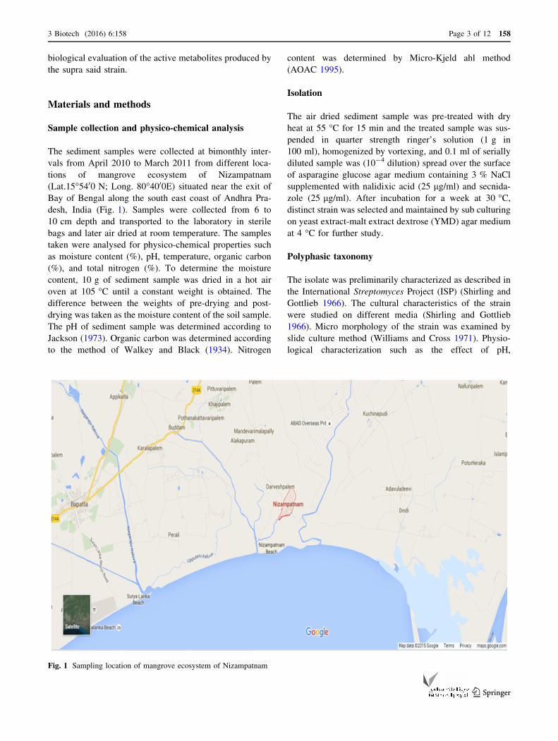
desh, India (Fig. 1). Samples were collected from 6 to
10 cm depth and transported to the laboratory in sterile
bags and later air dried at room temperature. The samples
taken were analysed for physico-chemical properties such
as moisture content (%), pH, temperature, organic carbon
(%), and total nitrogen (%). To determine the moisture
content, 10 g of sediment sample was dried in a hot air
oven at 105 �C until a constant weight is obtained. The
difference between the weights of pre-drying and post-
drying was taken as the moisture content of the soil sample.
The pH of sediment sample was determined according to
Jackson (1973). Organic carbon was determined according
to the method of Walkey and Black (1934). Nitrogen
content was determined by Micro-Kjeld ahl method
(AOAC 1995).
Isolation
The air dried sediment sample was pre-treated with dry
heat at 55 �C for 15 min and the treated sample was sus-
pended in quarter strength ringer’s solution (1 g in
100 ml), homogenized by vortexing, and 0.1 ml of serially
diluted sample was (10-4 dilution) spread over the surface
of asparagine glucose agar medium containing 3 % NaCl
supplemented with nalidixic acid (25 lg/ml) and secnida-
zole (25 lg/ml). After incubation for a week at 30 �C,distinct strain was selected and maintained by sub culturing
on yeast extract-malt extract dextrose (YMD) agar medium
at 4 �C for further study.
Polyphasic taxonomy
The isolate was preliminarily characterized as described in
the International Streptomyces Project (ISP) (Shirling and
Gottlieb 1966). The cultural characteristics of the strain
were studied on different media (Shirling and Gottlieb
1966). Micro morphology of the strain was examined by
slide culture method (Williams and Cross 1971). Physio-
logical characterization such as the effect of pH,
Fig. 1 Sampling location of mangrove ecosystem of Nizampatnam
3 Biotech (2016) 6:158 Page 3 of 12 158
123

temperature, and salinity tolerance was analysed (Ellaiah
et al. 2004). Biochemical tests of the strain were also
evaluated (Cowan 1974; Gordon 1966; Jones 1949;
Waksman 1961). Carbohydrate utilization was determined
by growing the strain on carbon utilization medium (ISP-9)
(Pridham and Gottlieb 1948). Enzymatic profile of the
strain was studied as described previously (Holding and
Collee 1971; Gulati et al. 1997; Yeoh et al. 1985).
Molecular genomic identification of the strain was carried
out according to the procedure of Nilsson and Strom
(2002).
Extraction and secondary metabolite profiling
The seed broth was prepared by culturing Pseudonocardia
endophytica VUK-10 on ISP-2 medium and incubated on a
rotary shaker (250 rpm) at 35 �C for 48 h. Seed culture at
the concentration of 10 % was transferred into fermenta-
tion broth consisting of glucose (8 %), soy-peptone (1 %),
yeast extract (0.2 %), meat extract (0.1 %), CaCO3
(0.3 %), K2HPO4 (0.03 %), MgSO4 (0.1 %), FeSO4
(0.005 %), and NaCl (3 %) with pH adjusted to 7.0. The
culture filtrates (40 L) obtained after cultivation of the
strain for 96 h were extracted twice with ethyl acetate and
concentrated in rotavap, freeze dried to yield a dark brown
residue. The weight of the total crude extract was 5.5 g.
Purification and structure elucidation
The ethyl acetate extract was subjected to Sephadex LH-20
gel filtration chromatography (36 9 2.5 cm, Sephadex
G-15) using 1:1 DCM:MeOH (dichloromethane: methanol)
as eluent yielded VII fractions. Based on the 1H-NMR
spectral data and Bio-active screening, the fraction V
(2.2 g) was selected for further study and subjected to silica
gel column chromatography (25 9 5 cm, Silica gel 60,
Merck) which afforded six fractions. Based on TLC mon-
itoring and NMR spectral data, the sub fractions 2 and 5
were selected for further purification.
In the course of separation, initially the sub fraction 2
(369 mg) was subjected to dinitrobenzylation as it is dif-
ficult to isolate the compound, followed by purification on
silica gel column chromatography using Dichloromethane:
Acetone (92:8) as eluent and further purified by employing
Chloroform: Isopropanol (80:20) yielded 54 mg of pure
compound 1. The sub fraction 5 (247 mg) was subjected to
purification on silica gel column chromatography with
dichloromethane: acetone (85:15) as eluent and further
purified by employing chloroform: isopropanol (80:20)
yielded 32 mg of pure compound 2.
The structures of the isolated compounds were eluci-
dated on the basis of 1D and 2D NMR spectral data cor-
relations in combination with FT-IR spectra (Model:
Thermo Nicolet Nexus 670 spectrophotometer with NaCl
optics), Mass spectroscopy (EIMS; spectrum was measured
on VG-7070H Micro mass spectrometer), 1D and 2D
NMRs [1H, 13C, DEPT, HMQC, HMBC, and COSYNMR]
were recorded on Avance 300 MHz spectrometer and
CDCl3 was used as solvent.
Test micro-organisms
Gram-positive bacteria: Bacillus cereus (MTCC 430),
Streptococcus mutans (MTCC 497), Staphylococcus aur-
eus (MTCC 3160), Staphylococcus epidermis (MTCC
120), Bacillus subtilis (ATCC 6633), Bacillus megaterium
(NCIM 2187); Gram-negative bacteria: Escherichia coli
(ATCC 35218), Pseudomonas aeruginosa (ATCC 9027),
Proteus vulgaris (MTCC 7299), Serratia marcescens
(MTCC 118) Xanthomonas campestris (MTCC 2286),
Xanthomonas malvacearum (NCIM 2954) and Salmonella
typhi (ATCC 14028); Medically important dermatophytes:
Candida albicans (ATCC 10231) and Epidermophyton
floccosum (MTCC 145); Medically and agriculturally
important filamentous fungi: Aspergillus niger (ATCC
1015), Aspergillus flavus (ATCC 9643), Fusarium oxys-
porum (MTCC 3075), Fusarium solani (MTCC 4634),
Penicillum citrinum (MTCC 6489), Verticillium alboatrum
and Alternaria alternata (MTCC 6572). The test micro
organisms used in the present study were procured from
ATCC, University Boulevard, Manassas, USA and MTCC,
Chandigarh, NCIM, Pune, India and preserved at 4 �C.
MIC assay
The antimicrobial spectra of the bioactive compounds
produced by the strain were determined in terms of mini-
mum inhibitory concentration (MIC) against a wide variety
of bacteria and fungi using the broth dilution method
(Andrews 2001). The purified compounds were dissolved
in ethyl acetate while the antibiotics were dissolved in
sterile distilled water to give stock concentrations of
2000 lg/ml. Twofold serial dilutions of the compounds
and antibiotics were made with broth media at concentra-
tions ranging from 1 to 1000 lg/ml. The test concentration
of the compound along with the 100 ll of the microbial
suspension (for bacteria 1 9 108 and 1 9 107 CFU/ml for
yeast and fungi) was transferred aseptically to 10 ml of
broth and incubated for 24 h at 37 �C for bacteria and
48–72 h at 28 �C for fungi. The growth was observed both
visually and by measuring the OD at 600 nm against
uninoculated broth. Triplicate sets of the tubes were
maintained for each concentration of the test sample. The
lowest concentration of the bioactive metabolites exhibit-
ing significant antimicrobial activity against the test
microorganisms was taken as the MIC of the compound.
158 Page 4 of 12 3 Biotech (2016) 6:158
123

Cell lines and culture conditions
Cell lines for testing in vitro cytotoxicity: human breast
adeno carcinoma cell line (MDA-MB-231), human cervical
cancer cell line (HeLa), human ovarian cyst adenocarci-
noma cell line (OAW-42) and human breast adenocarci-
noma cell lines (MCF-7) (cell lines reported to be resistant
to cancer drugs). The cell lines used in the present study
were obtained from National Centre for Cell Science, Pune,
India. Cell lines MDA-MB-231, HeLa, and OAW-42 were
cultured on Dulbecco’s modified Eagle’s medium supple-
mented with fetal bovine serum (10 % (v/v)), L-glutamine
(2 mM), penicillin (10 units/ml), and streptomycin (10 lg/ml), while breast cancer cell line MCF-7 was cultured on
Roswell Park Memorial Institute medium 1640 supple-
mented with fetal bovine serum (10 %; (v/v)), L-glutamine
(2 mM), penicillin (10 units/ml), and streptomycin (10 lg/ml), all in a humidified atmosphere (95 %) with 5 % of
CO2 at 37 �C.
MTT assay
The cytotoxicity of the compounds was assessed on the
basis of the measurement of the in vitro growth in 96-well
plates by cell-mediated reduction of tetrazolium salt to
water insoluble formazan crystals, as per the micro culture
MTT assay (Mosmann 1983). Cells were seeded in 96-well
micro titer plates at a density of 5 9 103 per well (100 ll)containing 0.1 ml of medium. After overnight incubation,
the cells were treated with different test concentrations of
bioactive compounds (10, 100, 1000, and 5000 nM) at
identical conditions with three replicates of each concen-
tration. After 24 h of incubation, the cell viability was
assessed by adding 20 ll of MTT (5 mg/ml in PBS) per
well and the plates were incubated at 37 �C for 4 h. The
formazan crystals formed in the cells were dissolved with
100 ll of 0.1 % acidified isopropanol, and the rate of color
development was measured at 570 nm using a micro plate
reader. The IC50 values (50 % inhibitory concentration) of
the compounds were calculated using Sigma Plot software
with reference to that of Taxol as standard. All the
experiments were carried out in triplicates.
Results
Physico-chemical analysis
Physico-chemical characteristics of sediment samples col-
lected from Nizampatnam mangrove ecosystem were anal-
ysed and the parameters were recorded. The soil was slightly
alkaline in nature with pH 7.6. The temperature of the site at
the time of sample collection was measured at 32 �C. The
moisture content of the soil sample was 15 %, organic carbon
7.2 mg/g, and the total nitrogen content was 4.5 lg/g. Soilswith slightly alkaline or neutral pH together with 10–15 %
moisture content and high organic content were reported to
support high incidence of actinobacteria (De andGupta 1993).
Isolation and polyphasic taxonomy
Screening of soil samples from Nizampatnam mangrove
ecosystem for potent actinomycetes led to the isolation of
morphologically distinct actinomycete isolate VUK-10.
The strain exhibited good growth on ISP-1, ISP-2, ISP-3,
ISP-5, Czapek-Dox agar, and maltose tryptone agar,
moderate on ISP-4 and ISP-7 agar, while growth was poor
on nutrient agar media. Morphological and micro mor-
phological observations of the strain revealed abundant
aerial and vegetative hyphae which are well developed and
fragmented to form rod shaped spores with smooth surface.
Pigment production by the strain was not found on the
culture media tested except melanin pigmentation on tyr-
osine (ISP-7) agar media. Detailed morphology of the
strain was examined through scanning electron microscopy
(SEM). Figure 2 depicts the SEM image of the strain at
65009 magnification. Growth of the strain occurred in the
pH range of 6–9 with an optimum growth at pH 7. The
temperature range for growth was 20–45 �C with the
optimum temperature being 35 �C. The strain exhibited
salt tolerance up to 10 % with optimum growth at 3 %
NaCl. VUK-10 exhibited positive response to catalase
production, starch hydrolysis and citrate utilization but
negative for urease, hydrogen sulphide production, nitrate
reduction, gelatine liquefaction, indole, methyl red, and
Vogues-Proskauer tests. The strain efficiently utilized D-
glucose, lactose, maltose, sucrose, galactose, fructose, and
starch as carbon sources but not xylose, arabinose and
Fig. 2 Scanning electron microscope image of Pseudonocardia
endophytica VUK-10 (96500)
3 Biotech (2016) 6:158 Page 5 of 12 158
123

mannitol. The strain could produce commercially impor-
tant enzymes like amylase, cellulase, chitinase, and L-as-
paraginase but it was negative for protease, DNase, and
RNase. 16S rDNA gene sequence of the isolate VUK-10
showed a close relation with Pseudonocardia endophytica.
The rDNA sequence was deposited in the NCBI Gen Bank
with an accession number JN087501 (Ushakiranmayi et al.
2012).
Purification and structure elucidation
Culture filtrates obtained after 96 h fermentation were
extracted with ethyl acetate and concentrated to yield a dark
brown residue, which in turn was subjected to gel filtration
chromatography using the solvent system of dichlor-
omethane/methanol. Among the seven fractions collected,
the fraction V exhibiting good antimicrobial activity was
rechromatographed on a silica gel column and yielded six
fractions. Based on TLCmonitoring and NMR spectral data,
the sub fractions 2 and 5 were selected for further purifica-
tion. The sub fraction 2 (369 mg) was subjected to dini-
trobenzylation followed by purification on silica gel column
chromatography which yielded compound 1. The sub frac-
tion 5was subjected to purification on silica gel column using
different eluents which yielded pure compound 2.
Compound (1) was obtained as white solid with m.p.
155–161 �C; a½ �25D ?20 (c 0.5, CDCl3). Its molecular for-
mula C15H20N4O5 was derived from HRESIMS m/z
359.1319 [M ? Na]? (calcd for C15H20NaN4O5 is
359.1331) and 13C NMR spectral data measurements. The
structural elucidation of 1 was established by detailed
interpretation of 1D and 2D NMR spectral data correlations
(Table 1), including 1H NMR (Supplementary Fig. A), 13C
NMR (Supplementary Fig. B), DEPT (Supplementary
Fig. C), HMBC (Supplementary Fig. D), HSQC (Supple-
mentary Fig. E) COSY (Supplementary Fig. F), and
HRESIMS (Supplementary Fig. G). The 13C NMR and
DEPT spectral data reveal the presence of 15 carbon res-
onances, including four of quarternary and six of methy-
lene carbons along with two methane carbons acquired
with nitrogen atoms. As the sub-fraction 5 (crude) was
treated with 3,5-Dinitro benzoic acid in the presence of
DCC/DMAP (solvent DCM) in DCM as solvent and stirred
at 0 �C for 12 h followed by work-up procedures. The
reaction mixture was monitored by TLC and purified on
silica gel column chromatography to obtain product 1, the13C NMR spectrum of the resultant product (1) also had
signals for tri-substituted aromatic ring carbons along with
an amide carbonyl carbon between dC 120 ppm and dC175 ppm. As the crude fraction (V) was treated with 3,5-
Table 1 1D and 2D NMR data correlations of N-(4-aminocyclooctyl)-3,5-dinitrobenzamide
S. no 13C and (DEPT) HSQC 1H-1H COSY HMBC
1 56.0 (CH) 4.23 (tt, J = 3.59, 2.09 Hz) 2-H, C-9, C-10, C-8, C-2
2 30.9 (CH2) 1.96 (d, J = 11.44 Hz)
1.74 (dt, J = 3.27, 12.45 Hz)
1-H, 3-H C-8, C-1, C-3
3 32.1 (CH2) 1.48 (d J = 9.80 Hz)
0.84 (qd, J = 2.61, 12.09 Hz)
4-H, 2-H C-4, C-5, C-2
4 50.6 (CH) 3.51 (m) 3-H C-3, C-5
5 25.5 (CH2) 1.71 (m)
1.19 (q, J = 13.07 Hz)
– C-7, C-2, C-4
6 25.4 (CH2) 1.56 (d, J = 13.07 Hz)
1.03 (m)
– C-3, C-5
7 24.9 (CH2) 1.58 (m)
1.19 (q, J = 13.07 Hz)
– C-5, C-2, C-4
8 26.2 (CH2) 1.87 (d, J = 13.07 Hz)
1.39 (q, J = 13.07 Hz)
8-H$ 8-H C-2, C-1,C-7
9 165.0 (Cq) – – –
10 153.9 (Cq) – – –
11 127.8 (2CH) 8.72 (d, J = 2.31 Hz) – C-13, C-15, C-12, C-9
12 148.4 (Cq) – – –
13 120.3 (CH) 9.09 (s) – C-11, C-12, C-14, C-15
14 148.4 (Cq) – – –
15 127.8 (2CH) 8.72 (d, J = 2.31 Hz) – C-13, C-11, C-12 (14), C-9
16 (N-H) 5.85 (d, J = 7.84 Hz) – C-3, C-4, C-10
158 Page 6 of 12 3 Biotech (2016) 6:158
123

dinitrobenzoic acid in DCC as solvent, the isolated
semisynthetic product of the natural compound comprised
with a 3,5-dinitrobenzoic acid ester residue in the structure.
The molecular formula (C15H20N4O5) and spectral data
account for six degrees of unsaturation including an aro-
matic ring and a carbonyl group; hence 1 is with di-sub-
stituted aliphatic monocyclic ring compound.
The 1H-1H COSY spectrum of 1 was consistent showing
the linear correlation between 1-H (dH 4.23, tt), 2-2H (dH 1.96,
d and 1.74, dt), 3-2H (dH 1.48, d and 0.84, qd) and 4-H (dH3.51, m) protons. The HMBC correlations of (1) consistently
suggesting cyclic-octane ring formulation (1-H/C2, C8, C9,
C10; 2-H/C8,C1; 3-H/C2,C4, C5; 4-H/C3, C5; 5-H/C-2, C-4,
C-7; 6-H/C-3, C-5; 7-H/C-2, C-4, C-5 and 8-H/C-1, C-2, C-7)
in addition to the dinitrobenzyl group in the structure (Fig. 3).
Meticulous analysis of 1D and 2D NMR spectral data corre-
lations confirmed that, (1) is a novel cyclooctane-1, 4-diamine
carbon skeleton derivative (Fig. 4).
Compound (2) was obtained as white amorphous solid
which was completely soluble in DMSO, methanol, etha-
nol, N–N-dimethyl formamide, and ethyl acetate but
insoluble in water and n–hexane. The 1H-NMR (300 MHz,
CDCl3) spectrum of the compound 2 showed signals at d8.46 (s, 1H); 7.57 (d, 1H, J = 7.7 Hz); 7.37 (d, 1H,
J = 8.1 Hz); 7.22 (t, 1H, J = 7.1 Hz); 7.16-7.08 (m, 2H);
5.84 (s, 1H); 4.37 (dd, 1H, J = 2.6, 10.5 Hz); 4.06 (t, 1H,
J = 6.6 Hz); 3.72 (dd, 1H, J = 3.3, 14.9 Hz); 3.66–3.48
(m, 2H); 2.96 (dd, 1H, J = 10.5, 14.9 Hz); 2.35–2.27 (s,
1H); 2.06–1.82 (m, 3H) (Supplementary Fig. H), while 13C
(75 MHz, CDCl3) exhibited signals at (75 MHz, CDCl3) d169.4, 165.4, 136.5, 126.6, 123.4, 122.5, 119.7, 118.4,
111.5, 109.6, 59.1, 54.5, 45.3, 28.1, 26.7, and 22.5
(Supplementary Fig. I). Mass spectrometric (EIMS) anal-
ysis of the compound gave a molecular ion peak at m/z 306
(M ? 23) (Supplementary Fig. J). In IR spectrum, the
compound exhibited absorption bands at Vmax 3280, 2924,
1658, 1430, 1103, and 745 cm-1 (Supplementary Fig. K).
On the basis of spectral data (C16H17N3O2) analysis and
literature survey (Ivanova et al. 2003), the compound 2 was
identified as (3-(1H-indol-6-yl) methyl) hexahydropyrrolo
[1, 2-a] pyrazine-1, 4-dione (Fig. 4).
MIC assay
Antibacterial activities of the bioactive compounds (1 and
2) in terms of MIC are shown in Table 2. Bioactive com-
pounds exhibited antibacterial activity against a variety of
Gram-positive and Gram-negative bacteria for which the
MIC values ranged from 4 to 64 lg/ml. Among the fac-
ultative and pathogenic Gram-positive bacteria, the com-
pound 1 was active against all the bacteria tested and the
best activity of this compound was recorded against
Streptococcus mutans (4 lg/ml) followed by Bacillus
cereus (8 lg/ml). Compound 2 presented highest activity
against Staphylococcus aureus (16 lg/ml) followed by
Bacillus megaterium (32 lg/ml). Of the Gram-negative
bacteria, the micro-organisms that presented highest sen-
sitivity towards compound 1 was Pseudomonas aeruginosa
(16 lg/ml) followed by Salmonella typhi, Escherichia coli,
and Xanthomonas malvacearum (32 lg/ml). Compound 2
recorded highest activity against Escherichia coli and
Xanthomonas malvacearum (16 lg/ml). Tetracycline
served as positive control for bacteria. Compared to stan-
dard drug tetracycline, compound 1 displayed high sensi-
tivity against Streptococcus mutans and Staphylococcus
aureus, while compound 2 recorded similar sensitivity like
positive control against Serratia marcescens (Table 2).
Tetracycline, in other cases showed good antibacterial
activity over the metabolites of the strain.
Antifungal activity against dermatophytes and filamen-
tous fungi and corresponding MIC values are recorded in
Table 3. The bioactive compounds showed significant
antifungal activity against the dermatophytes (Candida
albicans and Epidermophyton floccosum). Among the fil-
amentous fungi tested, Aspergillus niger recorded
NH
O
NO2
NO2
H2N
Fig. 3 Important COSY (–) and HMBC (?) correlations of semisyn-
thetic compound N-(4-aminocyclooctyl)-3,5 dinitrobenzamide
Fig. 4 Structures of the
compounds 1 and 2 isolated
from Pseudonocardia
endophytica VUK-10
3 Biotech (2016) 6:158 Page 7 of 12 158
123

sensitivity of 32 lg/ml followed by Aspergillus flavus,
Penicillium citrinum, and Verticillium alboatrum (64 lg/ml) towards compound 1. Compound 2 was active against
Aspergillus flavus and Fusarium solani at 64 lg/ml and for
this compound Verticillium alboatrum recorded no activity
up to 512 lg/ml. Both compounds recorded lower anti-
fungal activity than the standard fungicide amphotericin-B
against fungi.
MTT assay
The in vitro cytotoxicity evaluated against a panel of
human tumor cell lines indicated that both the compounds
had similar level of anti tumor activity against MDA-MB-
231, HeLa, OAW-42, and MCF-7 cell lines.
The activity of compound 1 against MDA-MB-231,
HeLa, MCF-7, and OAW-42 cell lines is presented in
Fig. 5a–d. Compound 1 exhibited potent cytotoxicity when
evaluated against MDA-MB-231, HeLa, MCF-7, and
OAW-42 cell lines displaying IC50 values of 100 nM (59.6,
58 %) (MDA-MB-231) (OAW-42), 10 nM (64.9 %)
(HeLa), and 5000 nM (53.4 %) (MCF-7).
The activity of compound 2 against MDA-MB-231,
HeLa, MCF-7, and OAW-42 cell lines is presented in
Fig. 6a–d. Compound 2 showed significant cytotoxicity
with MDA-MB-231, HeLa, MCF-7, and OAW-42 cell
lines. The cell lines exhibited IC50 values of 100 nM
(71.5 %, 52.7 %) (MDA-MB-231) (OAW-42), 10 nM
(60.6 %) (HeLa), and 5000 nM (41.6 %) (MCF-7), with
Taxol an anti-cancer drug used as standard that recorded an
Table 2 Minimum inhibitory
concentration (MIC) of
bioactive compounds isolated
from Pseudonocardia
endophytica VUK-10 [MIC-
(lg/ml)] against test bacteria
Test microorganisms Compound 1 Compound 2 Tetracycline
S. aureus 16 ± 0.02 16 ± 0.01 32 ± 0.03
S. mutans 4 ± 0.03 64 ± 0.00 32 ± 0.03
S. epidermis 16 ± 0.01 64 ± 0.02 16 ± 0.03
X. campestris 64 ± 0.00 32 ± 0.03 16 ± 0.03
X. malvacearum 32 ± 0.02 16 ± 0.01 8 ± 0.01
B. subtilis 16 ± 0.03 64 ± 0.03 32 ± 0.02
B. megaterium 16 ± 0.02 32 ± 0.02 16 ± 0.03
B. cereus 8 ± 0.01 64 ± 0.01 8 ± 0.02
E. coli 32 ± 0.01 16 ± 0.01 8 ± 0.01
P. aeruginosa 16 ± 0.02 64 ± 0.02 8 ± 0.03
S. marcescens 64 ± 0.01 32 ± 0.03 32 ± 0.01
P. vulgaris 64 ± 0.00 64 ± 0.01 16 ± 0.03
S. typhi 32 ± 0.03 64 ± 0.02 8 ± 0.02
Values are mean ± standard deviation (n = 3). Compound 1: N-(4-aminocyclooctyl)-3, 5-dinitrobenza-
mide. Compound 2: (3-((1H-indol-6-yl) methyl) hexahydropyrrolo [1, 2-a] pyrazine-1, 4-dione. Antibiotic:
tetracycline
Table 3 Minimum inhibitory
concentration (MIC) of
bioactive compounds isolated
from Pseudonocardia
endophytica VUK-10 [MIC-
(lg/ml)] against dermatophytes
and fungi
Compound 1 Compound 2 Antifungal agent
Dermatophytes
Candida albicans 16 ± 0.03 64 ± 0.02 16 ± 0.01
Epidermophyton floccosum 32 ± 0.02 32 ± 0.00 16 ± 0.01
Fungi
Aspergillus niger 32 ± 0.02 128 ± 0.03 16 ± 0.02
Aspergillus flavus 64 ± 0.00 64 ± 0.02 8 ± 0.00
Fusarium oxysporum 128 ± 0.01 256 ± 0.01 16 ± 0.00
Fusarium solani 128 ± 0.02 64 ± 0.02 32 ± 0.01
Penicillium citrinum 64 ± 0.00 256 ± 0.03 8 ± 0.01
Verticillium alboatrum 64 ± 0.03 [512 ± 0.02 64 ± 0.02
Alternaria alternata 128 ± 0.03 128 ± 0.01 32 ± 0.01
Values are mean ± standard deviation (n = 3). Compound 1: N-(4-aminocyclooctyl)-3, 5-dinitrobenza-
mide. Compound 2: 3-((1H-indol-6-yl) methyl) hexahydropyrrolo [1, 2-a] pyrazine-1, 4-dione. Antifungal
agent: griseofulvin against dermatophytes and amphotericin-B against fungi
158 Page 8 of 12 3 Biotech (2016) 6:158
123

IC50 value of 10 nM (59, 60, 57, and 63 %) against MDA-
MB-231, HeLa, OAW-42, and MCF-7 cell lines.
Discussion
Recent research efforts have focused on the exploration of
under explored habitats to discover novel bioactive sec-
ondary metabolites from the autochthonous microbiota
(Omar et al. 2015). Natural products are the important
assuring source for novel bioactive molecules especially
antimicrobial compounds over combinatorial chemistry
and fragment based drug designing that devote very little to
corner the spread of the antibiotic resistant pathogens
(Shuvankar et al. 2015). Antibiotics are the group of
important bioactive compounds for the treatment of
infectious diseases. Natural products remain as most
prominent source for the exploration of uncommon inno-
vative bioactive molecules especially antimicrobial com-
pounds due to the fact that the spread of the antibiotic
resistant pathogens cannot be counteracted and reversed
since combinatorial chemistry and fragment-based drug
designing contribute very little (Goodfellow and Fiedler
2010; Mikhail et al. 2015). Among the different niches,
mangrove forests are exclusively rare niches which harbor
a diverse range of microbes (Farshid and Faranak 2015).
Actinomycetes are the predominant members of marine
microbial communities and are also known to adapt to
harsh environmental conditions (Augustine et al. 2012).
In the present screening programme undertaken for
exploration of the novel bioactive secondary metabolites
from Nizampatnam mangrove ecosystem, a potent isolate
Fig. 5 Dose response curve of Compound 1 on the growth of a MDA-MB-231, b HeLa, c MCF-7 and d OAW-42 cell lines
3 Biotech (2016) 6:158 Page 9 of 12 158
123

with high antimicrobial and cytotoxic activities identified
as Pseudonocardia endophytica VUK-10 was isolated. A
semisynthetic derivative (N-(4-aminocyclooctyl)-3,
5-dinitrobenzamide) along with a known compound (3-
(1H-indol-6-yl) methyl) hexahydropyrrolo [1, 2-a] pyr-
azine-1, 4-dione) was isolated from the fermentation broth
of the strain grown on glucose-soy peptone broth. The
precursor of the semisynthetic compound 1 could be
cyclooctane-1, 4-diamine (3) (Fig. 7). A quick SciFinder
search reveals that this is an unknown compound that has
not been identified as a metabolite from any
microorganisms.
The compound 2, a non-ribosomal peptide is indistin-
guishable with cyclo (tryptophanyl prolyl), a cyclic
dipeptide between the amino acids tryptophan and proline.
Cyclic dipeptides (CDPs) are commonly biosynthesized
from amino acids by different organisms, including mam-
mals, and are considered to be secondary functional
metabolites or side products of terminal peptide cleavage
(Maristela and Ivone 2007). CDPs are more than simple
curiosities and are well known for their economically ben-
eficial biological activities (Prasad 1955). The therapeutic
potential of these compounds reflects their antibiotic,
antiviral, and antitumor properties and various cyclic
dipeptides are detectable in protein and polypeptide hydro-
lysates as well as in cultures of yeasts, lichens, and molds
(Otteinheijm et al. 1976; Graz et al. 1999; Katrin et al. 2002).
The isolation of compound 2 was previously reported as a
natural product from two Penicillium strains (Paterson et al.
1990).
Our results showed that compound 1 has a good
potential inhibitor against Streptococcus mutans (respon-
sible for human dental decay), Pseudomonas aeruginosa
Fig. 6 Dose response curve of Compound 2 on the growth of a MDB-MB-231, b HeLa, c MCF-7 and d OAW-42 cell lines
Fig. 7 Compound 3 (not been
isolated purely) is the precursor
of semi synthetic compound 1
158 Page 10 of 12 3 Biotech (2016) 6:158
123

(causes pneumonia, bacteremia, and endocarditis), Can-
dida albicans (causes oral thrush and vaginal infection) and
Aspergillus niger (causes black mold on grapes, onions,
and peanuts). Compound 2 showed to have antimicrobial
activity against Staphylococcus aureus (causes impetigo,
carbuncles, and abscesses), Escherichia coli (causes
cholecystitis, bacteremia, cholangitis, and urinary tract
infection), Xanthomonas malvacearum (causes angular leaf
spot of cotton) and Epidermophyton floccosum (causes
tinea pedis, tinea cruris, and tinea corporis).
The potential of compounds 1 and 2 was investigated to
inhibit cancer cell growth in MDA-MB-231, HeLa, OAW-
42, and MCF-7 cell lines. Our results showed that com-
pounds 1 and 2 displayed similar level of significant
cytotoxicity with variation in percentage of cell line inhi-
bition against MDA-MB-231, HeLa, OAW-42, and MCF-7
cell lines. To the best of our knowledge, anti cancer
activity of compounds 1 and 2 against the above said cell
lines are reported here for the first time. The data reported
in this paper clearly indicate that our compounds exhibit
significant anti cancer activities at nanomolar concentra-
tions. Contemporary identification of primary and sec-
ondary metabolites from taxonomically exclusive
population of actinomycetes disclosed that these microbes
could add a new element to microbial natural product
research (Jicheng et al. 2015).
Conclusion
Based on the distribution, actinomycetes have been vali-
dated as an important source of bioactive metabolites. The
present study is concluded that actinomycetes strain
Pseudonocardia endophytica VUK-10 with an interesting
bioactivity profile has been exploited vividly to study their
bioactive metabolites. The results of the present study are
amazing and the compounds 1 and 2 exhibited strong
effects against test microorganisms and cancer cell lines
even in impressive low concentrations. Therefore, the
bioactive compounds can be ideal ammunition to use as a
potential antimicrobial and anticancer agents and trusting it
as the first report of isolation and characterization of
compounds 1 and 2 from the genus Pseudonocardia.
Acknowledgments This work was supported by the Council of
Scientific and Industrial Research, New Delhi, Government of India
under Grant 38-1218-09-EMR-II.
Compliance with ethical standards
Conflict of interest All the authors declare that there is no conflict of
interest.
Open Access This article is distributed under the terms of the
Creative Commons Attribution 4.0 International License (http://
creativecommons.org/licenses/by/4.0/), which permits unrestricted
use, distribution, and reproduction in any medium, provided you give
appropriate credit to the original author(s) and the source, provide a
link to the Creative Commons license, and indicate if changes were
made.
References
Andrews JM (2001) Determination of minimum inhibitory concen-
trations. J Antimicrob Chemother 48:5–16
AOAC (1995) Official methods of analysis of the association of
official analytical chemists, 16th edn. AOAC, Arlington,
pp 483–486
Augustine N, Abraham WP, Kerkar S, Thomas S (2012) Arctic
actinomycetes as potential inhibitors of Vibrio cholerae biofilm.
Curr Microbiol 64:338–342
Balagurunathan R, Masilamani SM, Kathiresan K (2010) Bio-
prospecting of mangrove rhizosphere actinomycetes from
Pitchavaram with the special reference to antibacterial activity.
J Pharm Res 3:909–911
Ballav S, Dastager SG, Kerkar S (2012) Biotechnological significance
of actinobacterial research in India. Recent Res Sci Technol
4:31–39
Clardy J, Fischbach MA, Walsh CT (2006) New antibiotics from
bacterial natural products. Nat Biotechnol 24:1541–1550
Cowan ST (1974) Cowan and Steel’s manual for the identification of
medical bacteria, 2nd edn. University Press, Cambridge
Davies OF, Adeleye IA, Wang PG (2015) Hyoscyamine-producing
marine Actinomycetes from Lagos Lagoon sediment. Asian Pac J
Trop Biomed 5:196–201
De K, Gupta KD (1993) Actinomycetes from various micro sites of
crop plants and trees of Jabalpur. Ind J Microbiol 33:269–272
Dekker KA, Inagaki T, Gootz TD, Huang LH, Kozima Y, Kohl
Brenner WE, Matsunaga Y, Mcguirk PR, Nomurae E, Sakak-
ibara T, Sakemi S, Suzuki Y, Yamauchi Y, Kozima N (1998)
New quinolone compounds from Pseudonocardia sp. with
selective and potent anti- Helicobacter pylori activity. Taxon-
omy of producing strain, fermentation, isolation, structural
elucidation and biological activities. J Antibiot 51:145–152
Demain AL, Sanchez S (2009) Microbial drug discovery: 80 years of
progress. J Antibiot 62:5–16
Dhanasekaran D, Rajakumar G, Sivamani P, Selvamani S, Panneer-
selvam A, Thajuddin A (2005) Screening of salt pans actino-
mycetes for anti-bacterial agents. Internet J Microbiol 1:1–3
Ellaiah P, Ramana T, Bapiraju KVVS, Sujatha P, Umasankar A
(2004) Investigation on marine actinomycetes from Bay of
Bengal near Kakinada coast of Andhra Pradesh. Asian J
Microbiol Biotechnol Environ Sci 6:53–56
Farshid K, Faranak D (2015) Amylase activity of aquatic actino-
mycetes isolated from the sediments of mangrove forests in
south of Iran. Egypt J Aquat Res 41:197–201
Gavin C, Emily RD, Eric C, Cameron RC, Jon C (2012) Antibiotic
and antimalarial quinones from fungus-growing ant associated
Pseudonocardia sp. J Nat Product 75:1806–1809
Goodfellow M, Fiedler HP (2010) A guide to successful bioprospect-
ing: informed by actinobacterial systematic. Antonie Van
Leeuwenhoek 98:119–142
Gordon RE (1966) Some criteria for the recognition of Nocardia
madura (Vincent) Blanchord. J Gen Microbiol 45:355–364
Graz M, Hunt A, Jamie H, Grant G, Milne P (1999) Antimicrobial
activity of selective cyclic dipeptides. Pharmazie 54:772–775
Gulati R, Saxena RK, Gupta R (1997) A rapid plate assay for
screening L-asparaginase producing microorganisms. Lett Appl
Microbiol 24:23–26
3 Biotech (2016) 6:158 Page 11 of 12 158
123

Hayakawa M (2008) Studies on the isolation and distribution of rare
actinomycetes in soil. Actinomycetologica 22:12–19
Holding AJ, Collee JG (1971) Routine biochemical tests. In: Norris
JR, Ribbons DW (eds) Methods in microbiology. Academic
Press Inc, London, pp 1–32
Ivanova V, Graefe U, Schlegel R, Schlegel B, Gusterova A, Kolarova
M, Aleksieva K (2003) Isolation and structure elucidation of
Tyramine and Indole alkaloids from Antarctic Strain Microbis-
pora aerata IMBAS-11A. Biotechnol Biotec Eq 17:128–133
Jackson ML (1973) Soil chemical analysis. Prentice Hall of India Pvt.
Ltd, New Delhi
Jicheng Yu, Liu Z, Qiu L, Qi Xiaohui, Ji Ying, Beom SK (2015)
Isolation and characterization of actinobacteria from Yalujiang
coastal wetland, North China. Asian Pac J Trop Biomed
5:555–560
Jones K (1949) Fresh isolates of actinomycetes in which the presence
of sporogenous aerial mycelia is a fluctuating characteristics.
J Bacteriol 57:141–145
Katrin S, Jorgen S, Anders B, John S (2002) Lactobacillus plantarium
MiLAB-393 produce the antifungal cyclic dipeptides cyclo (L-
Phe-L-Pro) and cyclo (L-Phe-trans-4-OH-L-Pro) and 3-phenyl-
lactic acid. Appl Environ Microbiol 68:4322–4327
Khanna M, Solanki R, Rup L (2011) Selective isolation of rare
actinomycetes producing novel anti-microbial compounds. Int J
Adv Biotechnol Res 2:357–375
Kui H, An-Hui G, Qing-Yi X, Hao G, Ling Z, Hai-Ping Y, Jia L, Xin-
Sheng Y, Michael G, Ji-Sheng R (2009) Actinomycetes for
marine drug discovery isolated from mangrove soils and plants
in China. Mar Drugs 7:24–44
Lakshmipathy DT, Kannabiran K (2009) A morphological, biochem-
ical and biological studies of halophilic Streptomyces sp. isolated
from saltpan environment. Am J Infect Dis 5:200–206
Lee MJ, Kong D, Han K, Sherman DH, Bai L, Deng Z, Lin S, Kim ES
(2012) Structural analysis and biosynthetic engineering of a
solubility improved and less haemolytic nystatin like polyene in
Pseudonocardia autotrophica. Appl Microbiol Biotechnol
95:157–168
Li S, Tian X, Niu S, Zhang W, Chen Y, Zhang H, Yang X, Zhang W,
Li W, Zhang S, Ju J, Zhang C (2011) Pseudonocardians A–C,
new diazaanthraquinone derivatives from a deep sea actino-
mycetes Pseudonocardia sp. SCSIO 01299. Mar Drugs
9:1428–1439
Maristela BM, Ivone C (2007) Diketopiperazines: biological activity
and synthesis. Tetrahedron 63:9923–9932
Maskey RP, Kock I, Helmke E, Laatsch H (2003) Isolation and
structure determination of phenazostatin D, a new phenazine
from marine actinomycetes isolate Pseudonocardia sp. B6273.
Z Naturforsch 58:692–694
Mikhail M, Tietz JI, Melby JO, Blair PM, Zhu L, Livnat I, Severinov
K, Mitchell DA (2015) Structure, bioactivity and resistance
mechanism of streptomonomicin, an unusual lasso peptide from
an understudied halophilic actinomycete. Chem Biol 22:241–250
Mosmann T (1983) Rapid colorimetric assay for cellular growth and
survival: application to proliferation and cytotoxicity assays.
J Immunol Methods 65:55–63
Nayan RB, Shukla VJ (2001) Antibacterial and antifungal activities
from leaf extract of Cassia fistula I: an ethno medicinal plant.
J Adv Pharm Technol Res 2:104–109
Nilsson WB, Strom MS (2002) Detection and identification of
bacterial pathogens of fish in kidney tissue using terminal
restriction fragment length polymorphism (T-RFLP) analysis of
16S rRNA genes. Dis Aquat Organ 48:175–185
Oh DC, Scott JJ, Currie CR, Clardy J (2009) Mycangimycin, a
polyene peroxide from a mutualist Streptomyces sp. J Org Lett
11:633–636
Omar M, Mourad B, Ibrahim B, Djamal-Elddine A (2015) Identifi-
cation and preliminary characterization of non-polyene antibi-
otics secreted by new strain of actinomycete isolated from
sebkha of Kenadsa, Algeria. Asian Pac J Trop Biomed
5:438–445
Omura S, Tanaka H, Tanaka Y, Spiri-nakagawa P, Oiwa R, Takahashi
Y, Matsuyama K, Iwai Y (1979) Studies on bacterial cell wall
inhibitors. VII. Azureomycins A and B, new antibiotics
produced by Pseudonocardia azureanov sp. Taxonomy of the
producing organism, Isolation, characterization and biological
properties. J Antibiot 32:985–994
Otteinheijm HCJ, Herscheid JDM, Kerkhoff GPC, Spende TF (1976)
Preparation of episulfide DKPS from ZN2? catalyzed cyclization
procedure. J Org Chem 41:3433–3438
Paterson RRM, Simmonds MJS, Kemmelmeier C, Blaney WM
(1990) Effects of brevianamide A, its photolysis product
brevianamide D, and ochratoxin A from two Penicillium strains
on the insect pests Spodoptera frugiperda and Heliothis
virescens. Mycol Res 94:538–542
Piret JM, Demain AL (1988) Actinomycetes in biotechnology.
Academic Press, London
Prasad C (1955) Bioactive cyclic dipeptides. Peptides 16:151–164
Pridham TG, Gottlieb D (1948) The utilization of carbon compounds
by some actinomycetales as an aid for species determination.
J Bacteriol 56:107–114
Sadhegi A, Karimi E, Dahaji PA, Javid MG, Dalvand Y, Askari H
(2012) Plant growth promoting activity of an auxin and
siderophore producing isolate of Streptomyces under saline soil
conditions. World J Microbiol Biotechnol 28:1503–1509
Satyanarayana T, Raghukumar C, Shivaji S (2005) Extremophilic
microbes: diversity and perspectives. Curr Sci 89:78–90
Shirling EB, Gottlieb D (1966) Methods for characterization of
Streptomyces sp. Int J Syst Bacteriol 16:313–340
Shuvankar B, Savita K, Sabu T, Nimmy A (2015) Halophilic and
halotolerant actinomycetes from a marine saltern of Goa, India
producing anti-bacterial metabolites. J Biosci Bioeng
119:323–330
Solanki R, Khanna M, Lal R (2008) Bioactive compounds from
marine actinomycetes. Ind J Microbiol 48:410–431
Talbot GH, Bradely J, Edwards JE, Gilbert D, Scheldt M, Bartlett JG
(2006) Bad bugs need drugs: an update on the development
pipeline from the antimicrobial availability task force of the
infectious disease society of America. Clin Infect Dis
42:657–668
Ushakiranmayi M, Vijayalakshmi M, Sudhakar P, Sreenivasulu K
(2012) Isolation, identification and molecular characterization of
rare actinomycetes from mangrove ecosystem of Nizampatnam.
Malays J Microbiol 8:83–91
Waksman SA (1961) The actinomycetes classification, identification
and description of genera and species. Williams and Wilkins CO,
Baltimore
Walkey A, Black CA (1934) An examination of the Degtiareff
method for determining soil organic matter and proposed
modification of chromic valid titration method. Soil Sci
37:27–38
Williams ST, Cross T (1971) Isolation, purification, cultivation and
preservation of actinomycetes. In: Norris JR, Ribbons DW (eds)
Methods in microbiology. Academic Press, London, pp 295–334
Wise R (2008) The worldwide threat of antimicrobial resistance. Curr
Sci 95:181–187
Yeoh HH, Khew E, Lim G (1985) A simple method for screening
cellulolytic fungi. Mycologia 77:161–162
158 Page 12 of 12 3 Biotech (2016) 6:158
123