Is This A Vein Problem? · Lymphedema S Abnormal accumulation of interstitial fluid and...
38
S Is This A Vein Problem? Parker Truong, DO Oklahoma Heart Hospital Physicians
Transcript of Is This A Vein Problem? · Lymphedema S Abnormal accumulation of interstitial fluid and...

S
Is This A Vein Problem?Parker Truong, DO
Oklahoma Heart Hospital Physicians

S
Relevant Disclosure
Under the Oklahoma State Medical Association CME guidelines
disclosure must be made regarding relevant financial relationships
with commercial interests within the last 12 months.
Parker Truong, DO
I have no relevant financial relationships or affiliations with
commercial interests to disclose.
Objectives
S Differentiation of venous disease vs. arterial disease vs.
lymphatic disease vs. other disease states.
S General approach to venous disease.
S Diagnosis of superficial venous disease.
S Diagnosis of deep venous disease.
S General treatments of venous disease.


Differential Diagnosis
S Arterial disease
S Lymphatic disease
S Neuropathic disease
S Venous disease
S Others: Lipedema, myxedema, KTS, Raynaud’s, medication
side effects.
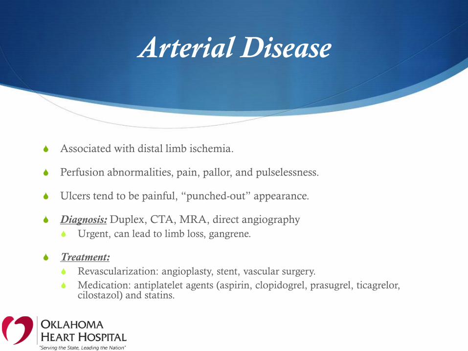
Arterial Disease
S Associated with distal limb ischemia.
S Perfusion abnormalities, pain, pallor, and pulselessness.
S Ulcers tend to be painful, “punched-out” appearance.
S Diagnosis: Duplex, CTA, MRA, direct angiography
S Urgent, can lead to limb loss, gangrene.
S Treatment:
S Revascularization: angioplasty, stent, vascular surgery.
S Medication: antiplatelet agents (aspirin, clopidogrel, prasugrel, ticagrelor, cilostazol) and statins.

Arterial Disease
S Symptoms: vary from asymptomatic, “intermittent claudication” or reproducible discomfort of a defined group of muscles with exertion that relieved with rest, rest pain, to ischemic limb.
S Risk factors: Age, smoking, diabetes, hypertension, hyperlipidemia, homocysteinemia, FH of atherosclerosis.
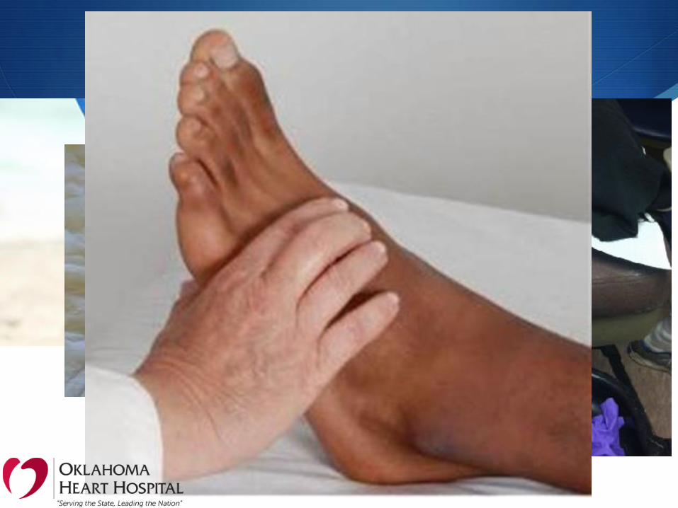
S Examination: diminished distal pulses, pallor, coolness, ischemic ulcers, and gangrene.
Arterial Disease
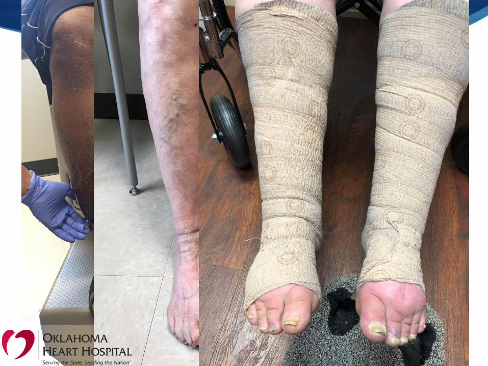
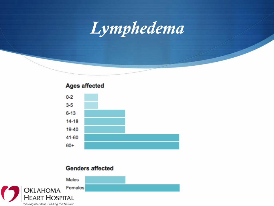
Lymphedema

Lymphedema
S Abnormal accumulation of interstitial fluid and fibroadipose tissues resulting from injury, infection, congenital abnormalities of the lymphatic system.
S Differential diagnosis: CVI, DVT, PTS, lipedema, limb hypertrophy (Klippel-Trenaunay syndrome), myxedema, tumor.
S Classified as primary or secondary depending on etiology and presentation.
S Diagnosis: History and physical, Stemmer’s sign, imaging mainly lymphoscintigraphy.
S Treatment: Lymphedema therapy (MLD Therapy)

Lipedema
S AKA “Painful Fat Syndrome”
S Disorder of fat metabolism, affects mainly women
S Onset during puberty, pregnancy, or menopause, in 4 stages
S Bilateral, symmetrical fatty tissue excess
S Mainly in hip, upper and lower legs, sparing the feet
S Pain, sensitivity, hypermobility, recurrent cellulitis
S There are treatments but no cure.
S Lipedema is not rare, but the diagnosis is rarely made!

Four Stages of Lipedema
“Cut-Off Sign”
“Lipo-Lymphedema”

Other causes of edema

Common “Edemagenic” Drugs
S Actos (pioglitazone)
S Lyrica (pregabalin)
S Neurontin (gabapentin)
S Procardia (nifedipine)
S Norvasc (amlodipine)
S Prednisone
S Long list…

Other causes of edema

Raynaud’s Phenomenon

Klippel-Trenaunay Syndrome

Venous Disease
S “A world of its own”
S Downstream disease
S Thin walled vessels – prone to compression or dilation
S Slow flow system
S Different clotting cascade
S Different disease etiologies vs. arterial or lymphatic diseases

Categories of Venous Disease
S Superficial venous disease
S Varicose veins, venous insufficiency, venous ulcers, phlebitis
S Symptoms – Aching, cramping, tired legs, swelling, heaviness, restless legs, itching (by order of frequency)
S Deep venous disease
S Deep venous thrombosis, deep venous insufficiency, malformation
S Symptoms – thrombotic vs. non-thrombotic
S Venous claudication
S Perforator venous disease
S Connects the superficial to the deep venous systems
S Mainly for vein specialists
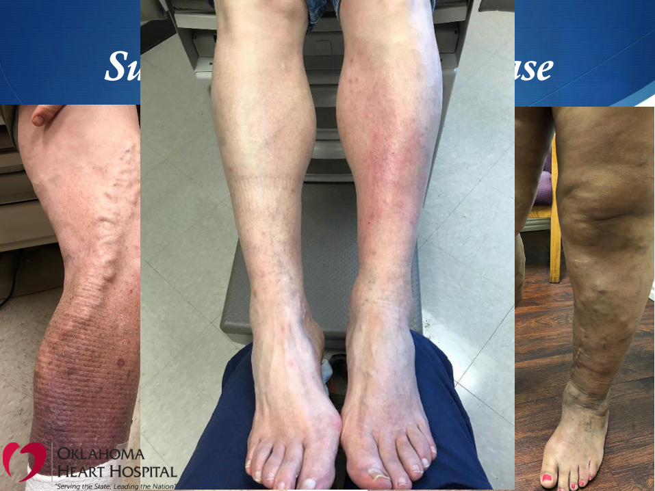
Superficial Venous Disease
Superficial Venous Disease
S Pathophysiology
S Inadequate muscle pump function
S Incompetent venous valves (reflux)
S Venous thrombosis or obstruction leading to venous hypertension
S Epidemiology
S Telangiectasias and reticular veins: most prevalent, 50-66% population, women 56-71%, men 36-44%.
S Varicose veins: > 3 mm, 10-30%, higher with age, W~M
S Chronic venous insufficiency: edema, skin changes, ulceration. 6-7 million in US affected at a given time, ulcers 1-5%.
Superficial Venous Disease
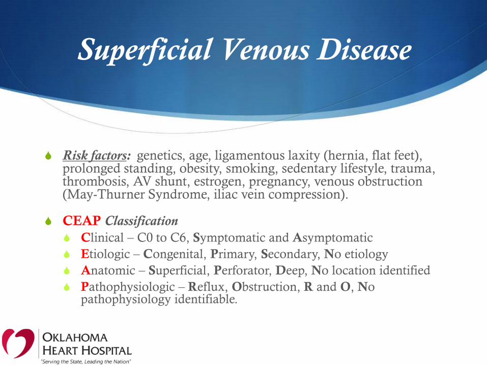
S Risk factors: genetics, age, ligamentous laxity (hernia, flat feet), prolonged standing, obesity, smoking, sedentary lifestyle, trauma, thrombosis, AV shunt, estrogen, pregnancy, venous obstruction (May-Thurner Syndrome, iliac vein compression).
S CEAP Classification
S Clinical – C0 to C6, Symptomatic and Asymptomatic
S Etiologic – Congenital, Primary, Secondary, No etiology
S Anatomic – Superficial, Perforator, Deep, No location identified
S Pathophysiologic – Reflux, Obstruction, R and O, No pathophysiology identifiable.

CEAP Classification

C4b,S, Ep, As,p Pr
Painful varicosities,
lipodermatosclerosis
reflux in superficial
and perforators by
duplex.

Superficial Venous Disease
S Diagnosis: Typical symptoms, venous reflux > 500 ms for superficial and > 1000 ms for deep veins.
S Treatments:
S Initial conservative measures
S Endovenous ablations
S Phlebectomy
S Valvular reconstruction
S Contraindications: Pregnancy, acute venous thrombosis, severe PAD (ABI < 0.5), Klippel-Trenaunay Syndrome, advanced systemic disease with poor prognosis.

Ambulatory Phlebectomy

Deep Venous Disease
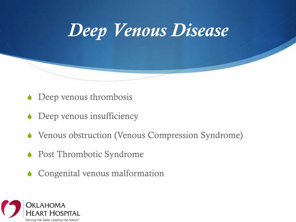
Deep Venous Disease
S Deep venous thrombosis
S Deep venous insufficiency
S Venous obstruction (Venous Compression Syndrome)
S Post Thrombotic Syndrome
S Congenital venous malformation

Deep Venous Thrombosis
S DVT and PE: Comprise Venous ThromboEmbolism (VTE)
S Symptoms sensitivity and specificity
S Swelling: 97 and 33 percent
S Pain: 86 and 19 percent
S Warmth: 72 and 48 percent
S Risk factors: Immobility, trauma, surgery, obesity, previous VTE, malignancy, OC, pregnancy, age > 65, FH, inflam. bowel disease.
Left vs. Right vs. Bilateral DVT: May have different causes.
Deep Venous Thrombosis
S Physical examination: can be benign, edema, erythema, calf pain with dorsiflexion (Homan’s Sign), bulging veins.
S Larger calf diameter – most usual finding
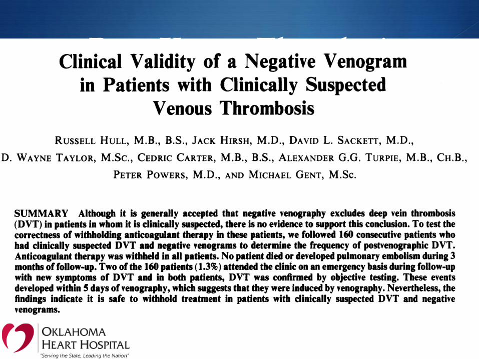
S Testing: Venous duplex, D-Dimer (high negative predictive value)
S Differential diagnosis: 160 patients with suspected DVT but negative venograms
S Muscle strain/tear 40% Baker’s cyst 5%
S Paralyzed limb swelling 9% Cellulitis 3%
S Lymph obstruction 7% Knee abnormality 2%
S Venous insufficiency 7% Unknown 26%

Deep Venous Thrombosis
S Special populations:
S Phlegmasia cerulea dolens
S Upper extremity DVT
S IVC and IVC filter thrombosis
S Pregnancy

A word on MTS
S May Thurner Syndrome – Iliac vein compression syndrome
S Usually: young female (20-40 yo) with left leg swelling,
pain, redness, discomfort, and or DVT.
S Due to compression of left common iliac vein between the
L5 vertebral body and the right common iliac artery.
S Treatment – depends on the patient’s clinical scenario.

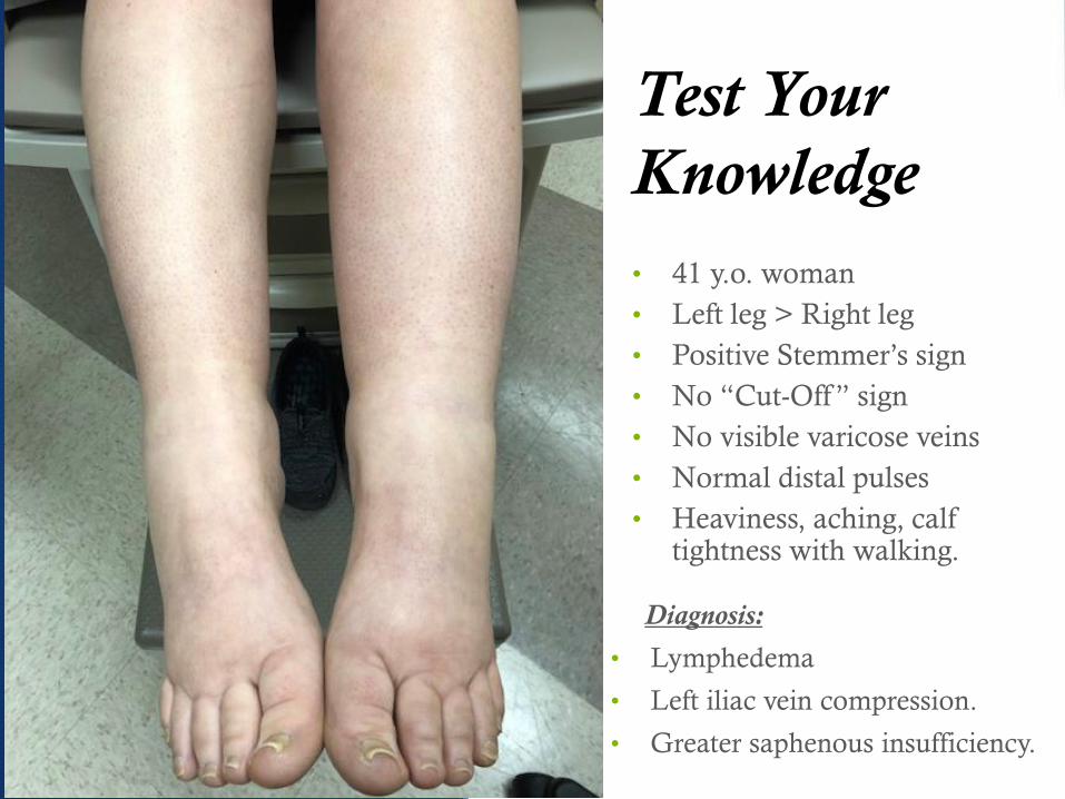
Test Your
Knowledge
• 41 y.o. woman
• Left leg > Right leg
• Positive Stemmer’s sign
• No “Cut-Off ” sign
• No visible varicose veins
• Normal distal pulses
• Heaviness, aching, calf tightness with walking.
Diagnosis:
• Lymphedema
• Left iliac vein compression.
• Greater saphenous insufficiency.

Venous vs. Arterial vs. Lymphatic
Final Thoughts
S Venous vs. Arterial: Nothing alike except for vessel names.
S Arterial disease is upstream.
S Venous disease is downstream.
S Diagnosis and treatments are very different.
S Venous research and literature are not as abundant as arterial.
S There is a need for more venous research and specialists.
References
S UpToDate 2019
S Creager MA, et al. Acute limb ischemia. N Engl J Med. 2012 June; 366(23):2198-206.
S Fatdisorders.org
S Lipedema.org
S Suter LG, et al. The incidence and natural history of Raynaud’s phenomenon in the community. Arthritis Rheum: 2005; 5(4):1259.
S Wassef M, et al. Vascular anomalies classification. Pediatrics. July 2015; 136 (1), e203-14.
S Glovickzki P, et al. The care of patients with varicose veins and associated chronic venous diseases. J. Vasc Surg 2011:53:2S.
S Wittens C, et al. Management of chronic venous disease. Eur J Vasc Endovasc Surg 2015;49:678.
S Sandler et al., Diagnosis of deep venous thrombosis. Lancet, 1984; 2:716.
S May R, Thurner J. The cause of the predominantly sinistral occurrence of thronbosis of the pelvic veins. Angiology 1957;8:419.
S Kearon C, et al. Categorization of patients as having provoked or unprovoked venous thromboembolism: guidance from the SSC of ISTH. J Thromb Haemost 2016;14:1480.
Thank You!


![[T] Treatment of upper limb lymphedema with low …...lymphatic drainage, compression and elastic bandages Fisioter Mov. 2014 out/dez;27(4):663-74 Treatment of upper limb lymphedema](https://static.fdocuments.us/doc/165x107/5f9ae2b4ec3eff620160143f/t-treatment-of-upper-limb-lymphedema-with-low-lymphatic-drainage-compression.jpg)


![Lymphatic System. Functions of the Lymphatic System Fluid Balance - returns interstitial fluid to the blood Fat [and fat soluble vitamins] Absorption.](https://static.fdocuments.us/doc/165x107/56649d6b5503460f94a4a17d/lymphatic-system-functions-of-the-lymphatic-system-fluid-balance-returns.jpg)