IS THIS A CN III PALSY / ANEURYSM? - Pennsylvania...
29
1/21/2017 1 IS THIS A CN III PALSY / ANEURYSM? Presented by Kelly A. Malloy, OD (no financial interests to disclose) March 5, 2017 Nothing to Disclose What are the features of a CN III Palsy? • Symptoms • Signs Features of CN III Palsy • Diplopia – Vertical (SR, IR – reversing hyper deviation) – Horizontal (MR – adduction deficit) • Ptosis – Levator (partial or complete) • Pupil Involvement (more concerning for aneurysm) – Anisocoria - greater in bright illumination (parasympathetic) – Possibly fixed, dilated pupil • Pain – Possibly (more concerning for aneurysm) PUPIL IN CN III Palsy • INVOLVED = ANEURYSM (86%) • SPARED = VASCULOPATHIC (77%) • DOES NOT APPLY IF – COMPLICATED CNIII – INCOMPLETE CNIII – RELATIVE SPARING – 20-50 YEARS OF AGE PAIN in CN III Palsy ANEURYSM = 95%+ DIABETES = 80% PITUITARY APOPLEXY GIANT CELL CAVERNOUS SINUS VASCULOPATHIC ANEURYSM EMERGENCY : Sub-Arachnoid Heme 20% die in 48 hrs!
Transcript of IS THIS A CN III PALSY / ANEURYSM? - Pennsylvania...

1/21/2017
1
IS THIS A CN III PALSY / ANEURYSM? Presented by Kelly A. Malloy, OD (no financial interests to disclose) March 5, 2017
Nothing to Disclose
What are the features of a CN III Palsy?
• Symptoms
• Signs
Features of CN III Palsy • Diplopia
– Vertical (SR, IR – reversing hyper deviation) – Horizontal (MR – adduction deficit)
• Ptosis – Levator (partial or complete)
• Pupil Involvement (more concerning for aneurysm) – Anisocoria - greater in bright illumination
(parasympathetic) – Possibly fixed, dilated pupil
• Pain – Possibly (more concerning for aneurysm)
PUPIL IN CN III Palsy • INVOLVED = ANEURYSM (86%) • SPARED = VASCULOPATHIC (77%)
• DOES NOT APPLY IF – COMPLICATED CNIII – INCOMPLETE CNIII – RELATIVE SPARING – 20-50 YEARS OF AGE
PAIN in CN III Palsy ANEURYSM = 95%+ DIABETES = 80% PITUITARY APOPLEXY GIANT CELL CAVERNOUS SINUS
VASCULOPATHIC
ANEURYSM
EMERGENCY: Sub-Arachnoid Heme 20% die in 48 hrs!

1/21/2017
2
PSEUDO – GRAEFE SIGN
EYELID SYNKINESIA
ABERRANT REGENERATION OF CN III Aneurysm, Tumor, Trauma NEVER Vasculopathic !
LIGHT-NEAR DISSOCIATION PUPILS CN III PALSY WORK-UP
• 20-50 YEARS – CT, MRI, MRA,
A-GRAM
• 50+ YEARS (pupil, palsy, pain) – NEUROIMAGING – VASCULOPATHIC
EVALUATION – R/O GCA
ADULTS CONGENITAL (MRI)
ACQUIRED •EXCLUDE TRAUMA OR MIGRAINE
•CONSIDER LP IF MRI (-)
•IF MRI & LP ARE NEGATIVE > 10 years, ARTERIOGRAM TO LOOK FOR ANEURYSM
CHILDREN
Importance of Accurate Diagnosis • CN III could be a sign of aneurysm, and therefore a
medical emergency • Don’t want to miss an aneurysm – if it ruptures, pt
could die in 24-48 hours • However, want to be sure we are really dealing with a
CNIII palsy, and not something that needs to be treated differently (and can also be an urgent or emergent situation)
• Don’t want to put patient through unnecessary expensive and /or invasive testing
CASE 1
48 year old woman
Headache and pain OD x 2 weeks
Went to ER
- told symptoms were from the flu
- sent home
Symptoms persisted
Systemic history: + HTN, + DM

1/21/2017
3
What is the cause of complete ptosis ?
• CN III Palsy • Myasthenia Gravis • Others?
• How do we differentiate among these?
What is the cause of complete ptosis ? • CN III Palsy
– Associated motility abnormalities – Can have associated pain – Can have associated pupil abnormalities
• Myasthenia Gravis
– Associated motility abnormalities – No associated pain – No associated pupil abnormalities
• Others?
What do we expect to see when we lift the right eyelid?
Reversing hyper deviation Exo worse away from vertically limited eye

1/21/2017
4
?’s
• Is it partial or complete? • Pupil involved or not? • Is this a vasculopathic CN VI palsy? • Why or why not? • What is the management plan?
• DIAGNOSIS: – Pupil involved, painful, right CN III palsy – Aneurysm until proven otherwise
• WORK-UP: – To ER immediately
• MRI, MRA – Aneurysm confirmed
• TREATMENT: – coiling
CASE 2
55 year-old woman
• sudden onset diplopia and headache – symptoms began 4-5 days prior
• headache and pressure in the right side of head/face – 8/10 on pain scale
• Chills, body ache, and nausea -thought she had the flu • more tired than usual • Diplopia
– Horizontal, sometimes diagonal – At distance and near – Worse in right gaze
– used 800 mg Motrin every 8 hours
• Went to the ER 2 days ago because of headache
• had a non contrast CT – reportedly negative – Patient sent home
• SYSTEMIC HISTORY • Graves disease, diagnosed 7 years ago
– Initially hyperthyroid – had radioactive iodine ablation – treated on Synthroid since
• dosage recently adjusted downward • Prior to that, experiencing weight gain, joint pain,
fatigue x 3 mos • Was feeling better since change in med dosage

1/21/2017
5
• SYSTEMIC HISTORY: • Hypercholesterolemia, controlled by diet and exercise • sickle cell trait • osteoarthritis • MEDICATIONS: • Lexapro, levothyroxine, hydrocortisone and ibuprofen • SOCIAL HISTORY: • smoking 4-6 cigarettes per day x 10-15 years • FAMILY HISTORY:
– Diabetes – Hypertension – Stroke
• VA: 20/20- OD 20/20 OS • Color 14/14 OD 13/14 OS • (-) RAPD by reverse testing
• CF: normal OU
• Normal SLE and DFE OU • Normal neurologic examination
• Blood pressure was 150/87 right arm sitting
What is the cause of the diplopia and eyelid asymmetry?
• CN III Palsy
• Thyroid orbitopathy
• Myasthenia Gravis
• Others?
What is the cause of the diplopia and eyelid asymmetry?
• CN III Palsy – Eyelid asymmetry from ptosis – May or may not be associated pupil involvement – Reversing hyper deviation – Exo worse away from affected eye
• Aneurysm • Vasculopathic • Others
• Thyroid orbitopathy
– Eyelid asymmetry from proptosis – No pupil involvement – Any motility pattern
• Myasthenia Gravis – Eyelid asymmetry from ptosis – No pupil involvement – Any motility pattern
• Others?
Palpebral apertures of 10 mm OD and 12 mm OS Exophthalmometry 22 mm OD and 22 mm OS

1/21/2017
6
Ocular Motility
OD OS
60
90
60
100
100
100 100 25exo 6 exo 40ex
100
30exo
20 LH
20exo
10 RH
6.25 OD and 3.25 OS in bright illumination 6.5 OD and 4.0 OS in dim illumination
• DIAGNOSIS: – Pupil involved partial right cranial nerve III
palsy • No vasculopathic risk factors • severe headaches x 5 days
– highly concerning for aneurysm
• WORK-UP: – To ER immediately
• MRI of the brain and MRA / CTA of the Circle of Willis
• If negative, formal angiography
55 year-old woman follow-up • 2 weeks after initial presentation • Work-up results
– MRI, CTA, and cerebral angiogram all normal – Labs
• elevated TSH (44.75), low T3 (0.60), and slightly low Free T4 (0.93)
– Levothyroxine dosage was increased from 125 to 150 mg – scheduled to follow-up with endocrinologist
• hemoglobin A1C was elevated at 6.1%. – Lumbar puncture - normal
• Diplopia and headaches improved • Proposed viral cause of CN III palsy

1/21/2017
7
pupil sizes : 3.5 OD and 3.0 OS in bright illumination 4.25 OD and 3.75 OS in dim illumination palpebral apertures: 10 mm OD and 11 mm OS.
Ocular Motility
OD OS
95
90
95
100
100
100 100 ortho 1exo 16ex
100
6 LH
4 RH
• Resolving presumed viral CN III Palsy
• Never could make this diagnosis without first r/o aneurysm
• RX’d 4 Fresnel prism diopters BU over the left eye bifocal to help alleviate symptoms of diplopia when reading and looking down
CASE 3
68 year-old man
-Diplopia x 4 days
-Undergoing chemo (Rituxan) for non-Hodgkin’s Lymphoma
-D/C chemo due to diplopia
-Had MRI of brain without contrast (no etiology for diplopia)
-Constant frontal headache x 7 months
-Pain worse x 4 days – over and behind right eye
-Has been closing right eye to avoid diplopia

1/21/2017
8
• + DM x 10 yrs, HTN since age 18 • + hypercholesterolemia • S/p MI x 2 • S/p CABG X 4 • + Atrial Fibrillation (on Coumadin) • S/p Parathyroidectomy • MEDS: Glucotrol, Coreg, Digoxin,
Pravachol, Zoloft, Synthroid, Coumadin, amitriptyline, Protonix, Colchicine
• Partial pupil spared R CN III palsy
• Pt already had MRI • Pt has vasculopathic risk factors • So …..do we need to do any more work-
up in this case? Why or why not?
• Partial – Can’t use pupil as guide
• Pain • Hx of cancer • MRI was without contrast • MRA / CTA was not done
• WORK-UP
MRI brain with and without contrast • MRA of head
• Both normal
1 month later
CASE 4

1/21/2017
9
56 Year old man
• Pain in and behind OD x 3 days • Pain radiating to back of head • Pain up to a 7/10 on pain scale • He used friend’s unknown eye drop and ung OD • 2 days ago –felt dizzy and nauseous • Then diplopia began • Went to ER – put “drop” in eye, and prescribed
ibuprofen for pain
• Hx of elevated BP (not compliant with meds) • He snores and stops breathing while
sleeping, but was never tested for sleep apnea
• Smokes ½ pack of cigarettes per day since age 16
• Hx of cellulitis of right foot a few years ago, for which he was on IV antibiotics
• VA: 20/20 OD 20/20 OS • Color 14/14 OD 14/14 OS • Right pupil non-reactive • CF: full OU • IOP: 20 mm Hg OD, 17 mm Hg OS • FE: Healthy optic discs OU • (-) edema OU , (-) pallor OU • BP: 147/91 • Neurologic examination
– Unremarkable
Ocular Motility
OD OS
95
90
90
100
100
95 100 8 RH
4 exo
16 eso
8 RH
16exo
4 RH
70
12 LH
8 exo
4 RH
No torsion

1/21/2017
10
What is the cause of an apparent CN III and CN VI palsy?
• CN III Palsy and CN VI palsy – Cavernous sinus – Orbital apex syndrome
• Thyroid orbitopathy
• Myasthenia Gravis
• Idiopathic Orbital inflammatory Pseudotumor
What is the cause of the diplopia and eyelid asymmetry?
• CN III Palsy and CN VI palsy – Eyelid asymmetry from ptosis – May or may not be associated pupil involvement – Reversing hyper deviation – More Exo worse away from affected eye – More Eso toward affected eye
• Cavernous Sinus • Orbital apex
• Thyroid orbitopathy
– Eyelid asymmetry from proptosis – No pupil involvement – Any motility pattern
• Myasthenia Gravis – Eyelid asymmetry from ptosis – No pupil involvement – Any motility pattern
• Eyelid Measurements – Palpebral apertures OD 10 mm, OS 9 mm – Lid crease: symmetric – Levator function: symmetric
Exophthalmometry OD 23 mm, OS 18 mm

1/21/2017
11
What is the cause of the apparent CN III and VI palsy with proptosis?
• Orbital Apex / Cavernous Sinus Syndrome
– Unilateral – Can be painful
• Thyroid orbitopathy
– Unilateral or bilateral – Can be painful – Eyelid retraction
Work-Up • Right CN III and CN VI palsy
– Localizes to right orbital apex or cavernous sinus • Work-up Needed
– CT, MRI with and without contrast, MRA, MRV • DDX
– Tolosa-Hunt syndrome, Cavernous sinus thrombosis, cavernous sinus fistula, sarcoid, Wegener’s granulomatosis, aneurysm, infectious, other inflammatory
• Urgency – Emergent – send to ER – Call ahead and let ER know of the findings, localization,
and differential diagnosis
Results / Diagnosis
• MRI report – Inflammatory process in right cavernous sinus
• Other testing – No indication of vascular process, or any other
etiology
-Diagnosed with Tolosa-Hunt Syndrome, and started on IV steroids
Tolosa-Hunt Syndrome
• Unilateral • Intense pain around eye • Ophthalmoplegia • Possible involvement of CN III, IV, V,
and VI • Can have proptosis, fatigue, vertigo
Tolosa-Hunt Syndrome
• Exact cause is unknown • Inflammation of cavernous sinus and/or
superior orbital fissure • Diagnosis of exclusion • Need to rule out all other causes with labs,
imaging, and LP • Treat with steroids / good prognosis • Can recur in 30-40% of cases
CASE 5

1/21/2017
12
47 year old woman
• intermittent blurring of vision • headaches x 4 weeks, located behind eyes
– Mainly behind OS – worse with reading • 9 out of 10 on a pain scale • headaches can last up to two hours.
• Systemic history: – Asthma – s/p 10 abdominal surgeries – Percocet due to back pain
• head trauma to left nose / cheek (3 years ago) – industrial kitchen accident – collided with a cart and broke her nose – underwent trans-nasal surgery
• VA: 20/20- OD 20/20- OS • Color 14/14 OD 14/14 OS • CF: full OU • IOP: 14 mm Hg OU • DFE: Healthy optic discs, small cupping OU • (-) edema OU , (-) pallor OU • Normal neurologic examination
OD – no reaction to light, questionable minimal reaction to near
Ocular Motility
OD OS
100
100
100
100
100
95 100 10exo 6exo 4exo
100
10exo
12exo
1LH
What could be the cause of the dilated pupil?

1/21/2017
13
What could be the cause of the dilated pupil?
• CNIII / aneurysm • Pharmacologic dilation • Tonic pupil
How can we distinguish among these?
What could be the cause of the dilated pupil?
• PHARMACOLOGIC TESTING: – CNIII / aneurysm
• Do NOT use any drops if you think it IS a CN III palsy • Would constrict with 1% Pilocarpine • Would NOT constrict with 0.125% Pilocarpine
– Pharmacologic dilation • Would NOT constrict to either 1% or 0.125% Pilocarpine
– Tonic pupil • Would constrict to 0.125% Pilocarpine • Supersensitivity reaction
After instillation of 0.125% Pilocarpine
FEATURES OF TONIC PUPIL
MID-DILATED LIGHT NEAR DISASSOCIATION
“3 S’s” Sector paralysis Stromal spread
Stromal steaming

1/21/2017
14
Clinical Features
• “Flat” edges • “Vermiform” iris movement • Poor response to light & near or LND • “Dilation lag” following prolonged near effort • “Paradoxical Pupil” - aniso greater in light & dim
– IF PUPIL IS MID-DILATED
Pathogenesis of Tonic Pupil
• DAMAGE TO CILIARY GANGLION • Ciliary ganglion
– 90% CB – 3% iris
• Aberrant regeneration of CB fibers to iris sphincter (light-near/gaze pupil dissociation)
Adie WJ. Brain 1932
LOCAL TONIC PUPIL • VARICELLA • RETROBULBAR • ORBITAL TUMOR • ORBITAL SURGERY • ORBITAL TRAUMA
NEUROPATHIC TONIC PUPIL
•DIABETES SYPHILIS
•SARCOID LYME
IDIOPATHIC TONIC PUPIL •“ADIE’S”
•UNKNOWN ETIOLOGY
Management
• Lab testing – Negative for systemic causes of tonic pupil
• Updated refraction – Unequal adds due to unequal accommodation – Should relieve headaches and eyestrain
1/21/2017
15
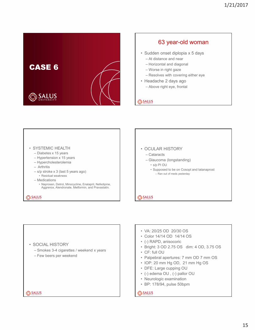
CASE 6
63 year-old woman
• Sudden onset diplopia x 5 days – At distance and near – Horizontal and diagonal – Worse in right gaze – Resolves with covering either eye
• Headache 2 days ago – Above right eye, frontal
• SYSTEMIC HEALTH – Diabetes x 15 years – Hypertension x 15 years – Hypercholesterolemia – Arthritis – s/p stroke x 3 (last 5 years ago)
• Residual weakness – Medications
• Naprosen, Detrol, Minocycline, Enalapril, Nefedipine, Aggrenox, Alendronate, Metformin, and Pravastatin.
• OCULAR HISTORY – Cataracts – Glaucoma (longstanding)
• s/p PI OU • Supposed to be on Cosopt and latanaprost
– Ran out of meds yesterday
• SOCIAL HISTORY – Smokes 3-4 cigarettes / weekend x years – Few beers per weekend
• VA: 20/25 OD 20/30 OS • Color 14/14 OD 14/14 OS • (-) RAPD, anisocoric • Bright: 3 OD 2.75 OS dim: 4 OD, 3.75 OS • CF: full OU • Palpebral apertures: 7 mm OD 7 mm OS • IOP: 20 mm Hg OD, 21 mm Hg OS • DFE: Large cupping OU • (-) edema OU , (-) pallor OU • Neurologic examination • BP: 178/94, pulse 50bpm

1/21/2017
16
Ocular Motility
OD OS
90
100
85
100
100
100 100 12 RH
10 eso
20 RH
10 eso
6 RH
10 exo
100
4 RH
8 eso
6 RH
6 eso
Head tilt testing demonstrated 16 right hyper on right head tilt, and 12 right hyper and 14 eso on left head tilt
What is the cause of the adduction deficit and infraduction deficit?
What is the cause of the adduction deficit and infraduction deficit?
• CN III Palsy • Thyroid Orbitopathy • Myasthenia Gravis • INO and Skew Deviation
What is the cause of the adduction deficit and infraduction deficit?
• CN III Palsy – Negative forced duction test
• Thyroid Orbitopathy
– Positive forced duction test • Myasthenia Gravis
– Negative forced duction test – Fatigue
• INO and Skew Deviation
– Abducting nystagmus – Higher eye intorted, Lower eye extorted – Negative forced duction test
Double Maddox rod testing : - 15-20 degrees of incyclotorsion OD - 15-20 degrees of excyclotorsion OS

1/21/2017
17
Management
• INO / Skew – Likely secondary to stroke
• In setting of elevated BP and low pulse
• Admit to hospital for work-up – MRI
• Acute brainstem lesion noted
CASE 7
42 year old woman • Noticed right eye has been pointing upward x 1 month
– Getting progressively worse • Denies diplopia • History of poor vision OD due to macular scar
• Recently saw PCP due to not feeling well • Right arm weakness • Slurring words • Difficulty swallowing • Some breathing difficulties
• PCP ordered an MRI to r/o MS – Reported to be normal
• Pt also went to local ER – another MRI – Normal - no etiology determined
– Pt scheduled for neurology consult in 2
months

1/21/2017
18
What is the cause of the eyelid asymmetry and the diplopia?
• Neurologic Exam – Weakness of neck flexor muscles – Weakness of right upper extremity
Myasthenia Gravis • Acetylcholine receptor antibodies • Binding **** 1500 (normal < 0.30) • Blocking *** positive 75 (normal <15) • Modulating *** positive • All thyroid tests negative • Chest CT
• Admitted to hospital for evaluation / treatment for
fear of myasthenic crisis • Plasmapheresis (ICU), IVIG, Mestinon, Prednisone

1/21/2017
19
Other ways to Assess for MG
• Fatigue • Ice Pack Testing • Orbicularis oculi testing
CASE 8
47 year old man • Sharp pain OS x 2-3 weeks • Constant, with varying intensity • Pain around OS, left face, left temple region • Increased pain when blows nose or lies on left side • Left eyelid seems droopy x 2 weeks
• OCULAR HISTORY • Dx with glaucoma elsewhere 2-3 years ago
– Not compliant with meds • Dx with glaucoma at TEI late 2011
– Put on Travatan Z, later Trusopt added – Switched to Travatan and Combigan – More recently switched from Combigan to Cosopt due
to cost – Pt confused about drops
• Using Travatan BID OU, d/c generic Cosopt due to symptoms
• Using Visene Maximum redness relief BID x 2-3 weeks
• SYSTEMIC HISTORY – Unremarkable
• Multiple episodes of head / eye trauma in past
• hit with a pipe in forehead • hit in the left eye with a fist • hit in the back of the head with a black jack
• No recent trauma
• VA: 20/20- OD 20/20- OS • Color 14/14 OD 14/14 OS • (-) RAPD • CF: full OU HVF: no definite pattern • IOP: 8 mm Hg OU • DFE: Large cupping OU • (-) edema OU , (-) pallor OU • Neurologic examination

1/21/2017
20
Ocular Motility
OD OS
100
100
100
100
100
100 100 ortho 1 LH
2 eso
4 eso 95
ortho
1 eso
What is the cause of ptosis and pain?
What is the cause of ptosis and pain?
• CN III Palsy • Horner syndrome (Carotid dissection) • Others
What is the cause of ptosis and pain?
• CN III Palsy – Can be any degree of ptosis – If anisocoria, will be greater in bright
• Horner syndrome (Carotid dissection)
– Only few mm of ptosis – If anisocoria will be greater in dim
• Others
Palpebral apertures: 13 mm OD and 11 mm OS
Exophthalmometry : 19 mm OD and 17.5 mm OS

1/21/2017
21
• Anisocoria apparently greatest in dim illumination
– Sympathetic system issue
• Horner Syndrome
A PAINFUL Horner
Syndrome is a Carotid
Dissection Until Proven Otherwise !
Diagnostic Test For Horner Syndrome
• 0.5% or 1.0% Apraclonidine (Iopidine) – Alpha agonist – Weak alpha 1 agonist
• No effect on normal pupil • Dilates Horner pupil (supersensitivity)
• Look for REVERSAL OF ANISOCORIA • May NOT be positive in acute Horner Syndrome
– carotid dissection
Normal Eye • Norepinephrine production controlled by
alpha 2 receptors, which work by down-regulation of alpha 1 receptors responsible for dilation
Horner Eye • Norepinephrine amount is reduced, so alpha 2 receptors are not activated, resulting in up-regulation of alpha 1 receptors, leading to denervation sensitivity
Post- Apraclonidine z

1/21/2017
22
1.Telendiencephalic 2. Nuclear CNIV
5.Wallenberg’s
6. Brachial Plexus
7.Pancoast’s
8. Phrenic Nerve Syn
9. Carotid Dissection
11. Palatine tonsil
12.Caroticotympanic
13. Cavernous Sinus
10.Vernet’s
C6-8
4. RAPD
3. INTERNUC OPH
Carotid Artery Dissection
• Can occur with or without trauma
• Medical Emergency • Horner’s with eye, head, neck pain
• Could cause stroke, CRAO, etc. – Work-up: (MRI, MRA, CTA, angiogram)
FOLLOW-UP
• MRI and MRA head and neck with contrast – Normal (No dissection)
• Using Travatan Z qhs OU – Pain returns and mild ptosis OS – eyes red OS > OD – IOP 13OD and 15 OS
FOLLOW-UP
• DIAGNOSIS: • Prostaglandin likely causing redness and pain • Plan
• D/C Travatan (Prostaglandin) • RX Combigan bid OU • RX artificial tears
• Horner’s may be longstanding from past trauma
– multiple traumatic episodes
CASE 9

1/21/2017
23
93 year old woman • Sudden onset vertical diplopia
– X 1 week – At distance and near – Relief with closing either eye
• Associated blur • Left upper eyelid droopy
– x 1 week
• Eye pain OU x 1 week
• Generalized weakness x 1 week
• Systemic history – Hypertension x 30 years – Hypercholesterolemia – Arthritis – Ovarian cancer 30 years ago
• VA: 20/25- OD 20/20- OS • Color 13/14 OD 13/14 OS • (-) RAPD • CF: full OU • IOP: 10 mm Hg OU • DFE: Healthy optic discs, small cupping OU • (-) edema OU , (-) pallor OU • Neurologic examination
– Mild left upper extremity weakness
Palpebral apertures: 11 mm OD and 7 mm OS
Exophthalmometry: 17 mm OD and 18 mm OS

1/21/2017
24
Ocular Motility
OD OS
100
100
100
60-70
100
100 100 18 RH
2 eso
25RH 18RH 100
30 RH
ortho
Head tilt: 20 right hyper on right head tilt, and 18 right hyper on left head tilt
What is the cause of the supraduction deficit?
What is the cause of the vertical misalignment / supraduction deficit? • CN III Palsy – superior division • Myasthenia Gravis • Thyroid Orbitopathy • Skew Deviation • CN IV Palsy
What is the cause of the vertical misalignment, supraduction deficit, ptosis?
• CN III Palsy – partial ? superior division? – Forced Duction: negative (no restriction)
• Myasthenia Gravis
– Forced Duction: negative (no restriction)
• Orbital Mass – Forced Duction: positive (restriction)
• Thyroid Orbitopathy
– Forced Duction: positive (restriction) • Skew Deviation
– Torsion: higher eye intorted • CN IV Palsy
– Torsion: higher eye extorted
Work-Up • Could be a partial CN III palsy
– Get MRI / MRA to r/o aneurysm • Get labs to r/o other causes
– CBC with differential, platelet count, ESR (Westergren), C-reactive protein
– Lyme titer – RPR, FTA-ABS – ACE – Acetylcholine receptor antibody testing (binding,
blocking and modulating), – TSH, t4 and anti-thyroid antibodies (anti-
thyroperoxidase and anti-thyroglobulin)

1/21/2017
25
Results • MRI report
– mild bilateral proptosis – prominent orbital fat – mild fatty atrophy of inferior, lateral, and medial rectus
muscles OU
• Lab testing – elevated TSH at 9.55 – T3 at 37 – thyroglobulin antibodies at 1790 – thyroid peroxidase antibodies at > 1000
Confirms thyroid dysfunction and likely thyroid eye disease / Grave’s disease.
Diagnosis / Treatment
• DX: autoimmune thyroid disease with subclinical hypothyroidism and thyroid orbitopathy
• Endocrine consult • started on levothyroxine 25mcg daily
6 week follow-up Pt reports no diplopia x 2 weeks
CASE 10
63 year old woman • 3-week history of left eye and head pain • The pain keeps her up at night • Then, the left eyelid began to droop • 2 weeks ago, she noticed double vision • Decreased appetite • Weakness and fatigue
• SYSTEMIC HISTORY: • - Lung cancer 2 years ago • - surgery, radiation, chemo • - still undergoing treatment
• Otherwise unremarkable

1/21/2017
26
• VA OD 20/40 OS 20/40 • Normal color vision • (-)APD • CF: Full OU • IOP normal • BP normal • ONH: (-) pallor OU, (-)edema OU
• Emergent hospitalization to rule out aneurysm • MRI and MRA negative
• While in hospital CN III palsy progressively
worsened
• Cerebral angiogram negative
• Lumbar puncture • Diagnosed with meningeal carcinomatosis
LEPTOMENINGEAL CARCINOMATOSIS
• Also called neoplastic or carcinomatous meningitis • invasion to and subsequent proliferation of
neoplastic cells in the subarachnoid space • substantial rates of morbidity and mortality • MRI with contrast can detect leptomeningeal
enhancement • Diagnosis confirmed with CSF cytometry • Sites of origin
– Lung – Breast – Melanoma – Medulloblastoma
LEPTOMENINGEAL CARCINOMATOSIS
• Because the leptomeninges cover the cranial nerve roots, tumor seeding of the cranial nerves is not uncommon.
• Can cause symptoms either from encasement of the nerve or by direct invasion with subsequent axonal destruction and demyelination.
– Can have cranial nerve palsies • Symptoms include headache (50%), nausea,
vomiting, seizures

1/21/2017
27
CASE 11
66 year-old Asian man • referred for a fixed dilated pupil OS • Until recently, no eye exam in > 10 years • c/o pain, redness and reduced vision OS • ? Yellow discharge • Referring doctor rx’d Vigamox / Maxitrol
– over the course of two weeks – Pain a bit better – Vision OS is still intermittently blurry
• Headaches x 1 year – Frontal and in the sinus area. – His PCP ordered a CT without contrast of the
head • unremarkable.
• SYSYTEMIC HEALTH – abdominal surgery done in Hong Kong
• Medications – Amoxicillin and methylpred
• Prescribed by his PCP 1 day prior because of a presumed sinusitis
• CT report does not mention any sinus involvement.
• Ocular history is otherwise unremarkable • Social history is remarkable for smoking x 50 years.
• VA: 20/30- OD 20/60- OS • Color 14/14 OD 14/14 OS • (-) RAPD • Normal neurologic examination
• Blood pressure was 128/78 right arm sitting.
• Pupil size: bright: OD 3 OS 6 dim: OD 4 OS 6 – (-) Light-near dissociation
What is the cause of the painful dilated pupil?

1/21/2017
28
What is the cause of the fixed dilated pupil?
• CN III Palsy
• Pharmacologic dilation
• Tonic Pupil
• Others?
What is the cause of the fixed dilated pupil?
• CN III Palsy – Look for ptosis, reversing hyper deviation – Would NOT constrict with 0.125% Pilocarpine – Would constrict with 1% Pilocarpine
• Pharmacologic dilation – Would NOT constrict with 0.125% Pilocarpine – Would NOT constrict with 1% Pilocarpine
• Tonic Pupil – Would constrict with 0.125% Pilocarpine
• Others?
• Palpebral apertures – 8 mm OD and 8 mm OS
• Ocular motility testing
– normal ductions, versions, and saccades
• Cover testing at distance – 1 eso deviation – comitant in all positions of gaze
• Cover testing at near – 7 exo deviation – comitant
• Slit lamp examination – OD:
• superior temporal corneal scar • moderate nuclear lenticular changes • Narrow Angles
– OS: • mild corneal edema • endothelial pigment • inferior nasal posterior synechia of the iris • pigment on the lens • Moderate nuclear lens changes • Very Narrow Angles
• Gonioscopy – OS:
• chronic angle closure / peripheral anterior synechiae of nasal angle
• remaining angles showed only a small portion of anterior TM

1/21/2017
29
• Applanation tensions – initially 26 mmHg OD and 60 mmHg OS
• Treatment: – 250 mg of Diamox PO – Iopidine OU, Azopt OS and Timolol OS – The synechia was broken with 1% tropicamide and 2.5%
phenylephirine – After 2.5 hours the intraocular pressures were ultimately
lowered to 12 mmHg OD and 20 mmHg OS – Referred for peripheral iridotomy OU
• Visual fields – possible early superior and inferior arcuate defects
OD – superior arcuate defect and inferior nasal step OS
• Fundus examination – (-) edema OU – 0.5 x 0.6 cupping OD with thinning superiorly – 0.55 x 0.70 cupping OS with thinning inferiorly – Neuro retinal rim intact and pink OU
CONCLUSIONS • Always consider a CN III palsy in your differential with any of
the following features – Ptosis – Vertical misalignment – Reversing hyper deviation – Fixed dilated pupil – Anisocoria greater in bright illumination
– Know how to use your exam findings to rule in or rule out a CN
III palsy – CN III palsy may signify a medical emergency, and should be
managed as such – Other emergent conditions may mimic a CN III palsy, and need
to be managed differently; an accurate diagnosis is essential
ANY QUESTIONS?
THANK YOU.