Is there a role of interstitial cells of Cajal and mast...
4
800 http://journals.tubitak.gov.tr/medical/ Turkish Journal of Medical Sciences Turk J Med Sci (2015) 45: 800-803 © TÜBİTAK doi:10.3906/sag-1408-21 Is there a role of interstitial cells of Cajal and mast cells and eosinophils in appendicitis in children? Esra KARAKUŞ 1, *, Müjdem Nur AZILI 2 , Bilge KARABULUT 2 , Gülşah BAYRAM KABAÇAM 3 , Resul KARAKUŞ 4 1 Department of Pathology, Ankara Children’s Hematology and Oncology, Research and Training Hospital, Ankara, Turkey 2 Department of Pediatric Surgery, Ankara Children’s Hematology and Oncology, Research and Training Hospital, Ankara, Turkey 3 Department of Radiology, Ankara Children’s Hematology and Oncology, Research and Training Hospital, Ankara, Turkey 4 Department of Immunology Faculty of Medicine, Gazi University, Ankara, Turkey * Correspondence: [email protected] 1. Introduction e etiology and pathogenesis of appendicitis are multifactorial. e cause of appendicitis is considered to be obstruction of the appendiceal lumen and subsequent onset of ischemia, mucosal disintegration, and bacterial invasion. Obstruction and inflammation are implicated in the pathogenesis of appendicitis (1). Interstitial cells of Cajal (ICC) are important for intestinal motility and may also play a role in dysmotility (2,3). e number of eosinophils and mast cells (MCs) may change during acute and chronic inflammatory conditions. In appendicitis, levels of eosinophil and MCs are not clearly defined (4). ICC, MCs, and eosinophils may play an important role in appendicitis pathogenesis. e aim of the present study was to compare the distribution of ICC, eosinophils, and MCs in normal and inflamed appendices, and to evaluate the correlation of presence of these cells with inflammation severity in appendicitis. 2. Materials and methods is study was conducted on 40 samples obtained from appendices. e specimens were classified into two groups (Table 1). e appendicitis group (n = 30) was divided into three groups, namely simple, suppurative, and gangrenous perforated, according to the macroscopic description and histologic findings. Ten normal appendices served as controls. Tissue samples were processed for routine histological examination with standard formalin fixation and paraffin embedding, and 5 µm thin sections were stained with hematoxylin–eosin. In addition, all sections were immunohistochemically stained as follows: 3 µm thin sections were cut, dried, and deparaffinized before placing them on Ventana Benchmark GX immunostainer (Ventana, Tucson, AZ, USA). Diaminobenzidine was used as chromogen. e antibody panel included CD117 (T595, ready to use, Leica, Newcastle, United Kingdom) and mast cell Background/aim: e aim of this study was to compare the distribution of interstitial Cajal cells, eosinophils, and mast cells in normal and inflamed appendices, and to evaluate the correlation of presence of these cells with severity of inflammation in appendicitis. Materials and methods: e appendicitis group (n = 30) was divided further into three groups according to the macroscopic description and the histological findings. Ten normal appendices served as controls. Tissue samples were processed for routine histological examination. Additionally, all sections were immunohistochemically stained with CD117 and mast cell tryptase antibodies. Results: When specimens were compared in terms of Cajal cells, the observed mean number for the appendicitis group was 4.9 and for the control group it was 8.3. In contrast, eosinophils and mast cells were significantly increased in the appendicitis group when compared with the control group. Conclusion: We detected that eosinophils and mast cells are increased in appendicitis, and correlate with the degree of inflammation of the appendix. e density of interstitial Cajal cells was significantly lower in patients with severe appendix inflammation compared to controls. e histopathological differences observed in this study may help elucidate the pathophysiology of appendicitis. Key words: Appendicitis, mast cell, eosinophil, interstitial cells of Cajal Received: 07.08.2014 Accepted/Published Online: 03.12.2014 Printed: 30.07.2015 Research Article
Transcript of Is there a role of interstitial cells of Cajal and mast...

800
http://journals.tubitak.gov.tr/medical/
Turkish Journal of Medical Sciences Turk J Med Sci(2015) 45: 800-803© TÜBİTAKdoi:10.3906/sag-1408-21
Is there a role of interstitial cells of Cajal and mast cells and eosinophilsin appendicitis in children?
Esra KARAKUŞ1,*, Müjdem Nur AZILI2, Bilge KARABULUT2, Gülşah BAYRAM KABAÇAM3, Resul KARAKUŞ4
1Department of Pathology, Ankara Children’s Hematology and Oncology, Research and Training Hospital, Ankara, Turkey2Department of Pediatric Surgery, Ankara Children’s Hematology and Oncology, Research and Training Hospital, Ankara, Turkey
3Department of Radiology, Ankara Children’s Hematology and Oncology, Research and Training Hospital, Ankara, Turkey4Department of Immunology Faculty of Medicine, Gazi University, Ankara, Turkey
* Correspondence: [email protected]
1. IntroductionThe etiology and pathogenesis of appendicitis are multifactorial. The cause of appendicitis is considered to be obstruction of the appendiceal lumen and subsequent onset of ischemia, mucosal disintegration, and bacterial invasion. Obstruction and inflammation are implicated in the pathogenesis of appendicitis (1).
Interstitial cells of Cajal (ICC) are important for intestinal motility and may also play a role in dysmotility (2,3). The number of eosinophils and mast cells (MCs) may change during acute and chronic inflammatory conditions. In appendicitis, levels of eosinophil and MCs are not clearly defined (4). ICC, MCs, and eosinophils may play an important role in appendicitis pathogenesis. The aim of the present study was to compare the distribution of ICC, eosinophils, and MCs in normal and inflamed appendices, and to evaluate the correlation of presence of these cells with inflammation severity in appendicitis.
2. Materials and methodsThis study was conducted on 40 samples obtained from appendices. The specimens were classified into two groups (Table 1). The appendicitis group (n = 30) was divided into three groups, namely simple, suppurative, and gangrenous perforated, according to the macroscopic description and histologic findings. Ten normal appendices served as controls.
Tissue samples were processed for routine histological examination with standard formalin fixation and paraffin embedding, and 5 µm thin sections were stained with hematoxylin–eosin. In addition, all sections were immunohistochemically stained as follows: 3 µm thin sections were cut, dried, and deparaffinized before placing them on Ventana Benchmark GX immunostainer (Ventana, Tucson, AZ, USA). Diaminobenzidine was used as chromogen.
The antibody panel included CD117 (T595, ready to use, Leica, Newcastle, United Kingdom) and mast cell
Background/aim: The aim of this study was to compare the distribution of interstitial Cajal cells, eosinophils, and mast cells in normal and inflamed appendices, and to evaluate the correlation of presence of these cells with severity of inflammation in appendicitis.
Materials and methods: The appendicitis group (n = 30) was divided further into three groups according to the macroscopic description and the histological findings. Ten normal appendices served as controls. Tissue samples were processed for routine histological examination. Additionally, all sections were immunohistochemically stained with CD117 and mast cell tryptase antibodies.
Results: When specimens were compared in terms of Cajal cells, the observed mean number for the appendicitis group was 4.9 and for the control group it was 8.3. In contrast, eosinophils and mast cells were significantly increased in the appendicitis group when compared with the control group.
Conclusion: We detected that eosinophils and mast cells are increased in appendicitis, and correlate with the degree of inflammation of the appendix. The density of interstitial Cajal cells was significantly lower in patients with severe appendix inflammation compared to controls. The histopathological differences observed in this study may help elucidate the pathophysiology of appendicitis.
Key words: Appendicitis, mast cell, eosinophil, interstitial cells of Cajal
Received: 07.08.2014 Accepted/Published Online: 03.12.2014 Printed: 30.07.2015
Research Article

801
KARAKUŞ et al. / Turk J Med Sci
tryptase (AA1, 1:100, Thermo Scientific, USA). We used skin samples as positive controls for CD117 and reactive lymph node for mast cell tryptase. For negative controls, the primary antibodies were omitted. ICC, MCs, and eosinophils were counted per high-power field (original magnification ×400; objective ×40, and eyepiece ×10) and mean numbers were derived per 10 high-power fields. Muscularis mucosa and submucosa were not included for eosinophil and mast cell counts (Figure 1). The ICC were present in both the circular and longitudinal muscle layers (Figure 2). Mast cells also stained positive for CD117. However, these were easy to distinguish because of their characteristic size and shape; their cell body and nucleus are round and big and are mostly localized in the mucosa and submucosa. Mast cells contained numerous metachromatic granules. We also performed mast cell tryptase staining to identify and distinguish c-kit-positive MCs from ICC (Figure 3).
Table 1. Basic features of the study and control groups.
Groups Diagnosis n
Control a
Ovarian cystsFamilial Mediterranean feverIntussusceptionNecrotizing enterocolitis strictureMalrotationSmall bowel obstruction
212122
Acute appendicitis bSimpleSuppurativeGangrenous perforated
101010
aThe specimens of the control group revealed a normal appendix histology.bThe specimens of the acute appendicitis group were histopathologically grouped according to different degrees of inflammation.
Figure 1. Distribution of eosinophils in the appendiceal wall (HE, ×400).
Figure 2. The ICC were present in both the circular and longitudinal muscle layers (CD117, ×400).
Figure 3. Distribution of mast cell tryptase positive mast cells (mast cell tryptase, ×100).
802
KARAKUŞ et al. / Turk J Med Sci
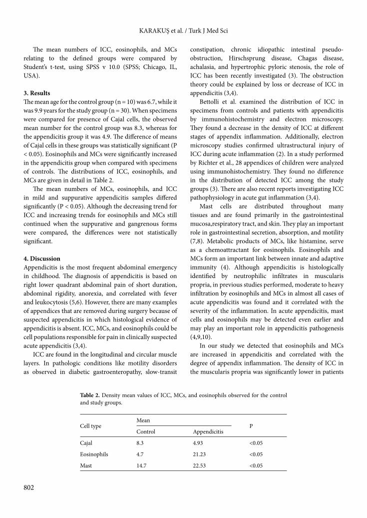
The mean numbers of ICC, eosinophils, and MCs relating to the defined groups were compared by Student’s t-test, using SPSS v 10.0 (SPSS; Chicago, IL, USA).
3. ResultsThe mean age for the control group (n = 10) was 6.7, while it was 9.9 years for the study group (n = 30). When specimens were compared for presence of Cajal cells, the observed mean number for the control group was 8.3, whereas for the appendicitis group it was 4.9. The difference of means of Cajal cells in these groups was statistically significant (P < 0.05). Eosinophils and MCs were significantly increased in the appendicitis group when compared with specimens of controls. The distributions of ICC, eosinophils, and MCs are given in detail in Table 2.
The mean numbers of MCs, eosinophils, and ICC in mild and suppurative appendicitis samples differed significantly (P < 0.05). Although the decreasing trend for ICC and increasing trends for eosinophils and MCs still continued when the suppurative and gangrenous forms were compared, the differences were not statistically significant.
4. DiscussionAppendicitis is the most frequent abdominal emergency in childhood. The diagnosis of appendicitis is based on right lower quadrant abdominal pain of short duration, abdominal rigidity, anorexia, and correlated with fever and leukocytosis (5,6). However, there are many examples of appendices that are removed during surgery because of suspected appendicitis in which histological evidence of appendicitis is absent. ICC, MCs, and eosinophils could be cell populations responsible for pain in clinically suspected acute appendicitis (3,4).
ICC are found in the longitudinal and circular muscle layers. In pathologic conditions like motility disorders as observed in diabetic gastroenteropathy, slow-transit
constipation, chronic idiopathic intestinal pseudo-obstruction, Hirschsprung disease, Chagas disease, achalasia, and hypertrophic pyloric stenosis, the role of ICC has been recently investigated (3). The obstruction theory could be explained by loss or decrease of ICC in appendicitis (3,4).
Bettolli et al. examined the distribution of ICC in specimens from controls and patients with appendicitis by immunohistochemistry and electron microscopy. They found a decrease in the density of ICC at different stages of appendix inflammation. Additionally, electron microscopy studies confirmed ultrastructural injury of ICC during acute inflammation (2). In a study performed by Richter et al., 28 appendices of children were analyzed using immunohistochemistry. They found no difference in the distribution of detected ICC among the study groups (3). There are also recent reports investigating ICC pathophysiology in acute gut inflammation (3,4).
Mast cells are distributed throughout many tissues and are found primarily in the gastrointestinal mucosa,respiratory tract, and skin. They play an important role in gastrointestinal secretion, absorption, and motility (7,8). Metabolic products of MCs, like histamine, serve as a chemoattractant for eosinophils. Eosinophils and MCs form an important link between innate and adaptive immunity (4). Although appendicitis is histologically identified by neutrophilic infiltrates in muscularis propria, in previous studies performed, moderate to heavy infiltration by eosinophils and MCs in almost all cases of acute appendicitis was found and it correlated with the severity of the inflammation. In acute appendicitis, mast cells and eosinophils may be detected even earlier and may play an important role in appendicitis pathogenesis (4,9,10).
In our study we detected that eosinophils and MCs are increased in appendicitis and correlated with the degree of appendix inflammation. The density of ICC in the muscularis propria was significantly lower in patients
Table 2. Density mean values of ICC, MCs, and eosinophils observed for the control and study groups.
Cell typeMean
PControl Appendicitis
Cajal 8.3 4.93 <0.05
Eosinophils 4.7 21.23 <0.05
Mast 14.7 22.53 <0.05

803
KARAKUŞ et al. / Turk J Med Sci
with severe appendix inflammation than in controls. The number of ICC may have an impact on appendix motility. The histopathological differences observed in this study may help elucidate the pathophysiology of appendicitis.
Although the mechanism of pain in suspected acute appendicitis is still unclear, cellular products of mast cells, eosinophils, and ICC might have an impact, which may inspire further studies.
References
1. Ergul E. Heredity and familial tendency of acute appendicitis. Scand J Surg 2007; 96: 290–302.
2. Bettolli M, De Carli C, Cornejo-Palma D, Jolin-Dahel K, Wang XY, Huizinga J, Krantis A, Rubin S, Staines WA. Interstitial cell of Cajal loss correlates with the degree of inflammation in the human appendix and reverses after inflammation. J Pediatr Surg 2012; 47: 1891–1899.
3. Richter A, Wit C, Vanderwinden JM, Wit J, Barthlen W. Interstitial cells of Cajal in the vermiform appendix in childhood. Eur J Pediatr Surg 2009; 19: 30–33.
4. Singh UR, Malhotra A, Bhatia A. Eosinophils, mast cells, nerves and ganglion cells in appendicitis. Indian J Surg 2008; 70: 231–234.
5. Ruffolo C, Fiorot A, Pagura G, Antoniutti M, Massani M, Caratozzolo E, Bonariol L, Calia di Pinto F, Bassi N. Acute appendicitis: what is the gold standard of treatment? World J Gastroenterol 2013; 19: 8799–8807.
6. Kim JS. Acute abdominal pain in children. Pediatr Gastroenterol Hepatol Nutr 2013; 16: 219–224.
7. Lee H, Park JH, Park DI, Kim HJ, Cho YK, Sohn CI, Jeon WK, Kim BI, Chae SW. Mucosal mast cell count is associated with intestinal permeability in patients with diarrhea predominant irritable bowel syndrome. J Neurogastroenterol Motil 2013; 19: 244–250.
8. Jakate S, Demeo M, John R, Tobin M, Keshavarzian A. Mastocytic enterocolitis: increased mucosal mast cells in chronic intractable diarrhea. Arch Pathol Lab Med 2006; 130: 362–367.
9. Mysorekar VV, Chanda S, Dandeka CP. Mast cells in surgically resected appendices. Indian J Pathol Microbiol 2006; 49: 229–333.
10. Coşkun N, Indel SM, Elpek GO. Mast cell density and neuronal hypertrophy in patients with acute appendicitis. Turk J Gastroenterol 2003; 14: 54–58.