
Is the course of the mandibular nerve deducible from the shape - ZIB
5
Is the course of the inferior alveolar nerve deducible from the shape of the mandible? S. Zachow a , H. Lamecker a , B. Elsholtz b , M. Stiller b a Zuse-Institute Berlin (ZIB), Medical Planning Systems, zachow@zib.de b Universitätsmedizin Berlin, Charité, Campus Benjamin Franklin Abstract. In a predominant amount of cases in orthodontics and orthognathic surgery, from the removal of wisdom teeth or cysts up to osteotomies, two-dimensional imaging is the first, and often the only method of the diagnostic process. However, without knowing the exact course of the mandibular nerve, the risk of an unintended lesion during an intervention is considerably high. This becomes even more relevant with an increasing number of cases, and complications, in dental implantology and orthognathic surgery. Hence, three-dimensional imaging such as cone beam tomography or DVT is increasingly employed. Within this study we are going to analyze the three-dimensional courses of the mandibular nerves statistically, aiming to provide knowledge about the potential course of the mandibular canal for two-dimensional imaging, thus extending diagnostics and therapy planning in dento-maxillofacial surgery or dental implantology. Keywords: orthognathic surgery, dental implantology, OPG, mandibular nerve, statistical shape model 1. Introduction In dental implantology a primary concern is an optimal and stable placement of im- plants within the jaw-bone without any impairment of the facial nerves. Typically im- plant planning is performed on the basis of panoramic X-ray images (so called ortho- pantomograms, OPG). However, such OPGs do not provide information on the thick- ness or width of the mandibular corpus and the lateral position of the inferior alveolar nerve (IAN), which is located between the mandibular and mental foramina. The only information that can be perceived from such 2D images is the height in cranial-caudal direction and the vertical position of the nerve. A decision, whether or not an implant could pass a nerve, which might be beneficial for an increased stability, cannot be made. With dental CT scanners one can overcome this problem, thus a more reliable 3D planning with regard to the mandibular nerve becomes possible. But, not in all cases a 3D scan is indicated, and time and effort acquiring a DVT is currently higher than that of an OPG. In this study we develop a statistical 3D shape model of a human mandible including the IAN, aiming to evaluate the variations in the courses of the mandibular canals, and whether or not these courses are deducible from the shape of the mandible. 2. Previous Works An early classification of the vertical positions of the course of the alveolar nerve was reported by Carter and Keen [1]. Three main courses were identified on basis of eight dissected mandibles (Fig. 1, left). In a study of Nortje et al. the course of the IAN was evaluated with the help of 3612 panoramic radiographs [2]. In conclusion three distinct variations between the apices of the roots of the teeth and the lower border of the mandible were described, predominantly being bilaterally symmetrical. In about 3% of the mandibles duplication or division of the canal, partial or complete absence, or lack of symmetry was found.
Transcript of Is the course of the mandibular nerve deducible from the shape - ZIB

Is the course of the inferior alveolar nerve deducible from the shape of the mandible?
S. Zachow a, H. Lamecker a, B. Elsholtz b, M. Stiller b
a Zuse-Institute Berlin (ZIB), Medical Planning Systems, [email protected] b Universitätsmedizin Berlin, Charité, Campus Benjamin Franklin
Abstract. In a predominant amount of cases in orthodontics and orthognathic surgery, from the removal of wisdom teeth or cysts up to osteotomies, two-dimensional imaging is the first, and often the only method of the diagnostic process. However, without knowing the exact course of the mandibular nerve, the risk of an unintended lesion during an intervention is considerably high. This becomes even more relevant with an increasing number of cases, and complications, in dental implantology and orthognathic surgery. Hence, three-dimensional imaging such as cone beam tomography or DVT is increasingly employed. Within this study we are going to analyze the three-dimensional courses of the mandibular nerves statistically, aiming to provide knowledge about the potential course of the mandibular canal for two-dimensional imaging, thus extending diagnostics and therapy planning in dento-maxillofacial surgery or dental implantology. Keywords: orthognathic surgery, dental implantology, OPG, mandibular nerve, statistical shape model
1. Introduction In dental implantology a primary concern is an optimal and stable placement of im-
plants within the jaw-bone without any impairment of the facial nerves. Typically im-plant planning is performed on the basis of panoramic X-ray images (so called ortho-pantomograms, OPG). However, such OPGs do not provide information on the thick-ness or width of the mandibular corpus and the lateral position of the inferior alveolar nerve (IAN), which is located between the mandibular and mental foramina. The only information that can be perceived from such 2D images is the height in cranial-caudal direction and the vertical position of the nerve. A decision, whether or not an implant could pass a nerve, which might be beneficial for an increased stability, cannot be made. With dental CT scanners one can overcome this problem, thus a more reliable 3D planning with regard to the mandibular nerve becomes possible. But, not in all cases a 3D scan is indicated, and time and effort acquiring a DVT is currently higher than that of an OPG. In this study we develop a statistical 3D shape model of a human mandible including the IAN, aiming to evaluate the variations in the courses of the mandibular canals, and whether or not these courses are deducible from the shape of the mandible.
2. Previous Works An early classification of the vertical positions of the course of the alveolar nerve was
reported by Carter and Keen [1]. Three main courses were identified on basis of eight dissected mandibles (Fig. 1, left). In a study of Nortje et al. the course of the IAN was evaluated with the help of 3612 panoramic radiographs [2]. In conclusion three distinct variations between the apices of the roots of the teeth and the lower border of the mandible were described, predominantly being bilaterally symmetrical. In about 3% of the mandibles duplication or division of the canal, partial or complete absence, or lack of symmetry was found.

Fig. 1. Left) variation of the vertical position of the inferior alveolar nerve according to [1] (reproduced from [5]), right) mental foramen view, preparation of an inferior alveolar nerve for transposition
Multiple mandibular canals, characterized by a single mandibular foramen and two nearly equal canals are rather unusual. Duplication or division of the canal was found in 0.9% (33/3612) of all cases. This observation was confirmed in an evaluation of 6000 panoramic radiographs, with 57 (0.95%) cases of so called bifid mandibular canals [3]. In a review by Anderson et al. was stated, that the buccal-lingual and superior-inferior positions of the IAN were not consistent among mandibles. The IAN frequently ran a concave curve with a posterior segment descending as it progressed anteriorly, and an anterior segment that ascended to the mental foramen (cf. Fig. 1, right). A bony canal was not always visible and the canal itself frequently lacked definite walls, especially in the vicinity of the mental foramen. Bilateral symmetry was commonly observed, where on the other hand duplications of the canal were rare [4].
More recent studies also tried to investigate the three-dimensional course of the mandibular canal using either 3D imaging techniques or dissections. In a study of Mraiwa et al. 50 mandibles of adult human cadavers (40 edentulous, 10 partially den-tate) were evaluated with panoramic and tomographic imaging, as well as vertical dissections [6]. The objective of this study was to evaluate the presence and course of the incisive canal in the mental interforaminal region of the human mandible. Results indicated a well-defined incisive canal with a mean inner diameter of 1.8 mm (SD 0.5 mm), being located on average 9.7 mm (SD 1.8 mm) from the lower cortical border and continued towards the incisor region in a slightly downward direction, with a mean distance to the lower cortical border of 7.2 mm (SD 2.1 mm). Kieser et al. investigated the vertical positioning and intra-bony branching patterns of the IAN in 39 edentulous mandibles of human cadavers by buccal micro-dissections [7]. Dissected mandibles were classified on the basis of the height of the IAN within the body of the mandible and the branching pattern of the IAN. In 30.7% (12/39) of the cases, the IAN was located in the superior part of the body of the mandible, and in 69.3% (27/39) of the cases the IAN was half-way or closer to the inferior border of the mandible. In a more recent study with 107 human cadaveric mandibles, the authors found that for 73% of males and 70% of females the IAN was located in the lower half of the mandible, thus showing that the pattern of distribution does not significantly differ between the sexes, between sides of the jaw, or with age [9]. An evaluation of the position of the mandi-bular foramen and the course of the IAN was performed on 12 right and 14 left cadaveric hemimandibles by Narayana et al. [8]. The bone was chiseled from its lingual
surface to expose the mandibular canal, and the distances from the nerve to the alveolar and inferior borders were measured. The distances from the nerve to the borders did not show any significant side differences. The study concludes that the canal and conse-quently the nerve do not maintain a constant position in the mandible and the location of the mandibular foramen varies from bone to bone despite its bilateral symmetry [8]. Tsuji et al. investigated the position and the course of the mandibular canal through the mandibular ramus using computed tomography imaging of 35 patients with skeletal Class III prognathism, in order to relate their findings to performing sagittal split ramus osteotomies [10]. Their conclusion was that special care must be taken when low sagittal splitting of the mandibular ramus is performed, due to the variability of the course of the IAN and the risk of its unintended lesion.
3. Material and Methods
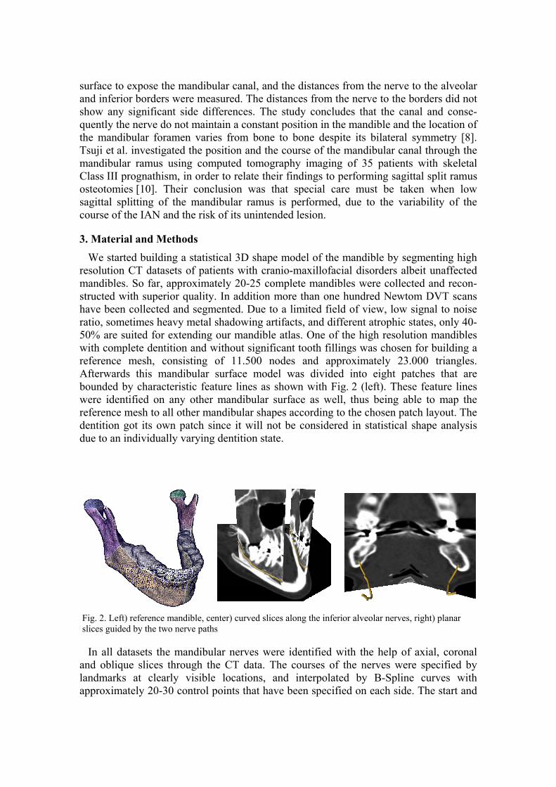
We started building a statistical 3D shape model of the mandible by segmenting high resolution CT datasets of patients with cranio-maxillofacial disorders albeit unaffected mandibles. So far, approximately 20-25 complete mandibles were collected and recon-structed with superior quality. In addition more than one hundred Newtom DVT scans have been collected and segmented. Due to a limited field of view, low signal to noise ratio, sometimes heavy metal shadowing artifacts, and different atrophic states, only 40-50% are suited for extending our mandible atlas. One of the high resolution mandibles with complete dentition and without significant tooth fillings was chosen for building a reference mesh, consisting of 11.500 nodes and approximately 23.000 triangles. Afterwards this mandibular surface model was divided into eight patches that are bounded by characteristic feature lines as shown with Fig. 2 (left). These feature lines were identified on any other mandibular surface as well, thus being able to map the reference mesh to all other mandibular shapes according to the chosen patch layout. The dentition got its own patch since it will not be considered in statistical shape analysis due to an individually varying dentition state.
Fig. 2. Left) reference mandible, center) curved slices along the inferior alveolar nerves, right) planar slices guided by the two nerve paths
In all datasets the mandibular nerves were identified with the help of axial, coronal and oblique slices through the CT data. The courses of the nerves were specified by landmarks at clearly visible locations, and interpolated by B-Spline curves with approximately 20-30 control points that have been specified on each side. The start and
end points of the nerves were defined by the mandibular and mental foramen, both being clearly visible on the surface reconstruction of the mandible. In order to verify the correctness of the nerve segmentation, a curved slice was computed on the basis of the smoothly interpolated curve, visualizing the complete course of the respective nerve. The control points were adjusted with the help of this immediately updated curved slice visualization as well as another orthogonal slice that can be shifted along the actual path of the nerve (Fig. 2). Finally the nerve segmentation led to a set of 500 sample points for each nerve. Each mandibular surface including the two sampled nerves has been added to our statistical 3D shape model.
4. Results A principal component analysis enables us to provide a compact description of a mean
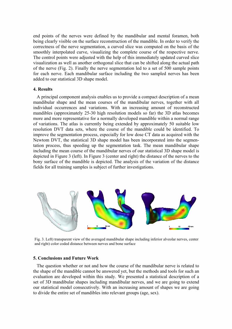
mandibular shape and the mean courses of the mandibular nerves, together with all individual occurrences and variations. With an increasing amount of reconstructed mandibles (approximately 25-30 high resolution models so far) the 3D atlas becomes more and more representative for a normally developed mandible within a normal range of variations. The atlas is currently being extended by approximately 50 suitable low resolution DVT data sets, where the course of the mandible could be identified. To improve the segmentation process, especially for low dose CT data as acquired with the Newtom DVT, the statistical 3D shape model has been incorporated into the segmen-tation process, thus speeding up the segmentation task. The mean mandibular shape including the mean course of the mandibular nerves of our statistical 3D shape model is depicted in Figure 3 (left). In Figure 3 (center and right) the distance of the nerves to the bony surface of the mandible is depicted. The analysis of the variation of the distance fields for all training samples is subject of further investigations.
Fig. 3. Left) transparent view of the averaged mandibular shape including inferior alveolar nerves, center and right) color coded distance between nerves and bone surface
5. Conclusions and Future Work The question whether or not and how the course of the mandibular nerve is related to
the shape of the mandible cannot be answered yet, but the methods and tools for such an evaluation are developed within this study. We presented a statistical description of a set of 3D mandibular shapes including mandibular nerves, and we are going to extend our statistical model consecutively. With an increasing amount of shapes we are going to divide the entire set of mandibles into relevant groups (age, sex).

Furthermore, we are preparing a setup to use our statistical 3D shape model of normally developed mandibles for the segmentation process. We will thoroughly investigate the segmentation and prediction quality, i.e. the deviation between manual and atlas based segmentation results as well as the correspondence of the estimated nerve positions with the real positions that are manually traced within the image data.
In addition, the statistical 3D shape model will be registered with computed panoramic X-rays from CT-Data in a ‘leave-one-out’ test, such that the bone contours optimally coincide with the intersection of the 3D shape model and the curved image slice. Again the correlation between predicted and true course of the mandibular nerve will be evaluated. The envisaged goal of our investigation is to extend the 2D image information of conventional OPGs or multi-planar dental imaging (with recorded imaging parameters) by a statistical shape model of the mandible, reliably representing the 3D shape of the mandible as well as the courses of the inferior alveolar nerves.
References [1] Carter RB and Keen EN: The intramandibular course of the inferior dental nerve. J Anat. 108(Pt 3),
pp. 433-440 (1971) [2] Nortje CJ, AG Farman, and FW Grotepass: Variations in the normal anatomy of the inferior dental
(mandibular) canal: a retrospective study of panoramic radiographs from 3612 routine dental patients. Br J Oral Surg 15(1), pp. 55-63 (1977)
[3] Langlais RP, R Broadus, and BJ Glass: Bifid mandibular canals in radiographs. J Am Dent Assoc 110(6), pp. 923-926 (1985)
[4] Anderson LC and TF Kosinski: A review of the intraosseous course of the nerves of the mandible. J Oral Implantology 17(4), pp. 394-403 (1991)
[5] McManners J: Mandibular body osteotomy. In: Fonseca RJ (ed) Oral and maxillofacial surgery, WB Saunders Co, Philadelphia, p. 330 (2000)
[6] Mraiwa N, R Jacobs, P Moerman, I Lambrichts, D van Steenberghe, and M Quirynen: Presence and course of the incisive canal in the human mandibular interforaminal region: two-dimensional imaging versus anatomical observations. Surg Radiol Anat. 25(5-6), pp. 416-423 (2003)
[7] Kieser JA, M Paulin, and B Law: Intrabony course of the inferior alveolar nerve in the edentulous mandible. Clin Anat. 17(2), pp. 107-111 (2004)
[8] Narayana K and S Vasudha: Intraosseous course of the inferior alveolar (dental) nerve and its relative position in the mandible. Indian J Dent Res. 15(3), pp. 99-102 (2004)
[9] Kieser J, D Kieser, and T Hauman: The course and distribution of the inferior alveolar nerve in the edentulous mandible. J Craniofac Surg. 16(1), pp. 6-9 (2005)
[10] Tsuji Y, T Muto, J Kawakami, and S Takeda: Computed tomographic analysis of the position and course of the mandibular canal: relevance to the sagittal split ramus osteotomy. Int J Oral Maxillofac Surg. 34(3), pp. 243-246 (2005)


